Article

Functional Analyses of Rare Germline Missense BRCA1 Variants Located within and outside Protein Domains with Known Functions

by
Henrikke Nilsen Hovland
1,2,3,
Eunice Kabanyana Mchaina
1,2,
Hildegunn Høberg-Vetti
1,2,4,
Sarah Louise Ariansen
5,
Wenche Sjursen
6,7,
Marijke Van Ghelue
8,9,
Bjørn Ivar Haukanes
2,
Per Morten Knappskog
2,3,
Ingvild Aukrust
2,3,*,† and
Elisabet Ognedal
1,2,†
1
Western Norway Familial Cancer Center, Haukeland University Hospital, 5021 Bergen, Norway
2
Department of Medical Genetics, Haukeland University Hospital, 5021 Bergen, Norway
3
Department of Clinical Science, University of Bergen, 5021 Bergen, Norway
4
Faculty of Health Studies, VID Specialized University, 5009 Bergen, Norway
5
Department of Medical Genetics, Oslo University Hospital, 0424 Oslo, Norway
6
Department of Medical Genetics, St. Olavs University Hospital, 7006 Trondheim, Norway
7
Department of Clinical and Molecular Medicine, Norwegian University of Science and Technology, 7491 Trondheim, Norway
8
Department of Medical Genetics, University Hospital of North Norway, 9038 Tromsø, Norway
9
Department of Clinical Science, UiT The Arctic University of Norway, 9019 Tromsø, Norway
*
Author to whom correspondence should be addressed.
These authors contributed equally to this work.
Genes 2023, 14(2), 262; https://doi.org/10.3390/genes14020262
Submission received: 14 December 2022 / Revised: 16 January 2023 / Accepted: 17 January 2023 / Published: 19 January 2023

Abstract

:
The BRCA1 protein is implicated in numerous important cellular processes to prevent genomic instability and tumorigenesis, and pathogenic germline variants predispose carriers to hereditary breast and ovarian cancer (HBOC). Most functional studies of missense variants in BRCA1 focus on variants located within the Really Interesting New Gene (RING), coiled-coil and BRCA1 C-terminal (BRCT) domains, and several missense variants in these regions have been shown to be pathogenic. However, the majority of these studies focus on domain specific assays, and have been performed using isolated protein domains and not the full-length BRCA1 protein. Furthermore, it has been suggested that BRCA1 missense variants located outside domains with known function are of no functional importance, and could be classified as (likely) benign. However, very little is known about the role of the regions outside the well-established domains of BRCA1, and only a few functional studies of missense variants located within these regions have been published. In this study, we have, therefore, functionally evaluated the effect of 14 rare BRCA1 missense variants considered to be of uncertain clinical significance, of which 13 are located outside the well-established domains and one within the RING domain. In order to investigate the hypothesis stating that most BRCA1 variants located outside the known protein domains are benign and of no functional importance, multiple protein assays including protein expression and stability, subcellular localisation and protein interactions have been performed, utilising the full-length protein to better mimic the native state of the protein. Two variants located outside the known domains (p.Met297Val and p.Asp1152Asn) and one variant within the RING domain (p.Leu52Phe) were found to make the BRCA1 protein more prone to proteasome-mediated degradation. In addition, two variants (p.Leu1439Phe and p.Gly890Arg) also located outside known domains were found to have reduced protein stability compared to the wild type protein. These findings indicate that variants located outside the RING, BRCT and coiled-coiled domains could also affect the BRCA1 protein function. For the nine remaining variants, no significant effects on BRCA1 protein functions were observed. Based on this, a reclassification of seven variants from VUS to likely benign could be suggested.

1. Introduction

Through interaction with myriad protein partners, the multifunctional BRCA1 protein is involved in numerous important cellular processes to prevent genomic instability and tumorigenesis. While pathogenic germline alterations including missense variants in BRCA1 predispose carriers to hereditary breast and ovarian cancer (HBOC), the role of variants of uncertain significance (VUSs) is unclear [1]. Rare missense variants constitute a major part of all BRCA1 VUSs, and are particularly challenging to classify due to limited or conflicting evidence.
The BRCA1 gene encodes a large protein of 220 kDa, primarily located in the nucleus, which consists of several functional domains (Figure 1). The N-terminal Really Interesting New Gene (RING) domain (aa 22–64) binds to BRCA1-Associated RING Domain protein 1 (BARD1), where heterodimerisation of the complex provides E3 ubiquitin ligase activity [2,3,4]. Two nuclear localisation sequences (NLS) (aa 503–508 and 607–614) allocate the BRCA1 protein to the nucleus where it exerts its functions. The coiled-coil domain (aa 1364–1437) located towards the C-terminal is involved in binding to the Partner and Localiser of BRCA2 (PALB2). Through the BRCA1 C-terminal (BRCT) domain (aa 1646–1736 and 1760–1855), BRCA1 interacts with multiple proteins involved in transcription and DNA damage response [5,6]. In addition to the established protein domains, BRCA1 contains an approximately 1500 residue unstructured central non-conserved region, of which very little is known [7].
Functional assays are considered as evidence of supportive to very strong strength for variant classification in the ACMG-AMP guidelines (BS3 or PS3 evidence) [9,10]. According to the BRCA1 specific guideline for variant interpretation from CanVIG-UK, five functional protein studies are suggested with specific recommendations regarding the strength of their respective functional evidence [11,12,13,14,15,16]. However, only two of these studies use the full-length BRCA1 protein [11,12]. This is also the case for several other BRCA1 functional studies published to date, which focus primarily on variants located in the RING and BRCT domains using plasmid constructs expressing only parts of the full-length protein [17,18,19,20,21,22]. In addition, several of the previously published studies perform assays to study only one of the multiple functional characteristics of the BRCA1 protein separately, such as ubiquitination, transcriptional activation or homologous recombination repair (HRR). However, as BRCA1 VUSs are distributed throughout the entire protein including regions outside well-established domains, examining only a single assay may be misleading [23]. Hence, to clarify how variants in the more non-conserved parts of the protein can potentially affect its functions, there is a need for several functional assays utilising the full-length protein to mimic the more native state of the BRCA1 protein. Some of the protein functions of BRCA1 also involve several domains of the protein, and consequently there is a need for multiple functional assays covering different activities.
Several publications have suggested that most BRCA1 missense substitutions located outside the well-established and conserved RING, coiled-coil and BRCT domains could be classified as (likely) benign, arguing that pathogenic missense variants are infrequent in these regions, which are thought to tolerate variations and be without essential functions [24,25,26]. In a recent publication, classification of BRCA1 missense variants available in the public database ClinVar was used to illustrate this, and the authors suggest the incorporation of criteria regarding coldspots to improve the ACMG-AMP guidelines for BRCA1 variant interpretation, as a counterweight to hotspots [27]. Noteworthily, a coldspot criterion is included in the BP1 evidence in the BRCA1/BRCA2 gene-specific guidelines for variant interpretation from CanVIG-UK, which states that the location of a missense variant outside the RING, coiled-coil and BRCT domains is supporting evidence towards benign effect [28]. On the other hand, the approximately 1500 residue central region of BRCA1 has been suggested to act as a long flexible scaffold for intermolecular interactions which obtain a more ordered structure upon binding to protein partners, and may, thus, still be functionally important in the DNA damage response [7,29,30,31,32]. Furthermore, amino acid residues located outside well-established domains in the primary structure of the polypeptide chain can potentially interact with or become part of important structural and functional elements in the native folded three-dimensional structure of the BRCA1 protein. This indicates that replacing amino acid residues located outside an important protein domain could still possibly affect both the structure and function of the protein.
The purpose of this study was, therefore, to functionally characterise a set of 14 BRCA1 VUSs, of which 13 variants are located outside the known domains, by multiple different protein assays utilising the full-length BRCA1 protein. The BRCA1 VUSs were selected from our recently published study of BRCA1 variants detected in families with suspected HBOC in Norway, “BRCA1 Norway” [33]. Since the majority of the VUSs investigated in this study are located outside the known protein domains of BRCA1, we aimed to use not only BRCA1 specific assays but also more general protein assays to assess their impact on protein expression, protein stability and subcellular localisation. Based on this, we wanted to investigate the hypothesis stating that BRCA1 variants located outside the known protein domains are benign and of no functional importance. Furthermore, we aimed to use the data gathered from the different functional assays, in combination with other available information, as a tool to clarify the pathogenicity of these variants.

2. Materials and Methods

2.1. Plasmids and Construction of BRCA1 Variants

The plasmid pDEST-mCherry-LacR-BRCA1 encoding mCherry-tagged wild type (WT) full-length human BRCA1 protein was a gift from Daniel Durocher (Addgene plasmid #71115; http://n2t.net/addgene:71115 (accessed on 2 January 2020); RRID:Addgene_71115) [34]. This plasmid will hereafter be assigned BRCA1 WT, or WT only. The BRCA1 missense variants (listed in Supplementary Table S1) were introduced in the WT plasmid using the QuikChange II XL Site Directed Mutagenesis Kit (Agilent Technologies, Santa Clara, CA, USA). Primers used to produce variants of interest and control variants are available upon request. The empty vector (EV) plasmid pDEST-mCherry-LacR, hereafter assigned EV, was also kindly provided by Daniel Durocher [34]. All plasmids were prepared by QIAfilter Plasmid Maxi Kit (QIAGEN, Hilden, Germany), and the presence of the altered variants, in addition to the whole BRCA1 insert, were verified by Sanger sequencing. The variants were all selected from our previous study, “BRCA1 Norway”, and were reported as VUSs in ClinVar or classified as VUS by one or more of the Norwegian medical genetic departments at the time of selection [33]. Some of the variants were classified as both VUS and likely benign by different departments, and these were specifically included, aiming to harmonise the variant classification between the different departments. Intentionally, variants throughout the whole BRCA1 gene were selected, and all variants except one (located within the RING domain) are located outside the well-established RING, coiled-coil and BRCT domains (Figure 1). For each assay, benign and pathogenic control variants were chosen. If possible, variants tested previously by the same type of assay were preferred as controls. A recurring issue and a limitation for all assays performed in this study was the lack of well-established relevant pathogenic missense control variants located in the regions outside of the known domains. No pathogenic variants outside of these regions were found in ClinVar or the literature. This made it difficult to fulfil the requirement of a sufficient number of control variants as suggested by Brnich et al. [10]. For investigations of co-immunoprecipitation assays with BARD1 and PALB2, controls were chosen from the relevant regions (RING and coiled-coiled domain, respectively).
In the co-immunoprecipitation assay, the plasmids pcDNA6.2-BARD1-V5, hereafter called BARD1-V5 WT, and pDEST-FRT/T0-Flag-PALB2, hereafter called Flag-PALB2 WT, were used. BARD1-V5 WT was a gift from Masanori Kurihara and Atsushi Iwata [35], and Flag-PALB2 WT was a gift from Daniel Durocher (Addgene plasmid #71114; http://n2t.net/addgene:71114 (accessed on 2 January 2020); RRID:Addgene_71114) [34]. The corresponding empty vectors (pcDNA6.2-V5 and pDEST FRT/TO-FLAG) were used as controls.

2.2. Cell Culture and Transfection

HEK293FT and MDA-MB-231 cells were cultured in DMEM high glucose GlutaMAX™ medium or DMEM medium (Thermo Fisher Scientific, Waltham, MA, USA), respectively, supplemented with 10% FBS (Thermo Fisher Scientific) and 1% PenStrep (Sigma-Aldrich, St. Louis, MO, USA). Both cell lines were maintained in 5% CO2 at 37 °C. JetPrime® (Polyplus-Transfection, Illkirch-Graffenstaden, France) was used for transient transfection of the cells according to the manufacturer’s protocol.

2.3. Assessment of BRCA1 Protein Expression by Immunoblotting

For Western blot analyses, cells were lysed in RIPA buffer (supplemented with complete Mini EDTA-free Protease inhibitor cocktail tablets, Roche, Basel, Switzerland) 48 h post transfection, and centrifuged at 13,000× g for 10 min at 4 °C. Following measurements of the protein concentration by Pierce BCA protein assay kit (Thermo Fisher Scientific), 5 µg total protein were analysed by SDS-PAGE using 3–5% Tris-Acetate gels (150 V, 75 min) and transferred to a nitrocellulose membrane (30 V, 60 min). One BRCA1 WT sample was always included in each gel to ensure comparable results with the variants investigated. To detect BRCA1 protein, the following antibodies were used: primary anti-BRCA1 (sc-6954, Santa Cruz Biotechnology, Dallas, TX, USA) and secondary m-IgGκ BP-HRP (sc-516102, Santa Cruz). Anti-β-Actin antibody (sc-47778, Santa Cruz) was used as loading control and for quantification of relative BRCA1 protein expression levels. Proteins were visualised using SuperSignalTM West Pico PLUS Chemiluminescent Substrate (Thermo Fisher Scientific) and the ChemiDOCTM MP imaging system. The signals were quantified using the Image LabTM Software from BioRad (version 6.0). As benign controls for protein expression, the variants p.Lys45Gln, p.Arg504His, and p.Val1378Ile were included (all classified as benign by the ENIGMA expert panel) [36]. As negative controls for protein expression, empty vector and the pathogenic variants p.Ala1708Glu and p.Val1838Gly were included [18,37].

2.4. RNA Purification and qPCR

HEK293FT cells were seeded in 12-well plates (0.35 × 10⁶ cells/well), and transfected with BRCA1 WT and variant plasmids. Forty-eight hours after transfection, RNA was purified using RNeasy Mini Kit (QIAGEN) as described by the manufacturer. The quality of the RNA samples was analysed by the Agilent RNA 2200 ScreenTape System. Purified RNA (1 µg) was used to synthesise single-stranded cDNA, applying the SuperScript™ VILO™ cDNA Synthesis Kit (Invitrogen, Waltham, MA, USA). The synthesised cDNA was then used as a template for the analysis of expression of BRCA1 variants and the housekeeping gene β-actin by qPCR using TaqMan® Gene Expression Assays (Applied Biosystems, Life Technologies).

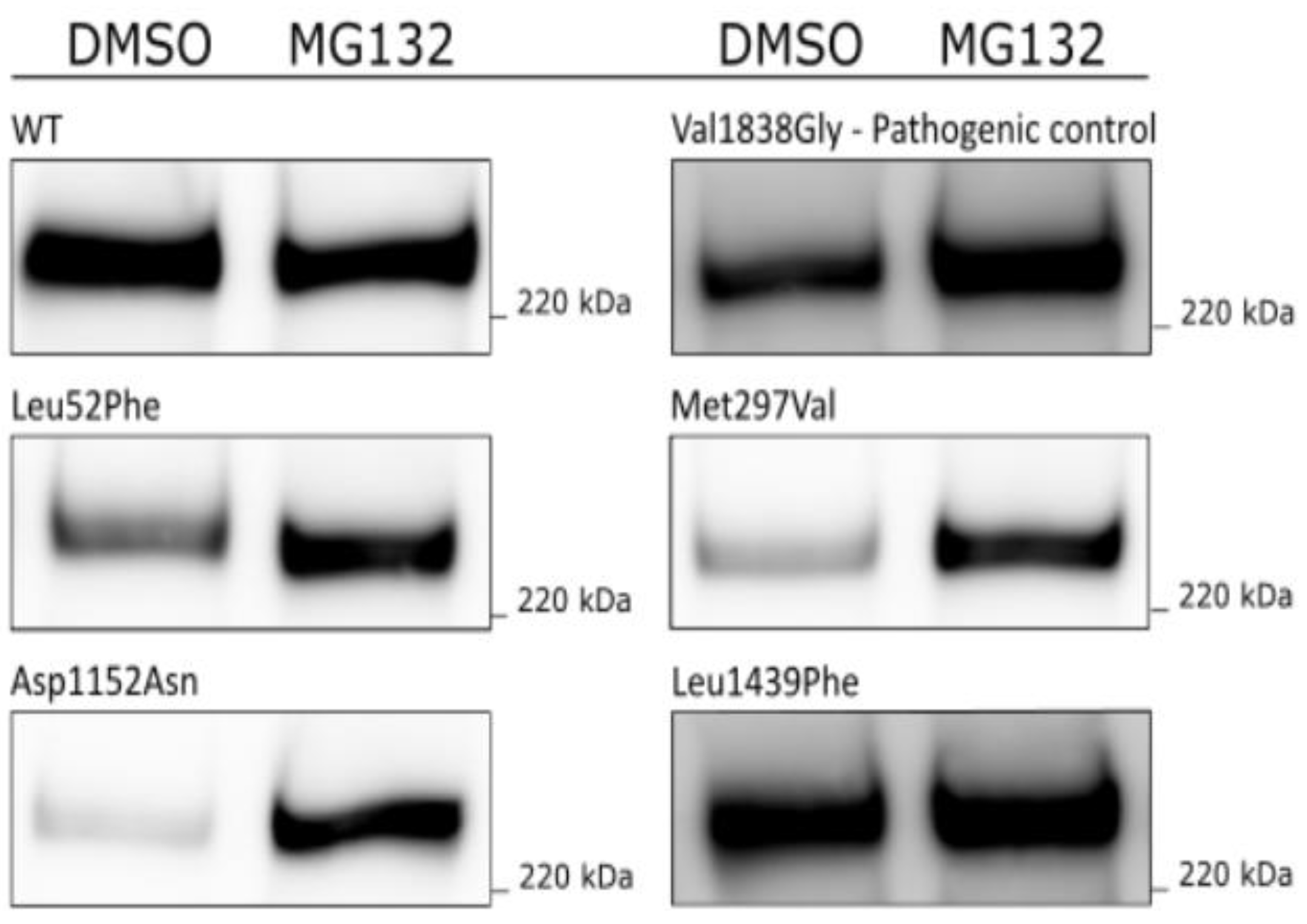
2.5. MG132 Assay for Assessment of Proteasomal Degradation

HEK293FT cells were seeded in 12-well plates (0.35 × 10⁶ cells/well) and transfected with BRCA1 WT and variants. Twenty-four hours post transfection, cells were incubated with 20 µM MG132 (Sigma-Aldrich) dissolved in DMSO or DMSO only for 24 h. Cells were then lysed in 100 µL RIPA buffer (with protease inhibitor). Samples containing 10 µg of protein were analysed by Western blotting, and compared to WT and p.Val1838Gly used as benign and pathogenic controls, respectively.

2.6. Cycloheximide Chase Assay for Measurement of BRCA1 Protein Stability

HEK293FT cells were seeded in 12-well plates (0.35 × 10⁶ cells/well) and transfected with BRCA1 WT and variants. Twenty-four hours post transfection, the medium was removed and replaced with fresh medium containing 50 µg/mL cycloheximide (Sigma-Aldrich) dissolved in DMSO or DMSO only. Cells were harvested after 0, 2 and 8 h of treatment. For each time point, the cells were lysed in RIPA buffer (supplemented with protease inhibitor) and frozen at −20 °C immediately after harvest. Centrifugation (13,000 g, 10 min, 4 °C) was performed for all samples in parallel >24 h post freezing. Samples containing 5 µg of protein were analysed by Western blotting. As benign controls for protein stability, WT and three benign variants (p.Lys45Gln, p.Arg504His, p.Vall378Ile) were included. As pathogenic controls for protein stability, two variants known to harbour reduced protein stability were used (p.Cys49Tyr and p.Ala1708Glu) [37,38,39]. The resulting % protein expressions presented are relative to the protein levels for the corresponding variant at the starting point (0 h, corresponding to 100%).

2.7. Fractionation Assay for Assessment of Subcellular Localisation

Subcellular localisation was tested by a fractionation assay separating the cytosolic and nuclear cell fractions [40,41]. HEK293FT cells seeded in 10 cm dishes (4.8 × 10⁶ cells/dish) were transfected with 10 µg plasmid encoding BRCA1 WT or variants. Forty-eight hours post transfection, the cells were washed in PBS and pelleted at 1200 rpm for 5 min, before resuspending the cells in 250 µL buffer A (10 µM HEPES pH 7.8, 1.5 mM MgCl2, 10 mM KCl, 0.10% IGEPAL, 0.5 mM DTT, EDTA free protease inhibitor) and incubating for 30 min. The suspension was then pelleted at 13,000 rpm for 5 min at 4 °C. The resulting supernatant, which is the cytosolic fraction, was then frozen at −80 °C for later analyses. The pellet was washed once with 100 µL buffer A and resuspended in 100 µL buffer B (20 mM HEPES pH 7.8, 420 mM NaCl, 1.5 mM MgCl2, 0.2 mM EDTA, 0.5 mM DTT, EDTA free protease inhibitor) by pipetting up/down 30 times. After 30 min incubation on ice with vortexing every minute, the resuspension was centrifuged at 13,000 rpm for 15 min at 4 °C. The resulting supernatant, which is the nuclear fraction, was then frozen at −80 °C for later analyses. Cytosolic and nuclear samples containing 5 µg of total protein were analysed by Western blotting. Anti-Topoisomerase IIα (D10G9, Cell Signaling Technology, Danvers, MA, USA) and Anti-HSP 90α/β (sc-13119, Santa Cruz) were used to confirm the purity of the nuclear and cytosol fractions, respectively, and were used for normalisation. The % of BRCA1 protein in each fraction was then calculated, and the level of protein in the nucleus was presented.

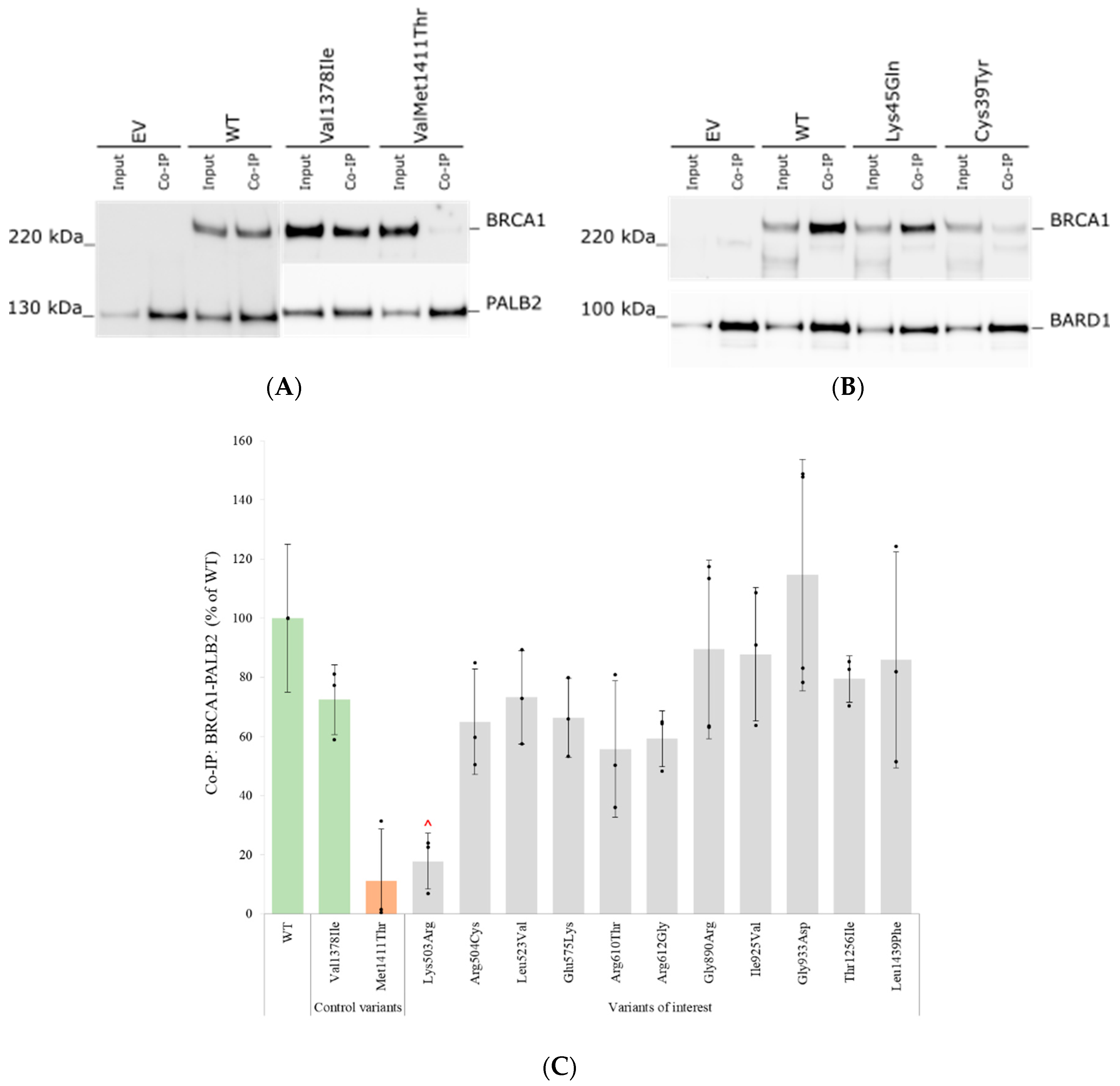
2.8. Co-Immunoprecipitation Assay

HEK293FT cells were seeded in 10 cm Petri dishes (4.8 × 10⁶), and co-transfected with 5 µg plasmid encoding BRCA1 WT or the selected BRCA1 variants in combination with either 5 µg plasmid encoding BARD1-V5 WT or Flag-PALB2 WT. After 48 h, cells were lysed in 500 µL ice-cold IP Lysis/Wash Buffer (supplemented with protease inhibitor) per dish. The cell lysate was centrifuged at 13,000× g for 10 min at 4 °C, before measuring protein concentration by Pierce BCA protein assay kit. Co-immunoprecipitation (Co-IP) was performed using the DynabeadsTM Protein G Immunoprecipitation Kit (Invitrogen) according to the manufacturer’s protocol, with the following specifications: 5 µg of V5 antibody (for WT BARD1-V5) or 5 µg of Flag antibody (for WT Flag-PALB2) was coupled to 50 µL magnetic beads. Equal amounts of cell lysate proteins (2 mg, input) were incubated with the antibody-coupled beads for 90 min at 4 °C. After non-denaturing elution of the protein complexes, the proteins bound to the beads (IP) were separated by SDS-PAGE, and BRCA1 WT or variants in combination with BARD1-V5 or Flag-PALB2 were visualised by Western blotting using anti-V5 (46-0705, Invitrogen) or anti-Flag (F1804, Sigma-Aldrich), respectively. BRCA1 protein levels in the IP samples were quantified and normalised to the anti-V5 signal or anti-Flag signal in the IP samples. The data for each of the variants were presented as % compared to WT (set to 100%). As controls for the Co-IP assay with BRCA1-PALB2, the benign variant p.Val1378Ile and the pathogenic variant p.Met1411Thr (both located in the coiled-coil domain of BRCA1), were included [42,43]. As controls for the Co-IP assay with BRCA1-BARD1, the benign variant p.Lys45Gln and the pathogenic variant p.Cys39Tyr (both located in the RING domain of BRCA1) were included [38,39].

2.9. Statistics

All experiments were carried out on at least three independent occasions unless otherwise specified in the figure legends, with the exception of the empty vector, which was performed in one replicate only. The standard deviations were calculated for WT and each variant in all assays. The statistical significance was evaluated with the Student’s t-test with p values < 0.05.

2.10. Assessment of Variant Classifications

The Alamut Software (Version 2.15, SOPHiA GENETICS) and the Human Gene Mutation Database (HGMD) professional 2022.1 (QIAGEN) were used for gathering information on the BRCA1 variants. Reinterpretation of the variants was performed based on new knowledge using the ACMG-AMP criteria supplemented with the BRCA1/BRCA2 gene-specific criteria by CanVIG-UK [9,16].

3. Results

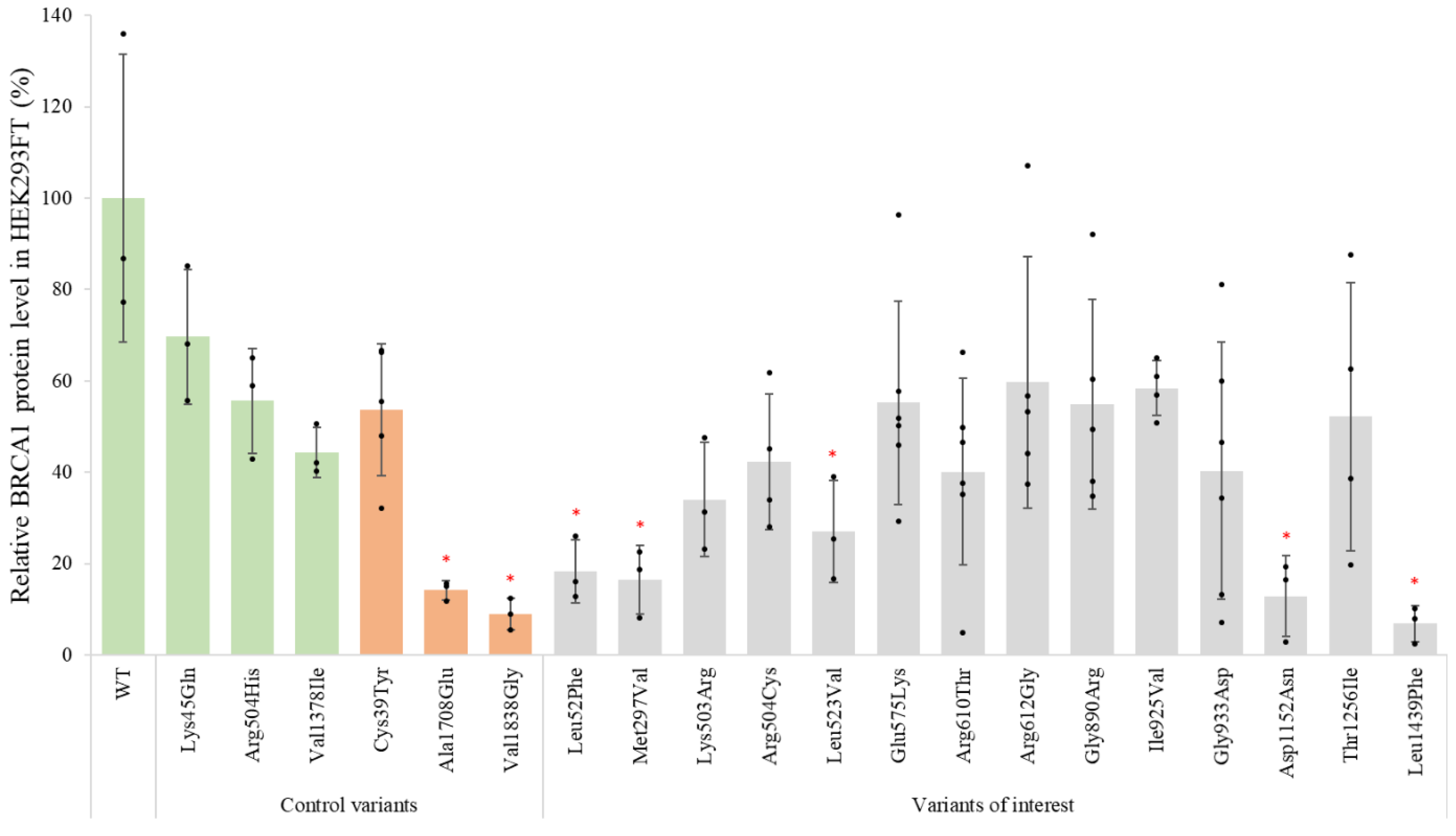
3.1. Effects of BRCA1 Variants on Protein Expression

To test the effect of the selected BRCA1 missense variants (Figure 1) on the protein expression level, the corresponding plasmids were transfected into HEK293FT cells and the cell lysates were analysed by Western blot analysis (Supplementary Figure S1A). As expected, a band located just above 220 kDa, corresponding to the theoretical molecular weight of mCherry-BRCA1 (248 kDa), was detected for both the WT and variants. Nine variants had similar relative expression levels as the WT (100%) and/or benign controls (44–70%). Four of the variants (p.Leu52Phe, p.Met297Val, p.Asp1152Asn and p.Leu1439Phe) displayed severely reduced protein levels, i.e., <20% protein compared to WT, similar to the included pathogenic controls (9–14%) (Figure 2). In addition, the variant p.Leu523Val was found to have reduced protein expression (27%) compared to the WT, at an intermediate expression level between pathogenic (9–14%) and benign controls (44–70%). For comparison, the assay was repeated in MDA-MB-231 cells, where a similar trend for protein expression was seen (Supplementary Figure S1B)

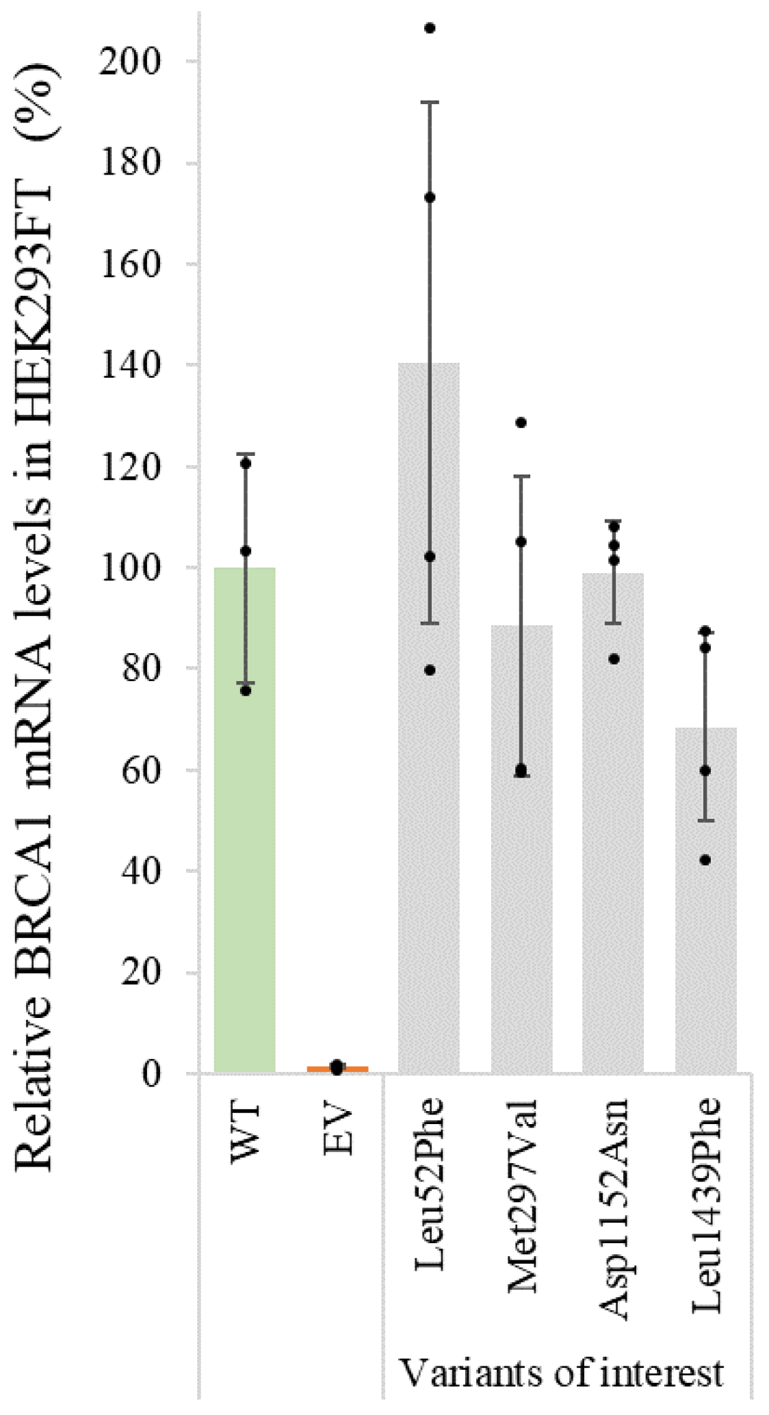
3.2. qPCR for Assessment of mRNA Levels

The four protein variants (p.Leu52Phe, p.Met297Val, p.Asp1152Asn and p.Leu1439Phe) found to be expressed at levels lower or similar to the included pathogenic controls in HEK293FT cells were subsequently analysed by qPCR to investigate if the low protein expression was caused by a reduction of the mRNA levels. After normalisation of the data by actin, the relative mRNA levels for each variant compared to the BRCA1 WT were calculated (Figure 3). The results suggest that the plasmids encoding p.Leu52Phe, p.Met297Val, p.Asp1152Asn and p.Leu1439Phe produce similar amounts of mRNA as the WT plasmid. Thus, for these variants, reduced protein levels are unlikely to be caused by reduced transcription or transfection efficiency, but are more likely caused by increased protein degradation or reduced stability.

3.3. MG132 Assay for Assessment of Proteasomal Degradation

To check whether the low protein levels in HEK293FT cells observed for p.Leu52Phe, p.Met297Val, p.Asp1152Asn and p.Leu1439Phe could be due to degradation by the ubiquitin–proteasome system, transfected HEK293FT cells were treated with proteasome inhibitor MG132 for 24 h. As shown in Figure 4, protein expression clearly increased for the pathogenic control (p.Val1838Gly) and three of the variants (p.Leu52Phe, p.Met297Val and p.Asp1152Asn) after treatment with MG132, compared to the control samples treated with DMSO only. For the variant p.Leu1439Phe, comparable amounts of protein were observed in the MG132 treated sample and the DMSO control sample.

3.4. Cycloheximide Chase Assay for Assessment of Protein Stability

For the BRCA1 variants which showed protein expression levels above 20% compared to the BRCA1 WT protein in the Western blot analysis (Figure 2), including the variant p.Leu1439Phe, which showed equal amounts of protein in the MG132 assay (Figure 4), the protein stability was analysed by cycloheximide chase assay to follow protein degradation over time in transfected HEK293FT cells. The results from one representative replicate after 0, 2 and 8 h of treatment with the protein synthesis inhibitor cycloheximide, compared to DMSO only for a minor selection of variants (p.Gly890Arg and p.Leu1439Phe) and controls (p.Arg504His and p.Ala1708Glu), are shown in Supplementary Figure S2. Figure 5 shows the mean percentage BRCA1 protein level remaining for each variant in transfected HEK293FT cells treated with cycloheximide for 8 h. For the BRCA1 WT, the protein expression level decreased to 83% after 8 h treatment with cycloheximide. For all the variants, including the benign controls, a more prominent degradation of BRCA1 protein was observed during cycloheximide treatment. The protein levels for all the benign controls were reduced to 28–34%, while there was, respectively, 0% and 9% protein detected for the pathogenic controls p.Ala1708Glu and p.Cys39Tyr. Protein levels of the two BRCA1 VUSs, p.Gly890Arg and p.Leu1439Phe, were reduced to 11% and 10%, respectively, after 8 h of cycloheximide treatment, similar to the pathogenic controls. The nine remaining variants showed a reduction in protein levels comparable to the benign controls after 8 h of treatment.

3.5. Assessment of Subcellular Localisation by Fractionation Assay

According to the literature, the BRCA1 protein is known to be mainly located in the nucleus, and the two NLS of BRCA1 are located at aa 503–508 and 607–614 [44]. To investigate if some of the variants of interest could alter the nuclear localisation of the protein, subcellular localisation was assessed by a nuclear/cytosol fractionation assay. This was especially interesting to evaluate for the variants p.Lys503Arg, p.Arg504Cys, p.Arg610Thr and p.Arg612Gly, which are all located within the nuclear localisation sequences. Similar to the BRCA1 WT protein (84% located in the nucleus), all analysed variants were found to be mainly located in the nucleus fraction (Supplementary Figure S3).

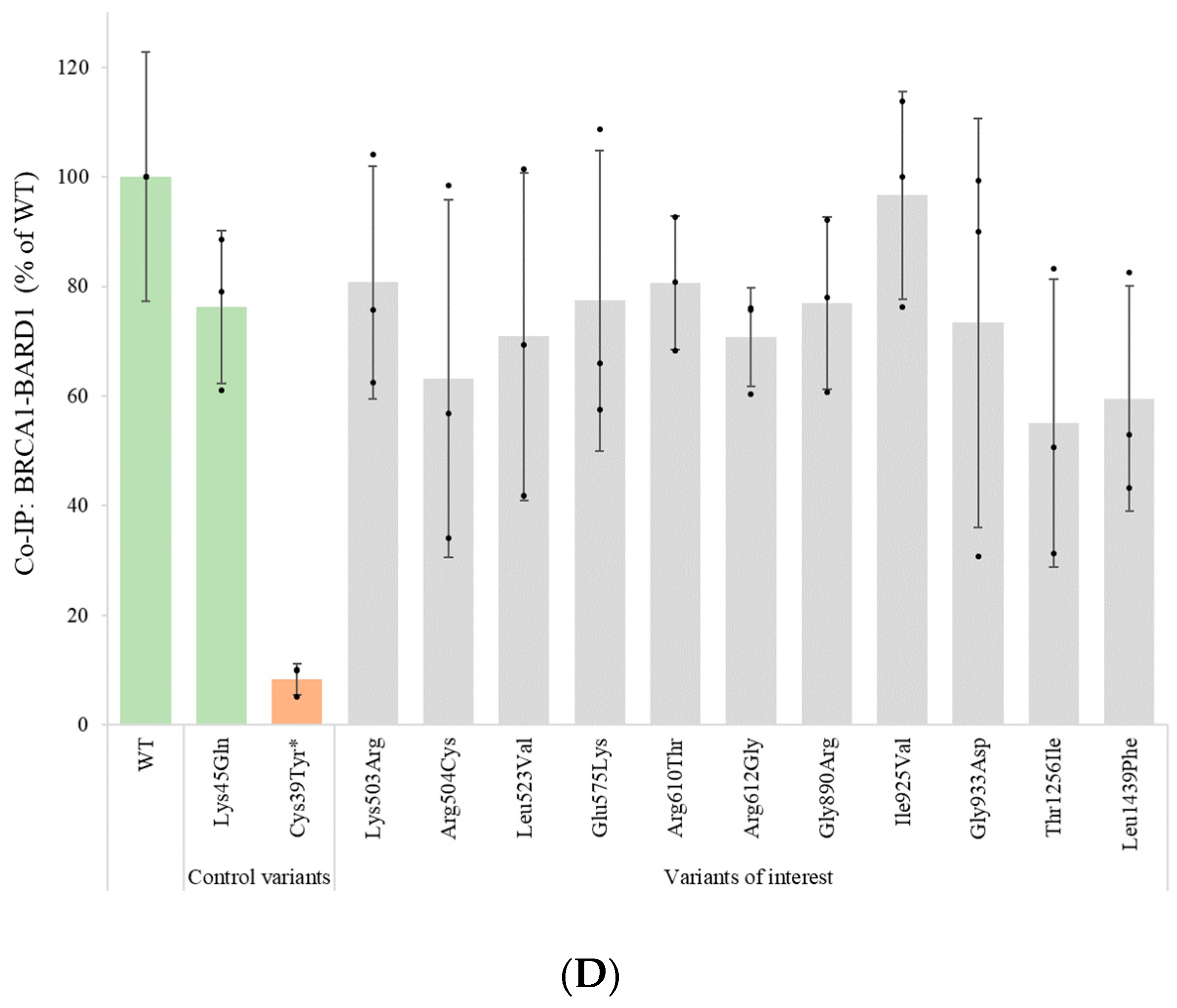
3.6. Assessment of Protein–Protein Interactions with BARD1 and PALB2 by Co-Immunoprecipitation

Co-IP assays were performed to test the potential effect of the VUSs on the binding of the BRCA1 protein to two of its binding partners: BARD1 and PALB2. The resulting Western blots for the WT, EV, control variants and a selection of the analysed VUSs are illustrated in Figure 6A,B. As seen in the blots, the BRCA1 WT protein captured both PALB2 (Figure 6A) and BARD1 (Figure 6B). A strong binding to the respective benign controls p.Val1378Ile and p.Lys45Gln was observed, and a weak binding to the respective pathogenic controls p.Met1411Thr and p.Cys39Tyr. Mean values for all variants (% binding capacity compared to WT) are shown in Figure 6C,D. In the initial analysis, the variant p.Lys503Arg appeared to have a reduced binding to PALB2 (Figure 6C), but this interaction was shown to be similar to the WT/benign controls when quantifying against the amount of the variant input sample. Thus, none of the variants of interest were showed to have significantly reduced binding to either BARD1 or PALB2.

3.7. Summary of Functional Assays

When summarising the data from each of the different functional assays (Table 1), five BRCA1 variants were found to have one or more features strongly deviating from the WT protein, while the remaining nine variants showed no or only minor deviations. The data gathered throughout the study were combined with other relevant information and used to suggest an updated classification of the pathogenicity of these rare BRCA1 variants (Table 2). The molecular properties and previously known information on each of these variants will be discussed below.

4. Discussion

In this study, we have examined the effects of 14 rare BRCA1 missense VUSs to investigate the hypothesis stating that no pathogenic BRCA1 missense variants are present outside of known protein domains in BRCA1 [24,25,26]. We have investigated the effect of each variant by multiple functional protein assays, using the full-length BRCA1 protein to better mimic the native state of the protein.

4.1. BRCA1 Protein Domains and Amino Acid Conservation

Use of the BP1 criteria in the gene-specific BRCA1/BRCA2 guidelines from CanVIG-UK, indicating that no pathogenic BRCA1 missense variants are present outside of known protein domains, was debated in our “BRCA1 Norway” publication [16,33]. Counterarguments stated that amino acid residues located outside well-established domains in the primary structure of the polypeptide chain can potentially interact with or become part of important structural and functional elements in the native folded three-dimensional structure of the BRCA1 protein. Thus, the replacement of amino acid residues located outside an important domain in the primary structure could possibly affect both structure and function of the protein. In fact, it has been suggested that the majority of the loss of function missense mutation is indirectly caused by the destabilisation of the protein’s three-dimensional-structure, rather than directly disrupting important functional characteristics such as binding sites [37,49,50,51]. In contrast to the highly conserved RING and BRCT domains, for which the structure is known, it has been suggested that the central 1500 residue region of BRCA1 acts as a long flexible scaffold for intermolecular interactions, even though the central region lacks substantial conserved motifs [7]. Such intrinsically non-conserved disordered regions are known to obtain a more folded structure upon interaction with its protein partners [7,29,30,31,32]. Thus, although a disordered region in the absence of its binding partners, this central region might still be functionally important in the DNA damage response [7].

4.2. Protein Expression and Protein Stability of the BRCA1 Variants

For many genes, the protein expression level of a variant is known to correlate with the pathogenicity of the variant [40,41]. In contrast, it has been demonstrated that BRCA1 protein variants displaying low protein levels may still sustain structure/function similar to the WT protein, and that variants with protein levels similar to the WT protein may fail to sustain function [13,52]. These studies were, however, performed by expression of isolated protein domains and not the full-length BRCA1 protein. The effect of missense changes on the expression of full-length BRCA1 protein, particularly those located outside of the known domains, has to our knowledge not previously been thoroughly investigated. We, therefore, aimed to investigate the effect of our selected BRCA1 variants on protein expression levels in HEK293FT cells by Western blotting. The benign control variants showed reduced protein level (44–70%) compared to the WT protein, but considerably higher levels than the pathogenic control variants (9–14%). In concordance with previous studies [52], this indicates that even significantly reduced BRCA1 expression levels are sufficient to maintain BRCA1 protein functions, and that protein expression levels do not necessarily correlate with the level of protein activity. In addition, the lower threshold for BRCA1 protein expression associated with pathogenicity is currently unknown. Nine of the investigated variants showed protein expression levels comparable to the benign controls/WT protein (27–60%), while the four variants (p.Leu52Phe, p.Met297Val, p.Asp1152Asn and p.Leu1439Phe) showed reduced protein levels in the range of the pathogenic controls (7–18%). Low protein expression can be caused by, among others, low transcription levels, protein instability or increased protein degradation. Although protein expression analyses alone are not adequate to distinguish between benign and pathogenic variants, protein expression analysis can, in combination with additional protein assays, still provide important insights regarding the underlying mechanism for the loss of protein function. To investigate the cause of the reduced protein levels, therefore, we analysed the four variants, p.Leu52Phe, p.Met297Val, p.Asp1152Asn and p.Leu1439Phe, using qPCR. The mRNA levels for the four variants were found to be in the same range as the WT, indicating that the underlying mechanism for the low protein expression levels is at the protein level.
It has previously been shown that several missense variants in the BRCT domain lead to increased susceptibility to degradation of BRCA1 and destabilisation of the protein structure, by, among others, the ubiquitin–proteasome system [53,54,55,56,57,58]. When investigating the aforementioned four variants by inhibiting the ubiquitin–proteasome degradation pathway by MG132, an increased protein level was observed for three of the variants (p.Leu52Phe, p.Met297Val and p.Asp1152Asn) and the pathogenic control p.Val1838Gly, which has been previously shown to have reduced protein levels in HEK293 cells [18]. This indicates that these variants, of which two are located outside of known protein domains, make the BRCA1 protein more prone to ubiquitin-mediated degradation. In contrast, the variant p.Leu1439Phe seems not to be removed by the proteasomal system.
To evaluate the protein stability of the BRCA1 variants over time, a cycloheximide chase assay was performed for the BRCA1 variants showing protein expression levels comparable to the benign controls/WT protein. In addition, the p.Leu1439Phe variant, which was found not to be removed by the proteasomal system, was included. The BRCA1 WT protein showed a stability of 83% after treatment with cycloheximide. All benign control variants, surprisingly, illustrated 28–34% protein levels compared to the WT protein after eight hours, indicating that a protein variant could have pronounced reduction in stability without affecting the pathogenicity. In comparison, the pathogenic control variants p.Ala1708Glu and p.Cys39Tyr showed, respectively, 0% and 9% protein expression after treatment with cycloheximide. Similar to the two pathogenic controls, the two VUSs, p.Gly890Arg and p.Leu1439Phe, showed severely reduced protein stability compared to the WT protein (11% and 10%, respectively). After cycloheximide treatment, four of the VUSs (p.Lys503Arg, p.Ile925Val, p.Gly933Asp and p.Thr1256Ile) demonstrated protein levels in the range 19–23% of the WT, at an intermediate level between the pathogenic and benign controls. The remaining five VUSs showed protein levels comparable to the benign controls (29–48%). In order to improve the capacity of the assay to better discriminate the benign/pathogenic thresholds, more pathogenic and benign controls should be included in this assay [10].

4.3. The Effect of BRCA1 Variants on BARD1 and PALB2 Interaction

The BRCA1 protein is known to interact with a myriad of other proteins. Among others, BRCA1 interacts with BARD1 though the RING domain, and with PALB2 through the coiled-coil domain. Although only one of the 14 VUSs analysed in this study is located in the RING domain, and none in the coiled-coil domain, we wanted to investigate if any of our variants of interest could alter these interactions. The effect of an abolished BRCA1–BARD1 interaction was illustrated by the pathogenic BRCA1 control variant p.Cys39Tyr, located in the RING domain. Even though initial protein levels of the variant appeared to be within the normal range (Figure 2, this variant demonstrated reduced binding to BARD1 during Co-IP (Figure 6D) and severely reduced protein stability in the cycloheximide chase assay (Figure 5). The reduced stability can potentially be explained by the fact that variants impairing the interaction between BRCA1 and BARD1 can result in the proteolytic degradation of both proteins and, thus, our results are in agreement with previously published data [38,39]. In contrast, the benign control p.Lys45Gln, which is also located within the RING domain, showed normal protein expression levels, stability and BRCA1-BARD1 binding. In the BRCA1-PALB2 assay, the variant p.Met1411Thr located in the coiled-coil domain was used as a pathogenic control. This missense variant has, in agreement with our results, previously been shown to abolish BRCA1 interaction with PALB2 [42,43]. The benign control p.Val1378Ile, equally located in the coiled-coil domain, showed normal BRCA1-PALB2 binding. However, none of the variants of interest showed significantly reduced binding to either BARD1 or PALB2.

4.4. Variant Interpretation of the Investigated VUSs

Even though the general protein-based analyses performed in this study are not among the functional assays suggested by CanVIG-UK, our data indicate that the new knowledge could provide useful information regarding the pathogenicity of variants located outside of the known protein domains of BRCA1. We, therefore, wanted to investigate whether our newly achieved functional data could contribute to the re-classification of the 14 investigated VUSs (Table 2). In our study, the three variants p.Leu52Phe, p.Met297Val and p.Asp1152Asn were shown to have reduced protein expression levels (<20% protein compared to WT), probably due to removal by proteasomal degradation. The p.Leu52Phe variant has previously been functionally assessed by others, with conflicting results. This variant has been shown to have normal binding to BARD1 [59,60], normal HRR activity [59,61], and normal saturation genome editing assay [11]. However, defective ubiquitination [19], impact on centrosome duplication [62], and changes in E3 ligase activity [60] have also been reported. Furthermore, the variant allele frequency in the East Asian population is 0.09% according to the gnomAD database, which is above the expected frequency of a pathogenic variant (BS1 criteria) [16]. In all cases where there were conflicts between our newly achieved functional data and the data in any of the five functional BRCA1 protein studies suggested by the CanVIG-UK, we chose not to include the functional evidence criteria (BS3 or PS3) when classifying the variants [11,12,13,14,15,16]. Due to the conflicting evidence from functional studies, we, therefore, still classify p.Leu52Phe as a VUS. For the p.Met297Val variant, no previous experimental evidence demonstrating its impact on protein function has been reported. In cases where the variants of interest were not investigated in any of the functional studies recommended by CanVIG-UK, we chose to apply the functional criteria (PS3 or BS3) as supportive strength. Thus, due to a lack of evidence, p.Met297Val is also still assessed as a VUS. The variant p.Asp1152Asn is predicted as benign by in silico tools, and according to CanVIG-UK this variant could, therefore, theoretically be classified as likely benign (BP1 and BP4 criteria). This variant has been shown to harbour normal HRR activity [12,62], and to be neutral in cisplatin and olaparib assays [12], which would qualify for the BS3 criteria. However, in our study, the variant showed low protein expression when analysed in HEK293FT cells (Figure 2), and even lower in MDA-MB-231 cells (Supplementary Figure S1B). Due to conflicting functional evidence, we therefore still chose to classify p.Asp1152Asn as a VUS due to the remarkably low protein expression levels and increased proteasomal degradation, and suggest that the variant should be analysed by further studies. In the initial Western blot analysis, the variant p.Gly890Arg showed similar protein expression levels as the benign controls, but a severely reduced protein stability over time compared to the WT in the cycloheximide chase assay. The variant p.Leu1439Phe was shown to have both reduced initial protein expression levels and reduced protein stability over time. Both p.Gly890 and p.Leu1439 are weakly conserved amino acids. For p.Gly890Arg, no experimental evidence demonstrating its impact on protein function has previously been reported. The variant p.Leu1439Phe has been found to be neutral in HRR and a cisplatin sensitivity assay, but showed inconclusive results in an olaparib sensitivity assay. Due to our findings of reduced protein stability, we still assess these variants as VUSs.
For the nine remaining variants, no significant effect on the BRCA1 protein expression, protein stability, subcellular localisation or BARD1/PALB2 interaction was observed. When including information on allele frequency, conservation, the literature, and in silico predictions, seven of these variants were suggested to be reclassified as likely benign. The original and new classifications for each variant are summarised in Table 2.

5. Conclusions

In this study, we have assessed the effect of 14 BRCA1 missense VUSs using the full-length protein and multiple functional assays, aiming to investigate the hypothesis stating that no pathogenic BRCA1 missense variants are present outside of protein domains with known function. Although our findings should be confirmed using additional pathogenic and benign control variants to improve the discrimination, the findings indicate that variants located outside the RING, BRCT and coiled-coiled domains could also affect the BRCA1 protein, and that the BP1 criteria should be used with care. This study also illustrates the importance of not relying on one functional assay only, but rather including several assays when investigating variants in the multifunctional BRCA1 protein.

Supplementary Materials

The following supporting information can be downloaded at: https://www.mdpi.com/article/10.3390/genes14020262/s1, Table S1: BRCA1 variants included in this study; Figure S1—Protein expression levels of BRCA1 variants determined by western blot analysis; Figure S2—Assessment of protein stability after 0, 2 and 8 hours treatment with cycloheximide or DMSO only; Figure S3—Assessment of nuclear localisation of BRCA1 variants by cellular fractionation assay.

Author Contributions

Conceptualization, I.A. and E.O.; methodology, H.N.H. and E.K.M.; software, H.N.H.; formal analysis, H.N.H., I.A. and E.O.; data curation, H.N.H.; writing—original draft preparation, H.N.H.; writing—review and editing, H.N.H., H.H.-V., S.L.A., W.S., M.V.G., B.I.H., P.M.K., I.A. and E.O.; visualization, H.N.H.; supervision, B.I.H., P.M.K., I.A. and E.O.; project administration, I.A. and E.O. All authors have read and agreed to the published version of the manuscript.

Funding

This study was funded by the Western Norway Regional Health Authority and the Western Norway Familial Cancer Center (Grant Number F-10199/4800001941).

Institutional Review Board Statement

The study was approved by the Regional Committee for Medical and Health Research Ethics of Haukeland University Hospital (2018/2467).

Informed Consent Statement

Not applicable.

Data Availability Statement

The data generated during the current study are available from the corresponding author upon request.

Acknowledgments

The authors thank Hilde Eldevik Rusaas, Sigrid Erdal and Birgitt Løkhaug Gjerde from the Department of Medical Genetics, Haukeland University Hospital for assistance in laboratory experiments.

Conflicts of Interest

The authors declare that they have no conflict of interest.

References

  1. Antoniou, A.; Pharoah, P.D.P.; Narod, S.; Risch, H.A.; Eyfjord, J.E.; Hopper, J.L.; Loman, N.; Olsson, H.; Johannsson, O.; Borg, Å.; et al. Average Risks of Breast and Ovarian Cancer Associated with BRCA1 or BRCA2 Mutations Detected in Case Series Unselected for Family History: A Combined Analysis of 22 Studies. Am. J. Hum. Genet. 2003, 72, 1117–1130. [Google Scholar] [CrossRef] [PubMed] [Green Version]
  2. Baer, R.; Ludwig, T. The BRCA1/BARD1 heterodimer, a tumor suppressor complex with ubiquitin E3 ligase activity. Curr. Opin. Genet Dev. 2002, 12, 86–91. [Google Scholar] [CrossRef] [PubMed]
  3. Mallery, D.L.; Vandenberg, C.J.; Hiom, K. Activation of the E3 ligase function of the BRCA1/BARD1 complex by polyubiquitin chains. EMBO J. 2002, 21, 6755–6762. [Google Scholar] [CrossRef] [PubMed] [Green Version]
  4. Xia, Y.; Pao, G.M.; Chen, H.W.; Verma, I.M.; Hunter, T. Enhancement of BRCA1 E3 Ubiquitin Ligase Activity through Direct Interaction with the BARD1 Protein. J. Biol. Chem. 2003, 278, 5255–5263. [Google Scholar] [CrossRef] [Green Version]
  5. De Siervi, A.; De Luca, P.; Byun, J.S.; Di, L.J.; Fufa, T.; Haggerty, C.M.; Vazquez, E.; Moiola, C.; Longo, D.L.; Gardner, K. Transcriptional Autoregulation by BRCA1. Cancer Res. 2010, 70, 532–542. [Google Scholar] [CrossRef] [PubMed] [Green Version]
  6. Roy, R.; Chun, J.; Powell, S.N. BRCA1 and BRCA2: Different roles in a common pathway of genome protection. Nat. Rev. Cancer 2011, 12, 68–78. [Google Scholar] [CrossRef] [Green Version]
  7. Mark, W.-Y.; Liao, J.C.C.; Lu, Y.; Ayed, A.; Laister, R.; Szymczyna, B.; Chakrabartty, A.; Arrowsmith, C.H. Characterization of Segments from the Central Region of BRCA1: An Intrinsically Disordered Scaffold for Multiple Protein–Protein and Protein–DNA Interactions? J. Mol. Biol. 2005, 345, 275–287. [Google Scholar] [CrossRef]
  8. Anantha, R.W.; Simhadri, S.; Foo, T.K.; Miao, S.; Liu, J.; Shen, Z.; Ganesan, S.; Xia, B. Functional and mutational landscapes of BRCA1 for homology-directed repair and therapy resistance. Elife 2017, 6, e21350. [Google Scholar] [CrossRef]
  9. Richards, S.; Aziz, N.; Bale, S.; Bick, D.; Das, S.; Gastier-Foster, J.; Grody, W.W.; Hegde, M.; Lyon, E.; Spector, E.; et al. Standards and guidelines for the interpretation of sequence variants: A joint consensus recommendation of the American College of Medical Genetics and Genomics and the Association for Molecular Pathology. Genet. Med. 2015, 17, 405–424. [Google Scholar] [CrossRef] [Green Version]
  10. Brnich, S.E.A.T.; Ahmad, N.; Couch, F.J.; Cutting, G.R.; Greenblatt, M.S.; Heinen, C.D.; Kanavy, D.M.; Luo, X.; McNulty, S.M.; Starita, L.M.; et al. Recommendations for application of the functional evidence PS3/BS3 criterion using the ACMG/AMP sequence variant interpretation framework. Genome Med. 2019, 12, 3. [Google Scholar] [CrossRef]
  11. Findlay, G.M.; Daza, R.M.; Martin, B.; Zhang, M.D.; Leith, A.P.; Gasperini, M.; Janizek, J.D.; Huang, X.; Starita, L.M.; Shendure, J. Accurate classification of BRCA1 variants with saturation genome editing. Nature 2018, 562, 217–222. [Google Scholar] [CrossRef] [PubMed]
  12. Bouwman, P.; van der Heijden, I.; van der Gulden, H.; De Bruijn, R.; Braspenning, M.E.; Moghadasi, S.; Wessels, L.F.A.; Vreeswijk, M.P.G.; Jonkers, J. Functional categorization of BRCA1 variants of uncertain clinical significance in homologous recombination repair complementation assays. Clin. Cancer Res. 2020, 26, 4559–4568. [Google Scholar] [CrossRef]
  13. Fernandes, V.C.; Golubeva, V.A.; Di Pietro, G.; Shields, C.; Amankwah, K.; Nepomuceno, T.C.; de Gregoriis, G.; Abreu, R.B.V.; Harro, C.; Gomes, T.T.; et al. Impact of amino acid substitutions at secondary structures in the BRCT domains of the tumor suppressor BRCA1: Implications for clinical annotation. J. Biol. Chem. 2019, 294, 5980–5992. [Google Scholar] [CrossRef] [PubMed] [Green Version]
  14. Petitalot, A.; Dardillac, E.; Jacquet, E.; Nhiri, N.; Guirouilh-Barbat, J.; Julien, P.; Bouazzaoui, I.; Bonte, D.; Feunteun, J.; Schnell, J.; et al. Combining Homologous Recombination and Phosphopeptide-binding Data to Predict the Impact of BRCA1 BRCT Variants on Cancer Risk. Mol. Cancer Res. 2019, 17, 54–69. [Google Scholar] [CrossRef] [PubMed] [Green Version]
  15. Starita, L.M.; Islam, M.M.; Banerjee, T.; Adamovich, A.I.; Gullingsrud, J.; Fields, S.; Shendure, J.; Parvin, J.D. A Multiplex Homology-Directed DNA Repair Assay Reveals the Impact of More Than 1,000 BRCA1 Missense Substitution Variants on Protein Function. Am. J. Hum. Genet. 2018, 103, 498–508. [Google Scholar] [CrossRef] [PubMed] [Green Version]
  16. CanVIG-UK Gene Specific Recommendations: BRCA1/BRCA2. Available online: https://www.cangene-canvaruk.org/gene-specific-recommendations (accessed on 1 June 2022).
  17. Langerud, J.; Jarhelle, E.; Van Ghelue, M.; Ariansen, S.L.; Iversen, N. Trans-activation-based risk assessment of BRCA1 BRCT variants with unknown clinical significance. Hum. Genom. 2018, 12, 51. [Google Scholar] [CrossRef]
  18. Jarhelle, E.; Stensland, H.M.F.R.; Mæhle, L.; Van Ghelue, M. Characterization of BRCA1 and BRCA2 variants found in a Norwegian breast or ovarian cancer cohort. Fam. Cancer 2016, 16, 1–16. [Google Scholar] [CrossRef] [Green Version]
  19. Morris, J.R.; Pangon, L.; Boutell, C.; Katagiri, T.; Keep, N.H.; Solomon, E. Genetic analysis of BRCA1 ubiquitin ligase activity and its relationship to breast cancer susceptibility. Hum. Mol. Genet. 2006, 15, 599–606. [Google Scholar] [CrossRef] [Green Version]
  20. Lee, M.S.; Green, R.; Glover, J.N.M.; Marsillac, S.M.; Coquelle, N.; Williams, R.S.; Yeung, T.; Foo, D.; Hau, D.D.; Hui, B.; et al. Comprehensive Analysis of Missense Variations in the BRCT Domain of BRCA1 by Structural and Functional Assays. Cancer Res. 2010, 70, 4880–4890. [Google Scholar] [CrossRef] [Green Version]
  21. Glover, J.N.M.; Williams, R.S.; Lee, M.S.; Hau, D.D. Structural basis of phosphopeptide recognition by the BRCT domain of BRCA1. Nat. Struct. Mol. Biol. 2004, 11, 519–525. [Google Scholar]
  22. Adamovich, A.I.; Diabate, M.; Banerjee, T.; Nagy, G.; Smith, N.; Duncan, K.; Mendoza, M.E.; Prida, G.; Freitas, M.A.; Starita, L.M.; et al. The functional impact of BRCA1 BRCT domain variants using multiplexed DNA double-strand break repair assays. Am. J. Hum. Genet. 2022, 109, 618–630. [Google Scholar] [CrossRef] [PubMed]
  23. Christou, C.M.; Hadjisavvas, A.; Kyratzi, M.; Flouri, C.; Neophytou, I.; Anastasiadou, V.; Loizidou, M.A.; Kyriacou, K. The BRCA1 Variant p.Ser36Tyr Abrogates BRCA1 Protein Function and Potentially Confers a Moderate Risk of Breast Cancer. PLoS ONE 2014, 9, e93400. [Google Scholar] [CrossRef] [PubMed]
  24. Easton, D.F.; Deffenbaugh, A.M.; Pruss, D.; Frye, C.; Wenstrup, R.J.; Allen-Brady, K.; Tavtigian, S.V.; Monteiro, A.N.; Iversen, E.S.; Couch, F.J.; et al. A Systematic Genetic Assessment of 1,433 Sequence Variants of Unknown Clinical Significance in the BRCA1 and BRCA2 Breast Cancer–Predisposition Genes. Am. J. Hum. Genet. 2007, 81, 873–883. [Google Scholar] [CrossRef] [PubMed] [Green Version]
  25. Tavtigian, S.V.; Byrnes, G.B.; Goldgar, D.E.; Thomas, A. Classification of rare missense substitutions, using risk surfaces, with genetic- and molecular-epidemiology applications. Hum. Mutat. 2008, 29, 1342–1354. [Google Scholar] [CrossRef] [PubMed] [Green Version]
  26. Vallée, M.P.; Di Sera, T.L.; Nix, D.A.; Paquette, A.M.; Parsons, M.T.; Bell, R.; Hoffman, A.; Hogervorst, F.B.L.; Goldgar, D.E.; Spurdle, A.B.; et al. Adding In Silico Assessment of Potential Splice Aberration to the Integrated Evaluation of BRCA Gene Unclassified Variants. Hum. Mutat. 2016, 37, 627–639. [Google Scholar] [CrossRef] [Green Version]
  27. Dines, J.N.; Shirts, B.H.; Slavin, T.P.; Walsh, T.; King, M.-C.; Fowler, D.M.; Pritchard, C.C. Systematic misclassification of missense variants in BRCA1 and BRCA2 “coldspots”. Genet. Med. 2020, 22, 825–830. [Google Scholar] [CrossRef] [Green Version]
  28. Garrett, A.; Callaway, A.; Durkie, M.; Cubuk, C.; Alikian, M.; Burghel, G.J.; Robinson, R.; Izatt, L.; Talukdar, S.; Side, L.; et al. Cancer Variant Interpretation Group UK (CanVIG-UK): An exemplar national subspecialty multidisciplinary network. J. Med. Genet. 2020, 57, 829–834. [Google Scholar] [CrossRef] [Green Version]
  29. Radhakrishnan, I.; Pérez-Alvarado, G.C.; Parker, D.; Dyson, H.J.; Montminy, M.R.; Wright, P.E. Solution Structure of the KIX Domain of CBP Bound to the Transactivation Domain of CREB: A Model for Activator:Coactivator Interactions. Cell 1997, 91, 741–752. [Google Scholar] [CrossRef] [Green Version]
  30. Rosen, M.K.; Kim, A.S.; Kakalis, L.T.; Abdul-Manan, N.; Liu, G.A. Autoinhibition and activation mechanisms of the Wiskott-Aldrich syndrome protein. Nature 2000, 404, 151–158. [Google Scholar]
  31. Kriwacki, R.W.; Lacy, E.R.; Filippov, I.; Lewis, W.S.; Otieno, S.; Xiao, L.; Weiss, S.; Hengst, L. p27 binds cyclin-CDK complexes through a sequential mechanism involving binding-induced protein folding. Nat. Struct. Mol. Biol. 2004, 11, 358–364. [Google Scholar]
  32. Kriwacki, R.W.; Hengst, L.; Tennant, L.; Reed, S.I.; Wright, P.E. Structural studies of p21Waf1/Cip1/Sdi1 in the free and Cdk2-bound state: Conformational disorder mediates binding diversity. Proc. Natl. Acad. Sci. USA 1996, 93, 11504–11509. [Google Scholar] [CrossRef] [PubMed] [Green Version]
  33. Hovland, H.N.; Al-Adhami, R.; Ariansen, S.L.; Van Ghelue, M.; Sjursen, W.; Lima, S.; Bolstad, M.; Berger, A.; Høberg-Vetti, H.; Knappskog, P.; et al. BRCA1 Norway: Comparison of classification for BRCA1 germline variants detected in families with suspected hereditary breast and ovarian cancer between different laboratories. Fam. Cancer 2022, 21, 389–398. [Google Scholar] [CrossRef] [PubMed]
  34. Orthwein, A.; Noordermeer, S.M.; Wilson, M.D.; Landry, S.; Enchev, R.I.; Sherker, A.; Munro, M.; Pinder, J.; Salsman, J.; Dellaire, G.; et al. A mechanism for the suppression of homologous recombination in G1 cells. Nature 2015, 528, 422–426. [Google Scholar] [CrossRef]
  35. Kurihara, M.; Mano, T.; Saito, Y.; Murayama, S.; Toda, T.; Iwata, A. Colocalization of BRCA1 with Tau Aggregates in Human Tauopathies. Brain Sci. 2019, 10, 7. [Google Scholar] [CrossRef] [Green Version]
  36. Evidence-based Network for the Interpretation of Germline Mutant Alleles. Available online: https://enigmaconsortium.org/library/general-documents/enigma-classification-criteria/z (accessed on 24 January 2022).
  37. Gaboriau, D.C.A.; Rowling, P.J.; Morrison, C.G.; Itzhaki, L.S. Protein stability versus function: Effects of destabilizing missense mutations on BRCA1 DNA repair activity. Biochem. J. 2015, 466, 613–624. [Google Scholar] [CrossRef] [PubMed]
  38. Tarsounas, M.; Sung, P. The antitumorigenic roles of BRCA1-BARD1 in DNA repair and replication. Nat. Rev. Mol. Cell Biol. 2020, 21, 284–299. [Google Scholar] [CrossRef] [PubMed]
  39. Ruffner, H.; Joazeiro, C.A.; Hemmati, D.; Hunter, T.; Verma, I.M. Cancer-Predisposing Mutations within the RING Domain of BRCA1: Loss of Ubiquitin Protein Ligase Activity and Protection from Radiation Hypersensitivity. Proc. Natl. Acad. Sci. USA 2001, 98, 5134–5139. [Google Scholar] [CrossRef] [Green Version]
  40. Malikova, J.; Kaci, A.; Dusatkova, P.; Aukrust, I.; Torsvik, J.; Vesela, K.; Kankova, P.D.; Njølstad, P.R.; Pruhova, S.; Bjørkhaug, L. Functional analyses of HNF1A-MODY variants refine the interpretation of identified sequence variants. J. Clin. Endocrinol. Metab. 2020, 105, e1377–e1386. [Google Scholar] [CrossRef]
  41. Althari, S.; Najmi, L.A.; Bennett, A.J.; Aukrust, I.; Rundle, J.K.; Colclough, K.; Molnes, J.; Kaci, A.; Nawaz, S.; van der Lugt, T.; et al. Unsupervised Clustering of Missense Variants in HNF1A Using Multidimensional Functional Data Aids Clinical Interpretation. Am. J. Hum. Genet. 2020, 107, 670–682. [Google Scholar] [CrossRef]
  42. Sy, S.M.H.; Huen, M.S.Y.; Chen, J. PALB2 Is an Integral Component of the BRCA Complex Required for Homologous Recombination Repair. Proc. Natl. Acad. Sci. USA 2009, 106, 7155–7160. [Google Scholar] [CrossRef] [Green Version]
  43. Woods, N.T.; Baskin, R.; Golubeva, V.; Jhuraney, A.; De-Gregoriis, G.; Vaclova, T.; Goldgar, D.E.; Couch, F.J.; Carvalho, M.A.; Iversen, E.S.; et al. Functional assays provide a robust tool for the clinical annotation of genetic variants of uncertain significance. NPJ Genom. Med. 2016, 1, 16001. [Google Scholar] [CrossRef] [PubMed] [Green Version]
  44. Clark, S.L.; Rodriguez, A.M.; Snyder, R.R.; Hankins, G.D.; Boehning, D. Structure-Function Of The Tumor Suppressor BRCA1. Comput. Struct. Biotechnol. J. 2012, 1, e201204005. [Google Scholar] [CrossRef] [PubMed] [Green Version]
  45. Karczewski, K.J.; Francioli, L.C.; Tiao, G.; Cummings, B.B.; Alföldi, J.; Wang, Q.; Collins, R.L.; Laricchia, K.M.; Ganna, A.; Birnbaum, D.P.; et al. Combining evidence for and against pathogenicity for variants in cancer susceptibility genes: CanVIG-UK consensus recommendations. J. Med. Genet. 2021, 58, 297–304. [Google Scholar]
  46. CanVIG-UK Consensus Specification for Cancer Susceptibility Genes ACGS Best Practice Guidelines for Variant Classification (v2.16). Available online: https://www.cangene-canvaruk.org/canvig-uk-guidance (accessed on 1 June 2022).
  47. Ioannidis, N.M.; Rothstein, J.H.; Pejaver, V.; Middha, S.; McDonnell, S.K.; Baheti, S.; Musolf, A.; Li, Q.; Holzinger, E.; Karyadi, D.; et al. REVEL: An Ensemble Method for Predicting the Pathogenicity of Rare Missense Variants. Am. J. Hum. Genet. 2016, 99, 877–885. [Google Scholar] [CrossRef] [PubMed]
  48. Yue, P.; Li, Z.; Moult, J. Loss of Protein Structure Stability as a Major Causative Factor in Monogenic Disease. J. Mol. Biol. 2005, 353, 459–473. [Google Scholar] [CrossRef]
  49. Shi, Z.; Sellers, J.; Moult, J. Protein stability and in vivo concentration of missense mutations in phenylalanine hydroxylase. Proteins 2012, 80, 61–70. [Google Scholar] [CrossRef] [Green Version]
  50. Yates, C.M.; Sternberg, M.J.E. The Effects of Non-Synonymous Single Nucleotide Polymorphisms (nsSNPs) on Protein–Protein Interactions. J. Mol. Biol. 2013, 425, 3949–3963. [Google Scholar] [CrossRef]
  51. Nepomuceno, T.C.; dos Santos, A.P.P.; Fernandes, V.C.; Elias, A.B.R.; Gomes, T.T.; Suarez-Kurtz, G.; Iversen, E.S.; Couch, F.J.; Monteiro, A.N.A.; Carvalho, M.A. Assessment of small in-frame indels and C-terminal nonsense variants of BRCA1 using a validated functional assay. Sci. Rep. 2022, 12, 16203. [Google Scholar] [CrossRef]
  52. Kim, S.; Jin, H.; Seo, H.-R.; Lee, H.J.; Lee, Y.-S. Regulating BRCA1 protein stability by cathepsin S-mediated ubiquitin degradation. Cell Death Differ. 2019, 26, 812–825. [Google Scholar] [CrossRef] [Green Version]
  53. Rowling, P.J.E.; Cook, R.; Itzhaki, L.S. Toward Classification of BRCA1 Missense Variants Using a Biophysical Approach. J. Biol. Chem. 2010, 285, 20080–20087. [Google Scholar] [CrossRef] [Green Version]
  54. Glover, J.N.M.; Williams, R.S.; Green, R. Crystal structure of the BRCT repeat region from the breast cancer-associated protein BRCA1. Nat. Struct. Biol. 2001, 8, 838–842. [Google Scholar]
  55. Williams, R.S.; Chasman, D.I.; Hau, D.D.; Hui, B.; Lau, A.Y.; Glover, J.N. Detection of Protein Folding Defects Caused by BRCA1-BRCT Truncation and Missense Mutations. J. Biol. Chem. 2003, 278, 53007–53016. [Google Scholar] [CrossRef] [PubMed] [Green Version]
  56. Wang, X.; Lu, G.; Li, L.; Yi, J.; Yan, K.; Wang, Y.; Zhu, B.; Kuang, J.; Lin, M.; Zhang, S.; et al. HUWE1 interacts with BRCA1 and promotes its degradation in the ubiquitin–proteasome pathway. Biochem. Biophys. Res. Commun. 2014, 444, 549–554. [Google Scholar] [CrossRef] [PubMed]
  57. Miyahara, K.; Takano, N.; Yamada, Y.; Kazama, H.; Tokuhisa, M.; Hino, H.; Fujita, K.; Barroga, E.; Hiramoto, M.; Handa, H.; et al. BRCA1 degradation in response to mitochondrial damage in breast cancer cells. Sci. Rep. 2021, 11, 8735. [Google Scholar] [CrossRef] [PubMed]
  58. Ransburgh, D.J.R.; Chiba, N.; Ishioka, C.; Toland, A.E.; Parvin, J.D. Identification of Breast Tumor Mutations in BRCA1 That Abolish Its Function in Homologous DNA Recombination. Cancer Res. 2010, 70, 988–995. [Google Scholar] [CrossRef]
  59. Starita, L.M.; Young, D.L.; Islam, M.; Kitzman, J.O.; Gullingsrud, J.; Hause, R.J.; Fowler, D.M.; Parvin, J.D.; Shendure, J.; Fields, S. Massively Parallel Functional Analysis of BRCA1 RING Domain Variants. Genetics 2015, 200, 413–422. [Google Scholar] [CrossRef] [Green Version]
  60. Towler, W.I.; Zhang, J.; Ransburgh, D.J.R.; Toland, A.E.; Ishioka, C.; Chiba, N.; Parvin, J.D. Analysis of BRCA1 Variants in Double-Strand Break Repair by Homologous Recombination and Single-Strand Annealing. Hum. Mutat. 2013, 34, 439–445. [Google Scholar] [CrossRef] [Green Version]
  61. Kais, Z.; Chiba, N.; Ishioka, C.; Parvin, J.D. Functional differences among BRCA1 missense mutations in the control of centrosome duplication. Oncogene 2012, 31, 799–804. [Google Scholar] [CrossRef] [Green Version]
  62. Lu, C.; Xie, M.; Wendl, M.C.; Wang, J.; McLellan, M.D.; Leiserson, M.D.M.; Huang, K.-L.; Wyczalkowski, M.A.; Jayasinghe, R.; Banerjee, T.; et al. Patterns and functional implications of rare germline variants across 12 cancer types. Nat. Commun. 2015, 6, 10086. [Google Scholar] [CrossRef]
Figure 1. Schematic presentation of BRCA1 and location of the investigated missense variants. RING = Really Interesting New Gene, NES = Nuclear Export Signal, NLS = Nuclear Localisation Signal, BRCT = BRCA1 C-terminal. Figure adapted from [8].
Genes 14 00262 g001
Figure 2. Protein expression levels of BRCA1 variants determined by Western blot analysis: HEK293FT cells were transiently transfected with BRCA1 WT, known benign and pathogenic control variants and 14 missense BRCA1 VUSs. The black dots represent individual normalised band intensities. Each column represents the mean of three to six independent replicates (n = 3–6). The benign (green) and pathogenic (orange) control variants are grouped to the left. Variants marked with a red * indicate p < 0.05. Error bars represent standard deviation.
Genes 14 00262 g002
Figure 3. mRNA levels of BRCA1 variants in HEK293FT cells determined by qPCR: HEK293FT cells were transfected with plasmids encoding BRCA1 WT and the four variants found to be expressed at protein levels lower or similar to the included pathogenic controls, as shown in Figure 2. Each column represents the mean of three or four independent replicates (n = 3–4), and the black dots represent individual values after normalisation using actin. Error bars represent standard deviation.
Genes 14 00262 g003
Figure 4. Assessment of proteasomal degradation of BRCA1 variants by treatment with MG132: HEK293FT cells were transiently transfected with BRCA1 WT, the pathogenic control p.Val1838Gly, and four missense BRCA1 VUSs. Cells were treated with 20 µM MG132 or DMSO for 8 h after transfection. BRCA1 (220 kDa) was detected with anti-BRCA1 antibody.
Genes 14 00262 g004
Figure 5. Assessment of BRCA1 protein variant stability after 8 h by cycloheximide chase assay: HEK293FT cells were transiently transfected with BRCA1 WT, known benign and pathogenic control variants and 11 missense BRCA1 VUSs. The columns show normalised mean protein levels of three to five independent replicates (n = 3–5) after 8 h of treatment with cycloheximide relative to the levels at 0 h of treatment (100%) for each individual variant. The black dots represent individual normalised band intensities. Error bars represent standard deviation. The benign and pathogenic control variants are coloured green and orange, respectively. Variants marked with a red * indicate a significant reduction in protein stability compared with WT protein (p < 0.05).
Genes 14 00262 g005
Figure 6. Assessment of protein interactions between BRCA1 and BARD1 or PALB2 by Co-IP assay: (A) HEK293FT cells were transiently co-transfected with EV or BRCA1 construct together with Flag-PALB2. Cells were harvested 48 h post transfection and co-immunoprecipitation (Co-IP) was performed. Input = input cell lysates, Co-IP = eluates from the Flag-column. BRCA1 (220 kDa) was detected with anti-BRCA1. PALB2-Flag (130 kDa) was detected with anti-Flag. Representative results from one of in total three experiments are shown. (B) Identical experiment to (A), with BARD1-V5 and V5 antibody coupled to the magnetic beads. BARD1-V5 (100 kDa) was detected with anti-V5. (C) Quantified results from BRCA1-PALB2 Co-IP. Western blot bands from three biological replicates were quantified by Image Lab software (n = 3). Black dots represent individual normalised band intensities. Graphs represent mean % compared to the WT. Error bars represent standard deviation. The benign (green) and pathogenic (orange) control variants are grouped to the left. In the initial analysis, the variant p.Lys503Arg appeared to have a reduced binding to PALB2, but this interaction was shown to be similar to the WT/benign controls when quantifying against the amount of the variant input samplemarked by a red ∧). (D) Identical experiment to (C), but with BRCA1-BARD1 Co-IP.
Genes 14 00262 g006aGenes 14 00262 g006b
Table 1. Summary of results from functional assays.
Variant cDNAProteinProtein Expression (% of WT)qPCR (% of WT)% Reduction in Protein Levels (after 8 h CHX Treatment)Inhibition of Proteolytic Degradation (MG132)Co-IP BARD1 (% of WT)Co-IP PALB2 (% of WT)Nuclear Localization (% of Total)Summary Functional Assays
WT protein100 ± 31100 ± 2383 ± 23Equal amounts100 ± 23100 ± 2584 ± 10
c.154C > Tp.Leu52Phe18 ± 7141 ± 51 Increased protein levels 77 ± 5Reduced protein expression
c.889A > Gp.Met297Val16 ± 889 ± 30 Increased protein levels 82 ± 7Reduced protein expression
c.1508A > Gp.Lys503Arg34 ± 12 22 ± 11 81 ± 2118 ± 981 ± 12No deviations detected
c.1510C > Tp.Arg504Cys42 ± 15 48 ± 23 63 ± 3365 ± 1871 ± 16No deviations detected
c.1567T > Gp.Leu523Val27 ± 11 42 ± 20 71 ± 3073 ± 1688 ± 1No deviations detected
c.1723G > Ap.Glu575Lys55 ± 22 43 ± 24 77 ± 2766 ± 1387 ± 10No deviations detected
c.1829G > Cp.Arg610Thr40 ± 20 39 ± 34 81 ± 1256 ± 2387 ± 2No deviations detected
c.1834A > Gp.Arg612Gly60 ± 28 29 ± 13 71 ± 959 ± 988 ± 9No deviations detected
c.2668G > Ap.Gly890Arg55 ± 23 11 ± 6 77 ± 1689 ± 3090 ± 10Reduced protein stability
c.2773A > Gp.Ile925Val58 ± 6 19 ± 7 97 ± 1988 ± 2395 ± 6No deviations detected
c.2798G > Ap.Gly933Asp40 ± 28 19 ± 6 73 ± 37115 ± 3988 ± 12No deviations detected
c.3454G > Ap.Asp1152Asn13 ± 999 ± 10 Increased protein levels 88 ± 10Reduced protein expression
c.3767C > Tp.Thr1256Ile52 ± 29 23 ± 7 55 ± 2679 ± 890 ± 2No deviations detected
c.4315C > Tp.Leu1439Phe7 ± 469 ± 1910 ± 13Equal amounts60 ± 2086 ± 3782 ± 13Reduced protein expression and stability
Benign control variants
c.133A > Cp.Lys45Gln70 ± 15 28 ± 14 76 ± 14
c.1511G > Ap.Arg504His56 ± 11 33 ± 5
c.4132G > Ap.Val1378Ile44 ± 5 34 ± 17 72 ± 11
Pathogenic control variants
c.116G > Ap.Cys39Tyr 9 ± 7 8 ± 3
c.4232T > Cp.Met1411Thr 11 ± 18
c.5513T > Gp.Val1838Gly9 ± 3 Increased protein levels
c.5123C > Ap.Ala1708Glu14 ± 2 0 ± 0
Abbreviations: CHX, cycloheximide; WT, wild type.
Table 2. Characteristics and resulting reclassification for the studied BRCA1 variants.
VariantRegion/DomainFunctional Studies Recommended by CanVIG-UK *Results in Functional Assays in This StudyGnomAD MAF % (Allele Count)
**
REVEL
***
Splicing
****
ClinVar ClassificationsCanVIG-UK CriteriaOriginal Class
*****
Revised Class
c.154C > T p.(Leu52Phe)RINGFunctional HRR [15], functional saturating genome assay [11]Reduced protein expression and increased proteasomal degradation0.09354
(24)
0.68Possible effectVUSx7,
LBx4,
Bx2
BS1_strongVUSVUS
c.889A > G p.(Met297Val) Reduced protein expression and increased proteasomal degradation-
(1)
0.58 VUSx6BP1, PS3_sup, PM2_supLB, VUSVUS
c.1508A > G p.(Lys503Arg)NLS No deviations detected0.0003240
(2)
0.76 VUSx4,
LBx3
BP1, BS3_sup, PP3,
PM2_sup
LB, VUSVUS
c.1510C > T p.(Arg504Cys)NLS No deviations detected0.0003240
(4)
0.65 VUSx10BP1, BS3_sup,
PM2_sup
VUSLB
c.1567T > G p.(Leu523Val) No deviations detected-
(1)
0.61 VUSx2BP1, BS3_sup,
PM2_sup
VUSLB
c.1723G > A p.(Glu575Lys) No deviations detected0.0009700
(5)
0.62 VUSx6BP1, BS3_sup,
PM2_sup
VUSLB
c.1829G > C p.(Arg610Thr)NLS No deviations detected-
(0)
0.74 VUSx1BP1, BS3_sup, PP3,
PM2_mod
VUSVUS
c.1834A > G p.(Arg612Gly)NLS No deviations detected-
(0)
0.60 VUSx6,
LBx1,
Bx1 (ENIGMA)
BS3_sup, BP6,
PM2_mod
VUSLB
c.2668G > A p.(Gly890Arg) Reduced protein stability-
(0)
0.39Possible effectVUSx3,
LBx5
PS3_sup,
PM2_mod
VUSVUS
c.2773A > G p.(Ile925Val) No deviations detected0.0003240
(2)
0.19 VUSx4,
LBx3
BP1,
BP4, BS3_sup,
PM2_sup
LB, VUSLB
c.2798G > A p.(Gly933Asp) Neutral in cisplatin, olaparib and DR-GFP HRR assays [12]No deviations detected-
(0)
0.29Possible effectVUSx5,
LBx1
BS3_strong, BS4_sup, PM2_modLB, VUSLB
c.3454G > A p.(Asp1152Asn) Neutral in cisplatin, olaparib and DR-GFP HRR assays [12]Reduced protein expression and increased proteasomal degradation0.004516
(10)
0.37 VUSx10,
LBx6,
Bx1
BP1,
BP4
LBVUS •
c.3767C > T p.(Thr1256Ile) No deviations detected-
(0)
0.42 VUSx1BP1, BS3_sup,
PM2_mod
VUSLB
c.4315C > T p.(Leu1439Phe) Neutral in cisplatin and DR-GFP HRR assays, not clear in olaparib assay [12]Reduced protein expression and stability0.0007760(3)0.25 VUSx3,
LBx1
BP1,
BP4,
PM2_sup
LB, VUSVUS •
* According to the CanVIG-UK BRCA1 specific guideline for variant interpretation, five functional protein studies are suggested with specific recommendations regarding the strength of their respective functional evidence [11,12,13,14,15,16]. ** Minor allele frequencies were retrieved from GnomAD (v2.1.1., non-cancer) Popmax Filtering AF (95% confidence) [45]. As recommended by Garrett et al., the PM2 evidence was ignored when determining the final variant classifications in the presence of evidence towards benignity [46]. *** REVEL was used to assess in silico predictions with a benign cut off at or below 0.4 and a pathogenic cut off at or above 0.7 as recommended in the Best Practice Guidelines for Variant Classification by CanVIG-UK [47,48]. **** When investigating the variants’ effect on splicing, SpliceSiteFinder-like and MaxEntScan in Alamut were used as recommended by CanVig-UK [16]. ***** Original classification in the “BRCA1 Norway” study [33]. • These variants could theoretically be classified as likely benign (BP1 and BP4 criteria) according to CanVIG-UK [16,47], but due to conflicting functional evidence they were still classified as VUSs. Abbreviations: B, Benign; LB, Likely Benign; MAF, Minor Allele Frequency; VUS, Variant of Uncertain Significance.
Disclaimer/Publisher’s Note: The statements, opinions and data contained in all publications are solely those of the individual author(s) and contributor(s) and not of MDPI and/or the editor(s). MDPI and/or the editor(s) disclaim responsibility for any injury to people or property resulting from any ideas, methods, instructions or products referred to in the content.

