Review

Sex Differences in Colon Cancer: Genomic and Nongenomic Signalling of Oestrogen

by
Brian J. Harvey
1,* and
Harry M. Harvey
2
1
Faculty of Medicine, Royal College of Surgeons in Ireland, RCSI University of Medicine and Health Sciences, D02 YN77 Dublin, Ireland
2
Princess Margaret Cancer Centre, Toronto, ON M5G 1Z5, Canada
*
Author to whom correspondence should be addressed.
Genes 2023, 14(12), 2225; https://doi.org/10.3390/genes14122225
Submission received: 22 November 2023 / Revised: 10 December 2023 / Accepted: 14 December 2023 / Published: 16 December 2023
(This article belongs to the Special Issue Signaling Pathway of Cancer)

Abstract

:
Colon cancer (CRC) is a prevalent malignancy that exhibits distinct differences in incidence, prognosis, and treatment responses between males and females. These disparities have long been attributed to hormonal differences, particularly the influence of oestrogen signalling. This review aims to provide a comprehensive analysis of recent advances in our understanding of the molecular mechanisms underlying sex differences in colon cancer and the protective role of membrane and nuclear oestrogen signalling in CRC development, progression, and therapeutic interventions. We discuss the epidemiological and molecular evidence supporting sex differences in colon cancer, followed by an exploration of the impact of oestrogen in CRC through various genomic and nongenomic signalling pathways involving membrane and nuclear oestrogen receptors. Furthermore, we examine the interplay between oestrogen receptors and other signalling pathways, in particular the Wnt/β-catenin proliferative pathway and hypoxia in shaping biological sex differences and oestrogen protective actions in colon cancer. Lastly, we highlight the potential therapeutic implications of targeting oestrogen signalling in the management of colon cancer and propose future research directions to address the current gaps in our understanding of this complex phenomenon.

Graphical Abstract

1. Introduction

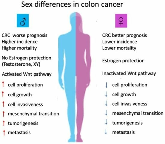
1.1. Sex Differences/Sexual Dimorphism/Gender Differences in Cancer

Sex differences and sexual dimorphism refer to biological and morphological differences, respectively, between males and females in various traits or characteristics. While sexual dimorphism refers to the form or appearances such as height and morphology (evolutionary adaptations), sex differences are primarily associated with biological features, and their impact on cancer is an exciting and rapidly developing area of ongoing research. These terms have been used interchangeably, and not without some misunderstanding, in the scientific literature with reference to male/female differences in cancer [1,2,3]. Gender differences, on the other hand, commonly refer to behavioural and lifestyle traits but are often confused with biological sex differences [4,5,6]. Several types of cancer have demonstrated differences in incidence, presentation, and outcomes between males and females [7,8]. Certain cancers are more prevalent in one sex compared to the other due to anatomical differences, for example, prostate cancer in males, and ovarian or uterine cancer in females, whereas others may be associated with hormonal and genetic factors such as breast cancer [9]. In a survey of the most recent data from the Global Cancer Observatory, it can be noted that the top 20 most common nonreproductive tissue cancers worldwide show sex differences both in incidence and mortality, with females showing lower age-standardized incidence and mortality for all cancers (excluding those of anatomical differences) and for all cancers in every global region surveyed (Figure 1). While hormone and genetic factors contribute to these differences, other nonbiological risk factors can increase the likelihood of incidence and mortality, including smoking and excessive alcohol consumption, particularly for lung, stomach, oesophagus, and liver cancer [10,11]. Here, we discuss the evidence for genomic and nongenomic biological actions of oestrogen underpinning biological sex differences in colon cancer. We focus on more recent reports and try to integrate current knowledge into a holistic understanding of the role of oestrogen, its receptors, and molecular targets, in order to explain this very complex phenomenon for which many mechanistic questions still remain unanswered.

1.2. Sex Differences and Regional Variances in Colon Cancer

Colon cancer, also known as colorectal cancer (CRC), is one of the most common types of cancer worldwide. The exact prevalence may vary depending on factors such as age, gender, geographical location, and lifestyle choices. According to the World Health Organization, colon cancer is the second-most commonly diagnosed cancer globally and the third-leading cause of cancer-related deaths (Figure 1). Sex differences showing a female advantage are is a feature of CRC [12]. Although the main risk factor for CRC is age (about 90% of patients are older than 50 years), sex differences in CRC are evident in age-matched male and female patients [13]. The age-standardized mortality rate for men is 50% higher (10.8 per 100,000 person-year) than for women (7.2 per 100,000 person-year) [14].
The CRC incidence rates can differ significantly between countries, but all countries show a male predominance in both incidence and mortality of CRC (Figure 2) [15]. Moreover, the trends for the next 20 years predict a continuing sex difference with more males than females affected by CRC both in its incidence and mortality (Globocan 2020, https://gco.iarc.fr, accessed on 29 July 2023) [16]. Certain risk factors can increase the likelihood of developing colon cancer, including a family history of the disease, a personal history of inflammatory bowel disease, a sedentary lifestyle, a diet high in processed meats and fat and low in fruits, vegetables, and fibre, smoking, and excessive alcohol consumption. It is important to note that advancements in early detection and improved treatment options have contributed to higher survival rates and better outcomes for individuals diagnosed with colon cancer. Regular screenings such as colonoscopies can help detect precancerous polyps or early-stage colon cancer, improving the chances of successful treatment. Biological sex differences, however, are too often a neglected factor in both clinical trials and treatment of colon cancer, although its added value to personalized medicine is incontestable [17]. Sex differences exist at multiple levels in colon cancer, and women have a lower risk of developing CRC than men. Females at a younger age are less likely to die from CRC than age-matched male patients, and certain types of CRC occur predominantly in women. The biological differences in CRC mortality are noticeable in the survival advantage of women during premenopause (18–44 y) compared to men of the same age or to older women post-menopause [18]. These premenopausal advantages and the observations that hormone replacement therapy (HRT) may also be protective in CRC [19,20,21] indicate a role for the female sex steroid hormone oestrogen in delaying the onset and reducing mortality in females with colon cancer [22,23]. Sexual disparity has also been shown in epidemiological studies to be an important factor in the site of onset and metastases in CRC [24]. Women show a higher frequency of right-sided tumours (proximal colon) than men who present more commonly with left-sided (distal colon) tumours [25].
Right-sided CRC tumours arise from the ascending colon and proximal two-thirds of the transverse colon, whereas left-sided CRC tumours arise from the descending and sigmoid colon and distal one-third of the transverse colon. The regional origin of proximal (right) or distal (left) colon in CRC can impact prognosis, with CRC of proximal origin being associated with a worse prognosis [26]. Right-sided proximal tumours are harder to detect and diagnose [27] and tend to be more advanced, larger, and poorly differentiated at first diagnosis [28]. Moreover, right-sided proximal tumours tend to be more aggressive and resistant to chemotherapy, which may confound the advantages conferred by oestrogen protection. These regional and sex differences in CRC prognosis may result from differences in cellular molecular subtypes between proximal and distal colon [29].
One highly significant candidate is the Wnt signalling pathway and its regional variation along the length of the intestine. There is evidence for Wnt signalling gradients along the intestinal tract and region-specific differences in Wnt responsiveness in CRC [30]. Differential Wnt gene expression profiles and signalling potential have been demonstrated along the colonic crypt–villus axis and throughout the length of the colon, particularly following β-catenin stabilization [31]. This regional variation in Wnt signalling could influence tumour susceptibility between the proximal and distal colon. Genes in the Wnt signalling pathway are more enriched in male compared to female CRC colonic cells and their enhanced sensitization in males may be related to the higher risk and lower survival rates observed in colon cancer in males [32]. Sex differences in CRC patient survival with a female advantage exist for the expression of the Wnt receptor gene FZD1. Thus, sex differences and differential expression between proximal and distal colon in Wnt receptor expression and signalling may influence its potential as a therapeutic target [33] Although proximal tumours are more frequent in females, the oestrogen-induced quiescence of Wnt/β-catenin signalling in female CRC [12] will favour normal growth, morphology, and epithelial cell differentiation, whereas a lower activation threshold for Wnt signalling in male CRC will promote cell proliferation, EMT, and tumorigenesis.

1.3. Oestrogen and Sex Differences in Colon Cancer

Oestrogen is the main sex hormone that controls physiological functions of the female reproductive system, as well as the development of secondary sexual characteristics during female puberty. The predominant circulating oestrogen in humans is 17β-estradiol (E2), which is the most physiologically relevant oestrogen during the female reproductive years. In human females during the reproductive years, the plasma levels of E2 fluctuate over the oestrous cycle, reaching peak concentrations in the follicular phase; 150 to 300 pg/mL (0.5–1 nM) 24 h before ovulation [34], while the highest likely E2 level in the plasma during pregnancy is 8 ng/mL (30 nM) [35]. Oestrogen plasma levels fall off dramatically post-menopause (>45 y), decreasing to below 30 pg/mL (110 pM), which is close to levels found in age-matched males (10–50 pg/mL). Oestrogen at subnanomolar concentrations has been shown to exert multiple sexually distinct physiological actions, with a female advantage in tissues and organs outside the reproductive system, including brain, skeleton, muscle, cardiovascular, immune system, intestine, kidney, liver, and pancreas to influence, respectively, memory, bone strength, cardiac, and skeletal muscle contractility, immunity, intestinal secretion, renal Na+ handling, blood pressure, and metabolism [36]. These pleiotropic effects of oestrogen are thought to underpin sex differences and reinforce female physiology, reproduction, longevity, and healthy aging [37]. The beneficial biological effects of oestrogen are lost and often only become apparent after the menopause [38] and may be restored with hormone replacement therapy [39], but only if started early, for example, as in treating cardiovascular disease [40]. Pathophysiological effects of oestrogen are observed in some cancers, notably breast, uterine, and cervical cancers [41], whereas in nonreproductive organ cancers, oestrogen can have protective effects against morbidity and mortality [42]. Oestrogen signalling via specific oestrogen receptors has emerged as a crucial factor in sex differences in the development and progression of colon cancer [12,22,23].

2. Oestrogen Receptors in Colon Cancer

Oestrogen receptors determine the biological sex differences, specificity, and transduction of the multiple signalling responses to oestrogen [43]. Probably the most important factor in sex differences in colon cancer development is ligand activation of oestrogen receptors (ER), primarily ERα and ERβ. These receptors are expressed in both males and females but can have differential expression levels and activity patterns, depending on circulating levels of E2, as well as prognostic value in CRC [44]. The role of oestrogen in CRC progression is dependent on the relative abundance of the ER subtypes and their hormone responsiveness. The colonic crypt epithelium expresses both ERα and ERβ [45]. Positional differential expression of ERs has been reported along the colon length and within the crypt axis. ERα is expressed more highly at the base of the crypt of the proximal colon, while ERβ expression is predominant in the midsection of the crypt and in the apical surface cells [46]. This spatial partitioning of ER isoform expression indicates antagonistic roles for ERα and ERβ in transducing differential effects of oestrogen on the physiological function of the epithelial cells located at different sites along the crypt. For example, progenitor cell proliferation at the base of the crypt gives way to enterocyte differentiation in the midsection and shedding of senescent or apoptotic cells at the lumen surface. There is strong evidence that ERβ is more highly expressed in colon cancer tissues from females compared to males, while ERα expression levels can vary among the sexes [44,47]. ERβ has been proposed as a tumour suppressor in CRC, and ERβ Erβ expression is selectively lost during tumour progression through methylation-dependent gene silencing [48]. Thus, the ratio between ERα and ERβ expression and balance in their cell signalling may contribute to sex differences in CRC [49,50].

2.1. Nuclear Oestrogen Receptors in Colon Cancer

The two main subtypes of ERs are ERα (encoded by oestrogen receptor 1, ESR1) and ERβ (encoded by oestrogen receptor 2, ESR2), which are differentially expressed in normal colon tissue as well as in colon cancer cells. These two oestrogen receptor subtypes are often referred to as canonical or nuclear ERs, eliciting latent genomic responses to oestrogen and working in an antagonistic fashion on cell biology [51]. The natural ligand for all ERs is the biologically active form of oestrogen, 17β-estradiol (E2). Nuclear ERs play a complex role in colon cancer, influencing various aspects of tumour development and progression [42]. Colon cancer cells expressing ERs can be hormone-responsive, meaning they can respond to oestrogen stimulation. Oestrogen can directly bind to ERs in these cells and trigger a cascade of signalling events leading to changes in gene expression, protein synthesis, and cell behaviour (Figure 3). It is worth repeating that the specific roles and effects of ERs in colon cancer can vary depending on factors such as the subtype of ER (ERα vs. ERβ), their relative expression levels, and the interplay with other signalling pathways. For example, while ERα has been associated with promoting tumour growth and progression, ERβ has been suggested to have a potentially protective or inhibitory effect on colon cancer development [51,52]. Importantly, nuclear ERs are proven prognostic and therapeutic targets in colon cancer [53,54].

2.2. Membrane Oestrogen Receptors in Colon Cancer

Membrane oestrogen-sensitive receptors have been implicated in CRC [12,23,42], which, unlike the classical nuclear ERs, involve oestrogen-liganded membrane-initiated cell signalling pathways (Figure 3) [55]. Membrane-initiated oestrogen responses in colonic epithelium are typified by rapid nongenomic actions on protein kinase cell signalling pathways, intracellular Ca2+ activity, and ion channel function [56], which distinguish these events from the more latent genomic responses to oestrogen in a wide range of normal and cancerous cell types. The presence and role of membrane oestrogen receptors in colon cancer, and cancer in general, are a hot topic of ongoing research and have been the subject of dedicated international meetings continuously since 1992 (RRSH, FASEB SRC Meetings) [57,58,59]. The two most well-characterized membrane receptors for oestrogen are membrane ERα (mERα]) [60] and G protein-coupled oestrogen receptor 1 (GPER1), also known as GPR30 [61], which are discussed in detail below.

3. Genomic and Nongenomic Oestrogen Signalling Pathways in Colon Cancer

Steroid hormone signalling occurs via nongenomic membrane-initiated pathways and genomic nuclear transcriptional pathways working in a coordinated fashion (Figure 3) [62,63]. Oestrogen can trigger cellular signalling in both healthy and cancerous colonic epithelial cells via genomic and nongenomic mechanisms [4,14,42] in a sexually differentiated manner, which also shows dependency on the stage of the oestrous cycle [64]. Both genomic and nongenomic oestrogen signalling pathways in the colon are cell-type-specific and may differ among healthy and cancerous colonic tissues. Genomic oestrogen signalling pathways are characterized by long latency (hours/days) and involve nuclear ERs that bind oestrogen in the cytosol which then dimerize and translocate to the nucleus to bind to specific DNA sites to trigger synthesis of proteins which regulate cell growth, differentiation, and proliferation [65]. Nongenomic oestrogen signalling, on the other hand, is characterized by rapid onset of cell signalling responses (s/min) involving oestrogen binding to a membrane receptor, which, through phosphorylation reactions, can transactivate other membrane receptors and trigger a myriad of cellular signalling pathways impacting upon ion channels, protein kinases, and transcription factors to extend its rapid actions into genomic events [42,66]. The genomic and nongenomic receptor signalling pathways of oestrogen in colonic crypt cells are summarized in Figure 3, showing both protective and exacerbating arms of these pathways in colon cancer.

3.1. Genomic Mechanisms of Oestrogen Signalling in Colon Cancer

The genomic actions of oestrogen in colon cancer are not as well studied compared to its effects on other hormone-responsive cancers like breast or ovarian cancer. While oestrogen receptors have been found in the colon and rectal tissue, the specific role and mechanisms of oestrogen and ERs in colon cancer development and progression between the sexes are still not well understood. Several studies, however, over the past 20 years have suggested potential genomic actions of oestrogen in colon cancer that could underpin sex differences: oestrogen may promote or inhibit cell proliferation and reduce or stimulate cell death (apoptosis) depending on the ER subtype expression in colon cancer cells (Figure 3) [67]. E2–ERα interactions can activate signalling pathways, such as the PI3K/Akt pathway, leading to increased CRC cell growth and survival [68]. Oestrogen has been implicated in promoting angiogenesis, the formation of new blood vessels, which is essential for tumour growth and metastasis [69] and can upregulate the expression of vascular endothelial growth factor (VEGF), a key factor involved in angiogenesis [70]. Oestrogen may also influence epigenetic modifications in colon cancer cells [71] to effect post-translational DNA methylation patterns, histone modifications, and chromatin remodelling, potentially impacting gene expression and cellular behaviour [72]. Nuclear oestrogen receptors may also play a role in inducing or influencing the process of epithelial–mesenchymal transition (EMT) [73], which is involved in tumour metastases in colon cancer [74]. Oestrogen signalling through ERα can regulate the expression of genes associated with EMT, leading to increased cell motility and invasiveness in prostate and breast cancer cells [75,76] and in CRC [12]. Oestrogen signalling has also been found to impact the function and activation of immune cells, including macrophages and T-cells, and regulate the production of cytokines and other immune-related molecules [77] that can affect the tumour microenvironment and immune surveillance against cancer cells [78]. In this regard, oestrogen via ERβ has been shown to modulate the immunogenicity of the tumour microenvironment and immune responses in colon cancer [79].

3.2. Nongenomic Mechanisms of Oestrogen Signalling in Colon Cancer

It has been known for over two decades that oestrogen can exert sexually segregated rapid nongenomic actions independent of gene transcription on cell signalling in colon [80]. Nongenomic actions of oestrogen in CRC involve signalling pathways that do not require changes in gene expression or protein synthesis [12,23]. Although the nongenomic actions of oestrogen in colon cancer are less well studied compared to its genomic actions, here, we outline some of the potential mechanisms (Figure 3). Activation of membrane receptors: Oestrogen binding to ERα or GPER can trigger intracellular signalling cascades, including activation of protein kinase pathways, calcium mobilization, and stimulation of cyclic adenosine monophosphate (cAMP) production [81]. Rapid kinase activation: Oestrogen can rapidly (s/min) activate various kinases, such as mitogen-activated protein kinases (MAPKs), including extracellular signal-regulated kinase (ERK), c-Jun N-terminal kinase (JNK), and p38 MAPK [82]. These kinases play crucial roles in cell proliferation, survival, and migration, which are important in colon cancer progression [83]. Ion channels: Oestrogen modulates the activity and expression of ion channels in colon cancer cells, such as calcium and potassium channels, leading to changes in intracellular ion concentrations and membrane potential which can affect cell proliferation, apoptosis, and migration [84,85,86]. Activation of second messenger systems: Oestrogen can stimulate the production of second messengers, including cyclic adenosine monophosphate (cAMP) cyclic guanosine monophosphate (cGMP) and inositol trisphosphate (IP3) [87]. These signalling intermediates can regulate various cellular processes, including gene expression, protein phosphorylation, and intracellular calcium release [88]. It is important to note that both genomic and nongenomic actions of oestrogen can contribute to the overall effects of oestrogen on colon cancer cells. The balance between these actions and the interplay with other signalling pathways determine the ultimate impact of oestrogen on colon cancer development and progression.

3.3. Cooperativity between Genomic and Nongenomic Oestrogen Signalling in Colon Cancer

Multiple studies in a wide variety of tissues (brain, vascular, epithelial) have shown that genomic and nongenomic cellular responses to oestrogen are not mutually exclusive and can cooperate to produce synergistic effects in physiological functions and in cancer biology (Figure 3) [89]. In this context, it is important to note that a strict dichotomy does not exist between membrane nongenomic and nuclear genomic actions of oestrogen, and evidence exists for cross-talk and integration of these rapid and latent pathways to amplify biological responses to oestrogen [90]. Nongenomic actions of oestrogen have been shown to be both permissive and potentiating for genomic responses [91]. This type of cross-talk can be rapidly initiated by oestrogen actions through membrane ERs to transactivate growth factor receptor tyrosine kinases (EGF and IGF-I receptors) [92], which can produce rapid activation of ERK MAPK and phosphorylation of cytosolic ER to allow its translocation into the nucleus [93] and also cause phosphorylation and recruitment of coactivators (AP-1, Sp-1) to the nuclear transcriptome to amplify the genomic response [94]. Oestrogen signalling through membrane oestrogen receptors can also involve activation (phosphorylation) of protein kinase targets to induce gene transcription and latent nuclear transcriptional activity via ERK, MAPK, c-fos, PI3K/AKT, CREB, and JNK [95,96]. Conversely, genomic actions may amplify nongenomic responses to oestrogen in a sex-specific manner, for example, by stimulating the transcription of protein kinase intermediates of the membrane-initiated signalling pathways in colonocytes [97]. Thus, nuclear ERs are necessary for the expression of proteins that transduce oestrogen effects at the membrane [98]. An important caveat, therefore, in the debate on genomic versus nongenomic oestrogen responses is the accumulating evidence that these are not standalone independent events but show integration and cooperativity and, if considered separately, do not encompass the full range of oestrogen actions in physiology nor in cancer. Another important principle is that labelling nongenomic steroid hormone responses as being solely “rapid” no longer holds true, as latent genomic events may be initiated by earlier membrane–cell signalling [99,100]. Thus, nongenomic and genomic actions of oestrogen can be integrated in targeting cell proliferation pathways in colon cancer biology.

4. Oestrogen Signalling via ERα and ERβ in Colon Cancer

When oestrogen binds to ERα in the cytosol, the hormone receptor complex dimerizes and translocates to the nucleus where it interacts with specific DNA sequences, oestrogen response elements (ERE), or non-ERE transcription factors, such as c-Jun and c-Fos of the activating protein-1 complex (AP-1), and transcription factor specificity protein 1 (SP1) and NFκB, which extend E2–ERα cell proliferation and proinflammatory actions in colon cancer cells [101]. ERα activation can also initiate signalling pathways that promote cell cycle progression, such as the PI3K/Akt and MAPK/ERK pathways [102]. These pathways, in turn, can stimulate cell growth and survival. From these studies, we can reasonably conclude that oestrogen signal transduction via ERα is protumorigenic in CRC (Figure 3).
In contrast, oestrogen signalling through ERβ produces antitumorigenic cell signalling responses in a broad range of cancers including CRC by repressing ERα transcription and activating antiproliferative cell signalling pathways (Figure 3) [103]. ERβ upon binding to its ligand oestrogen, dimerizes and translocates to the nucleus where the E2–ERβ complex transcriptionally upregulates target genes which, unlike E2–ERα, promote proapoptotic and antiproliferative responses in CRC. The E2–ERβ complex binds to DNA elements such as ERE or AP1 which activate the FOXO3a gene. Activated FOXO3a in turn transcriptionally upregulates PUMA, p21, and p27, which have been shown to induce apoptosis of CRC cells [104]. ERβ also inhibits the expression of cell proliferating genes such as c-Myc and p45Skp2 [105]. Moreover, some of the EMT and metastasis genes, such as β-catenin, Slug, and Twist, are inhibited by ERβ [106]. Nuclear ERβ can also exert proapoptotic responses in colon cancer cells through increased Caspase-3 activity [107] and inhibit cell proliferation by locking the cell cycle in G1-S phase [108]. There is evidence that ERβ may also inhibit colon cancer cell growth through autophagy mediated by suppression of the mammalian target of rapamycin (mTOR) through Cyclin-D1 degradation [109,110]. In addition to these antitumorigenic actions, E2–ERβ may have immunosuppressive effects in CRC [111] as well as preserving an epithelial phenotype through stimulating the expression of tight junction proteins occludin-1 and JAMA [112] and inhibiting dedifferentiation and epithelial–mesenchymal transition via upregulation of E-cadherin and α-catenin while inhibiting β-catenin [113]. E2–ERβ may also induce microRNA protection by repression of the oncogenic prospero homebox 1 (PROX1) through the upregulation of miR-205, which inhibits colon cancer cell migration and invasiveness [114]. A protective role of ERβ in reducing colon crypt proliferation and inflammatory responses resulting from a high-fat diet has recently been demonstrated both in male and female mice [115].
There is evidence for cross-talk between the ER subtypes, the most important for CRC being the inhibition of ERα transcription by ERβ with oestrogen binding [116] and the negative regulation of the expression of ERα by ERβ [117] as colonocytes differentiate to a mature tight-junction epithelium [118]. Increased ERβ expression could also lead to ERα–ERβ heterodimerization, thereby skewing the expression pattern of target genes from proliferative, antiapoptotic towards an antiproliferative, proapoptotic, and antitumorigenic profile [46]. Disruption of ERβ expression, but not of ERα, increased intestinal neoplasia and promoted tumourigenesis in APC-/- mice [119]. A combined high ERβ expression together with negative ERα expression was found to be correlated with a better prognosis for CRC patients [120]. ERβ expression predominates over ERα in the normal healthy colon and in the initial stages of adenocarcinoma, with progressive loss of ERβ and increased ERα expression observed in colon biopsies in later stages of tumour development [38]. Moreover, the loss of ERβ is associated with enhanced CRC proliferation potential [121], leading to the hypothesis that high ERβ expression may not only be protective against developing CRC but also a prognostic marker and molecular target in the treatment of colon cancer [122,123]. Indeed, a predominant expression of ERβ may show sexual differentiation for its protective role in the early stages of CRC [124].

4.1. Nonligand Activation of Nuclear ERs in Colon Cancer

In addition to ligand-binding activation, nuclear ERs can be phosphorylated and transactivated by EGFR activated tyrosine kinases without requiring binding to oestrogen. For example, EGFR can activate the Ras/Raf/MAPK pathway, which phosphorylates ERs, resulting in dimerization and ligand-independent activation of target gene expression [125]. Moreover, the membrane HER2 receptor (erbB-2 receptor tyrosine kinase 2) when bound to EGFR can also activate tyrosine kinases signalling pathways RAS/RAF/ERK, PIK3K/AKT/mTOR, JAK/STAT3 producing a hyperactivation of mitogenic signals leading to uncontrolled cell proliferation and tumorigenesis in CRC [126]. Currently, there are no studies reported in the literature directly linking nonligand activation of ERs to EGFR/MAPK signalling. However, ligand (estradiol) activation of membrane and nuclear ERα has been shown to be linked to EGFR/MAPK signalling to drive proliferation, invasion, and angiogenesis in CRC [42,127].

4.2. Membrane Oestrogen Receptors mERα and mERβ in Colon Cancer

While ERα is traditionally recognized as a nuclear receptor that regulates gene expression, detailed evidence accumulated over the past 20 years has demonstrated that a small percentage (approximately 1%) of ERα can also be localized to the cell membrane in certain contexts to produce physiological and clinically relevant biological responses [128]. The membrane ERα (mERα) was first discovered in 1999 [129] and characterized in breast cancer cells by Levin and colleagues [130,131]. The molecular identity was shown to be a palmitoylated variant of the full-length (66 kDa) nuclear ERα receptor which allowed its tethering at the cell membrane in a caveolin-1 signalosome [132,133]. The understanding of the role of the mERα in physiology and cancer biology was hampered by the lack of specific inhibitors and agonists which could distinguish E2–mERα ligand binding from nuclear ERα signal transduction [134]. The nongenomic and rapid actions (s/min) of oestrogen on protein kinases, intracellular calcium, and ion channel activity in colonic crypts [135,136,137] distinguish it from canonical genomic E2–ERα signal transduction which shows typical long latency of hours/days to generate transcriptional responses [138]. It is important to note that the rapid membrane-initiated responses to oestrogen can be elicited at physiological subnanomolar concentrations of oestrogen such that dose–response is of little value in distinguishing nongenomic from nuclear E2–ERα actions [139]. One way to overcome this difficulty is the use of oestrogen analogues which penetrate poorly, if at all, the cell membrane, or the generation of nuclear excluded ERα mutants [140]. In this regard, certain specific membrane-impeded analogues of oestrogen such as E2–BSA [141] and oestrogen dendrimer conjugates (EDC) [142], which do not enter the cytosol to bind nuclear ERα, have been shown to replicate the rapid nongenomic actions of free unbound oestrogen (17β-estradiol). The most significant advance, however, in understanding the physiological and pathological roles of mERα have resulted from the generation by the Levin group of selective mouse models with membrane-only mERα (MOER) or nuclear-only ERα (NOER) expression [143]. Mutations of the palmitoylation site of ERα have also provided a useful tool to dissect membrane-initiated and nuclear actions of oestrogen [144]. These studies have shown the absolute requirement for mERα expression in transducing rapid actions of oestrogen on protein kinases and tyrosine kinase signalling pathways in cancer cell proliferation but not in the development of reproductive organs and tissues [143,144,145]. The presence of ERα on the cell membrane, in addition to its primarily nuclear localization, expands the range of oestrogen actions through combined nongenomic and genomic regulation of cell differentiation and proliferation through an expanded signalosome [146,147].
Regarding the role of membrane ERα in colon cancer, research in this specific area is still evolving, and the understanding of its implications is not yet fully established. However, many of the signalling pathways regulated by membrane oestrogen actions in colon (EGFR, ERK-MAPK, PI3K-Akt, and Wnt) are involved in cell proliferation, survival, and migration in colon cancer cells (Figure 3) [12]. For example, activation of membrane ERα in colonic epithelial cells isolated from females has been associated with the rapid activation of mitogenic signalling cascades, including the MAPK pathway, PI3K/Akt pathway, and Src kinase signalling [80,88,148,149], which can promote cell growth and survival. Additionally, membrane ERα has been implicated in modulating epithelial–mesenchymal transition in breast [150] and colon cancer cells [151], a process involved in CRC tumour invasion and metastasis [152].
The current data indicate that mERα is the primary endogenous ER mediator of rapid E2 responses, although a membrane ERβ (mERβ) is also found to be co-expressed with mERα in cancer cells to regulate cell proliferation [153]. The role of mERβ is less well studied in cancer biology, although ERβ may be present at the cell membrane in a palmitoylated form in colon cancer cells to inhibit cell proliferation [154]. Studies have demonstrated that mERβ can produce rapid nongenomic actions of oestrogen on ERK and JNK kinase activity when expressed in Chinese hamster ovarian cells [155], while other studies have shown E2–mERβ to rapidly activate p38 MAPK in human colon cancer cells [156]. The membrane ER subtypes appear to mimic their respective nuclear ER responses to oestrogen, i.e., proproliferative, protumorigenic effects via nuclear ERα and mERα while conversely producing antiproliferative, antitumorigenic effects via both nuclear ERβ and mERβ. In this way, oestrogen nongenomic interactions with membrane ERα and membrane ERβ can modulate cell proliferation, apoptotic pathways, and cell death in CRC [157].

4.3. Oestrogen Signalling via Truncated ERs in Colon Cancer

Several splice variants of full-length ERs have been reported in various healthy tissues and cancers [158,159]. These truncated ERα and ERβ receptor proteins arise from mutations in ESR1 and ESR2 genes but cannot form homodimers or recruit cofactors like full-length ERs [160]. Truncated ERs may form inactive heterodimers with full-length ERs [161] and collaborate with other oestrogen receptors such as GPER [162] to modulate proliferative and inflammatory responses in cancer. The best studied truncated ER has been ERα36 (36 kDa) in breast and gastric cancers [163,164], which transduces estrogenic nongenomic signalling to promote cell proliferation and metastatic potential [165] Apart from one study reporting decreased ERα36 mRNA expression with advanced Stage CRC [166], the relevance of truncated ERs to oestrogen signalling and sex differences in CRC is unknown and merits further investigation.

5. Oestrogen Signalling via G Protein-Coupled Oestrogen Receptor in Colon Cancer

G protein-coupled oestrogen receptor (GPER), also known as GPR30, first reported in 2005 [167], is a seven-transmembrane receptor that mediates membrane-initiated oestrogen signalling in a wide range of tissues [168]. Initially identified as an alternative oestrogen receptor in breast cancer [169], GPER signalling has emerged as a significant player in colon cancer [170], contributing to various aspects of tumour progression and estrogenic cell signalling responses [171]. While many studies over the past twenty years support an oestrogen-ligand receptor role for GPER [172,173], the involvement of GPER in transducing nongenomic actions of oestrogen in vivo has been challenged [174,175]. Several recent studies, however, have demonstrated the functional expression of GPER in colon cancer cells, which may vary among different colon cancer subtypes and individual tumours [176]. The role of GPER in CRC has garnered much interest as its expression predominates in colon cancer after the loss of nuclear ER, in particular after the loss of ERβ, which has been reported to negatively regulate GPER expression in breast cancer [177]. While the oestrogen protective effects in CRC have been mainly attributed to ERβ, its expression is lost during CRC progression, and this raises the possibility for a role in sex differences of GPER, which remains expressed after ERβ loss in CRC [178]. Chronic mucosal inflammation has been proposed as a precursor of CRC and it is interesting to note that GPER expression shows sex dependence in inflammatory bowel disease (IBD) [179] and may transduce oestrogen protective effects on mucosal barrier function in IBD [180]. The potential therapeutic implications of targeting GPER signalling in cancer have been recently reviewed. This includes the use of GPER agonists or antagonists, alone or in combination with other therapies [181].
GPER has been reported to have both tumorigenic and antitumorigenic roles in cancer progression [182,183]. Some studies have shown a protective role for GPER in CRC [184] and its activation has been reported to inhibit cell proliferation in CRC cell lines [185]. Furthermore, a low expression of GPER in CRC was associated with poor patient survival [171]. In contrast, other studies indicate GPER to be protumorigenic in CRC via oestrogen activation by steroid sulfatase [186] and to stimulate cell proliferation in CRC cell lines not expressing nuclear ERs [171]. In support of this hypothesis, GPER expression was shown to be upregulated and stimulated by MAPK signalling in mycotoxin-induced growth of colon cancer cells [187]. In addition, GPER can promote chromosomal instability in CRC, leading to neoplastic transformation and tumour development [188]. The impact of GPER signalling on angiogenesis may also be determinant in its tumour promoter actions [189,190]. GPER may have differential protective or exacerbating effects on CRC tumourigenesis, depending on the expression of ER and activation of cell proliferation signalling pathways.

Hypoxia and GPER Signalling in Colon Cancer

There is evidence that the divergent roles of GPER in CRC as being either protective or tumorigenic are dependent on the stage of the cancer and the level of hypoxia in the tumour microenvironment and on sex-specific factors influencing hypoxic signals [184]. Moreover, the actions of GPER in the regulation of vascular endothelial growth factor (VEGF) and hypoxia-inducible factor 1-α (HIF-1α) in CRC show sex dependence (Figure 3) [178].
Hypoxia is a key factor in promoting tumour growth through cell signalling involving HIF-1α [191] and its target VEGF, which are associated with poor clinical outcomes in CRC [192]. In a detailed study of the functional consequences of oestrogen actions within the hypoxic CRC cell microenvironment, Bustos et al. [178] found the pro- and antitumorigenic potential of GPER in CRC cell lines to be dependent on the level of oxygen exposure. Under normoxic conditions, oestrogen and the GPER agonist G1 both suppressed CRC cell proliferation. Under hypoxic conditions, however, GPER activation produced the opposite functional effect, with both oestrogen and G1 enhancing CRC cell proliferation, whereas the GPER antagonist G15 inhibited proliferation. Oestrogen treatment enhanced the hypoxia-induced expression of HIF-1α and VEGF, but repressed HIF-1α and VEGF expression under normoxic conditions. The expression or repression of VEGF by oestrogen were mediated by a GPER-dependent mechanism. Thus, GPER is essential in transducing the normoxic antiproliferative effects of oestrogen as well as its hypoxic proliferative actions in CRC cells. The latter response may be amplified by upregulation of GPER expression following exposure to hypoxia and oestrogen. Another protumorigenic amplification factor in the GPER response is the oestrogen-modulated gene, Ataxia Telangiectasia Mutated (ATM), which was shown to be repressed in hypoxia via GPER signalling [178]. Loss of ATM expression is associated with poor survival in CRC [193] and an increase in phosphorylated ATM protein levels has been observed in hypoxic colon cancer cells [194]. The modulation of ATM expression by GPER in low oxygen tension and the sensitivity of its expression to oestrogen in CRC provides an additional mechanism for protumorigenic actions of oestrogen via GPER in colon cancer under hypoxic conditions. Thus, it is important to take into account the CRC stage and tumour microenvironment when interpreting the role of E2–GPER interactions in colon cancer tumorigenesis, sex differences, and patient survival/treatment [170].
The involvement of GPER in CRC patient survival displays clear sex differences. In a cohort of 566 CRC patient tumour samples, GPER expression was significantly associated with poor survival in CRC Stages 3–4 females but not in the stage-matched male population [178]. Since a hypoxic tumour microenvironment is associated with late stages in CRC, we may conclude that sex differences in this case are underpinned by E2–GPER tumorigenic actions on HIF-1α/VEGF activation and on ATM suppression under hypoxia. The antiproliferative effects of E2-GPER signalling in normoxia may explain the observations of protective effects of GPER expression on CRC survival in the early stages of cancer development.

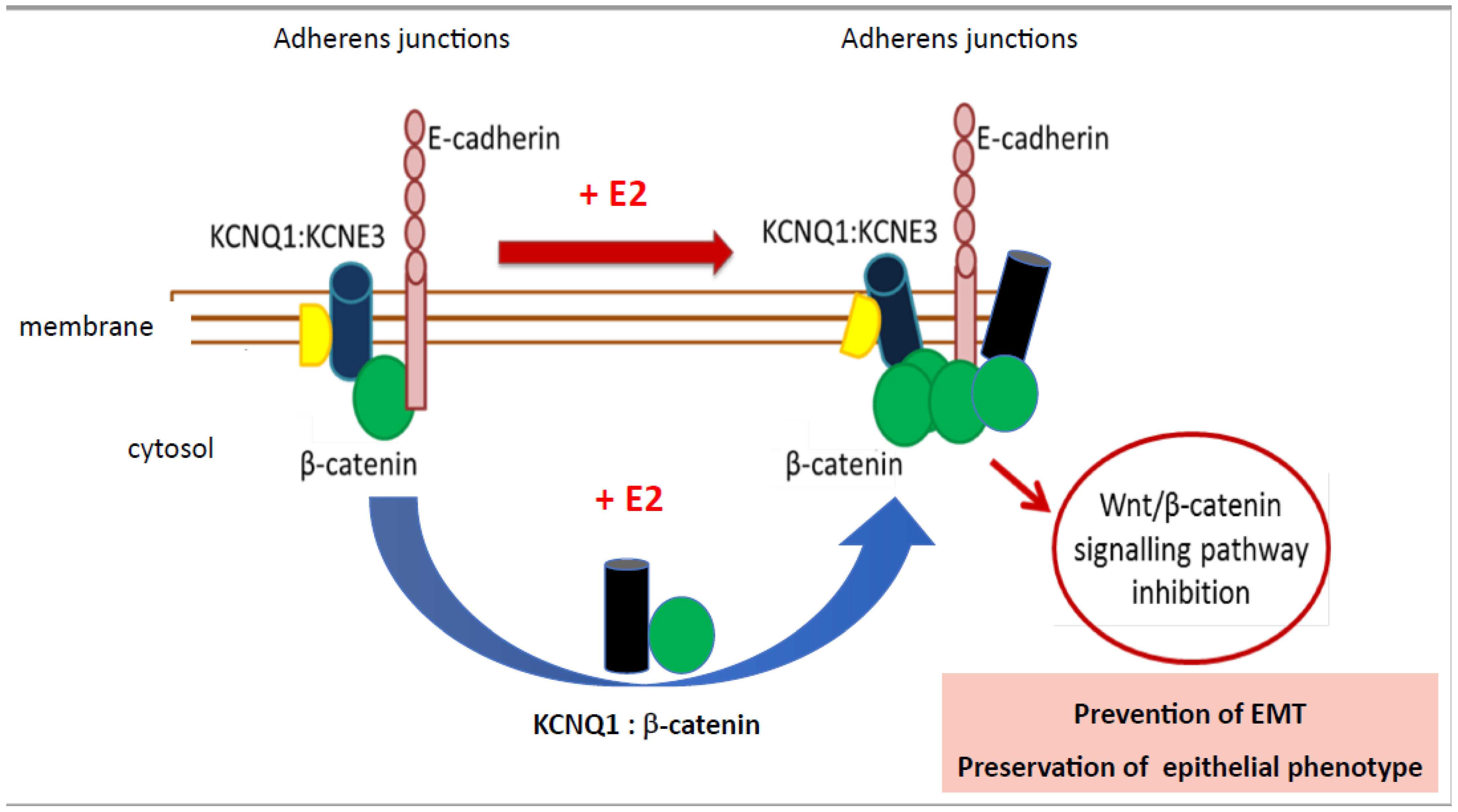
6. Oestrogen Regulation of Wnt/β-Catenin Signalling in Colon Cancer

Wnt/β-catenin signalling plays a key role in various biological processes, including embryonic development, tissue homeostasis, and cell proliferation [195,196]. Dysregulation of the Wnt/β-catenin signalling pathway has been strongly associated with the development and progression of colon cancer [197]. The Wnt/β-catenin signalling pathway is commonly hyperactivated in colon cancer and plays a crucial role in CRC development and progression [198]. Oestrogen has been shown to modulate this pathway in a sex-dependent manner in CRC [199] and in breast and endometrial cancers [200,201] through reciprocal interaction between ERα and β-catenin. In reproductive tissues, oestrogen promotes cell proliferation via ERα stimulation of β-catenin nuclear translocation [202]. In contrast, other studies indicate that ERα is inhibitory for Wnt/β-catenin-mediated proliferation and neoplasia in nonreproductive tissues, for example, in liver cancer [203]. Moreover, ERβ was reported in the latter study to have no role in oestrogen modulation of Wnt/β-catenin signalling. These studies suggest that oestrogen may have an inhibitory effect on Wnt/β-catenin signalling in nonreproductive tissue cancers in females, potentially contributing to sex differences in CRC incidence and outcomes. But how does this work in CRC and what are the factors that determine whether oestrogen will exert a protective or exacerbating role via Wnt/β-catenin pathway signalling in CRC, in particular when the expression of ER is lost with advancing tumorigenesis? To answer this question, we must first understand the possibility of cross-talk with other receptors and signalling pathways. The Wnt signalling pathway interacts with various signalling pathways, particularly K+ channels, implicated in colon cancer. Interaction between the Wnt/β-catenin pathway and the oestrogen-regulated K+ ion channel KCNQ1 in CRC is a major molecular mechanism that displays sex differences in the regulation of CRC cell proliferation and epithelial–mesenchymal transition [12].

6.1. Oestrogen Regulation of Wnt-KCNQ1 Interactions in Colon Cancer

KCNQ1 is a voltage-gated K+ channel (Kv7.1) expressed in the basolateral membranes of colonic crypts where it functions to provide the electrical driving force for Cl secretion [204]. In colon, the KCNQ1 channel is coexpressed with the β-regulatory subunit KCNE3, which greatly increases the ionic conductance of the channel and confers voltage and cAMP sensitivity to the channel [205]. KCNQ1 has been shown to have an antitumorigenic role in many gastrointestinal (GI) cancers, including colon cancer [206]. Moreover, relapse-free CRC patient survival was positively associated with high KCNQ1 gene expression, which displayed sex-dependence for a female advantage [207]. KCNQ1 channels modulate Wnt signalling in a wide number of GI cancers [208] and there is strong evidence for bidirectional interaction between KCNQ1 and β-catenin in normal healthy colon and in colon cancer cells [207]. Oestrogen regulates KCNQ1:KCNE3 channel function by uncoupling KCNQ1 from KCNE3 via PKCδ-dependent phosphorylation of KCNE3 at residue Ser82, which destabilizes the KCNE3:KCNQ1 channel complex [209]. The uncoupling of KCNQ1 from KCNE3 promotes KCNQ1 endocytosis and recycling in colonic crypts [210], which allow KCNQ1 to leave the plasma membrane and bind cytosolic activated β-catenin. KCNQ1 then returns β-catenin to the cell membrane to trap it in a complex with E-cadherin at adherens junctions (Figure 4) [207]. In this way, KCNQ1 anchors β-catenin at the plasma membrane, stabilizing adherens junctions, and promoting cell–cell adhesion. The association of KCNQ1 with β-catenin at the cell membrane is essential to preserve a well-differentiated epithelial phenotype by maintaining a stable KCNQ1: β-catenin:E-cadherin complex at adherens junctions and preventing epithelial–mesenchymal transition [211]. Trapping β-catenin with KCNQ1 at the cell membrane has been shown to retard the nuclear translocation of β-catenin and prevent the transcriptional activation of proliferative genes [207]. These observations suggest that oestrogen-regulated KCNQ1 channel quells the Wnt:β-catenin nuclear signalling pathway to suppress CRC cell proliferation and EMT, but only in females [212].

6.2. Oestrogen Regulation of Wnt Receptor Oncogenic Signalling in Colon Cancer

The receptor tyrosine kinase pathway, particularly the EGFR pathway, can cross-regulate Wnt signalling in colon cancer [213]. Additionally, the Transforming Growth Factor-β (TGF-β) pathway can modulate Wnt signalling through Smad proteins [214], all of which could amplify the cell proliferation responses in CRC. In contrast, a recent study has provided evidence for a role of GPER in protecting against CRC progression by selectively reducing the oncogenic effects of hyperactive Wnt/β-catenin signalling pathways in CRC [215]. Firstly, sex differences were observed in the gene expression of the Wnt receptor FZD1 (Frizzled 1) in Kaplan–Meier survival analyses across multiple CRC patient gene microarray datasets. High expression of FZD1 was associated with poor relapse-free survival rates in the male but not in the female CRC population. Secondly, activation of GPER with the G1 agonist prevented the Wnt-pathway-induced upregulation of the JUN oncogene. These novel findings indicate a mechanistic role for GPER in protecting against CRC progression in females by selectively reducing the tumorigenic effects of Wnt/β-catenin oncogenic signalling pathways.

7. Oestrogen Regulation of Epigenetic, Microbiome and Metabolic Factors in Colon Cancer

Other emerging factors underpinning sex differences in CRC include microRNAs and the gut microbiota. MicroRNAs (miRNAs) are small noncoding RNA molecules that regulate gene expression by targeting specific messenger RNAs for degradation or translational repression. Differential expression of miRNAs has been observed between males and females in CRC [216,217]. Oestrogen can modulate the expression and activity of specific miRNAs, which in turn influence the expression of genes involved in CRC development [218]. These sex-specific miRNA profiles may contribute to the oestrogen-mediated sex differences in CRC. This has been shown for miR-30c-5p expression which was associated with better survival in females and was downregulated in males [219]. Several miRNAs are currently under investigation for therapeutic applications in CRC [220].
Recent studies suggest that the gut microbiota can influence CRC development and responses to oestrogen [221]. The composition and function of the gut microbiota can differ between males and females [222], potentially affecting oestrogen metabolism and signalling. Thus, another contributory factor to sex disparities in CRC is the oestrogen–gut microbiome axis [223]. Microbial metabolites produced by the gut microbiota may modulate oestrogen receptor activity and alter oestrogen levels, contributing to sexual differentiation in CRC [224]. In addition, E2 may alter the gut microbiota to reduce the risk of developing CRC [225].
Another potential factor contributing to CRC sex differences is oestrogen metabolism. The metabolism of oestrogen is an important process that can influence its bioavailability and activity. In females, oestrogen is predominantly metabolized by cytochrome P450 enzymes, including CYP1A1 and CYP1B1, which convert estradiol to 2-hydroxyestradiol (2-OHE2) [226]. In males, oestrogen is mainly metabolized by CYP1A2 and CYP3A4, leading to the formation of 4-hydroxyestradiol (4-OHE2) [227]. The different metabolic pathways can result in distinct oestrogen metabolite profiles, which may contribute to sex variances in CRC susceptibility. There is some evidence that testosterone can exert neoplastic actions in CRC [227] and strong evidence for the presence of upregulated genes on the Y chromosome that contribute to colorectal cancer in males by driving tumour invasion and aiding immune escape [41,228,229].

8. Comparison of Oestrogen and Testosterone Signalling in Colon Cancer

Over 90% of CRC cases are sporadic, arising from transition from normal mucosa to adenoma, followed by the development of carcinoma usually involving mutations in antiproliferative genes, which include APC (adenomatous polyposis coli), and proliferative tumorigenic genes, such as K-ras and p53. CRC is not solely a hormone-dependent cancer compared to predominantly oestrogen- or androgen-driven cancers such as breast or prostate, respectively. However, oestrogen and testosterone have been implicated in modulating the initial adenoma and mesenchymal transitions through effects on tumour promoter genes, and on inflammatory, apoptosis, and angiogenesis signalling pathways.
Sex-based prevalence and mortality of colon cancer have been observed in many animal studies where ovariectomized rats and mice are more prone to develop CRC while castrated male animals have a lower risk in developing CRC in response to mutations in the Apc tumour-suppressor gene [230]. These studies indicate that female sex hormones are actively protective in CRC whereas male sex hormones can actively promote CRC [231]. More precisely, oestrogen protection appears to be conferred through cell signalling via ERβ receptors [232], while the nuclear androgen receptor A (AR-A) transduces the exacerbating effects of testosterone [233]. The B-isoform of the nuclear AR (AR-B) may confer protection in CRC as its loss is associated with adenoma formation, although the membrane-associated AR (mAR) may also confer protection in CRC [234]. These protective/exacerbating effects of oestrogen or androgens in CRC are also observed in human subjects supplemented with oestrogen therapy [235] or testosterone [236]; there is debate, however, on the exact role of testosterone in CRC [237]. Although the precise oestrogen and testosterone signalling pathways underpinning sex differences in CRC still remain elusive, we can make certain observations supported by the literature and highlight testable hypotheses on potential pathways that differentiate males and females in CRC outcomes, as summarized in schematic form in Figure 5.
Oestrogen may offer protection in females through switching off the Wnt/β-catenin signalling pathway, thereby reducing nuclear translocation of β-catenin (by promoting its localization at adherens junctions) with its transcription factor TCF-4 [201] and suppressing the activation of proliferative genes such as JUN, K-ras, CTNNB1.Myc, PROX1, and P35 [215,238,239,240,241]. Oestrogen can also reduce the potential for activation of Wnt signalling by reducing the expression of the WNT receptor FZD1 [215]. Moreover, oestrogen, by inactivating Wnt/β-catenin, may synergise with APC to negatively regulate canonical Wnt signalling and stabilization of β-catenin to counteract cell proliferation, invasion, and EMT by promoting epithelial differentiation and apoptosis, thereby suppressing tumour progression [242]. If some β-catenin:TCF-4 complex does escape into the nucleus, APC could further antagonize Wnt signalling by counteracting β-catenin in the nucleus. Oestrogen also has anti-inflammatory actions which may combat chronic inflammation, which, in some cases, can be a precursor of CRC. Oestrogen has been shown to reduce the activation of proinflammatory cytokines IL-1, IL-2, IL-6, IL-8, and monocyte chemoattractant protein-1 (MCP-1), as well as suppressing inflammatory transcription factor NFκB and inhibiting Tumour Necrosis Factor-α (TNF-α) [243,244,245,246,247]. Moreover, oestrogen activates anti-inflammatory IL-4 and IL-1 and prevents monocyte and neutrophil invasion [248]. Oestrogen also reinforces the immune system by activating Interferon-γ (IFN-γ) [249], which plays a key role in the activation of cellular immunity and the stimulation of antitumour immune-responses via helper T-cells and B-cells. In addition, the double XX chromosome also confers approximately 20% more immune genes to females [250] and reinforces the expression of immune genes [251] such as the X-linked immune gene TLR7 [252]. Finally, oestrogen inhibition of HIF-1α and VEGF in normoxia may confer a female advantage in early CRC stages by suppressing the initial transition from normal mucosa to adenoma [178].
In males, testosterone exerts pro-tumorigenic actions by stimulating the expression of proliferative genes and proinflammatory mediators that promote cell growth, cell survival [253], epithelial–mesenchymal transition, and cell invasiveness [254]. Moreover, it is unlikely that oestrogen can promote antitumorigenic actions in males due to the low circulating oestrogen levels and reduced oestrogen receptor expression. Estradiol produces a negative feedback inhibition of luteinizing hormone secretion from the pituitary, and subsequently testosterone, release [255], thus further lowering the production of oestrogen via aromatase activity in the adrenals and adipose tissue. Therefore, contrary to females, oestrogen may confer little or no protection against CRC in males [256].

9. Conclusions and Perspectives

An increasing number of epidemiological and molecular studies have demonstrated a female sex advantage in colon cancer such that the incidence. morbidity and mortality of CRC is lower in premenopausal females compared to males and postmenopausal females. The protective effect of oestrogen against CRC has been proposed as a possible explanation for this observation.
Membrane oestrogen receptors, particularly mERα, GPER, and nuclear oestrogen receptor ERα, ERβ, have been detected in colon cancer tissue. The presence of these receptors suggests that oestrogen nongenomic and genomic signalling could have an impact on CRC tumourigenesis in a sex-specific manner. Oestrogen signalling may protect against colon cancer development and growth through its effects on cell proliferation, apoptosis, cell cycle progression, and epithelial differentiation. These beneficial effects of oestrogen are absent in males, with the added disadvantage of testosterone and Y chromosome tumorigenic actions in CRC (Figure 5). It is worth emphasizing that in some cases, oestrogen signalling might be detrimental in CRC, as in a hypoxic tumour microenvironment which is a hallmark of late-stage CRC.
Oestrogen receptor status in CRC tumours may serve as a prognostic marker, providing information on the likelihood of disease progression and patient outcomes. ER-positive tumours in females might be associated with a better prognosis compared to ER-negative tumours. Some colon cancers that express oestrogen receptors may be candidates for targeted hormonal therapies. Selective oestrogen receptor modulators or aromatase inhibitors, which are used in hormone-receptor-positive breast cancer, have been explored in preclinical studies and early-phase clinical trials for colon cancer [257]. It is essential to note that not all colon cancers express oestrogen receptors, and the clinical relevance of oestrogen signalling targets may vary between individuals and tumour subtypes [258]. Therefore, identifying patients who may benefit from hormone-based therapies requires careful patient selection and further research.

Author Contributions

Conceptualization, B.J.H. and H.M.H.; writing—original draft preparation, B.J.H.; writing—reviewing and editing, B.J.H. and H.M.H. All authors have read and agreed to the published version of the manuscript.

Funding

This work was supported by a grant from the RCSI Academic Fund to B.J.H.

Institutional Review Board Statement

Not applicable.

Informed Consent Statement

Not applicable.

Data Availability Statement

Not applicable.

Acknowledgments

The authors thank numerous collaborators working in this field whose work is cited here.

Conflicts of Interest

The authors declare no conflict of interest.

References

  1. Rubin, J.B.; Lagas, J.S.; Broestl, L.; Sponagel, J.; Rockwell, N.; Rhee, G.; Rosen, S.F.; Chen, S.; Klein, R.S.; Imoukhuede, P.; et al. Sex differences in cancer mechanisms. Biol. Sex Differ. 2020, 11, 17. [Google Scholar] [CrossRef] [PubMed]
  2. Clocchiatti, A.; Cora, E.; Zhang, Y.; Dotto, G.P. Sexual dimorphism in cancer. Nat. Rev. Cancer 2016, 16, 330–339. [Google Scholar] [CrossRef] [PubMed]
  3. Dorak, M.T. Sexual dimorphism in molecular biology of cancer. In Principles of Gender-Specific Medicine, 4th ed.; Legato, M.J., Ed.; Academic Press: Cambridge, MA, USA, 2023; Chapter 29; pp. 463–476. [Google Scholar] [CrossRef]
  4. Lassek, W.D.; Gaulin, S.J.C. Substantial but Misunderstood Human Sexual Dimorphism Results Mainly From Sexual Selection on Males and Natural Selection on Females. Front. Psychol. 2022, 13, 859931. [Google Scholar] [CrossRef] [PubMed]
  5. Regitz-Zagrosek, V. Sex and gender differences in health. Science & Society Series on Sex and Science. EMBO Rep. 2012, 13, 596–603. [Google Scholar] [CrossRef] [PubMed]
  6. Maney, D.L. Perils and pitfalls of reporting sex differences. Phil. Trans. R. Soc. 2016, 371, 20150119. [Google Scholar] [CrossRef]
  7. Zheng, D.; Trynda, J.; Williams, C.; Vold, J.A.; Nguyen, J.H.; Harnois, D.M.; Bagaria, S.P.; McLaughlin, S.A.; Li, Z. Sexual dimorphism in the incidence of human cancers. BMC Cancer 2019, 19, 684. [Google Scholar] [CrossRef]
  8. Kim, H.I.; Lim, H.; Moon, A. Sex Differences in Cancer: Epidemiology, Genetics and Therapy. Biomol. Ther. 2018, 26, 335–342. [Google Scholar] [CrossRef]
  9. Rubin, J.B. The spectrum of sex differences in cancer. Trends Cancer 2022, 8, 303–315. [Google Scholar] [CrossRef]
  10. Jacob, L.; Freyn, M.; Kalder, M.; Dinas, K.; Kostev, K. Impact of tobacco smoking on the risk of developing 25 different cancers in the UK: A retrospective study of 422,010 patients followed for up to 30 years. Oncotarget 2018, 9, 17420–17429. [Google Scholar] [CrossRef]
  11. Rumgay, H.; Shield, K.; Charvat, H.; Ferrari, P.; Sornpaisarn, B.; Obot, I.; Islami, F.; Lemmens, V.E.P.P.; Rehm, J.; Soerjomataram, I. Global burden of cancer in 2020 attributable to alcohol consumption: A population-based study. Lancet Oncol. 2021, 22, 1071–1080. [Google Scholar] [CrossRef]
  12. Abancens, M.; Bustos, V.; Harvey, H.; McBryan, J.; Harvey, B.J. Sexual Dimorphism in Colon Cancer. Front. Oncol. 2020, 10, 607909. [Google Scholar] [CrossRef] [PubMed]
  13. Yang, Y.; Wang, G.; He, J.; Ren, S.; Wu, F.; Zhang, J.; Wang, F. Gender Differences in Colorectal Cancer Survival: A Meta-Analysis. Int. J. Cancer 2017, 141, 1942–1949. [Google Scholar] [CrossRef] [PubMed]
  14. Globocan. Estimated Cancer Incidence, Mortality and Prevalence Worldwide 2018. 2018. Available online: https://gco.iarc.fr/today/data/factsheets/cancers/10_8_9-Colorectum-fact-sheet.pdf (accessed on 29 July 2023).
  15. GBD 2019 Colorectal Cancer Collaborators. Global, regional, and national burden of colorectal cancer and its risk factors, 1990–2019: A systematic analysis for the Global Burden of Disease Study 2019. Lancet Gastroenterol. Hepatol. 2022, 7, 627–647. [Google Scholar] [CrossRef] [PubMed]
  16. Global Cancer Observatory. Available online: https://gco.iarc.fr/tomorrow/en/dataviz/trends?types=0_1&sexes=1_2&mode=population&group_populations=0&multiple_populations=1&multiple_cancers=1&cancers=8&populations=994&apc=cat_ca20v1.5_ca23v-1.5&group_cancers=1 (accessed on 29 July 2023).
  17. Hogan, A.M.; Collins, D.; Baird, A.W.; Winter, D.C. Oestrogen and gastrointestinal malignancy. Mol. Cell. Endocrinol. 2009, 307, 19–24. [Google Scholar] [CrossRef] [PubMed]
  18. Koo, J.H.; Jalaludin, B.; Wong, S.K.; Kneebone, A.; Connor, S.J.; Leong, R.W. Improved survival in young women with colorectal cancer. Am. J. Gastroenterol. 2008, 103, 1488–1495. [Google Scholar] [CrossRef] [PubMed]
  19. Rennert, G.; Rennert, H.S.; Pinchev, M.; Lavie, O.; Gruber, S.B. Use of hormone replacement therapy and the risk of colorectal cancer. J. Clin. Oncol. 2009, 27, 4542–4547. [Google Scholar] [CrossRef] [PubMed]
  20. Nelson, H.D.; Humphrey, L.L.; Nygren, P.; Teutsch, S.M.; Allan, J.D. Postmenopausal hormone replacement therapy: Scientific review. JAMA 2002, 288, 872–881. [Google Scholar] [CrossRef]
  21. Chlebowski, R.T.; Wactawski-Wende, J.; Ritenbaugh, C.; Hubbell, F.A.; Ascensao, J.; Rodabough, R.J.; Rosenberg, C.A.; Taylor, V.M.; Harris, R.; Chen, C.; et al. Oestrogen plus progestin and colorectal cancer in postmenopausal women. N. Engl. J. Med. 2004, 350, 991–1004. [Google Scholar] [CrossRef]
  22. Kennelly, R.; Kavanagh, D.O.; Hogan, A.M.; Winter, D.C. Oestrogen and the colon: Potential mechanisms for cancer prevention. Lancet Oncol. 2008, 9, 385–391. [Google Scholar] [CrossRef]
  23. Caiazza, F.; Ryan, E.J.; Doherty, G.; Winter, D.C.; Sheahan, K. Oestrogen receptors and their implications in colorectal carcinogenesis. Front. Oncol. 2015, 5, 19. [Google Scholar] [CrossRef]
  24. Perotti, V.; Fabiano, S.; Contiero, P.; Michiara, M.; Musolino, A.; Boschetti, L.; Cascone, G.; Castelli, M.; Tagliabue, G.; Cancer Registries Working Group. Influence of Sex and Age on Site of Onset, Morphology, and Site of Metastasis in Colorectal Cancer: A Population-Based Study on Data from Four Italian Cancer Registries. Cancers 2023, 15, 803. [Google Scholar] [CrossRef] [PubMed]
  25. Lee, M.S.; Menter, D.G.; Kopetz, S. Right Versus Left Colon Cancer Biology: Integrating the Consensus Molecular Subtypes. J. Natl. Compr. Cancer Netw. 2017, 15, 411–419. [Google Scholar] [CrossRef] [PubMed]
  26. Mik, M.; Berut, M.; Dziki, L.; Trzcinski, R.; Dziki, A. Right- and left-sided colon cancer—Clinical and pathological differences of the disease entity in one organ. Arch. Med. Sci. 2017, 13, 157–162. [Google Scholar] [CrossRef] [PubMed]
  27. White, A.; Ironmonger, L.; Steele, R.J.C.; Ormiston-Smith, N.; Crawford, C.; Seims, A. A review of sex-related differences in colorectal cancer incidence, screening uptake, routes to diagnosis, cancer stage and survival in the UK. BMC Cancer 2018, 18, 906. [Google Scholar] [CrossRef] [PubMed]
  28. Baran, B.; Mert Ozupek, N.; Yerli Tetik, N.; Acar, E.; Bekcioglu, O.; Baskin, Y. Difference Between Left-Sided and Right-Sided Colorectal Cancer: A Focused Review of Literature. Gastroenterol. Res. 2018, 11, 264–273. [Google Scholar] [CrossRef] [PubMed]
  29. Richman, S.; Adlard, J. Left and right sided large bowel cancer: Have significant genetic differences in addition to well-known clinical differences. BMJ Br. Med. J. 2002, 324, 931–932. [Google Scholar] [CrossRef] [PubMed]
  30. Adam, R.S.; van Neerven, S.M.; Pleguezuelos-Manzano, C.; Simmini, S.; Léveillé, N.; de Groot, N.E.; Holding, A.N.; Markowetz, F.; Vermeulen, L. Intestinal region-specific Wnt signalling profiles reveal interrelation between cell identity and oncogenic pathway activity in cancer development. Cancer Cell Int. 2020, 20, 578. [Google Scholar] [CrossRef] [PubMed]
  31. Leedham, S.J.; Rodenas-Cuadrado, P.; Howarth, K.; Lewis, A.; Mallappa, S.; Segditsas, S.; Davis, H.; Jeffery, R.; Rodriguez-Justo, M.; Keshav, S.; et al. A basal gradient of Wnt and stem-cell number influences regional tumour distribution in human and mouse intestinal tracts. Gut 2013, 62, 83–93. [Google Scholar] [CrossRef]
  32. Lopes-Ramos, C.M.; Kuijjer, M.L.; Ogino, S.; Fuchs, C.S.; DeMeo, D.L.; Glass, K.; Quackenbush, J. Gene Regulatory Network Analysis Identifies Sex-Linked Differences in Colon Cancer Drug Metabolism. Cancer Res. 2018, 78, 5538–5547. [Google Scholar] [CrossRef]
  33. Disoma, C.; Zhou, Y.; Li, S.; Peng, J.; Xia, Z. Wnt/β-catenin signalling in colorectal cancer: Is therapeutic targeting even possible? Biochimie 2022, 195, 39–53. [Google Scholar] [CrossRef]
  34. Dupon, C.; Hosseinian, A.; Kim, M.H. Simultaneous determination of plasma estrogens, androgens, and progesterone during the human menstrual cycle. Steroids 1973, 22, 47–61. [Google Scholar] [CrossRef] [PubMed]
  35. Shutt, D.A.; Smith, I.D.; Shearman, R.P. Oestrone, oestradiol-17beta and oestriol levels in human foetal plasma during gestation and at term. J. Endocrinol. 1974, 60, 333–341. [Google Scholar] [CrossRef] [PubMed]
  36. Kumar, A.; Banerjee, A.; Singh, D.; Thakur, G.; Kasarpalkar, N.; Gavali, S.; Gadkar, S.; Madan, T.; Mahale, S.D.; Balasinor, N.H.; et al. Estradiol: A Steroid with Multiple Facets. Horm. Metab. Res. 2018, 50, 359–374. [Google Scholar] [CrossRef] [PubMed]
  37. Brooks, R.C.; Garratt, M.G. Life history evolution, reproduction, and the origins of sex-dependent aging and longevity. Ann. N. Y. Acad. Sci. 2017, 1389, 92–107. [Google Scholar] [CrossRef] [PubMed]
  38. Hamilton, K.J.; Hewitt, S.C.; Arao, Y.; Korach, K.S. Oestrogen Hormone Biology. Curr. Top. Dev. Biol. 2017, 125, 109–146. [Google Scholar] [CrossRef] [PubMed]
  39. Barzi, A.; Lenz, A.M.; Labonte, M.J.; Lenz, H.J. Molecular pathways: Oestrogen pathway in colorectal cancer. Clin. Cancer Res. 2013, 19, 5842–5848. [Google Scholar] [CrossRef] [PubMed]
  40. Hashemzadeh, M.; Haseefa, F.; Peyton, L.; Park, S.; Movahed, M.R. The effects of oestrogen and hormone replacement therapy on platelet activity: A review. Am. J. Blood Res. 2022, 12, 33–42. [Google Scholar] [PubMed]
  41. Henderson, B.E.; Feigelson, H.S. Hormonal carcinogenesis. Carcinogenesis 2000, 21, 427–433. [Google Scholar] [CrossRef]
  42. Das, P.K.; Saha, J.; Pillai, S.; Lam, A.K.; Gopalan, V.; Islam, F. Implications of oestrogen and its receptors in colorectal carcinoma. Cancer Med. 2023, 12, 4367–4379. [Google Scholar] [CrossRef]
  43. Moggs, J.G.; Orphanides, G. Oestrogen receptors: Orchestrators of pleiotropic cellular responses. EMBO Rep. 2001, 2, 775–781. [Google Scholar] [CrossRef]
  44. Topi, G.; Ghatak, S.; Satapathy, S.R.; Ehrnström, R.; Lydrup, M.L.; Sjölander, A. Combined Oestrogen Alpha and Beta Receptor Expression Has a Prognostic Significance for Colorectal Cancer Patients. Front. Med. 2022, 9, 739620. [Google Scholar] [CrossRef] [PubMed]
  45. Thomas, M.L.; Xu, X.; Norfleet, A.M.; Watson, C.S. The presence of functional oestrogen receptors in intestinal epithelial cells. Endocrinology 1993, 132, 426–430. [Google Scholar] [CrossRef] [PubMed]
  46. Cho, N.L.; Javid, S.H.; Carothers, A.M.; Redston, M.; Bertagnolli, M.M. Oestrogen receptors α and β are inhibitory modifiers of Apc-dependent tumorigenesis in the proximal colon of Min/+ mice. Cancer Res. 2007, 67, 2366–2372. [Google Scholar] [CrossRef] [PubMed]
  47. Williams, C.; DiLeo, A.; Niv, Y.; Gustafsson, J.Å. Oestrogen receptor beta as target for colorectal cancer prevention. Cancer Lett. 2016, 372, 48–56. [Google Scholar] [CrossRef] [PubMed]
  48. Foley, E.F.; Jazaeri, A.A.; Shupnik, M.A.; Jazaeri, O.; Rice, L.W. Selective loss of oestrogen receptor β in malignant human colon. Cancer Res. 2000, 60, 245–248. [Google Scholar] [PubMed]
  49. Baraibar, I.; Ros, J.; Saoudi, N.; Salvà, F.; García, A.; Castells, M.; Tabernero, J.; Élez, E. Sex and gender perspectives in colorectal cancer. ESMO Open 2023, 8, 101204. [Google Scholar] [CrossRef] [PubMed]
  50. Matthews, J.; Gustafsson, J.A. Oestrogen Signalling: A Subtle Balance Between ER alpha and ER beta. Mol. Interv. 2003, 3, 281–292. [Google Scholar] [CrossRef]
  51. Ditonno, I.; Losurdo, G.; Rendina, M.; Pricci, M.; Girardi, B.; Ierardi, E.; Di Leo, A. Oestrogen Receptors in Colorectal Cancer: Facts, Novelties and Perspectives. Curr. Oncol. 2021, 28, 4256–4263. [Google Scholar] [CrossRef]
  52. Niv, Y. Estrogen receptor β expression and colorectal cancer: A systematic review and meta-analysis. Eur. J. Gastroenterol. Hepatol. 2015, 27, 1438–1442. [Google Scholar] [CrossRef]
  53. Nikolaou, S.; Qiu, S.; Fiorentino, F.; Rasheed, S.; Tekkis, P.; Kontovounisios, C. The prognostic and therapeutic role of hormones in colorectal cancer: A review. Mol. Biol. Rep. 2019, 46, 1477–1486. [Google Scholar] [CrossRef]
  54. Maingi, J.W.; Tang, S.; Liu, S.; Ngenya, W.; Bao, E. Targeting oestrogen receptors in colorectal cancer. Mol. Biol. Rep. 2020, 47, 4087–4091. [Google Scholar] [CrossRef] [PubMed]
  55. Pedram, A.; Razandi, M.; Levin, E.R. Nature of functional oestrogen receptors at the plasma membrane. Mol. Endocrinol. 2006, 20, 1996–2009. [Google Scholar] [CrossRef] [PubMed]
  56. O’Mahony, F.; Thomas, W.; Harvey, B.J. Novel female sex-dependent actions of oestrogen in the intestine. J. Physiol. 2009, 587 Pt 21, 5039–5044. [Google Scholar] [CrossRef] [PubMed]
  57. Jacquot, Y.; de Cremoux, P.; Harvey, B.J.; Wehling, M. The Rapid Responses to Steroid Hormones meetings: An important event for steroid science. Ann. Endocrinol. 2023, 84, 235–237. [Google Scholar] [CrossRef] [PubMed]
  58. Barton, M.; Frigo, D.E.; Madak-Erdogan, Z.; Mauvais-Jarvis, F.; Prossnitz, E.R. Steroid Hormones and Receptors in Health and Disease: A Research Conference Co-Organized by FASEB and the International Committee on Rapid Responses to Steroid Hormones [RRSH], May 25–27, 2021. FASEB J. 2021, 35, e21858. [Google Scholar] [CrossRef] [PubMed]
  59. Harvey, B.J. Guest editorial: 11th international meeting on rapid responses to steroid hormones RRSH2018. Steroids 2020, 154, 108552. [Google Scholar] [CrossRef] [PubMed]
  60. Levin, E.R. Plasma membrane oestrogen receptors. Trends Endocrinol. Metab. 2009, 20, 477–482. [Google Scholar] [CrossRef] [PubMed]
  61. Jacenik, D.; Beswick, E.J.; Krajewska, W.M.; Prossnitz, E.R. G Protein-Coupled Oestrogen Receptor in Colon Function, Immune Regulation and Carcinogenesis. World J. Gastroenterol. 2019, 25, 4092–4104. [Google Scholar] [CrossRef]
  62. Wilkenfeld, S.R.; Lin, C.; Frigo, D.E. Communication between genomic and non-genomic signalling events coordinate steroid hormone actions. Steroids 2018, 133, 2–7. [Google Scholar] [CrossRef]
  63. Levin, E.R.; Hammes, S.R. Nuclear receptors outside the nucleus: Extranuclear signalling by steroid receptors. Nat. Rev. Mol. Cell Biol. 2016, 17, 783–797. [Google Scholar] [CrossRef]
  64. O’Mahony, F.; Harvey, B.J. Sex and oestrous cycle-dependent rapid protein kinase signalling actions of oestrogen in distal colonic cells. Steroids 2008, 73, 889–894. [Google Scholar] [CrossRef]
  65. Fuentes, N.; Silveyra, P. Oestrogen receptor signalling mechanisms. Adv. Protein Chem. Struct. Biol. 2019, 116, 135–170. [Google Scholar] [CrossRef]
  66. Levin, E.R. Extranuclear oestrogen receptor’s roles in physiology: Lessons from mouse models. Am. J. Physiol. Endocrinol. Metab. 2014, 307, E133–E140. [Google Scholar] [CrossRef]
  67. Konstantinopoulos, P.A.; Kominea, A.; Vandoros, G.; Sykiotis, G.P.; Andricopoulos, P.; Varakis, I.; Sotiropoulou-Bonikou, G.; Papavassiliou, A.G. Oestrogen receptor beta (ERbeta) is abundantly expressed in normal colonic mucosa, but declines in colon adenocarcinoma paralleling the tumour’s dedifferentiation. Eur. J. Cancer 2003, 39, 1251–1258. [Google Scholar] [CrossRef]
  68. Koveitypour, Z.; Panahi, F.; Vakilian, M.; Peymani, M.; Forootan, F.S.; Esfahani, M.H.N.; Ghaedi, K. Signalling pathways involved in colorectal cancer progression. Cell Biosci. 2019, 9, 97. [Google Scholar] [CrossRef]
  69. Losordo, D.W.; Isner, J.M. Oestrogen and angiogenesis: A review. Arter. Thromb. Vasc. Biol. 2001, 21, 6–12. [Google Scholar] [CrossRef]
  70. Gargett, C.E.; Zaitseva, M.; Bucak, K.; Chu, S.; Fuller, P.J.; Rogers, P.A. 17-beta-estradiol up-regulates vascular endothelial growth factor receptor-2 expression in human myometrial microvascular endothelial cells: Role of oestrogen receptor-alpha and -beta. J. Clin. Endocrinol. Metab. 2002, 87, 4341–4349. [Google Scholar] [CrossRef]
  71. Grady, W.M.; Yu, M.; Markowitz, S.D. Epigenetic Alterations in the Gastrointestinal Tract: Current and Emerging Use for Biomarkers of Cancer. Gastroenterology 2021, 160, 690–709. [Google Scholar] [CrossRef]
  72. Cignarella, A.; Boscaro, C.; Albiero, M.; Bolego, C.; Barton, M. Post-transcriptional and epigenetic regulation of oestrogen signalling. J. Pharmacol. Exp. Ther. 2023, 386, 288–297. [Google Scholar] [CrossRef]
  73. Yoriki, K.; Mori, T.; Kokabu, T.; Matsushima, H.; Umemura, S.; Tarumi, Y.; Kitawaki, J. Oestrogen-related receptor alpha induces epithelial-mesenchymal transition through cancer-stromal interactions in endometrial cancer. Sci. Rep. 2019, 9, 6697. [Google Scholar] [CrossRef]
  74. Vu, T.; Datta, P.K. Regulation of EMT in Colorectal Cancer: A Culprit in Metastasis. Cancers 2017, 9, 171. [Google Scholar] [CrossRef]
  75. Di Zazzo, E.; Galasso, G.; Giovannelli, P.; Di Donato, M.; Bilancio, A.; Perillo, B.; Sinisi, A.A.; Migliaccio, A.; Castoria, G. Oestrogen Receptors in Epithelial-Mesenchymal Transition of Prostate Cancer. Cancers 2019, 11, 1418. [Google Scholar] [CrossRef] [PubMed]
  76. Bouris, P.; Skandalis, S.S.; Piperigkou, Z.; Afratis, N.; Karamanou, K.; Aletras, A.J.; Moustakas, A.; Theocharis, A.D.; Karamanos, N.K. Oestrogen receptor alpha mediates epithelial to mesenchymal transition, expression of specific matrix effectors and functional properties of breast cancer cells. Matrix Biol. 2015, 43, 42–60. [Google Scholar] [CrossRef] [PubMed]
  77. Orzołek, I.; Sobieraj, J.; Domagała-Kulawik, J. Estrogens, Cancer and Immunity. Cancers 2022, 14, 2265. [Google Scholar] [CrossRef] [PubMed]
  78. Zafari, N.; Khosravi, F.; Rezaee, Z.; Esfandyari, S.; Bahiraei, M.; Bahramy, A.; Ferns, G.A.; Avan, A. The role of the tumour microenvironment in colorectal cancer and the potential therapeutic approaches. J. Clin. Lab. Anal. 2022, 36, e24585. [Google Scholar] [CrossRef] [PubMed]
  79. Hases, L.; Archer, A.; Williams, C. ERβ and Inflammation. Adv. Exp. Med. Biol. 2022, 1390, 213–225. [Google Scholar] [CrossRef] [PubMed]
  80. Doolan, C.M.; Condliffe, S.B.; Harvey, B.J. Rapid non-genomic activation of cytosolic cyclic AMP-dependent protein kinase activity and [Ca2+]i by 17beta-oestradiol in female rat distal colon. Br. J. Pharmacol. 2000, 129, 1375–1386. [Google Scholar] [CrossRef] [PubMed]
  81. Arterburn, J.B.; Prossnitz, E.R. G Protein-Coupled Oestrogen Receptor GPER: Molecular Pharmacology and Therapeutic Applications. Annu. Rev. Pharmacol. Toxicol. 2023, 63, 295–320. [Google Scholar] [CrossRef]
  82. Simoncini, T.; Genazzani, A.R. Non-genomic actions of sex steroid hormones. Eur. J. Endocrinol. 2003, 148, 281–292. [Google Scholar] [CrossRef]
  83. Slattery, M.L.; Lundgreen, A.; Wolff, R.K. MAP kinase genes and colon and rectal cancer. Carcinogenesis 2012, 33, 2398–2408. [Google Scholar] [CrossRef]
  84. Irnaten, M.; Blanchard-Gutton, N.; Harvey, B.J. Rapid effects of 17beta-estradiol on epithelial TRPV6 Ca2+ channel in human T84 colonic cells. Cell Calcium 2008, 44, 441–452. [Google Scholar] [CrossRef]
  85. O’Mahony, F.; Alzamora, R.; Betts, V.; LaPaix, F.; Carter, D.; Irnaten, M.; Harvey, B.J. Female gender-specific inhibition of KCNQ1 channels and chloride secretion by 17beta-estradiol in rat distal colonic crypts. J. Biol. Chem. 2007, 282, 24563–24573. [Google Scholar] [CrossRef] [PubMed]
  86. Prevarskaya, N.; Skryma, R.; Shuba, Y. Ion Channels in Cancer: Are Cancer Hallmarks Oncochannelopathies? Physiol. Rev. 2018, 98, 559–621. [Google Scholar] [CrossRef] [PubMed]
  87. Marino, M.; Pallottini, V.; Trentalance, A. Estrogens cause rapid activation of IP3-PKC-alpha signal transduction pathway in HEPG2 cells. Biochem. Biophys. Res. Commun. 1998, 245, 254–258. [Google Scholar] [CrossRef] [PubMed]
  88. Doolan, C.M.; Harvey, B.J. Modulation of cytosolic protein kinase C and calcium ion activity by steroid hormones in rat distal colon. J. Biol. Chem. 1996, 271, 8763–8767. [Google Scholar] [CrossRef] [PubMed]
  89. Acconcia, F.; Marino, M. Synergism between genomic and non genomic oestrogen action mechanisms. IUBMB Life 2003, 55, 145–150. [Google Scholar] [CrossRef] [PubMed]
  90. Pedram, A.; Razandi, M.; Aitkenhead, M.; Hughes, C.C.; Levin, E.R. Integration of the non-genomic and genomic actions of oestrogen. Membrane-initiated signalling by steroid to transcription and cell biology. J. Biol. Chem. 2002, 277, 50768–50775. [Google Scholar] [CrossRef] [PubMed]
  91. Björnström, L.; Sjöberg, M. Mechanisms of Oestrogen Receptor Signalling: Convergence of Genomic and Nongenomic Actions on Target Genes. Mol. Endocrinol. 2005, 19, 833–842. [Google Scholar] [CrossRef]
  92. Martin, M.B.; Franke, T.F.; Stoica, G.E.; Chambon, P.; Katzenellenbogen, B.S.; Stoica, B.A.; McLemore, M.S.; Olivo, S.E.; Stoica, A. A role for Akt in mediating the estrogenic functions of epidermal growth factor and insulin-like growth factor I. Endocrinology 2000, 141, 4503–4511. [Google Scholar] [CrossRef]
  93. Kato, S.; Endoh, H.; Masuhiro, Y.; Kitamoto, T.; Uchiyama, S.; Sasaki, H.; Masushige, S.; Gotoh, Y.; Nishida, E.; Kawashima, H.; et al. Activation of the oestrogen receptor through phosphorylation by mitogen-activated protein kinase. Science 1995, 270, 1491–1494. [Google Scholar] [CrossRef]
  94. Levin, E.R. Integration of the Extranuclear and Nuclear Actions of Oestrogen. Mol. Endocrinol. 2005, 19, 1951–1959. [Google Scholar] [CrossRef] [PubMed]
  95. Madak-Erdogan, Z.; Lupien, N.; Stossi, F.; Brown, M.; Katzenellenbogen, B.S. Genomic Collaboration of Oestrogen Receptor α and Extracellular Signal-Regulated Kinase 2 in Regulating Gene and Proliferation Programs. Mol. Cell. Biol. 2011, 31, 226–236. [Google Scholar] [CrossRef] [PubMed]
  96. Hennessy, B.A.; Harvey, B.J.; Healy, V. 17beta-Estradiol rapidly stimulates c-fos expression via the MAPK pathway in T84 cells. Mol. Cell. Endocrinol. 2005, 229, 39–47. [Google Scholar] [CrossRef] [PubMed]
  97. O’Mahony, F.; Alzamora, R.; Chung, H.L.; Thomas, W.; Harvey, B.J. Genomic priming of the antisecretory response to oestrogen in rat distal colon throughout the oestrous cycle. Mol. Endocrinol. 2009, 23, 1885–1899. [Google Scholar] [CrossRef] [PubMed]
  98. Wade, C.B.; Robinson, S.; Shapiro, R.A.; Dorsa, D.M. Oestrogen receptor [ER]alpha and ERbeta exhibit unique pharmacologic properties when coupled to activation of the mitogen-activated protein kinase pathway. Endocrinology 2001, 142, 2336–2342. [Google Scholar] [CrossRef] [PubMed]
  99. Levin, E.R. Membrane oestrogen receptors signal to determine transcription factor function. Steroids 2018, 132, 1–4. [Google Scholar] [CrossRef] [PubMed]
  100. Cato, A.C.; Nestl, A.; Mink, S. Rapid actions of steroid receptors in cellular signaling pathways. Sci. STKE 2002, 2002, re9. [Google Scholar] [CrossRef] [PubMed]
  101. Marino, M.; Galluzzo, P.; Ascenzi, P. Oestrogen signalling multiple pathways to impact gene transcription. Curr Genom. 2006, 7, 497–508. [Google Scholar] [CrossRef]
  102. Khatpe, A.S.; Adebayo, A.K.; Herodotou, C.A.; Kumar, B.; Nakshatri, H. Nexus between PI3K/AKT and Oestrogen Receptor Signalling in Breast Cancer. Cancers 2021, 13, 369. [Google Scholar] [CrossRef]
  103. Mal, R.; Magner, A.; David, J.; Datta, J.; Vallabhaneni, M.; Kassem, M.; Manouchehri, J.; Willingham, N.; Stover, D.; Vandeusen, J.; et al. Oestrogen Receptor beta [ERβ]: A Ligand Activated Tumour Suppressor. Front Oncol. 2020, 10, 587386. [Google Scholar] [CrossRef]
  104. Yu, J.; Zhang, L.; Hwang, P.M.; Kinzler, K.W.; Vogelstein, B. PUMA induces the rapid apoptosis of colorectal cancer cells. Mol. Cell 2001, 7, 673–682. [Google Scholar] [CrossRef] [PubMed]
  105. Dey, P.; Barros, R.P.; Warner, M.; Ström, A.; Gustafsson, J.Å. Insight into the mechanisms of action of oestrogen receptor β in the breast, prostate, colon, and CNS. J. Mol. Endocrinol. 2013, 51, T61–T74. [Google Scholar] [CrossRef] [PubMed]
  106. Topi, G.; Satapathy, S.R.; Dash, P.; Fred Mehrabi, S.; Ehrnström, R.; Olsson, R.; Lydrup, M.L.; Sjölander, A. Tumour-suppressive effect of oestrogen receptor β in colorectal cancer patients, colon cancer cells, and a zebrafish model. J. Pathol. 2020, 251, 297–309. [Google Scholar] [CrossRef] [PubMed]
  107. Qiu, Y.; Waters, C.E.; Lewis, A.E.; Langman, M.J.; Eggo, M.C. Oestrogen-induced apoptosis in colonocytes expressing oestrogen receptor beta. J. Endocrinol. 2002, 174, 369–377. [Google Scholar] [CrossRef] [PubMed]
  108. Edvardsson, K.; Ström, A.; Jonsson, P.; Gustafsson, J.Å.; Williams, C. Estrogen receptor β induces antiinflammatory and antitumorigenic networks in colon cancer cells. Mol. Endocrinol. 2011, 25, 969–979. [Google Scholar] [CrossRef] [PubMed]
  109. Martineti, V.; Picariello, L.; Tognarini, I.; Sala, S.C.; Gozzini, A.; Azzari, C.; Mavilia, C.; Tanini, A.; Falchetti, A.; Fiorelli, G.; et al. ERbeta is a potent inhibitor of cell proliferation in the HCT8 human colon cancer cell line through regulation of cell cycle components. Endocr. Relat. Cancer 2005, 12, 455–469. [Google Scholar] [CrossRef] [PubMed]
  110. Wei, Y.; Huang, C.; Wu, H.; Huang, J. Oestrogen Receptor beta [ERβ] Mediated-CyclinD1 Degradation via Autophagy Plays an Anti-Proliferation Role in Colon Cells. Int. J. Biol. Sci. 2019, 15, 942–952. [Google Scholar] [CrossRef]
  111. Hases, L.; Indukuri, R.; Birgersson, M.; Nguyen-Vu, T.; Lozano, R.; Saxena, A.; Hartman, J.; Frasor, J.; Gustafsson, J.; Katajisto, P.; et al. Intestinal oestrogen receptor beta suppresses colon inflammation and tumorigenesis in both sexes. Cancer Lett. 2020, 492, 54–62. [Google Scholar] [CrossRef]
  112. Braniste, V.; Leveque, M.; Buisson-Brenac, C.; Bueno, L.; Fioramonti, J.; Houdeau, E. Oestradiol decreases colonic permeability through oestrogen receptor beta-mediated up-regulation of occludin and junctional adhesion molecule-A in epithelial cells. J. Physiol. 2009, 587 Pt 13, 3317–3328. [Google Scholar] [CrossRef]
  113. Silva, R.S.; Lombardi, A.P.G.; de Souza, D.S.; Vicente, C.M.; Porto, C.S. Activation of oestrogen receptor beta [ERβ] regulates the expression of N-cadherin, E-cadherin and β-catenin in androgen-independent prostate cancer cells. Int. J. Biochem. Cell Biol. 2018, 96, 40–50. [Google Scholar] [CrossRef]
  114. Nguyen-Vu, T.; Wang, J.; Mesmar, F.; Mukhopadhyay, S.; Saxena, A.; McCollum, C.W.; Gustafsson, J.Å.; Bondesson, M.; Williams, C. Oestrogen receptor beta reduces colon cancer metastasis through a novel miR-205—PROX1 mechanism. Oncotarget 2016, 7, 42159–42171. [Google Scholar] [CrossRef]
  115. Hases, L.; Archer, A.; Indukuri, R.; Birgersson, M.; Savva, C.; Korach-André, M.; Williams, C. High-fat diet and oestrogen impacts the colon and its transcriptome in a sex-dependent manner. Sci. Rep. 2020, 10, 16160. [Google Scholar] [CrossRef] [PubMed]
  116. Matthews, J.; Wihlén, B.; Tujague, M.; Wan, J.; Ström, A.; Gustafsson, J.A. Oestrogen receptor [ER] beta modulates ERalpha-mediated transcriptional activation by altering the recruitment of c-Fos and c-Jun to oestrogen-responsive promoters. Mol. Endocrinol. 2006, 20, 534–543. [Google Scholar] [CrossRef] [PubMed]
  117. Gougelet, A.; Mueller, S.O.; Korach, K.S.; Renoir, J.M. Oestrogen receptors pathways to oestrogen responsive elements: The transactivation function-1 acts as the keystone of oestrogen receptor [ER]beta-mediated transcriptional repression of ERalpha. J. Steroid Biochem. Mol. Biol. 2007, 104, 110–122. [Google Scholar] [CrossRef] [PubMed]
  118. Paech, K.; Webb, P.; Kuiper, G.G.; Nilsson, S.; Gustafsson, J.Å.; Kushner, P.J.; Scanlan, T.S. Differential ligand activation of oestrogen receptors ERalpha and ERbeta at AP1 sites. Science 1997, 277, 1508–1510. [Google Scholar] [CrossRef] [PubMed]
  119. Cleveland, A.G.; Oikarinen, S.I.; Bynoté, K.K.; Marttinen, M.; Rafter, J.J.; Gustafsson, J.; Roy, S.K.; Pitot, H.C.; Korach, K.S.; Lubahn, D.B.; et al. Disruption of oestrogen receptor signalling enhances intestinal neoplasia in Apc[Min/+] mice. Carcinogenesis 2009, 30, 1581–1590. [Google Scholar] [CrossRef] [PubMed]
  120. Nüssler, N.C.; Reinbacher, K.; Shanny, N.; Schirmeier, A.; Glanemann, M.; Neuhaus, P.; Nussler, A.K.; Kirschner, M. Sex-specific differences in the expression levels of estrogen receptor subtypes in colorectal cancer. Gend Med. 2008, 5, 209–217. [Google Scholar] [CrossRef]
  121. Xie, L.Q.; Yu, J.P.; Luo, H.S. Expression of oestrogen receptor beta in human colorectal cancer. World J. Gastroenterol. 2004, 10, 214–217. [Google Scholar] [CrossRef]
  122. 122. Grivas, P.D.; Tzelepi, V.; Sotiropoulou-Bonikou, G.; Kefalopoulou, Z.; Papavassiliou, A.G.; Kalofonos, H. Expression of ERalpha, ERbeta and co-regulator PELP1/MNAR in colorectal cancer: Prognostic significance and clinicopathologic correlations. Cell Oncol. 2009, 31, 235–247. [Google Scholar] [CrossRef] [PubMed]
  123. Stevanato Filho, P.R.; Aguiar Junior, S.; Begnami, M.D.; Ferreira, F.D.O.; Nakagawa, W.T.; Spencer, R.M.S.B.; Bezerra, T.S.; Boggiss, P.E.; Lopes, A. Oestrogen receptor β as a prognostic marker of tumour progression in colorectal cancer with familial adenomatous polyposis and sporadic polyps. Pathol. Oncol. Res. 2018, 24, 533–540. [Google Scholar] [CrossRef]
  124. Herichova, I.; Reis, R.; Hasakova, K.; Vician, M.; Zeman, M. Sex-dependent regulation of oestrogen receptor beta in human colorectal cancer tissue and its relationship with clock genes and VEGF-A expression. Physiol. Res. 2019, 68 (Suppl. 3), S297–S305. [Google Scholar] [CrossRef] [PubMed]
  125. Ellis, R. Levin, Bidirectional Signalling between the Oestrogen Receptor and the Epidermal Growth Factor Receptor. Mol. Endocrinol. 2003, 17, 309–317. [Google Scholar] [CrossRef]
  126. Ahcene Djaballah, S.; Daniel, F.; Milani, A.; Ricagno, G.; Lonardi, S. HER2 in Colorectal Cancer: The Long and Winding Road From Negative Predictive Factor to Positive Actionable Target. Am. Soc. Clin. Oncol. Educ. Book 2022, 42, 219–232. [Google Scholar] [CrossRef] [PubMed]
  127. Krasinskas, A.M. EGFR Signalling in Colorectal Carcinoma. Pathol. Res. Int. 2011, 2011, 932932. [Google Scholar] [CrossRef] [PubMed]
  128. Arnal, J.-F.; Lenfant, F.; Metivier, R.; Flouriot, G.; Henrion, D.; Adlanmerini, M.; Fontaine, C.; Gourdy, P.; Chambon, P.; Katzenellenbogen, B.; et al. Membrane and Nuclear Oestrogen Receptor Alpha Actions: From Tissue Specificity to Medical Implications. Physiol. Rev. 2017, 97, 1045–1087. [Google Scholar] [CrossRef] [PubMed]
  129. Norfleet, A.M.; Thomas, M.L.; Gametchu, B.; Watson, C.S. Oestrogen receptor-alpha detected on the plasma membrane of aldehyde-fixed GH3/B6/F10 rat pituitary tumour cells by enzyme-linked immunocytochemistry. Endocrinology 1999, 140, 3805–3814. [Google Scholar] [CrossRef] [PubMed]
  130. Levin, E.R. Extranuclear steroid receptors are essential for steroid hormone actions. Annu. Rev. Med. 2015, 66, 271–280. [Google Scholar] [CrossRef] [PubMed]
  131. Levin, E.R. Elusive extranuclear estrogen receptors in breast cancer. Clin. Cancer Res. 2012, 18, 6–8. [Google Scholar] [CrossRef]
  132. Acconcia, F.; Ascenzi, P.; Bocedi, A.; Spisni, E.; Tomasi, V.; Trentalance, A.; Visca, P.; Marino, M. Palmitoylation-dependent oestrogen receptor alpha membrane localization: Regulation by 17beta-estradiol. Mol. Biol. Cell 2005, 16, 231–237. [Google Scholar] [CrossRef]
  133. Marino, M.; Ascenzi, P. Steroid hormone rapid signalling: The pivotal role of S-palmitoylation. IUBMB Life 2006, 58, 716–719. [Google Scholar] [CrossRef]
  134. Evinger, A.J., 3rd; Levin, E.R. Requirements for oestrogen receptor alpha membrane localization and function. Steroids 2005, 70, 361–363. [Google Scholar] [CrossRef] [PubMed]
  135. Condliffe, S.B.; Doolan, C.M.; Harvey, B.J. 17beta-oestradiol acutely regulates Cl- secretion in rat distal colonic epithelium. J. Physiol. 2001, 530 Pt 1, 47–54. [Google Scholar] [CrossRef]
  136. McNamara, B.; Winter, D.C.; Cuffe, J.; Taylor, C.; O’Sullivan, G.C.; Harvey, B.J. Rapid activation of basolateral potassium transport in human colon by oestradiol. Br. J. Pharmacol. 2000, 131, 1373–1378. [Google Scholar] [CrossRef] [PubMed]
  137. Doolan, C.M.; Harvey, B.J. A Galphas protein-coupled membrane receptor, distinct from the classical oestrogen receptor, transduces rapid effects of oestradiol on [Ca2+]i in female rat distal colon. Mol. Cell. Endocrinol. 2003, 199, 87–103. [Google Scholar] [CrossRef] [PubMed]
  138. Adlanmerini, M.; Fontaine, C.; Gourdy, P.; Arnal, J.F.; Lenfant, F. Segregation of nuclear and membrane-initiated actions of estrogen receptor using genetically modified animals and pharmacological tools. Mol. Cell. Endocrinol. 2022, 539, 111467. [Google Scholar] [CrossRef] [PubMed]
  139. Jiang, Y.; Horkeby, K.; Henning, P.; Wu, J.; Lawenius, L.; Engdahl, C.; Gupta, P.; Movérare-Skrtic, S.; Nilsson, K.H.; Levin, E.; et al. Membrane oestrogen receptor α signalling modulates the sensitivity to estradiol treatment in a dose- and tissue- dependent manner. Sci. Rep. 2023, 13, 9046. [Google Scholar] [CrossRef] [PubMed]
  140. Stefkovich, M.L.; Arao, Y.; Hamilton, K.J.; Korach, K.S. Experimental models for evaluating non-genomic oestrogen signalling. Steroids 2018, 133, 34–37. [Google Scholar] [CrossRef] [PubMed]
  141. Pappas, T.C.; Gametchu, B.; Watson, C.S. Membrane oestrogen receptors identified by multiple antibody labeling and impeded-ligand binding. FASEB J. 1995, 9, 404–410. [Google Scholar] [CrossRef]
  142. Harrington, W.R.; Kim, S.H.; Funk, C.C.; Madak-Erdogan, Z.; Schiff, R.; Katzenellenbogen, J.A.; Katzenellenbogen, B.S. Oestrogen dendrimer conjugates that preferentially activate extranuclear, nongenomic versus genomic pathways of oestrogen action. Mol. Endocrinol. 2006, 20, 491–502. [Google Scholar] [CrossRef]
  143. Pedram, A.; Razandi, M.; Kim, J.K.; O’Mahony, F.; Lee, E.Y.; Luderer, U.; Levin, E.R. Developmental phenotype of a membrane only oestrogen receptor alpha [MOER] mouse. J. Biol. Chem. 2009, 284, 3488–3495. [Google Scholar] [CrossRef]
  144. Adlanmerini, M.; Solinhac, R.; Abot, A.; Fabre, A.; Raymond-Letron, I.; Guihot, A.-L.; Boudou, F.; Sautier, L.; Vessières, E.; Kim, S.H.; et al. Mutation of the palmitoylation site of oestrogen receptor α in vivo reveals tissue-specific roles for membrane versus nuclear actions. Proc. Natl. Acad. Sci. USA 2014, 111, E283–E290. [Google Scholar] [CrossRef]
  145. Abot, A.; Fontaine, C.; Buscato, M.; Solinhac, R.; Flouriot, G.; Fabre, A.; Drougard, A.; Rajan, S.; Laine, M.; Milon, A.; et al. The uterine and vascular actions of estetrol delineate a distinctive profile of oestrogen receptor α modulation, uncoupling nuclear and membrane activation. EMBO Mol. Med. 2014, 6, 1328–1346. [Google Scholar] [CrossRef] [PubMed]
  146. Levin, E.R. Rapid signalling by steroid receptors. Am. J. Physiol. Regul. Integr. Comp. Physiol. 2008, 295, R1425–R1430. [Google Scholar] [CrossRef]
  147. Acconcia, F.; Fiocchetti, M.; Busonero, C.; Fernandez, V.S.; Montalesi, E.; Cipolletti, M.; Pallottini, V.; Marino, M. The extra-nuclear interactome of the oestrogen receptors: Implications for physiological functions. Mol. Cell. Endocrinol. 2021, 538, 111452. [Google Scholar] [CrossRef] [PubMed]
  148. Chen, P.; Li, B.; Ou-Yang, L. Role of estrogen receptors in health and disease. Front Endocrinol (Lausanne). Front. Endocrinol. 2022, 13, 839005. [Google Scholar] [CrossRef] [PubMed]
  149. Winter, D.C.; Taylor, C.; CO’Sullivan, G.; Harvey, B.J. Mitogenic effects of oestrogen mediated by a non-genomic receptor in human colon. Br. J. Surg. 2000, 87, 1684–1689. [Google Scholar] [CrossRef] [PubMed]
  150. Jiménez-Salazar, J.E.; Posadas-Rodríguez, P.; Lazzarini-Lechuga, R.C.; Luna-López, A.; Zentella-Dehesa, A.; Gómez-Quiroz, L.E.; Königsberg, M.; Domínguez-Gómez, G.; Damián-Matsumura, P. Membrane-Initiated Estradiol Signalling of Epithelial-Mesenchymal Transition-Associated Mechanisms Through Regulation of Tight Junctions in Human Breast Cancer Cells. Horm. Cancer 2014, 5, 161–173. [Google Scholar] [CrossRef]
  151. Sabouni, E.; Nejad, M.M.; Mojtabavi, S.; Khoshdooz, S.; Mojtabavi, M.; Nadafzadeh, N.; Nikpanjeh, N.; Mirzaei, S.; Hashemi, M.; Aref, A.R.; et al. Unraveling the function of epithelial-mesenchymal transition [EMT] in colorectal cancer: Metastasis, therapy response, and revisiting molecular pathways. Biomed. Pharmacother. 2023, 160, 114395. [Google Scholar] [CrossRef]
  152. Wang, M.; Deng, C.; Yang, C.; Yan, M.; Lu, H.; Zhang, Y.; Liu, H.; Tong, Z.; Ma, J.; Wang, J.; et al. Unraveling temporal and spatial biomarkers of epithelial-mesenchymal transition in colorectal cancer: Insights into the crucial role of immunosuppressive cells. J. Transl. Med. 2023, 21, 794. [Google Scholar] [CrossRef]
  153. Marino, M.; Ascenzi, P. Membrane association of oestrogen receptor alpha and beta influences 17beta-estradiol-mediated cancer cell proliferation. Steroids 2008, 73, 853–858. [Google Scholar] [CrossRef]
  154. Galluzzo, P.; Caiazza, F.; Moreno, S.; Marino, M. Role of ERbeta palmitoylation in the inhibition of human colon cancer cell proliferation. Endocr. Relat. Cancer 2007, 14, 153–167. [Google Scholar] [CrossRef] [PubMed]
  155. Razandi, M.; Pedram, A.; Greene, G.L.; Levin, E.R. Cell membrane and nuclear oestrogen receptors [ERs] originate from a single transcript: Studies of ERα and ERβ expressed in Chinese hamster ovary cells. Mol. Endocrinol. 1999, 13, 307–319. [Google Scholar] [PubMed]
  156. Marino, M.; Galluzzo, P.; Leone, S.; Acconcia, F.; Ascenzi, P. Nitric oxide impairs the 17β-estradiol-induced apoptosis in human colon adenocarcinoma cells. Endocr. Relat. Cancer 2006, 13, 559–569. [Google Scholar] [CrossRef] [PubMed]
  157. Acconcia, F.; Totta, P.; Ogawa, S.; Cardillo, I.; Inoue, S.; Leone, S.; Trentalance, A.; Muramatsu, M.; Marino, M. Survival versus apoptotic 17beta-estradiol effect: Role of ER alpha and ER beta activated non-genomic signalling. J. Cell. Physiol. 2005, 203, 193–201. [Google Scholar] [CrossRef] [PubMed]
  158. Saito, K.; Cui, H. Oestrogen Receptor Alpha Splice Variants, Post-Translational Modifications, and Their Physiological Functions. Cells 2023, 12, 895. [Google Scholar] [CrossRef] [PubMed]
  159. Langdon, S.P. Oestrogen Receptor Signalling in Cancer. Cancers 2020, 12, 2744. [Google Scholar] [CrossRef]
  160. Resnick, E.M.; Schreihofer, D.A.; Periasamy, A.; Shupnik, M.A. Truncated oestrogen receptor product-1 suppresses oestrogen receptor transactivation by dimerization with oestrogen receptors alpha and beta. J. Biol. Chem. 2000, 275, 7158–7166. [Google Scholar] [CrossRef]
  161. Notas, G.; Panagiotopoulos, A.; Vamvoukaki, R.; Kalyvianaki, K.; Kiagiadaki, F.; Deli, A.; Kampa, M.; Castanas, E. ERα36-GPER1 Collaboration Inhibits TLR4/NFκB-Induced Pro-Inflammatory Activity in Breast Cancer Cells. Int. J. Mol. Sci. 2021, 22, 7603. [Google Scholar] [CrossRef]
  162. Romano, S.N.; Gorelick, D.A. Crosstalk between nuclear and G protein-coupled estrogen receptors. Gen. Comp. Endocrinol. 2018, 261, 190–197. [Google Scholar] [CrossRef]
  163. Pagano, M.T.; Ortona, E.; Dupuis, M.L. A Role for Oestrogen Receptor alpha36 in Cancer Progression. Front. Endocrinol. 2020, 11, 506. [Google Scholar] [CrossRef]
  164. Deng, H.; Huang, X.; Fan, J.; Wang, L.; Xia, Q.; Yang, X.; Wang, Z.; Liu, L. A variant of oestrogen receptor-alpha, ER-alpha36 is expressed in human gastric cancer and is highly correlated with lymph node metastasis. Oncol Rep. 2010, 24, 171–176. [Google Scholar] [PubMed]
  165. Thomas, C.; Gustafsson, J.Å. Oestrogen receptor mutations and functional consequences for breast cancer. Trends Endocrinol. Metab. 2015, 26, 467–476. [Google Scholar] [CrossRef] [PubMed]
  166. Jiang, H.; Teng, R.; Wang, Q.; Zhang, X.; Wang, H.; Wang, Z.; Cao, J.; Teng, L. Transcriptional analysis of oestrogen receptor alpha variant mRNAs in colorectal cancers and their matched normal colorectal tissues. J. Steroid Biochem. Mol. Biol. 2008, 112, 20–24. [Google Scholar] [CrossRef] [PubMed]
  167. Revankar, C.M.; Cimino, D.F.; Sklar, L.A.; Arterburn, J.B.; Prossnitz, E.R. A transmembrane intracellular oestrogen receptor mediates rapid cell signalling. Science 2005, 307, 1625–1630. [Google Scholar] [CrossRef] [PubMed]
  168. Filardo, E.J.; Thomas, P. Minireview: G Protein-Coupled Oestrogen Receptor-1, GPER-1: Its Mechanism of Action and Role in Female Reproductive Cancer, Renal and Vascular Physiology. Endocrinology 2012, 153, 2953–2962. [Google Scholar] [CrossRef] [PubMed]
  169. Thomas, P.; Pang, Y.; Filardo, E.J.; Dong, J. Identity of an oestrogen membrane receptor coupled to a G protein in human breast cancer cells. Endocrinology 2004, 164, 624–632. [Google Scholar]
  170. Qiu, Y.A.; Xiong, J.; Yu, T. Role of G Protein-Coupled Oestrogen Receptor in Digestive System Carcinomas: A Minireview. Onco Targets Ther. 2021, 14, 2611–2622. [Google Scholar] [CrossRef]
  171. Hall, K.A.; Filardo, E.J. The G Protein-Coupled Oestrogen Receptor (GPER): A Critical Therapeutic Target for Cancer. Cells 2023, 12, 2460. [Google Scholar] [CrossRef]
  172. Barton, M.; Filardo, E.J.; Lolait, S.J.; Thomas, P.; Maggiolini, M.; Prossnitz, E.R. Twenty years of the G protein-coupled oestrogen receptor GPER: Historical and personal perspectives. J. Steroid Biochem. Mol. Biol. 2018, 176, 4–15. [Google Scholar] [CrossRef]
  173. Prossnitz, E.R.; Barton, M. The G protein-coupled oestrogen receptor GPER in health and disease: An update. Nat. Rev. Endocrinol. 2023, 19, 407–424. [Google Scholar] [CrossRef]
  174. Levin, E.R. G protein-coupled receptor 30: Oestrogen receptor or collaborator? Endocrinology 2009, 150, 1563–1565. [Google Scholar] [CrossRef] [PubMed]
  175. Luo, J.; Liu, D. Does GPER Really Function as a G Protein-Coupled Oestrogen Receptor in vivo? Front Endocrinol. 2020, 11, 148. [Google Scholar] [CrossRef]
  176. Jacenik, D.; Krajewska, W.M. Significance of G Protein-Coupled Oestrogen Receptor in the Pathophysiology of Irritable Bowel Syndrome, Inflammatory Bowel Diseases and Colorectal Cancer. Front. Endocrinol. 2020, 11, 390. [Google Scholar] [CrossRef] [PubMed]
  177. Girgert, R.; Emons, G.; Gründker, C. Oestrogen Signalling in ERα-Negative Breast Cancer: ERβ and GPER. Front. Endocrinol. 2019, 9, 781. [Google Scholar] [CrossRef] [PubMed]
  178. Bustos, V.; Nolan, Á.M.; Nijhuis, A.; Harvey, H.; Parker, A.; Poulsom, R.; McBryan, J.; Thomas, W.; Silver, A.; Harvey, B.J. GPER mediates differential effects of oestrogen on colon cancer cell proliferation and migration under normoxic and hypoxic conditions. Oncotarget 2017, 8, 84258–84275. [Google Scholar] [CrossRef] [PubMed]
  179. Jacenik, D.; Cygankiewicz, A.I.; Mokrowiecka, A.; Małecka-Panas, E.; Fichna, J.; Krajewska, W.M. Sex- and Age-Related Oestrogen Signalling Alteration in Inflammatory Bowel Diseases: Modulatory Role of Oestrogen Receptors. Int. J. Mol. Sci. 2019, 20, 3175. [Google Scholar] [CrossRef] [PubMed]
  180. van der Giessen, J.; van der Woude, C.J.; Peppelenbosch, M.P.; Fuhler, G.M. A Direct Effect of Sex Hormones on Epithelial Barrier Function in Inflammatory Bowel Disease Models. Cells 2019, 8, 261. [Google Scholar] [CrossRef]
  181. Rouhimoghadam, M.; Lu, A.S.; Salem, A.K.; Filardo, E.J. Therapeutic Perspectives on the Modulation of G-Protein Coupled Oestrogen Receptor, GPER, Function. Front. Endocrinol. 2020, 11, 591217. [Google Scholar] [CrossRef]
  182. Skrzypczak, M.; Goryca, K.; Rubel, T.; Paziewska, A.; Mikula, M.; Jarosz, D.; Pachlewski, J.; Oledzki, J.; Ostrowsk, J. Modeling oncogenic signalling in colon tumours by multidirectional analyses of microarray data directed for maximization of analytical reliability. PLoS ONE 2010, 5, 12, Erratum in PLoS ONE 2010, 5, e13091. [Google Scholar] [CrossRef]
  183. Jung, J. Role of G Protein-Coupled Oestrogen Receptor in Cancer Progression. Toxicol. Res. 2019, 35, 209–214. [Google Scholar] [CrossRef]
  184. Pepermans, R.A.; Sharma, G.; Prossnitz, E.R. G Protein-Coupled Oestrogen Receptor in Cancer and Stromal Cells: Functions and Novel Therapeutic Perspectives. Cells 2021, 10, 672. [Google Scholar] [CrossRef]
  185. Liu, Q.; Chen, Z.; Jiang, G.; Zhou, Y.; Yang, X.; Huang, H.; Liu, H.; Du, J.; Wang, H. Epigenetic down regulation of G protein-coupled oestrogen receptor [GPER] functions as a tumour suppressor in colorectal cancer. Mol. Cancer 2017, 16, 87. [Google Scholar] [CrossRef] [PubMed]
  186. Gilligan, L.C.; Rahman, H.P.; Hewitt, A.M.; Sitch, A.J.; Gondal, A.; Arvaniti, A.; Taylor, A.E.; Read, M.L.; Morton, D.G.; Foster, P.A. Oestrogen Activation by Steroid Sulfatase Increases Colorectal Cancer Proliferation via GPER. J. Clin. Endocrinol. Metab. 2017, 102, 4435–4447. [Google Scholar] [CrossRef] [PubMed]
  187. Lo, E.K.K.; Lee, J.C.-Y.; Turner, P.C.; El-Nezami, H. Low dose of zearalenone elevated colon cancer cell growth through G protein-coupled estrogenic receptor. Sci. Rep. 2021, 11, 7403. [Google Scholar] [CrossRef] [PubMed]
  188. Bühler, M.; Fahrländer, J.; Sauter, A.; Becker, M.; Wistorf, E.; Steinfath, M.; Stolz, A. GPER1 links estrogens to centrosome amplification and chromosomal instability in human colon cells. Life Sci. Alliance 2022, 6, e202201499. [Google Scholar] [CrossRef] [PubMed]
  189. De Francesco, E.M.; Sims, A.H.; Maggiolini, M.; Sotgia, F.; Lisanti, M.P.; Clarke, R.B. GPER mediates the angiocrine actions induced by IGF1 through the HIF-1α/VEGF pathway in the breast tumour microenvironment. Breast Cancer Res. 2017, 19, 129. [Google Scholar] [CrossRef] [PubMed]
  190. Xu, F.; Ma, J.; Wang, X.; Wang, X.; Fang, W.; Sun, J.; Li, Z.; Liu, J. The Role of G Protein-Coupled Estrogen Receptor (GPER) in Vascular Pathology and Physiology. Biomolecules 2023, 13, 1410. [Google Scholar] [CrossRef] [PubMed]
  191. Harris, A.L. Hypoxia--a key regulatory factor in tumour growth. Nat. Rev. Cancer. 2002, 2, 38–47. [Google Scholar] [CrossRef] [PubMed]
  192. Cao, D.; Hou, M.; Guan, Y.; Jiang, M.; Yang, Y.; Gou, H. Expression of HIF-1alpha and VEGF in colorectal cancer: Association with clinical outcomes and prognostic implications. BMC Cancer 2009, 9, 432. [Google Scholar] [CrossRef]
  193. Beggs, A.D.; Domingo, E.; McGregor, M.; Presz, M.; Johnstone, E.; Midgley, R.; Kerr, D.; Oukrif, D.; Novelli, M.; Abulafi, M.; et al. Loss of expression of the double strand break repair protein ATM is associated with worse prognosis in colorectal cancer and loss of Ku70 expression is associated with CIN. Oncotarget 2012, 3, 1348–1355. [Google Scholar] [CrossRef]
  194. El-Khatib, M.; Geara, F.; Haddadin, M.; Gali-Muhtasib, H. Cell death by the quinoxaline dioxide DCQ in human colon cancer cells is enhanced under hypoxia and is independent of p53 and p21. Radiat. Oncol. 2010, 5, 107. [Google Scholar] [CrossRef] [PubMed]
  195. Clevers, H.; Nusse, R. Wnt/β-catenin signalling and disease. Cell 2012, 149, 1192–1205. [Google Scholar] [CrossRef] [PubMed]
  196. Liu, J.; Xiao, Q.; Xiao, J.; Niu, C.; Li, Y.; Zhang, X.; Zhou, Z.; Shu, G.; Yin, G. Wnt/β-catenin signalling: Function, biological mechanisms, and therapeutic opportunities. Signal Transduct. Target Ther. 2022, 7, 3. [Google Scholar] [CrossRef] [PubMed]
  197. Schatoff, E.M.; Leach, B.I.; Dow, L.E. Wnt Signalling and Colorectal Cancer. Curr. Colorectal. Cancer Rep. 2017, 13, 101–110. [Google Scholar] [CrossRef] [PubMed]
  198. Zhao, H.; Ming, T.; Tang, S.; Ren, S.; Yang, H.; Liu, M.; Tao, Q.; Xu, H. Wnt signalling in colorectal cancer: Pathogenic role and therapeutic target. Mol. Cancer 2022, 21, 144. [Google Scholar] [CrossRef] [PubMed]
  199. Song, H.; Sontz, R.A.; Vance, M.J.; Morris, J.M.; Sheriff, S.; Zhu, S.; Duan, S.; Zeng, J.; Koeppe, E.; Pandey, R.; et al. High-fat diet plus HNF1A variant promotes polyps by activating β-catenin in early-onset colorectal cancer. JCI Insight 2023, 8, e167163. [Google Scholar] [CrossRef]
  200. Kouzmenko, A.P.; Takeyama, K.-I.; Ito, S.; Furutani, T.; Sawatsubashi, S.; Maki, A.; Suzuki, E.; Kawasaki, Y.; Akiyama, T.; Tabata, T.; et al. Wnt/beta-catenin and oestrogen signalling converge in vivo. J. Biol. Chem. 2004, 279, 40255–40258. [Google Scholar] [CrossRef]
  201. Xiong, W.; Zhang, L.; Yu, L.; Xie, W.; Man, Y.; Xiong, Y.; Liu, H.; Liu, Y. Estradiol promotes cells invasion by activating β-catenin signalling pathway in endometriosis. Reproduction 2015, 150, 507–516. [Google Scholar] [CrossRef]
  202. Hou, X.; Tan, Y.; Li, M.; Dey, S.K.; Das, S.K. Canonical Wnt Signalling Is Critical to Oestrogen-Mediated Uterine Growth. Mol. Endocrinol. 2004, 18, 3035–3049. [Google Scholar] [CrossRef]
  203. Bhat, M.; Pasini, E.; Pastrello, C.; Angeli, M.; Baciu, C.; Abovsky, M.; Coffee, A.; Adeyi, O.; Kotlyar, M.; Jurisica, I. Oestrogen Receptor 1 Inhibition of Wnt/β-Catenin Signalling Contributes to Sex Differences in Hepatocarcinogenesis. Front. Oncol. 2021, 11, 777834. [Google Scholar] [CrossRef]
  204. Abbott, G.W. Biology of the KCNQ1 Potassium Channel. New J. Sci. 2014, 2014, 237431. [Google Scholar] [CrossRef]
  205. Schroeder, B.C.; Waldegger, S.; Fehr, S.; Bleich, M.; Warth, R.; Greger, R.; Jentsch, T.J. A constitutively open potassium channel formed by KCNQ1 and KCNE3. Nature 2000, 403, 196–199. [Google Scholar] [CrossRef] [PubMed]
  206. Than, B.L.; Goos, J.A.; Sarver, A.L.; O’Sullivan, M.G.; Rod, A.; Starr, T.K.; Fijneman, R.J.; Meijer, G.A.; Zhao, L.; Zhang, Y.; et al. The role of KCNQ1 in mouse and human gastrointestinal cancers. Oncogene 2014, 33, 3861–3868. [Google Scholar] [CrossRef] [PubMed]
  207. Rapetti-Mauss, R.; Bustos, V.; Thomas, W.; McBryan, J.; Harvey, H.; Lajczak, N.; Madden, S.F.; Pellissier, B.; Borgese, F.; Soriani, O.; et al. Bidirectional KCNQ1:β-catenin interaction drives colorectal cancer cell differentiation. Proc. Natl. Acad. Sci. USA 2017, 114, 4159–4164. [Google Scholar] [CrossRef] [PubMed]
  208. Shorthouse, D.; Zhuang, L.; Rahrmann, E.P.; Kosmidou, C.; Rahrmann, K.W.; Hall, M.; Greenwood, B.; Devonshire, G.; Gilbertson, R.J.; Fitzgerald, R.C.; et al. KCNQ potassium channels modulate Wnt activity in gastro-oesophageal adenocarcinomas. Life Sci. Alliance 2023, 6, e202302124. [Google Scholar] [CrossRef] [PubMed]
  209. Alzamora, R.; O’Mahony, F.; Bustos, V.; Rapetti-Mauss, R.; Urbach, V.; Cid, L.P.; Sepúlveda, F.V.; Harvey, B.J. Sexual dimorphism and oestrogen regulation of KCNE3 expression modulates the functional properties of KCNQ1 K⁺ channels. J. Physiol. 2011, 589 Pt 21, 5091–5107. [Google Scholar] [CrossRef]
  210. Rapetti-Mauss, R.; O’Mahony, F.; Sepulveda, F.V.; Urbach, V.; Harvey, B.J. Oestrogen promotes KCNQ1 potassium channel endocytosis and post-endocytic trafficking in colonic epithelium. J. Physiol. 2013, 591, 2813–2831. [Google Scholar] [CrossRef] [PubMed]
  211. Rapetti-Mauss, R.; Borgese, F.; Harvey, B.J.; Soriani, O. KCNQ1: A new regulator of the epithelio-mesenchymal transition in colorectal cancers. Med. Sci. 2018, 34, 21–24. [Google Scholar] [CrossRef]
  212. Rapetti-Mauss, R.; Berenguier, C.; Allegrini, B.; Soriani, O. Interplay Between Ion Channels and the Wnt/β-Catenin Signalling Pathway in Cancers. Front Pharmacol. 2020, 11, 525020. [Google Scholar] [CrossRef]
  213. Hu, T.; Li, C. Convergence between Wnt-β-catenin and EGFR signalling in cancer. Mol. Cancer 2010, 9, 236. [Google Scholar] [CrossRef] [PubMed]
  214. Luo, K. Signalling Cross Talk between TGF-β/Smad and Other Signalling Pathways. Cold Spring Harb. Perspect. Biol. 2017, 9, a022137. [Google Scholar] [CrossRef] [PubMed]
  215. Abancens, M.; Harvey, B.J.; McBryan, J. GPER Agonist G1 Prevents Wnt-Induced JUN Upregulation in HT29 Colorectal Cancer Cells. Int. J. Mol. Sci. 2022, 23, 12581. [Google Scholar] [CrossRef]
  216. Hasáková, K.; Bezakova, J.; Vician, M.; Reis, R.; Zeman, M.; Herichova, I. Gender-dependent expression of leading and passenger strand of miR-21 and miR-16 in human colorectal cancer and adjacent colonic tissues. Physiol. Res. 2017, 66 (Suppl. 4), S575–S582. [Google Scholar] [CrossRef] [PubMed]
  217. Hamfjord, J.; Stangeland, A.M.; Hughes, T.; Skrede, M.L.; Tveit, K.M.; Ikdahl, T.; Kure, E.H. Differential expression of miRNAs in colorectal cancer: Comparison of paired tumour tissue and adjacent normal mucosa using high-throughput sequencing. PLoS ONE 2012, 7, e34150. [Google Scholar] [CrossRef] [PubMed]
  218. Massaro, C.; Safadeh, E.; Sgueglia, G.; Stunnenberg, H.G.; Altucci, L.; Dell’Aversana, C. MicroRNA-Assisted Hormone Cell Signalling in Colorectal Cancer Resistance. Cells 2020, 10, 39. [Google Scholar] [CrossRef] [PubMed]
  219. Herichova, I.; Reis, R.; Hasakova, K.; Vician, M. Downregulation of miR-30c-5p expression in colorectal cancer tissue is sex-dependent. Physiol. Res. 2020, 69 (Suppl. 3), S479–S487. [Google Scholar] [CrossRef]
  220. Mehrgou, A.; Ebadollahi, S.; Seidi, K.; Ayoubi-Joshaghani, M.H.; Ahmadieh Yazdi, A.; Zare, P.; Jaymand, M.; Jahanban-Esfahlan, R. Roles of miRNAs in Colorectal Cancer: Therapeutic Implications and Clinical Opportunities. Adv. Pharm. Bull. 2021, 11, 233–247. [Google Scholar] [CrossRef]
  221. Kim, Y.S.; Unno, T.; Kim, B.Y.; Park, M.S. Sex Differences in Gut Microbiota. World J. Men’s Health 2020, 38, 48–60. [Google Scholar] [CrossRef]
  222. Markle, J.G.; Frank, D.N.; Mortin-Toth, S.; Robertson, C.E.; Feazel, L.M.; Rolle-Kampczyk, U.; von Bergen, M.; McCoy, K.D.; Macpherson, A.J.; Danska, J.S. Sex differences in the gut microbiome drive hormone-dependent regulation of autoimmunity. Science 2013, 339, 1084–1088. [Google Scholar] [CrossRef]
  223. Baker, J.M.; Al-Nakkash, L.; Herbst-Kralovetz, M.M. Oestrogen-gut microbiome axis: Physiological and clinical implications. Maturitas 2017, 103, 45–53. [Google Scholar] [CrossRef] [PubMed]
  224. Song, C.H.; Kim, N.; Nam, R.H.; Choi, S.I.; Lee, H.N.; Surh, Y.J. 17β-Estradiol supplementation changes gut microbiota diversity in intact and colorectal cancer-induced ICR male mice. Sci. Rep. 2020, 10, 12283. [Google Scholar] [CrossRef] [PubMed]
  225. Sánchez-Alcoholado, L.; Ramos-Molina, B.; Otero, A.; Laborda-Illanes, A.; Ordóñez, R.; Medina, J.A.; Gómez-Millán, J.; Queipo-Ortuño, M.I. The Role of the Gut Microbiome in Colorectal Cancer Development and Therapy Response. Cancers 2020, 12, 1406. [Google Scholar] [CrossRef] [PubMed]
  226. Tsuchiya, Y.; Nakajima, M.; Yokoi, T. Cytochrome P450-mediated metabolism of estrogens and its regulation in human. Cancer Lett. 2005, 227, 115–124. [Google Scholar] [CrossRef]
  227. Roshan, M.H.; Tambo, A.; Pace, N.P. The role of testosterone in colorectal carcinoma: Pathomechanisms and open questions. EPMA J. 2016, 7, 22. [Google Scholar] [CrossRef] [PubMed]
  228. Li, J.; Lan, Z.; Liao, W.; Horner, J.W.; Xu, X.; Liu, J.; Yoshihama, Y.; Jiang, S.; Shim, H.S.; Slotnik, M.; et al. Histone demethylase KDM5D upregulation drives sex differences in colon cancer. Nature 2023, 619, 632–639. [Google Scholar] [CrossRef] [PubMed]
  229. Ledford, H. How the Y chromosome makes some cancers more deadly for men. Nature 2023, 618, 898. [Google Scholar] [CrossRef] [PubMed]
  230. Amos-Landgraf, J.M.; Heijmans, J.; Wielenga, M.C.B.; Dunkin, E.; Krentz, K.J.; Clipson, L.; Ederveen, A.G.; Groothuis, P.G.; Mosselman, S.; Muncan, V.; et al. Sex disparity in colonic adenoma genesis involves promotion by male hormones, not protection by female hormones. Proc. Natl. Acad. Sci. USA 2014, 111, 16514–16519. [Google Scholar] [CrossRef]
  231. Harbs, J.; Rinaldi, S.; Gicquiau, A.; Keski-Rahkonen, P.; Mori, N.; Liu, X.; Kaaks, R.; Katzke, V.; Schulze, M.B.; Agnoli, C.; et al. Circulating Sex Hormone Levels and Colon Cancer Risk in Men: A Nested Case-Control Study and Meta-Analysis. Cancer Epidemiol. Biomark. Prev. 2022, 31, 793–803. [Google Scholar] [CrossRef]
  232. Slattery, M.L.; Sweeney, C.; Murtaugh, M.; Ni Ma, K.; Wolff, R.K.; Potter, J.D.; Caan, B.J.; Samowitz, W. Associations between ERalpha, ERbeta, and AR genotypes and colon and rectal cancer. Cancer Epidemiol. Biomark. Prev. 2005, 14, 2936–2942. [Google Scholar] [CrossRef]
  233. Catalano, M.G.; Pfeffer, U.; Raineri, M.; Ferro, P.; Curto, A.; Capuzzi, P.; Corno, F.; Berta, L.; Fortunati, N. Altered expression of androgen-receptor isoforms in human colon-cancer tissues. Int. J. Cancer 2000, 86, 325–330. [Google Scholar] [CrossRef]
  234. Gu, S.; Papadopoulou, N.; Nasir, O.; Föller, M.; Alevizopoulos, K.; Lang, F.; Stournaras, C. Activation of membrane androgen receptors in colon cancer inhibits the prosurvival signals Akt/Bad in vitro and in vivo and blocks migration via vinculin/actin signalling. Mol. Med. 2011, 17, 48–58. [Google Scholar] [CrossRef] [PubMed]
  235. Simon, M.S.; Chlebowski, R.T.; Wactawski-Wende, J.; Johnson, K.C.; Muskovitz, A.; Kato, I.; Young, A.; Hubbell, F.A.; Prentice, R.L. Estrogen plus progestin and colorectal cancer incidence and mortality. J. Clin. Oncol. 2012, 30, 3983–3990. [Google Scholar] [CrossRef] [PubMed]
  236. Patman, G. Colorectal cancer: Male hormones increase the incidence of colonic adenomas. Nat. Rev. Gastroenterol. Hepatol. 2015, 12, 4. [Google Scholar] [CrossRef] [PubMed]
  237. Krasanakis, T.; Nikolouzakis, T.K.; Sgantzos, M.; Mariolis-Sapsakos, T.; Souglakos, J.; Spandidos, D.A.; Tsitsimpikou, C.; Tsatsakis, A.; Tsiaoussis, J. Role of anabolic agents in colorectal carcinogenesis: Myths and realities (Review). Oncol. Rep. 2019, 42, 2228–2244. [Google Scholar] [CrossRef] [PubMed]
  238. Chen, Y.-C.; Young, M.-J.; Chang, H.-P.; Liu, C.-Y.; Lee, C.-C.; Tseng, Y.-L.; Wang, Y.-C.; Chang, W.-C.; Hung, J.-J. Estradiol-mediated inhibition of DNMT1 decreases p53 expression to induce M2-macrophage polarization in lung cancer progression. Oncogenesis 2022, 11, 25. [Google Scholar] [CrossRef] [PubMed]
  239. Liu, S.; Fan, W.; Gao, X.; Huang, K.; Ding, C.; Ma, G.; Yan, L.; Song, S. Oestrogen receptor alpha regulates the Wnt/β-catenin signalling pathway in colon cancer by targeting the NOD-like receptors. Cell Signal. Cell. Signal. 2019, 61, 86–92. [Google Scholar] [CrossRef] [PubMed]
  240. Edvardsson, K.; Nguyen-Vu, T.; Kalasekar, S.M.; Pontén, F.; Gustafsson, J.Å.; Williams, C. Oestrogen receptor β expression induces changes in the microRNA pool in human colon cancer cells. Carcinogenesis 2013, 34, 1431–1441. [Google Scholar] [CrossRef]
  241. Lu, M.H.; Huang, C.C.; Pan, M.R.; Chen, H.H.; Hung, W.C. Prospero homeobox 1 promotes epithelial-mesenchymal transition in colon cancer cells by inhibiting E-cadherin via miR-9. Clin. Cancer Res. 2012, 18, 6416–6425. [Google Scholar] [CrossRef]
  242. Hankey, W.; Frankel, W.L.; Groden, J. Functions of the APC tumour suppressor protein dependent and independent of canonical WNT signalling: Implications for therapeutic targeting. Cancer Metastasis Rev. 2018, 37, 159–172. [Google Scholar] [CrossRef]
  243. Xiao, L.; Luo, Y.; Tai, R.; Zhang, N. Oestrogen receptor β suppresses inflammation and the progression of prostate cancer. Mol. Med. Rep. 2019, 19, 3555–3563. [Google Scholar] [CrossRef] [PubMed]
  244. Shivers, K.Y.; Amador, N.; Abrams, L.; Hunter, D.; Jenab, S.; Quiñones-Jenab, V. Oestrogen alters baseline and inflammatory-induced cytokine levels independent from hypothalamic-pituitary-adrenal axis activity. Cytokine 2015, 72, 121–129. [Google Scholar] [CrossRef] [PubMed]
  245. Hsu, S.M.; Chen, Y.C.; Jiang, M.C. 17 beta-estradiol inhibits tumour necrosis factor-alpha-induced nuclear factor-kappa B activation by increasing nuclear factor-kappa B p105 level in MCF-7 breast cancer cells. Biochem. Biophys. Res. Commun. 2000, 279, 47–52. [Google Scholar] [CrossRef] [PubMed]
  246. Xing, D.; Oparil, S.; Yu, H.; Gong, K.; Feng, W.; Black, J.; Chen, Y.F.; Nozell, S. Oestrogen modulates NFκB signalling by enhancing IκBα levels and blocking p65 binding at the promoters of inflammatory genes via oestrogen receptor-β. PLoS ONE 2012, 7, e36890. [Google Scholar] [CrossRef] [PubMed]
  247. Xing, D.; Feng, W.; Miller, A.P.; Weathington, N.M.; Chen, Y.F.; Novak, L.; Blalock, J.E.; Oparil, S. Estrogen modulates TNF-alpha-induced inflammatory responses in rat aortic smooth muscle cells through estrogen receptor-beta activation. Am. J. Physiol. Heart Circ. Physiol. 2007, 292, H2607-12. [Google Scholar] [CrossRef] [PubMed]
  248. Rainer, H. Straub, The Complex Role of Estrogens in Inflammation. Endocr. Rev. 2007, 28, 521–574. [Google Scholar] [CrossRef]
  249. Karpuzoglu-Sahin, E.; Hissong, B.D.; Ansar Ahmed, S. Interferon-gamma levels are upregulated by 17-beta-estradiol and diethylstilbestrol. J. Reprod. Immunol. 2001, 52, 113–127. [Google Scholar] [CrossRef] [PubMed]
  250. Klein, S.; Flanagan, K. Sex differences in immune responses. Nat. Rev. Immunol. 2016, 16, 626–638. [Google Scholar] [CrossRef]
  251. Syrett, C.M.; Anguera, M.C. When the balance is broken: X-linked gene dosage from two X chromosomes and female-biased autoimmunity. J. Leukoc. Biol. 2019, 106, 919–932. [Google Scholar] [CrossRef]
  252. Sarmiento, L.; Svensson, J.; Barchetta, I.; Giwercman, A.; Cilio, C.M. Copy number of the X-linked genes TLR7 and CD40L influences innate and adaptive immune responses. Scand. J. Immunol. 2019, 90, e12776. [Google Scholar] [CrossRef]
  253. Feitelson, M.A.; Arzumanyan, A.; Kulathinal, R.J.; Blain, S.W.; Holcombe, R.F.; Mahajna, J.; Marino, M.; Martinez-Chantar, M.L.; Nawroth, R.; Sanchez-Garcia, I.; et al. Sustained proliferation in cancer: Mechanisms and novel therapeutic targets. Semin. Cancer Biol. 2015, 35, S25–S54. [Google Scholar] [CrossRef]
  254. Jiang, C.; Xu, F.; Yi, D.; Jiang, B.; Wang, R.; Wu, L.; Ding, H.; Qin, J.; Lee, Y.; Sang, J.; et al. Testosterone promotes the migration, invasion and EMT process of papillary thyroid carcinoma by up-regulating Tnnt1. J. Endocrinol. Investig. 2023, 1–18. [Google Scholar] [CrossRef]
  255. Nedresky, D.; Singh, G. Physiology, Luteinizing Hormone. In StatPearls [Internet]; StatPearls Publishing: Treasure Island, FL, USA, 2023. [Google Scholar]
  256. Cohen, P.G. Aromatase, adiposity, aging and disease. The hypogonadal-metabolic-atherogenic-disease and aging connection. Med. Hypotheses 2001, 56, 702–708. [Google Scholar] [CrossRef]
  257. Mahbub, A.A. Therapeutic Strategies and Potential Actions of Female Sex Steroid Hormones and Their Receptors in Colon Cancer Based on Preclinical Studies. Life 2022, 12, 605. [Google Scholar] [CrossRef]
  258. Andrei, P.; Battuello, P.; Grasso, G.; Rovera, E.; Tesio, N.; Bardelli, A. Integrated approaches for precision oncology in colorectal cancer: The more you know, the better. Semin. Cancer Biol. 2022, 84, 199–213. [Google Scholar] [CrossRef]
Figure 1. Age-standardized incidence and mortality rates for all cancers per 100,000 EU, USA, and Canada. Survey of most recent data from the Global Cancer Observatory Globocan 2020, https://gco.iarc.fr/overtime/en/dataviz/bars?sexes=1_2&sort_by=value2&mode=cancer, (accessed on 29 July 2023).
Genes 14 02225 g001
Figure 2. Age-standardized incidence and mortality rates for colorectal cancer per 100,000 across WHO regions. Survey of most recent data from the Global Cancer Observatory, Colon Cancer 1989–2012. https://gco.iarc.fr/overtime/en/dataviz/bars?sexes=1_2&sort_by=value2&cancers=5&types=1 (accessed on 29 July 2023).
Genes 14 02225 g002
Figure 3. Genomic and non-genomic estrogen (E2) signaling pathways in colon cancer. In the genomic pathway, estrogen interacts with estrogen receptors in the cytosol (cERα and cERβ) which translocate to the nucleus to activate (nERα) or repress (nERβ) cell proliferative genes. ERβ can also inhibit ERα nuclear transcription. Non-genomic to genomic cross-talk can occur when ERα transactivates EGFR to initiate cell proliferative signalling via MAPK, PI3K/Akt. Membrane estrogen receptors mERα and GPER signal through non-genomic pathways to activate protein kinases, calcium mobilization or intracellular pH, which in turn modulate ion channel activity or activate transcription factors such as CREB or HIF-1α and VEGF which can in turn cross-talk to genomic signaling pathways to increase the synthesis of protein kinases or ion channels. GPER activation can have dual effects on CRC cell proliferation depending on oxygen levels in the tumour microenvironment. In normoxia, E2-GPER inhibits HIF-1α, VEGF and c-Jun transcription of proliferative genes whereas in hypoxia E2-GPER activates these HIF-1α proliferative pathways. In addition Wnt/β-catenin proliferative pathways can be inhibited by the estrogen regulated ion channel KCNQ1 to trap β-catenin at adherens junctions and impede nuclear translocation of β-catenin. Red and green squares encompass signaling pathways which exacerbate CRC or protect against CRC, respectively.
Genes 14 02225 g003
Figure 4. KCNQ1 anchors β-catenin at the plasma membrane, stabilizing adherens junctions and promoting cell–cell adhesion to maintain a highly differentiated, tight junction epithelial phenotype in healthy colon. Oestrogen uncouples KCNQ1 from KCNE3, allowing the channel protein to enter an endocytosis recycling pathway and capture activated β-catenin in the cytosol, returning it to the cell membrane in association with E-cadherin. The trapping of β-catenin at the plasma membrane prevents its nuclear translocation and suppresses the Wnt pathway activation of cell proliferation genes as well as inhibiting epithelial–mesenchymal transition. Adapted from References [201,203,204].
Genes 14 02225 g004
Figure 5. Schematic representation of advantages for oestrogen and disadvantages for testosterone cell signalling mechanisms underpinning sex differences between females and males in colon cancer. In females, oestrogen inactivates the Wnt/β-catenin nuclear transcription pathway to suppress the expression of proliferative genes while allowing the expression of antiproliferative genes. Oestrogen also reinforces mucosal immunity while supressing inflammatory mediators. These oestrogen-induced antitumorigenic actions are permissive in maintaining a well-differentiated epithelial phenotype and enhancing CRC patient survival. In males, testosterone activates the expression of proliferative genes and proinflammatory mediators to promote cell growth, invasiveness, and epithelial–mesenchymal transition, leading to an enhanced tumorigenic potential and higher risk of mortality.
Genes 14 02225 g005
Disclaimer/Publisher’s Note: The statements, opinions and data contained in all publications are solely those of the individual author(s) and contributor(s) and not of MDPI and/or the editor(s). MDPI and/or the editor(s) disclaim responsibility for any injury to people or property resulting from any ideas, methods, instructions or products referred to in the content.

