Article

Intergenerational Influence of Gender and the DM1 Phenotype of the Transmitting Parent in Korean Myotonic Dystrophy Type 1

1
Department of Pediatrics, College of Medicine, The Catholic University of Korea, Seoul 06591, Korea
2
Department of Laboratory Medicine, Inha University School of Medicine, Incheon 22332, Korea
3
Department of Laboratory Medicine, Jeonbuk National University Medical School and Hospital, Jeonju 54907, Korea
4
Research Institute of Clinical Medicine of Jeonbuk National University, Biomedical Research Institute of Jeonbuk National University Hospital, Jeonju 54907, Korea
*
Authors to whom correspondence should be addressed.
Genes 2022, 13(8), 1465; https://doi.org/10.3390/genes13081465
Submission received: 25 July 2022 / Revised: 11 August 2022 / Accepted: 15 August 2022 / Published: 17 August 2022
(This article belongs to the Special Issue Genetics of Muscular Dystrophies from Pathogenesis to Gene Therapy)

Abstract

:
Myotonic dystrophy type 1 (DM1) is the most common autosomal-dominant disorder caused by the CTG repeat expansion of the DMPK, and it has been categorized into three phenotypes: mild, classic, and congenital DM1. Here, we reviewed the intergenerational influence of gender and phenotype of the transmitting parent on the occurrence of Korean DM1. A total of 44 parent–child pairs matched for the gender of the transmitting parent and the affected child and 29 parent–child pairs matched for the gender and DM1 phenotype of the transmitting parent were reviewed. The CTG repeat size of the DMPK in the affected child was found to be significantly greater when transmitted by a female parent to a female child (DM1-FF) (median, 1309 repeats; range, 400–2083) than when transmitted by a male parent to a male child (650; 160–1030; p = 0.038 and 0.048 using the Tukey HSD and the Bonferroni test) or by a male parent to a female child (480; 94–1140; p = 0.003). The difference in the CTG repeat size of the DMPK between the transmitting parent and the affected child was also lower when transmitted from a male parent with classic DM1 (−235; −280 to 0) compared to when it was transmitted from a female parent with mild DM1 (866; 612–905; p = 0.015 and 0.019) or from a female parent with classic DM1 (DM1-FC) (605; 10–1393; p = 0.005). This study highlights that gender and the DM1 phenotype of the transmitting parent had an impact on the CTG repeat size of the DMPK in the affected child, with greater increases being inherited from the DM1-FF or DM1-FC situations in Korean DM1.

1. Introduction

Myotonic dystrophy type 1 (DM1; OMIM #160900) is the most common type of muscular dystrophy in adults with a total age-standardized prevalence of 9.65/100,000 in the Rome province. It is mainly characterized by a multi-systemic progressive disease with symptoms such as muscular dystrophy, myotonia, hypogonadism, cataracts, and gastrointestinal, central nervous system, endocrine, cardiac conduction, and skin defects. DM1 is transmitted in an autosomal-dominant manner and results from the pathologic expansion of a polymorphic CTG repeat in the 3′ untranslated region of the DMPK, which encodes for the dystrophia myotonica protein kinase (OMIM * 605377). Patients with DM1 harbor alleles with expanded CTG repeats, ranging from 50 to >1000 CTG repeats, and have been categorized into three phenotypes correlating with their CTG repeat expansion size in the DMPK: mild, classic, and congenital DM1 [1,2,3]. The CTG repeat expansion of the DMPK shows both intergenerational and mitotic instability that are biased toward expansion. This results in interindividual variability due to the phenomena of anticipation and tissue mosaicism during linear transmission [4]. Several studies have reported that the CTG repeat length of the DMPK in peripheral leukocytes correlates inversely with the age of disease onset and is statistically correlated with both grip strength and myotonia [5,6].
The CTG repeats in the DMPK may further increase in length during gametogenesis, leading to the transmission of an allele with an expanded CTG repeat region that may be associated with a more severe DM1 phenotype and earlier onset of disease in the affected child [7,8]. There are two paternal factors that influence the degree of mutation expansion in the affected child: the gender of the transmitting parent and the repeat size in the transmitting parent. The mean intergenerational expansion is statistically high when DM1 is transmitted by a female; however, it is minimal when transmitted by a male regardless of the parent’s expansion size [8]. Other reports have found that patients born to affected mothers have the same tendency to show a CTG repeat expansion of the DMPK as those born to affected fathers [9,10]. On the other hand, a correlation between the frequency of congenital DM1 and the maternal CTG repeat size has been suggested [11]. In contrast, in the affected children of transmitting parents with small expansions (up to 100 CTG repeats), those with expanded alleles inherited paternally were found to have a larger CTG repeat expansion than those with maternally inherited expanded alleles [12].
Several studies have attempted to explain this discrimination and the clinical and genetic variable results, whereby the gender of the transmitting parent influences the CTG repeat size of the DMPK in the affected child, causing preferential transmission [13,14,15,16]. However, the gender and the DM1 phenotype of the transmitting parent have not been specifically studied in relation to Korean DM1. Here, we reviewed the intergenerational influence of the gender and phenotype of the transmitting parent with Korean DM1.

2. Intergenerational Data Collection and Analysis

Data on the gender, the CTG repeat size of the DMPK, and/or the DM1 phenotype of the transmitting parent and the affected child were collected to estimate the intergenerational influence of the gender and DM1 phenotype of the transmitting parent on the development of Korean DM1. The DM1 phenotype was classified as mild DM, classic DM1, or congenital DM. Mild DM1 was defined as cases with a CTG repeat size of 50–100 repeats displaying cataract or mild myotonia such as sustained muscle contraction. Classic DM1 was defined as cases with a CTG repeat size of 100–1000 repeats having cataract, cardiac conduction abnormalities, myotonia, muscle weakness, or wasting. Congenital DM1 was defined as cases with a CTG repeat size of >1000 repeats showing severe generalized weakness or hypotonia at birth, respiratory insufficiency, or intellectual disability. These data were clarified after combining clinical manifestations and molecular analysis results from our patients with the previous studies described. Data from eight patients from five unrelated families with DM1—confirmed by southern analysis after long-range PCR—were initially estimated at the Department of Pediatrics, Daejeon St. Mary’s Hospital (Daejeon, Korea). Southern analysis after long-range PCR was applied to patients referred for the genetic testing of DM1 at the Medical Genetics Center in the Asan Medical Center Children’s Hospital (Seoul, Korea). After pedigree analysis, five parent–child matched pairs with information about each gender and/or repeat size of DMPK were enrolled in this study.
In order to collect intergenerational data reported from previous studies, electronic journal databases, including PubMed (https://www.ncbi.nlm.nih.gov/pubmed, accessed on 2 June 2022) and KoreaMed (http://koreamed.org, accessed on 2 June 2022), containing information on the molecular genetics of DM1, were searched from 1990 to 2017. The following terms were used in search strategies: myotonic, dystrophy, type 1, and Korean. All related citations were retrieved to find other relevant articles that were not identified in the initial research. The literature search included Korean as well as English articles. Only reported cases with well-documented relevant information about gender, CTG repeat size of the DMPK, and/or the DM1 phenotype of the transmitting parent and the affected child were included. After a literature search related to Korean DM1 [17,18,19,20,21,22,23,24,25,26], 39 parent–child matched pairs with information about each gender, the CTG repeat size of the DMPK, and/or the DM1 phenotype were included in this study.
A one-way analysis of variance (ANOVA) with Tukey’s HSD and the Bonferroni test was used to evaluate the child’s CTG repeat size of the DMPK and the difference in the CTG repeat size of the DMPK between the transmitting parent and the affected child in four groups categorized according to the gender of the transmitting parent and the affected child and the gender and DM1 phenotype of the transmitting parent. The statistical analyses were carried out using MedCalc ver. 12.7.2 (MedCalc software, Mariakerke, Belgium), and a p value of <0.05 was considered to be statistically significant.

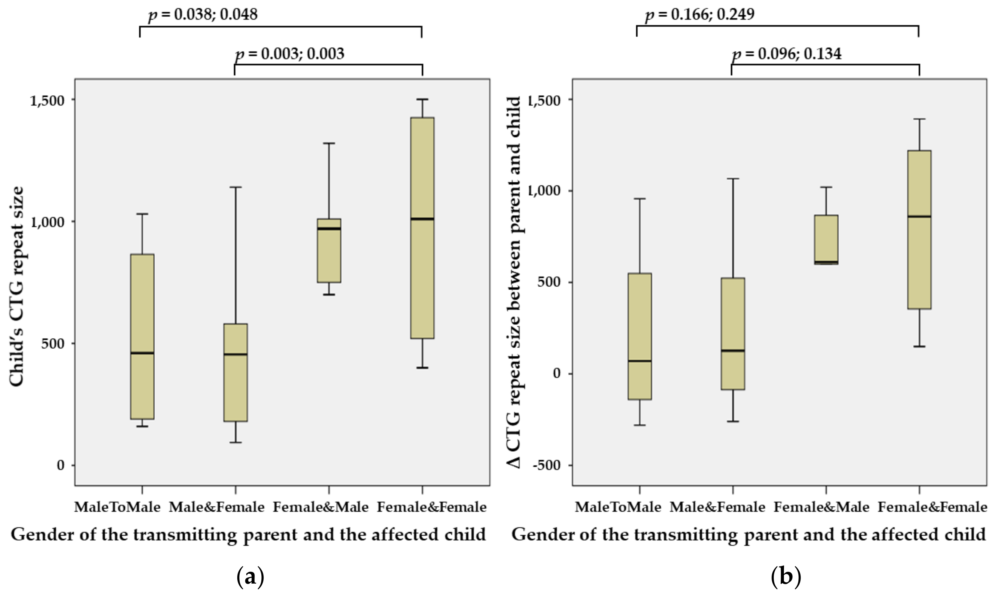
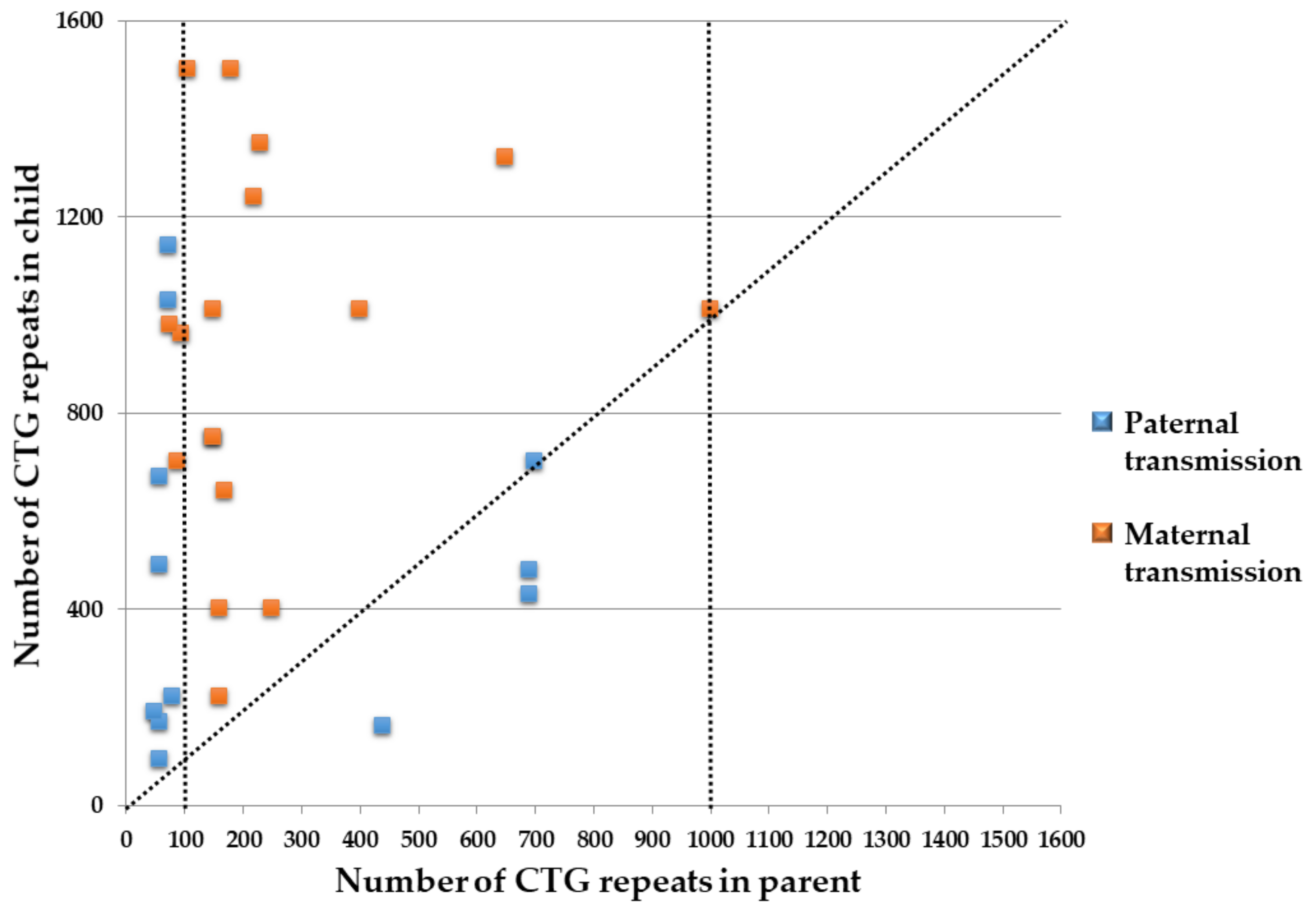
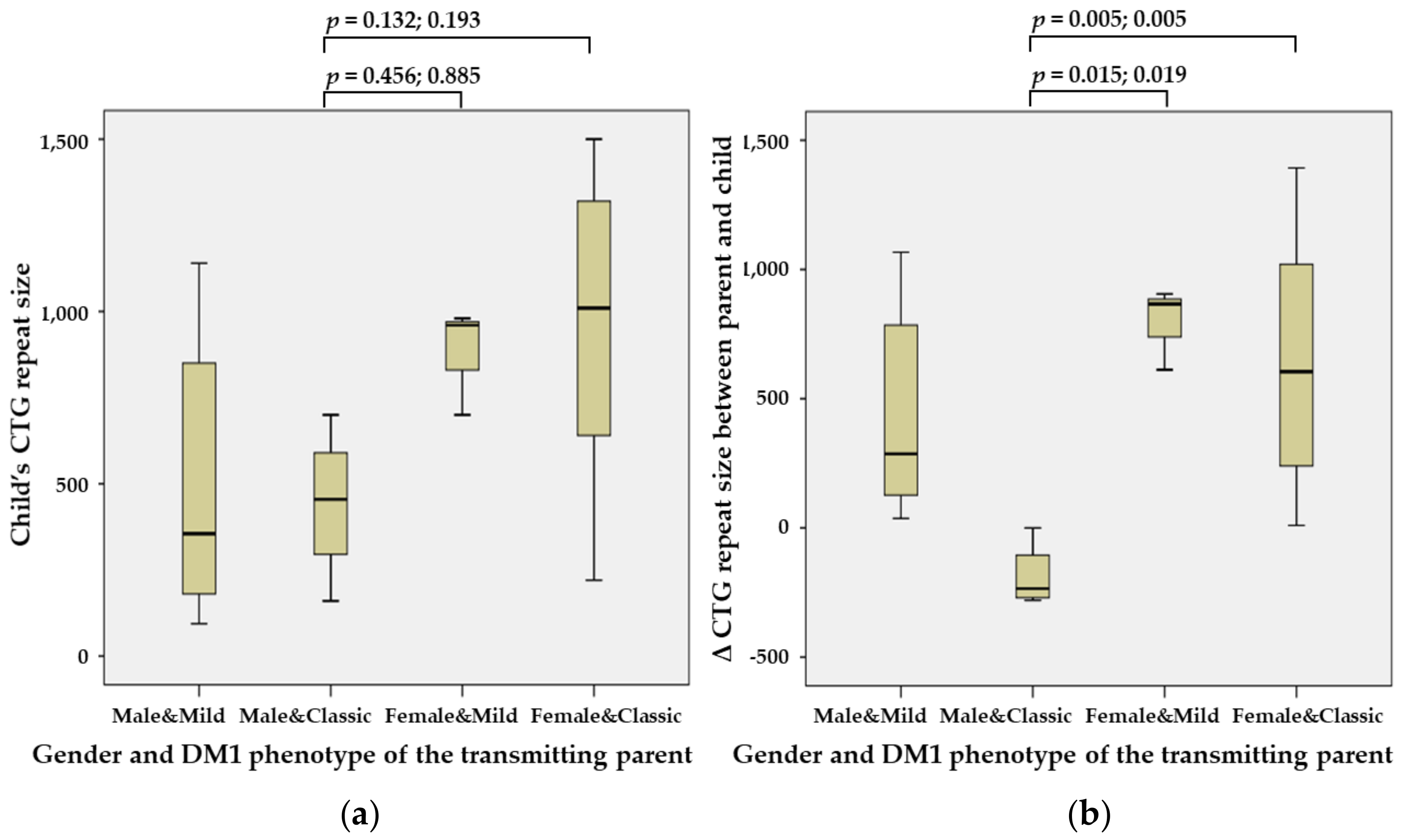
3. Intergenerational Influences in Korean DM1

A total of 44 parent–child pairs matched according to the gender of the transmitting parent and the affected child and 29 parent–child pairs matched according to the gender and the DM1 phenotype of the transmitting parent were used to estimate the intergenerational influence of the gender and DM1 phenotype of the transmitting parent on the development of Korean DM1.
To estimate the intergenerational influence of the gender of the transmitting parent and the affected child, 44 parent–child matched pairs were compared using four groups based on the gender of the transmitting parent and the affected child. As a result, maternal transmission (n = 28) was found to be 1.75 times more common than paternal transmission (n = 16) (Table 1).
The CTG repeat size of the DMPK in the affected child was significantly greater when the DMPK repeats were transmitted from a female parent to a female child (DM1-FF) (median, 1309 repeats; range, 400–2083) than when they were transmitted from a male parent to a male child (DM1-MM) (650; 160–1030; p = 0.038 and 0.048 using the Tukey HSD and the Bonferroni test) or from a male parent to a female child (DM1-MF) (480; 94–1140; p = 0.003) (Figure 1a). The difference in the CTG repeat size of the DMPK between the transmitting parent and the affected child was also higher in DM1-FF than in DM1-MM or DM1-MF, but this finding was not statistically significant (Figure 1b).
To evaluate the intergenerational influence of the gender and DM1 phenotype of the transmitting parent, 29 parent–child matched pairs were compared using four groups based on the gender and DM1 phenotype of the transmitting parent. The results show that the CTG repeat expansion of the DMPK always occurred when transmitted from a parent with mild DM1 regardless of their gender. On the contrary, the CTG repeat reduction or non-expansion of the DMPK only occurred when the condition was transmitted paternally, even though the transmitting father and the affected child were diagnosed with classic DM1 (Figure 2). However, two-thirds (62%, 18/29) of the affected children were found to have a more severe DM1 phenotype than their parent (Table 2).
The difference in the CTG repeat size of the DMPK between the transmitting parent and the affected child was lower when transmitted from a male parent with classic DM1 (DM1-MC) (455; 160–700) than when transmitted from a female parent with mild DM1 (DM1-Fm) (960; 700–980) or a female parent with classic DM1 (DM1-FC) (1010; 220–1500), although this result was not statistically significant. The difference in the CTG repeat size of the DMPK between the transmitting parent and the affected child was also lower in DM1-MC (−235; −280–0) compared with DM1-Fm (866; 612–905; p = 0.015 and 0.019) and DM1-FC (605; 10–1393; p = 0.005) (Figure 3).

4. Discussion

Geographically, large-scale population-based studies have been reported in Asia [27,28,29,30], Europe [31,32,33,34,35,36,37,38,39,40], North America [41], and Oceania [42]. Previous studies have reported that the incidence of DM1 is different in various ethnicities [43,44,45]. DM1 is more frequent in a European population; however, it is very rare in a Southern African population [44,45]. In an Asian population, the estimated incidence of DM1 was found to be low in Taiwan [28] with 0.45 patients per 100,000. The low incidence of DM1 in a Taiwanese population may be explained by the number of the CTG repeats of the DMPK with <18 [46]. In addition, the clinical manifestations of Chinese DM1 patients are distinguished from those of Caucasian DM1 patients [47], emphasizing the importance of appropriate molecular analysis for the diagnosis of DM1. Recently, Nicholas et al. demonstrated that founder effects are not a likely cause for the increased occurrence in which individuals with the CTG repeat expansion of the DMPK with ≥50 were not closely related and have diverse genetic ancestry, even though the incidence of DM1 may be higher in some populations than others due to founder effects [48]. Furthermore, the incidence of individuals with CTG repeat expansions of the DMPK is up to 5 times higher than previously reported estimates. These findings suggest that DM1, with multi-systemic characteristics, is likely underdiagnosed in practice [48].
We reviewed the intergenerational influence of the gender and DM1 phenotype of the transmitting parent on the development of Korean DM1. First, maternal transmission was related to the CTG repeat size of the DMPK and to the more severe DM1 phenotype, as shown in previous studies [2,8,14]. In children with congenital DM1, the condition is more frequently inherited maternally (n = 16) rather than paternally (n = 2). This phenomenon has been attributed to substantial DNA instability in the female germ cell lineage, leading to additional CTG repeat insertion during oogenesis [49]. DNA instability results in anticipation during maternal transmission, an occurrence corresponding to more severe DM1 phenotypes and earlier disease onset in consecutive generations. In contrast to our results, Dogan et al. reported that maternal transmission was observed in a minority of DM1 patients (37%). This probably resulted from perinatal lethality and increased miscarriage found in maternal DM1 transmitters [14].
Second, the gender difference suggested an unequal prevalence of several DM1 phenotypes in the affected child. Interestingly, paternal transmission (n = 8) was found to be 2.67 times more common than maternal transmission (n = 3) when the transmitting parent showed the mild DM1 phenotype in this study. However, this difference could not be explained by differences in the CTG repeat size of the DMPK or by male-to-female disproportion between the transmitting parent and the affected child in this study. The exact process underlying gender-dependent differences is unknown. Different characteristics of skeletal muscle tissue between males and females have been associated with significant differences in gene expression patterns and metabolic properties [50]. In myotonia congenita, sexual steroid hormones reflect testosterone modulation of CLCN1 chloride channel activity and affect clinical manifestations of myotonia congenita [51]. CTG repeat reduction and expansion of the DMPK could occur during the intergenerational transmission of DMPK expansion with variant CTG repeats in DM1 families. Cases of DMPK expansion with 50–100 CTG repeats are generally stable in maternal transmission and expansion frequently occurs in paternal transmission [52]. Compared with the maternal transmission of small-sized CTG repeats, the paternal transmission of DM1 pre-(CTG repeat size with 36–50 repeats) and proto-(CTG repeat size with 51–80 repeats) mutations is far more unstable [53]. This emphasizes that males with a small DMPK mutation have a higher risk of symptomatic offspring compared with females.
Third, DMPK expansions with >100–200 CTG repeats are volatile and are transmitted by both genders. In this size range, transmissions largely lead to CTG expansions of the DMPK; however, cases of stable CTG repeat reduction and expansion can occur, especially in paternal transmissions [54,55]. Large DMPK expansion with >1000 CTG repeats was found in immature and metaphase II oocytes; however, these large, mutated alleles were absent in the sperm of most male DM1 patients, consistent with the low frequency of paternal transmissions of congenital DM1 [49,56]. Methylation of the sequence located around the expanded CTG repeat of the DMPK might explain the maternal bias for the transmission of large expansions and congenital DM1, to a certain degree. A recent hypothesis to explain the almost exclusive maternal transmission of congenital DM1 suggests the prevention of the transmission of large repeat expansions after paternal transmissions, leading to the reduced expression of the SIX5 gene in the DM1 locus [57]. However, Yanovsky-Dagan et al. exclude the possibility that DMPK hypermethylation leads to selection against viable sperm cells in DM1 patients by assessing DNA methylation upstream to the CTG expansion of the DMPK in motile sperm cells of four DM1 patients [58]. Nevertheless, CTG repeat expansion of the DMPK harboring a single CAG interruption is characterized by stable transmission or CTG repeat reduction in successive generations [59], suggesting that a single nucleotide change within the CTG repeat may be enough to strengthen the meiotic stability of DMPK expansion. Although the transmission of interrupted DMPK expansions was not associated with the gender of the transmitting parent [60], it is noteworthy that all sporadically occurring variant DMPK expansions were transmitted by the male parent, whether it was an expansion harboring different patterns of CCG repeats [61,62] or a single CTC repeat [60].
A limitation of this study is the small study cohort of 44 patients and reported cases; nevertheless, the uniform clinical characterization of the participants, including their gender, the CTG repeat size of the DMPK, and/or the DM1 phenotype, is a strength of this study. Another limitation is the lack of detailed clinical information. The pathogenesis of DM1 is complex, with a pivotal role played by the pathogenic effect of the mutant DMPK pre-mRNAs harboring the expanded CUG stretch, which will eventually interrupt the expression of other genes in different tissues by damaging the functions of specific transcription factors controlling alternative splicing. Because the DMPK mRNA is expressed widely in various tissues, this finding explains the multi-organ involvement in patients with DM1, including the associated development of gastrointestinal disturbances, endocrine dysfunction, cardiac conduction abnormalities, premature cataracts, and behavioral and cognitive impairments [15]. Male patients with DM1 tended to show more apparent classical DM1 phenotypes, such as cardiac and respiratory involvement, significant myotonia, and cognitive impairment; however, female patients with DM1 had late-onset disease and more extra-muscular clinical features, characteristics which are less suggestive of DM1. These findings emphasize the significance of having greater recognition of preventive medical management in male individuals with DM1 [14]. Thus, the gender-associated differential risks of developing certain symptoms may require gender-orientated therapeutic care. In addition, we could not estimate the presence of common lifestyle risk factors in our cohort that may affect the risk of cancer in DM1, as reported previously [63].

5. Conclusions

In conclusion, the gender and DM1 phenotype of the transmitting parent impact the CTG repeat size of the DMPK in the affected child, with higher increases being inherited from DM1-FF or DM1-FC situations in Korean DM1. Thus, the gender and DM1 phenotype of the transmitting parent should be considered in the design of both stratified clinical trials and medical management. Further investigations are required to clarify the complex pathophysiology associated with the gender and DM1 phenotype of the transmitting parent and the affected child in order to make specific recommendations regarding the diagnostic assessment and medical care of the different DM1 phenotype distributions of Korean DM1 patients, as this often affects the quality of life of patients with DM1.

Author Contributions

Conceptualization, J.P.; Data curation, J.Y.H. and J.P.; Methodology, J.P. and W.J.; Formal analysis, J.P.; Original draft preparation, J.P.; Review and editing, J.Y.H. and W.J.; Supervision, J.P.; Funding acquisition, J.P. All authors have read and agreed to the published version of the manuscript.

Funding

This work was supported by a National Research Foundation of Korea (NRF) grant funded by the Korean government (MSIT; Ministry of Science and ICT) (2020R1F1A1077316).

Institutional Review Board Statement

This study was approved by the Institutional Review Board (IRB) of Daejeon St. Mary’s Hospital, The Catholic University of Korea (Approval number: DC22ZASE0026; Date of approval: 20 May 2022).

Informed Consent Statement

The requirement for written informed consent was waived by the IRB because of the retrospective nature of this study with analysis using anonymous clinical data.

Data Availability Statement

Not applicable.

Conflicts of Interest

The authors declare no conflict of interest.

References

  1. Aslanidis, C.; Jansen, G.; Amemiya, C.; Shutler, G.; Mahadevan, M.; Tsilfidis, C.; Chen, C.; Alleman, J.; Wormskamp, N.G.; Vooijs, M.; et al. Cloning of the essential myotonic dystrophy region and mapping of the putative defect. Nature 1992, 355, 548–551. [Google Scholar] [CrossRef] [PubMed]
  2. Brook, J.D.; McCurrach, M.E.; Harley, H.G.; Buckler, A.J.; Church, D.; Aburatani, H.; Hunter, K.; Stanton, V.P.; Thirion, J.P.; Hudson, T.; et al. Molecular basis of myotonic dystrophy: Expansion of a trinucleotide (CTG) repeat at the 3’ end of a transcript encoding a protein kinase family member. Cell 1992, 68, 799–808. [Google Scholar] [CrossRef]
  3. Harley, H.G.; Brook, J.D.; Rundle, S.A.; Crow, S.; Reardon, W.; Buckler, A.J.; Harper, P.S.; Housman, D.E.; Shaw, D.J. Expansion of an unstable DNA region and phenotypic variation in myotonic dystrophy. Nature 1992, 355, 545–546. [Google Scholar] [CrossRef] [PubMed]
  4. Cumming, S.A.; Jimenez-Moreno, C.; Okkersen, K.; Wenninger, S.; Daidj, F.; Hogarth, F.; Littleford, R.; Gorman, G.; Bassez, G.; Schoser, B.; et al. Genetic determinants of disease severity in the myotonic dystrophy type 1 OPTIMISTIC cohort. Neurology 2019, 93, e995–e1009. [Google Scholar] [CrossRef]
  5. Hogrel, J.Y.; Ollivier, G.; Ledoux, I.; Hébert, L.J.; Eymard, B.; Puymirat, J.; Bassez, G. Relationships between grip strength, myotonia, and CTG expansion in myotonic dystrophy type 1. Ann. Clin. Transl. Neurol. 2017, 4, 921–925. [Google Scholar] [CrossRef]
  6. André, L.M.; Ausems, C.R.M.; Wansink, D.G.; Wieringa, B. Abnormalities in Skeletal Muscle Myogenesis, Growth, and Regeneration in Myotonic Dystrophy. Front. Neurol. 2018, 9, 368. [Google Scholar] [CrossRef]
  7. Rakocevic-Stojanovic, V.; Savic, D.; Pavlovic, S.; Lavrnic, D.; Stevic, Z.; Basta, I.; Romac, S.; Apostolski, S. Intergenerational changes of CTG repeat depending on the sex of the transmitting parent in myotonic dystrophy type 1. Eur. J. Neurol. 2005, 12, 236–237. [Google Scholar] [CrossRef]
  8. Martorell, L.; Cobo, A.M.; Baiget, M.; Naudo, M.; Poza, J.J.; Parra, J. Prenatal diagnosis in myotonic dystrophy type 1. Thirteen years of experience: Implications for reproductive counselling in DM1 families. Prenat. Diagn. 2007, 27, 68–72. [Google Scholar] [CrossRef]
  9. Lavedan, C.; Hofmann-Radvanyi, H.; Shelbourne, P.; Rabes, J.P.; Duros, C.; Savoy, D.; Dehaupas, I.; Luce, S.; Johnson, K.; Junien, C. Myotonic dystrophy: Size- and sex-dependent dynamics of CTG meiotic instability, and somatic mosaicism. Am. J. Hum. Genet. 1993, 52, 875–883. [Google Scholar]
  10. Redman, J.B.; Fenwick, R.G., Jr.; Fu, Y.H.; Pizzuti, A.; Caskey, C.T. Relationship between parental trinucleotide GCT repeat length and severity of myotonic dystrophy in offspring. JAMA 1993, 269, 1960–1965. [Google Scholar] [CrossRef]
  11. Tsilfidis, C.; MacKenzie, A.E.; Mettler, G.; Barcelo, J.; Korneluk, R.G. Correlation between CTG trinucleotide repeat length and frequency of severe congenital myotonic dystrophy. Nat. Genet. 1992, 1, 192–195. [Google Scholar] [CrossRef]
  12. Pratte, A.; Prevost, C.; Puymirat, J.; Mathieu, J. Anticipation in myotonic dystrophy type 1 parents with small CTG expansions. Am. J. Med. Genet. Part A 2015, 167, 708–714. [Google Scholar] [CrossRef]
  13. Dean, N.L.; Loredo-Osti, J.C.; Fujiwara, T.M.; Morgan, K.; Tan, S.L.; Naumova, A.K.; Ao, A. Transmission ratio distortion in the myotonic dystrophy locus in human preimplantation embryos. Eur. J. Hum. Genet. 2006, 14, 299–306. [Google Scholar] [CrossRef]
  14. Dogan, C.; De Antonio, M.; Hamroun, D.; Varet, H.; Fabbro, M.; Rougier, F.; Amarof, K.; Bes, M.C.A.; Bedat-Millet, A.L.; Behin, A.; et al. Gender as a Modifying Factor Influencing Myotonic Dystrophy Type 1 Phenotype Severity and Mortality: A Nationwide Multiple Databases Cross-Sectional Observational Study. PLoS ONE 2016, 11, e0148264. [Google Scholar] [CrossRef]
  15. Perna, A.; Maccora, D.; Rossi, S.; Nicoletti, T.F.; Zocco, M.A.; Riso, V.; Modoni, A.; Petrucci, A.; Valenza, V.; Grieco, A.; et al. High Prevalence and Gender-Related Differences of Gastrointestinal Manifestations in a Cohort of DM1 Patients: A Perspective, Cross-Sectional Study. Front. Neurol. 2020, 11, 394. [Google Scholar] [CrossRef]
  16. Peric, S.; Pesovic, J.; Savic-Pavicevic, D.; Rakocevic Stojanovic, V.; Meola, G. Molecular and Clinical Implications of Variant Repeats in Myotonic Dystrophy Type 1. Int. J. Mol. Sci. 2021, 23, 354. [Google Scholar] [CrossRef]
  17. Nam, S.H.; Son, Y.B.; Lee, B.L.; Lee, J.; Ki, C.S.; Lee, M. Clinical characteristics of congenital myotonic dystrophy diagnosed by molecular genetic method. Korean J. Pediatr. 2007, 50, 868–874. [Google Scholar] [CrossRef]
  18. Kim, S.Y.; Kim, J.Y.; Kim, G.P.; Sung, J.J.; Lim, K.S.; Lee, K.W.; Chae, J.H.; Hong, Y.H.; Seong, M.W.; Park, S.S. Molecular and clinical characteristics of myotonic dystrophy type 1 in koreans. Korean J. Lab. Med. 2008, 28, 483–492. [Google Scholar] [CrossRef]
  19. Oh, J.H.; Lee, J.Y.; Lee, J.S.; Choi, J.C.; Kang, J.H.; Kang, S.Y. Congenital Myotonic Dystrophy Diagnosed in an Adolescent Period. J. Korean Neurol. Assoc. 2008, 26, 383–386. [Google Scholar]
  20. Yim, S.Y.; Cho, K.H.; Kim, J.Y.; Hong, J.Y.; Lee, I.Y. A Floppy Baby with Congenital Myotonic Dystrophy Complicated with Huge Subgaleal Hematoma Occurring in Non-instrumental Vaginal Delivery. J. Genet. Med. 2009, 6, 166–169. [Google Scholar]
  21. Kim, H.S.; Chung, K.W.; Kang, S.H.; Choi, S.K.; Cho, S.Y.; Koo, H.; Kim, S.B.; Choi, B.O. Myotonic dystrophy type I combined with X-linked dominant Charcot-Marie-Tooth neuropathy. Neurogenetics 2010, 11, 425–433. [Google Scholar] [CrossRef]
  22. Kang, S.Y.; Kang, H.J.; Chung, P.W.; Kim, K.K.; Lee, S.; Choi, B.O. Congestive heart failure after physical exercise in a young patient with myotonic dystrophy type 1. Int. J. Neurosci. 2011, 121, 637–639. [Google Scholar] [CrossRef]
  23. Son, S.B.; Chun, J.M.; Kim, K.A.; Ko, S.Y.; Lee, Y.K.; Shin, S.M. A case report on 30-week premature twin babies with congenital myotonic dystrophy conceived by in vitro fertilization. J. Korean Med. Sci. 2012, 27, 1269–1272. [Google Scholar] [CrossRef]
  24. Son, H.K.; Cha, Y.S.; Suh, H.; Ki, C.S.; Shin, Y.B. Upper thoracic myelopathy caused by delayed neck extensor weakness in myotonic dystrophy. Ann. Rehabil. Med. 2012, 36, 569–572. [Google Scholar] [CrossRef]
  25. Kim, H.N.; Cho, Y.K.; Cho, J.H.; Yang, E.M.; Song, E.S.; Choi, Y.Y. Transient complete atrioventricular block in a preterm neonate with congenital myotonic dystrophy: Case report. J. Korean Med. Sci. 2014, 29, 879–883. [Google Scholar] [CrossRef]
  26. Kim, H.J.; Oh, J.H.; Kang, S.Y. Bilateral Adduction Palsy in a Patient with Myotonic Dystrophy Type 1. Exp. Neurobiol. 2016, 25, 143–145. [Google Scholar] [CrossRef]
  27. Chung, B.; Wong, V.; Ip, P. Prevalence of neuromuscular diseases in Chinese children: A study in southern China. J. Child Neurol. 2003, 18, 217–219. [Google Scholar] [CrossRef]
  28. Hsiao, K.M.; Chen, S.S.; Li, S.Y.; Chiang, S.Y.; Lin, H.M.; Pan, H.; Huang, C.C.; Kuo, H.C.; Jou, S.B.; Su, C.C.; et al. Epidemiological and genetic studies of myotonic dystrophy type 1 in Taiwan. Neuroepidemiology 2003, 22, 283–289. [Google Scholar] [CrossRef]
  29. Lee, J.Y.; Jeong, H.N. Nationwide incidence of myotonic dystrophy type 1 and the status of multi-organ involvement. J. Neurol. 2022, 269, 2666–2672. [Google Scholar] [CrossRef]
  30. Sugimoto, M.; Kuru, S.; Takada, H.; Horie, R.; Yamauchi, K.; Kubota, T.; Matsumura, T.; Nakamura, H.; Kimura, E.; Takahashi, M.P. Characteristics of myotonic dystrophy patients in the national registry of Japan. J. Neurol. Sci. 2022, 432, 120080. [Google Scholar] [CrossRef]
  31. Medica, I.; Marković, D.; Peterlin, B. Genetic epidemiology of myotonic dystrophy in Istria, Croatia. Acta Neurol. Scand. 1997, 95, 164–166. [Google Scholar] [CrossRef] [PubMed]
  32. Siciliano, G.; Manca, M.; Gennarelli, M.; Angelini, C.; Rocchi, A.; Iudice, A.; Miorin, M.; Mostacciuolo, M. Epidemiology of myotonic dystrophy in Italy: Re-apprisal after genetic diagnosis. Clin. Genet. 2001, 59, 344–349. [Google Scholar] [CrossRef] [PubMed]
  33. Segel, R.; Silverstein, S.; Lerer, I.; Kahana, E.; Meir, R.; Sagi, M.; Zilber, N.; Korczyn, A.D.; Shapira, Y.; Argov, Z.; et al. Prevalence of myotonic dystrophy in Israeli Jewish communities: Inter-community variation and founder premutations. Am. J. Med. Genet. Part A 2003, 119, 273–278. [Google Scholar] [CrossRef] [PubMed]
  34. Mladenovic, J.; Pekmezovic, T.; Todorovic, S.; Rakocevic-Stojanovic, V.; Savic, D.; Romac, S.; Apostolski, S. Survival and mortality of myotonic dystrophy type 1 (Steinert’s disease) in the population of Belgrade. Eur. J. Neurol. 2006, 13, 451–454. [Google Scholar] [CrossRef]
  35. Norwood, F.L.; Harling, C.; Chinnery, P.F.; Eagle, M.; Bushby, K.; Straub, V. Prevalence of genetic muscle disease in Northern England: In-depth analysis of a muscle clinic population. Brain 2009, 132, 3175–3186. [Google Scholar] [CrossRef]
  36. Vanacore, N.; Rastelli, E.; Antonini, G.; Bianchi, M.L.; Botta, A.; Bucci, E.; Casali, C.; Costanzi-Porrini, S.; Giacanelli, M.; Gibellini, M.; et al. An Age-Standardized Prevalence Estimate and a Sex and Age Distribution of Myotonic Dystrophy Types 1 and 2 in the Rome Province, Italy. Neuroepidemiology 2016, 46, 191–197. [Google Scholar] [CrossRef]
  37. Lefter, S.; Hardiman, O.; Ryan, A.M. A population-based epidemiologic study of adult neuromuscular disease in the Republic of Ireland. Neurology 2017, 88, 304–313. [Google Scholar] [CrossRef]
  38. Lindberg, C.; Bjerkne, F. Prevalence of myotonic dystrophy type 1 in adults in western Sweden. Neuromuscul. Disord. 2017, 27, 159–162. [Google Scholar] [CrossRef]
  39. Pagola-Lorz, I.; Vicente, E.; Ibáñez, B.; Torné, L.; Elizalde-Beiras, I.; Garcia-Solaesa, V.; García, F.; Delfrade, J.; Jericó, I. Epidemiological study and genetic characterization of inherited muscle diseases in a northern Spanish region. Orphanet. J. Rare Dis. 2019, 14, 276. [Google Scholar] [CrossRef]
  40. Husebye, S.A.; Rebne, C.B.; Stokland, A.E.; Sanaker, P.S.; Bindoff, L.A. A hospital based epidemiological study of genetically determined muscle disease in south western Norway. Neuromuscul. Disord. 2020, 30, 181–185. [Google Scholar] [CrossRef]
  41. Hagerman, K.A.; Howe, S.J.; Heatwole, C.R. The myotonic dystrophy experience: A North American cross-sectional study. Muscle Nerve 2019, 59, 457–464. [Google Scholar] [CrossRef]
  42. Ford, C.; Kidd, A.; Hammond-Tooke, G. Myotonic dystrophy in Otago, New Zealand. New Zealand Med. J. 2006, 119, U2145. [Google Scholar]
  43. Fedorova, S.A.; Khusainova, R.I.; Kutuev, I.A.; Sukhomiatova, A.L.; Nikolaeva, I.A.; Kulichkin, S.S.; Akhmetova, V.L.; Salimova, A.Z.; Sviatova, G.S.; Berezina, G.M.; et al. Polymorphism of CTG-repeats in the DMPK gene in populations of Yakutia and central Asia. Mol. Biol. 2005, 39, 385–393. [Google Scholar] [CrossRef]
  44. Thornton, C.A. Myotonic dystrophy. Neurol. Clin. 2014, 32, 705–719. [Google Scholar] [CrossRef]
  45. Krause, A.; Seymour, H.; Ramsay, M. Common and Founder Mutations for Monogenic Traits in Sub-Saharan African Populations. Annu. Rev. Genom. Hum. Genet. 2018, 19, 149–175. [Google Scholar] [CrossRef]
  46. Pan, H.; Lin, H.M.; Ku, W.Y.; Li, T.C.; Li, S.Y.; Lin, C.C.; Hsiao, K.M. Haplotype analysis of the myotonic dystrophy type 1 (DM1) locus in Taiwan: Implications for low prevalence and founder mutations of Taiwanese myotonic dystrophy type 1. Eur. J. Hum. Genet. 2001, 9, 638–641. [Google Scholar] [CrossRef]
  47. Lu, H.; Li, Y.; Sadowsky, M.; Da, Y. Clinical characteristics of 37 Chinese patients with myotonic dystrophy Type 1. Brain Circ. 2016, 2, 95–98. [Google Scholar] [CrossRef]
  48. Johnson, N.E.; Butterfield, R.J.; Mayne, K.; Newcomb, T.; Imburgia, C.; Dunn, D.; Duval, B.; Feldkamp, M.L.; Weiss, R.B. Population-Based Prevalence of Myotonic Dystrophy Type 1 Using Genetic Analysis of Statewide Blood Screening Program. Neurology 2021, 96, e1045–e1053. [Google Scholar] [CrossRef]
  49. Dean, N.L.; Tan, S.L.; Ao, A. Instability in the transmission of the myotonic dystrophy CTG repeat in human oocytes and preimplantation embryos. Fertil. Steril. 2006, 86, 98–105. [Google Scholar] [CrossRef]
  50. Welle, S.; Tawil, R.; Thornton, C.A. Sex-related differences in gene expression in human skeletal muscle. PLoS ONE 2008, 3, e1385. [Google Scholar] [CrossRef]
  51. Fialho, D.; Kullmann, D.M.; Hanna, M.G.; Schorge, S. Non-genomic effects of sex hormones on CLC-1 may contribute to gender differences in myotonia congenita. Neuromuscul. Disord. 2008, 18, 869–872. [Google Scholar] [CrossRef]
  52. Savić, D.; Keckarević, D.; Branković-Srećković, V.; Apostolski, S.; Todorović, S.; Romac, S. Clinical case report atypical myopathy in a young girl with 91 CTG repeats in DM1 locus and a positive DM1 family history. Int. J. Neurosci. 2006, 116, 1509–1518. [Google Scholar] [CrossRef]
  53. Joosten, I.B.T.; Hellebrekers, D.; de Greef, B.T.A.; Smeets, H.J.M.; de Die-Smulders, C.E.M.; Faber, C.G.; Gerrits, M.M. Parental repeat length instability in myotonic dystrophy type 1 pre- and protomutations. Eur. J. Hum. Genet. 2020, 28, 956–962. [Google Scholar] [CrossRef]
  54. Ashizawa, T.; Anvret, M.; Baiget, M.; Barceló, J.M.; Brunner, H.; Cobo, A.M.; Dallapiccola, B.; Fenwick, R.G., Jr.; Grandell, U.; Harley, H.; et al. Characteristics of intergenerational contractions of the CTG repeat in myotonic dystrophy. Am. J. Hum. Genet. 1994, 54, 414–423. [Google Scholar]
  55. Salehi, L.B.; Bonifazi, E.; Stasio, E.D.; Gennarelli, M.; Botta, A.; Vallo, L.; Iraci, R.; Massa, R.; Antonini, G.; Angelini, C.; et al. Risk prediction for clinical phenotype in myotonic dystrophy type 1: Data from 2,650 patients. Genet. Test. 2007, 11, 84–90. [Google Scholar] [CrossRef]
  56. De Temmerman, N.; Sermon, K.; Seneca, S.; De Rycke, M.; Hilven, P.; Lissens, W.; Van Steirteghem, A.; Liebaers, I. Intergenerational instability of the expanded CTG repeat in the DMPK gene: Studies in human gametes and preimplantation embryos. Am. J. Hum. Genet. 2004, 75, 325–329. [Google Scholar] [CrossRef]
  57. Barbé, L.; Lanni, S.; López-Castel, A.; Franck, S.; Spits, C.; Keymolen, K.; Seneca, S.; Tomé, S.; Miron, I.; Letourneau, J.; et al. CpG Methylation, a Parent-of-Origin Effect for Maternal-Biased Transmission of Congenital Myotonic Dystrophy. Am. J. Hum. Genet. 2017, 100, 488–505. [Google Scholar] [CrossRef]
  58. Yanovsky-Dagan, S.; Cohen, E.; Megalli, P.; Altarescu, G.; Schonberger, O.; Eldar-Geva, T.; Epsztejn-Litman, S.; Eiges, R. DMPK hypermethylation in sperm cells of myotonic dystrophy type 1 patients. Eur. J. Hum. Genet. 2022, 30, 980–983. [Google Scholar] [CrossRef]
  59. Tomé, S.; Dandelot, E.; Dogan, C.; Bertrand, A.; Geneviève, D.; Péréon, Y.; Simon, M.; Bonnefont, J.P.; Bassez, G.; Gourdon, G. Unusual association of a unique CAG interruption in 5’ of DM1 CTG repeats with intergenerational contractions and low somatic mosaicism. Hum. Mutat. 2018, 39, 970–982. [Google Scholar] [CrossRef]
  60. Pešović, J.; Perić, S.; Brkušanin, M.; Brajušković, G.; Rakočević-Stojanović, V.; Savić-Pavićević, D. Molecular genetic and clinical characterization of myotonic dystrophy type 1 patients carrying variant repeats within DMPK expansions. Neurogenetics 2017, 18, 207–218. [Google Scholar] [CrossRef]
  61. Cumming, S.A.; Hamilton, M.J.; Robb, Y.; Gregory, H.; McWilliam, C.; Cooper, A.; Adam, B.; McGhie, J.; Hamilton, G.; Herzyk, P.; et al. De novo repeat interruptions are associated with reduced somatic instability and mild or absent clinical features in myotonic dystrophy type 1. Eur. J. Hum. Genet. 2018, 26, 1635–1647. [Google Scholar] [CrossRef] [PubMed]
  62. Mangin, A.; de Pontual, L.; Tsai, Y.C.; Monteil, L.; Nizon, M.; Boisseau, P.; Mercier, S.; Ziegle, J.; Harting, J.; Heiner, C.; et al. Robust Detection of Somatic Mosaicism and Repeat Interruptions by Long-Read Targeted Sequencing in Myotonic Dystrophy Type 1. Int. J. Mol. Sci. 2021, 22, 2616. [Google Scholar] [CrossRef] [PubMed]
  63. Bianchi, M.L.; Leoncini, E.; Masciullo, M.; Modoni, A.; Gadalla, S.M.; Massa, R.; Rastelli, E.; Terracciano, C.; Antonini, G.; Bucci, E.; et al. Increased risk of tumor in DM1 is not related to exposure to common lifestyle risk factors. J. Neurol. 2016, 263, 492–498. [Google Scholar] [CrossRef] [PubMed]
Figure 1. Intergenerational influence of gender between the transmitting parent and the affected child. (a) The CTG repeat size of the DMPK in the affected child was significantly greater when the DMPK repeats were transmitted from a female parent to a female child (DM1-FF) (median, 1309 repeats; range, 400–2083) than when they were transmitted from a male parent to a male child (DM1-MM) (650; 160–1030; p = 0.038 and 0.048 using the Tukey HSD and the Bonferroni test) and from a male parent to a female child (DM1-MF) (480; 94–1140; p = 0.003). (b) The difference in the CTG repeat size of the DMPK between the transmitting parent and the affected child was also higher in DM1-FF than in DM1-MM and in DM1-MF, but this result was not statistically significant.
Genes 13 01465 g001
Figure 2. The influence of the CTG repeat size of the DMPK of the transmitting parent on the affected child. CTG repeat reduction or no expansion only occurred following paternal transmission, even though the transmitting father and the affected child were diagnosed as having classic DM1. The vertical dotted line is used to indicate the mild (CTG repeat size with 50–100 repeats), classic (100–1000), and congenital (>1000) DM1 phenotypes. The diagonal dotted line indicates the influence of the CTG repeat size of the DMPK of the transmitting parent on that of the affected child.
Genes 13 01465 g002
Figure 3. Intergenerational influence of the gender and DM1 phenotype of the transmitting parent. (a) The influence of the CTG repeat size of the DMPK of the affected parent on the child was lower when the transmitting parent was male and when the condition was classic DM1 (DM1-MC) (455; 160−700) than for maternal transmission and mild DM1 (DM1-Fm) (960; 700–980) or for maternal transmission and classic DM1 (DM1-FC) (1010; 220–1500). (b) The difference in the CTG repeat size of the DMPK between the transmitting parent and the affected child was also lower in DM1-MC (−235; −280 to 0), compared with DM1-Fm (866; 612–905; p = 0.015 and 0.019) and DM1-FC (605; 10–1393; p = 0.005).
Genes 13 01465 g003
Table 1. DM1 phenotypes and CTG repeat size of the DMPK in the affected child according to the gender of the transmitting parent and the affected child in 44 Korean parent–child matched pairs diagnosed with DM1.
Parent
Gender
Child
Gender
Child DM1 PhenotypeChild’s Repeat Size;
Median (Range)
Δ Repeat Size;
Median (Range)
MildClassicCongenital
MaleMale041650 (160–1030)70 (−280–957)
MaleFemale191480 (94–1140)127 (−260–1067)
FemaleMale086970 (220–1667)611 (10–1020)
FemaleFemale04101309 (400–2083)860 (150–1393)
Δ Repeat size, difference between the transmitting parent and the affected child.
Table 2. DM1 phenotypes and CTG repeat size of the DMPK in the affected child according to gender and DM1 phenotype of the transmitting parent in 29 Korean parent–child matched pairs diagnosed with DM1.
Parent
Gender
Parent
Phenotype
Child DM1 PhenotypeChild’s Repeat Size;
Median (Range)
Δ Repeat Size;
Median (Range)
MildClassicCongenital
MaleMild152355 (94–1140)287 (37–1067)
MaleClassic040455 (160–700)−235 (−280–0)
FemaleMild030960 (700–980)866 (612–905)
FemaleClassic0681010 (220–1500)605 (10–1393)
Δ Repeat size, difference between the transmitting parent and the affected child.
Publisher’s Note: MDPI stays neutral with regard to jurisdictional claims in published maps and institutional affiliations.

