Article

The Effect of EGR1 on the Proliferation of Dermal Papilla Cells

1
College of Animal Science and Technology, Yangzhou University, Yangzhou 225009, China
2
International Joint Research Laboratory in Universities of Jiangsu Province of China for Domestic Animal Germplasm Resources and Genetic Improvement, Yangzhou 225009, China
3
Joint International Research Laboratory of Agriculture and Agri-Product Safety of Ministry of Education of China, Yangzhou University, Yangzhou 225009, China
4
International Centre for Agricultural Research in the Dry Areas, Addis Ababa 999047, Ethiopia
*
Authors to whom correspondence should be addressed.
Genes 2022, 13(7), 1242; https://doi.org/10.3390/genes13071242
Submission received: 10 June 2022 / Revised: 4 July 2022 / Accepted: 7 July 2022 / Published: 14 July 2022

Abstract

:
Early growth response factor 1 (EGR1) is a zinc-finger transcription factor that plays a vital role in the development of hair follicles. According to our previous studies, EGR1 is a transcriptional promoter of the bone morphogenetic protein 7 (BMP7), a candidate gene involved in the proliferation of dermal papilla cells. Since hair follicles are the basis of lambskin pattern formation and dermal papilla cells (DPCs) act on hair follicle growth, in order to elucidate the role of EGR1 and hair follicles, this study aimed to investigate the biological role of EGR1 in DPCs. In our study, the EGR1 coding sequence (CDS) region was firstly cloned by polymerase chain reaction, and bioinformatics analysis was performed. Then, the function of EGR1 was detected by 5-ethynyl-2’-deoxyuridine (EDU) and Cell Counting Kit-8 (CCK8), and Western blot (WB) was conducted to analyze the cellular effect of EGR1 on DPCs. The proliferative effect of EGR1 on DPCs was also further confirmed by detecting its expression by qPCR and WB on marker genes of proliferation, including PCNA and CDK2. The sequence of the EGR1 CDS region of a lamb was successfully cloned, and its nucleic acid sequence was analyzed and found to be highly homologous to Rattus norvegicus, Mus musculus, Bos taurus and Homo sapiens. Predictive analysis of the protein encoded by EGR1 revealed that it is an extra-membrane protein, and not a secretory protein, with subcellular localization in the nucleus and cytoplasm. The proliferative effect of DPCs was significantly stronger (p < 0.01) in EGR1 up-regulated DPCs compared to the controls, while the opposite result was observed in EGR1 down-regulated DPCs. Markers of proliferation including PCNA and CDK2 also appeared to be differentially upregulated in EGR1 gene overexpression compared to the controls, with the opposite result in EGR1 gene downregulation. In summary, our study revealed that EGR1 promotes the proliferation of DPCs, and we speculate that EGR1 may be closely associated with hair follicle growth and development.

1. Introduction

Hu sheep are famous for their white lambskin with a wavy pattern, which is generally classified into three types—small waves, medium waves, and large waves—determining the Hu lambskin quality (Figure 1). It is usually considered that small waves indicate the best quality [1]. With the continuous growth of mutton consumption, livestock producers have been more concerned with meat and reproductive traits, thus neglecting lambskin traits, resulting in the gradual decline of lambskin quality in the selection and breeding process, and the high-quality germplasm resources are threatened.
The type of lambskin pattern is determined by the different curling degree of the wool, which is influenced by the growth and development of the hair follicles. The hair follicle can be divided into epidermal and dermal components from a developmental biology perspective. Hair follicles are complex in composition, and include the epidermal component and the dermis. In particular, the dermis consists mainly of the dermal papilla (DP) [2]. Dermal papilla cells express exocrine signaling such as Wnts, FGF and Noggin in vivo, which can act on hair follicle biological activity [3,4]. Additionally, Nissiove et al. [5] proposed a “multiple papillary centres” (MPC) hypothesis, which suggests that dermal papilla play a positive role in hair follicle development and hair growth, and that each papillary structure acts independently, leading to differences in the growth rate of hair follicle cells and thus to curved hair growth. Therefore, we believe that the dermal papilla cells are important cells affecting the pattern formation of lambskin.
Early growth response factor 1 (EGR1) is a zinc-finger structure that is associated with diverse cellular functions, such as proliferation, apoptosis, and migration [6,7]. Activated EGR1 are involved in growth factors, inflammatory factors, reactive oxygen species, etc. [8]. The Ras-RAF-MEK1/2-ERK1/2 signaling pathway allows for successful transcription of EGR1, and EGR1 participate in target gene transcriptional regulation to regulate their expression [9]. Numerous studies have shown that the EGR1 gene is closely related to tumors [10], stomach cancer [11], glioma [12] and melanoma [13]; however, little is known about the direct effect of EGR1 on dermal papilla cells. Nevertheless, EGR1 was found to be an important transcription factor in the bone morphogenetic protein 7 (BMP7) promoter region, and BMP7 has been proved to be a candidate gene in the proliferation of dermal papilla cells, collectively. We speculated that EGR1 may also affect the proliferation of dermal papilla cells, which initially affects the pattern formation of lambskin [14]. In summary, we suggested that the important transcription factor EGR1 of BMP7 could regulate the proliferation of dermal papilla cells of Hu sheep, which in turn affects wool bending growth, leading to lambskin pattern formation.
To explore the biological role of EGR1 in DPCs, we cloned the sequence of the EGR1 CDS region and a series of bioinformatics analyses were conducted. The role of EGR1 in dermal papilla cells was further investigated by examining the tissue expression profile of EGR1, and the effects of EGR1 overexpression and interference on cells, thus speculating on the potential role of EGR1 in dermal follicle growth. Our results can provide the scientific basis for analyzing the molecular mechanism of lambskin pattern formation and pave the way for subsequent studies.

2. Materials and Methods

2.1. Experimental Animals

All lambs used in this experiment were from the Suzhou Stud Farm (Suzhou, China). A total of three pairs of full sibling individuals, including one straight hair and one small waves per pair. We collected approximately 0.8 cm2 of skin tissue from the same dorsal side of 6 three-day-old Hu lambs, which were rapidly stored in RNA preservation solution (TAKARA, Dalian, China). The hair follicles, heart, liver, spleen, lung, kidney, and muscle used in the tissue expression profile were obtained from laboratory preservation. Total RNA was extracted using Trizol (TIANGEN, Beijing, China). The first strand of cDNA was prepared using the FastKing gDNA Dispelling RT SuperMix (TIANGEN, Beijing, China), referring to the product instructions, and then stored at −20 °C.
Our experimental protocol was approved by the Animal Ethics Committee of Yangzhou University (No: NSFC2020-NFY-1).

2.2. Cloning of the EGR1 CDS Region

The primers were designed to amplify the EGR1 CDS sequence (1632 bp, GenBank accession number: EU552504.1) from the sheep EGR1 sequence provided by NCBI. The enzyme digestion sites were selected with EcoR I (G^AATTC) and Hind I III (A^AGCTT). The primer sequences were:
EGR1-F1: CTAGCGTTTAAACTTAAGCTTATGGCGGCAGCCAAGGC (5′→3′)
EGR1-R1: TGCTGGATATCTGCAGAATTCTTAGCAATTTCAATTGTCCTGGGA (5′→3′)
PCR amplification was performed according to the instructions of PrimeSTAR® Max DNA Polymerase (Takara Bio, Beijing, China), using cDNA as a template. We used 50 μL of PCR amplification system: PrimeSTAR Max Premix (2X) 25 μL, EGR1-F1 1 μL, EGR1-R1 1 μL, cDNA 2 μL, and ddH2O 21 μL. Reaction conditions: denaturation at 98 °C for 10 s, annealing at 60 °C for 15 s, extension at 72 °C for 90 s, 40 cycles. The products were identified by 1% agarose gel electrophoresis.

2.3. Bioinformatics Analysis of EGR1

According to the amino acid sequences of Hu sheep and other different species, the phylogenetic tree was calculated according to the adjacency method using Mega 11 software, including Homo sapiens (MK681487.1) (hsa), Bos taurus (AY924307.1) (bta), Mus musculus (BC138615.1) (mmu), and Rattus norvegicus (AY551092.1) (rno).
The following were conducted: analysis of basic protein chemistry using the ProtParam tool (https://web.expasy.org/protparam/, accessed on 3 April 2022); potential signal peptide cleavage site prediction using SignalP4.1 (https://services.healthtech.dtu.dk/service.php?SignalP-4.1, accessed on 3 April 2022); analysis of glycosylation sites using NetOGlyc4.0 (https://services.healthtech.dtu.dk/service.php?NetOGlyc-4.0, accessed on 3 April 2022); prediction of phosphorylation sites of amino acid sequences using NetPhos3.1 (https://services.healthtech.dtu.dk/service.php?NetPhos-3.1, accessed on 3 April 2022); prediction of conserved structural domains of amino acid sequences using SMART (http://smart.embl-heidelberg.de/, accessed on 3 April 2022); prediction of secondary structure using ProtScale (https://web.expasy.org/protscale/, accessed on 3 April 2022) to analyze the hydrophilicity of amino acid sequences; GOR IV (https://npsa-prabi.ibcp.fr/cgi-bin/npsa_automat.pl?page=npsa_gor4.html, accessed on 3 April 2022) to predict the secondary structure of proteins; transmembrane analysis of protein sequences using TMHMM-2.0 (https://services.healthtech.dtu.dk/service.php?TMHMM-2.0, accessed on 3 April 2022); and analysis of protein subcellular localization using PSORT II (https://psort.hgc.jp/form2.html, accessed on 3 April 2022).

2.4. Overexpression Vector Construction and siRNA Synthesis

The cloned product of the EGR1 CDS region was purified and ligated to the pcDNA3.1(+) vector. Then, recombinant vectors were sent to Qingke Biotechnology Co., Ltd. (Nanjing, China) for verification. The successful vector was named pcDNA3.1-EGR1. The siRNA of EGR1 was designed and synthesized by Suzhou GenePharma Co., Ltd. (Suzhou, China) (Table 1).

2.5. Cell Culture

DPCs were cultured in DMEM/F12 (HyClone, Logan, UT, USA) supplemented with 10% fetal bovine serum (Gibco, Grand Island, NY, USA) and 1% penicillin–streptomycin with 5% CO2 at 37 °C [15].
pcDNA3.1-EGR1 and the siRNA were introduced into DPCs. After that, DPCs were collected at 48 h post-transfection for subsequent studies, including negative controls (NC).

2.6. Cell Proliferation

Prior to cell proliferation assays, DPCs were transferred to 96-well plates. Then, 10 µL of Cell Counting Kit (CCK-8) was added to each well under the time period of 0, 24, 48 and 72 h. The Tecan Infinite F200/M200 microplate instrument (Tecan, Männedorf, Switzerland) was used to detect OD value at 450 nm. Additionally, EDU immunofluorescence was used to further detect proliferating cells.

2.7. RT-qPCR

The SYBR Green qPCR kit (Takara, Dalian, China) was used to detect the mRNA expression levels of the above reverse transcription products. To further validate the effect of this gene of EGR1 on dermal papilla cells, we also used proliferation-related markers, including proliferating cell nuclear antigen (PCNA), and cyclin-dependent kinase 2 (CDK2). According to the sequences information in GenBank, primers were designed and synthesized by Qingke Biotechnology Co., Ltd. (Nanjing, China). We used the house-keeping gene (GAPDH) as an internal reference. All treatments included three biological replicates, and the primers information is shown in Table 2.

2.8. Western Blot

After 48 h of transfection, the DPCs proteins were disposed of with RIPA lysis buffer (Beyotime, Shanghai, China), and concentrations were detected using the BCA method. The proteins were separated and then transferred to PVDF membranes, which were probed with 1:500 rabbit anti-EGR1 (Affinity, Melbourne, Australia), 1:2500 mouse anti-GAPDH (ABclonal, Wuhan, China), 1:1000 rabbit anti-PCNA (Abcam, Cambridge, UK), 1:1000 rabbit anti-CDK2 (Abcam, Cambridge, UK), 1:3000 goat anti-rabbit IgG HRG antibody (ABclon, Wuhan, China), and 1:3000 goat anti-mouse (ABclonal, Wuhan, China). The protein expressions were measured using the ECL Western Blot kit (BioSharp, Hefei, China), and analysed by the ChemiDocTM Analysis System (Bio-Rad, Hercules, CA, USA).

2.9. Analysis of EGR1 Expression Profiles in Different Tissues

RNA from various tissues of Hu sheep was used, reverse transcribed, and then RT-qPCR was carried out to detect the mRNA expression level of EGR1 in the heart, liver, spleen, lung, kidney, muscle, and hair follicle. We used an amplification system of 20 μL: 2×TSINGKE® Master qPCR Mix 10 μL, EGR1-F 1 μL, EGR1-R 1 μL, cDNA 1 μL, ddH2O 7 μL. Reaction procedure: 95 °C 1 min; 95 °C 10 s, 60 °C 10 s, 72 °C 10 s, total 40 cycles.

2.10. Statistical Analysis

All the above experiments were processed using the 2−∆∆CT method [16]. Statistical analyses were performed using SPSS 26.0 software. Independent samples t-test and ANOVA were used for variance and significance testing. All experimental data were expressed as mean ± standard error (SEM).

3. Results

3.1. Cloning of the EGR1 CDS Region

According to the previously designed specific primers and the cDNA of the small waves as a template, the EGR1 CDS region was amplified. After gel electrophoresis, the amplified product was observed using a UV gel imager as a clear band around 1632 bp with good specificity, and the sequencing results were consistent with the expected results.

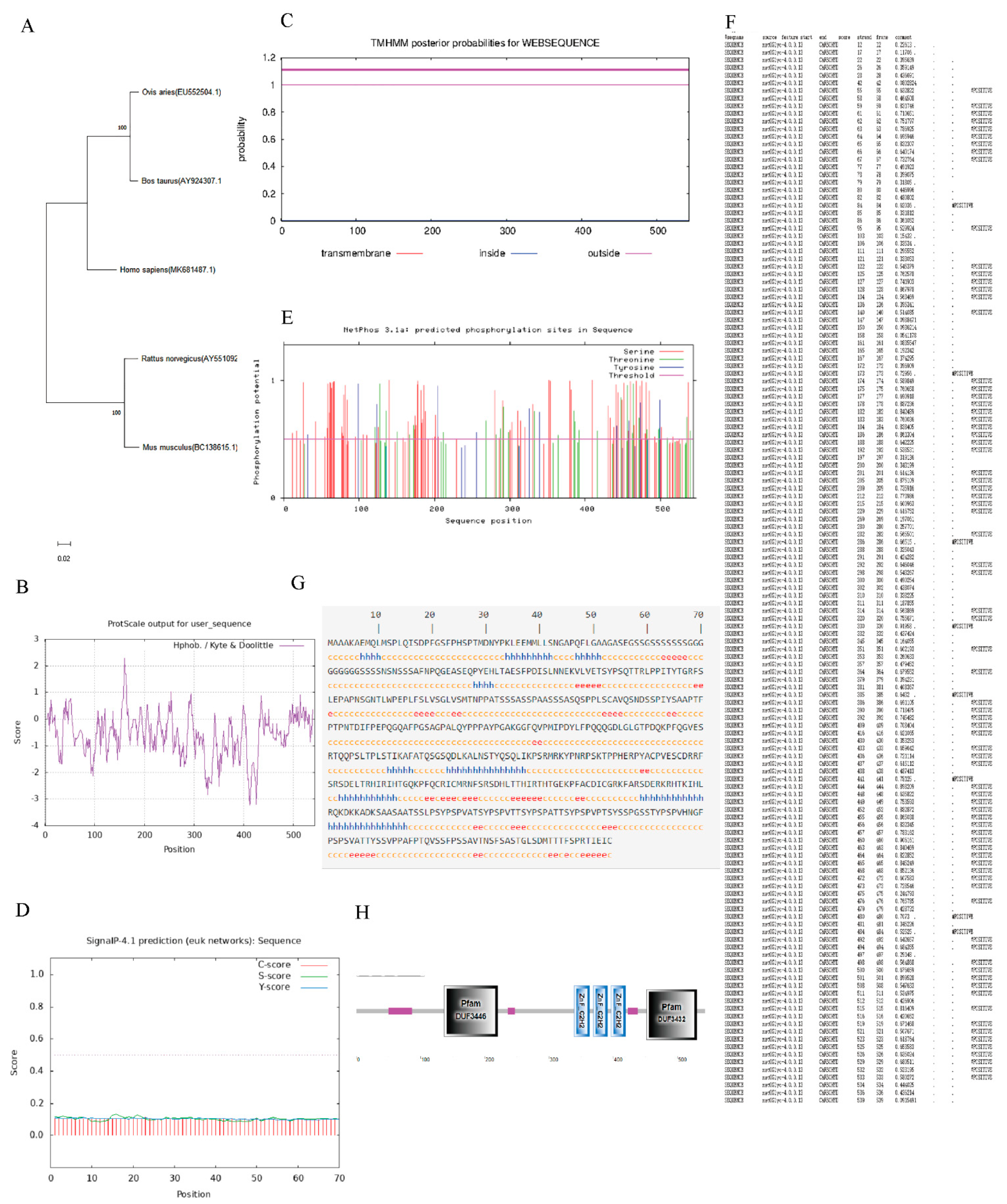
3.2. Bioinformatics Analysis of EGR1

The CDS nucleotide sequence of the sheep EGR1 gene (EU552504.1) was compared with that of bta, hsa, rno and mmu using BLAST in NCBI, and the results showed that the homology between sheep and bta hsa rno and mmu was 97.06%, 92.03%, 84.85% and 84.70% respectively (Table 3). This result demonstrated that the gene is relatively conserved during biological evolution. A nucleotide phylogenetic tree model was constructed using MEGA11, and it was found that the sheep were most closely related to bta during the evolution of the species (Figure 2A).
As previously mentioned, bioinformatics analysis revealed that the EGR1 CDS region of Hu sheep is 1632 bp and encodes 543 amino acids. The relative molecular mass is 57,512.70 Da and the isoelectric point is 8.50. In mammalian red blood cells, the protein has a half-life of 30 h and an instability coefficient of 75.20, indicating that the protein is unstable. The lipolysis index was 49.32 and the total average hydrophilicity (GRAVY) was −0.572, indicating that the protein is hydrophilic. Further hydrophobic analysis of the protein according to the ProtScale online software showed that the hydrophobic region was above the vertical coordinate of 0 (the higher the score, the stronger the hydrophobicity), and the hydrophilic region was below 0 (the lower the score, the stronger the hydrophilicity). This shows that the amino acid sequence has more hydrophilic residues than hydrophobic residues, so the overall performance is hydrophilic, which is the same as the hydrophilic result predicted by the ProtParam software (Figure 2B). The TMHMM-2.0 online software predicted the transmembrane structure of the EGR1 protein. The structure showed that all amino acids of the protein are outside the membrane (Figure 2C) and there is no transmembrane structure, so the protein is presumed to be an extramembrane protein. Prediction of the signal peptide of the EGR1 protein by the online software SignalP4.1 showed that no signal peptide sequence exists for this protein, which means that it is not a secreted protein (Figure 2D). Subcellular localization analysis using PSORT II prediction showed that the protein was localised to the nucleus and cytoplasm, with 95.7% and 4.3% of the protein, respectively. NetPhos 3.1 was used to predict the phosphorylation sites of the protein’s amino acid sequence, and these results showed that there were 115 potential phosphorylation sites, most of which were serine (Figure 2E). NetOGlyc 4.0 was used to predict the glycosylation sites of the amino acid sequence of the protein, and the results showed that 86 glycosylation sites were present (Figure 2F). Using GOR IV to analyze the EGR1 protein secondary structure (Figure 2G), it was found that 14.73% of the protein was in the alpha helix (h), 12.52% was in the extended chain (e), and 72.74% was in the random coiled coil (c). The structural domains of the amino acid sequence of this protein were predicted by SMART online software, and the results showed that there is a Pfam:DUF3446 structural domain between amino acids 137 and 220, respectively, and a Pfam:DUF3432 structural domain between amino acids 452 and 530, respectively, and that the EGR1 protein structure also contains three low-complexity regions and three ZnF_C2H2 (Figure 2H)

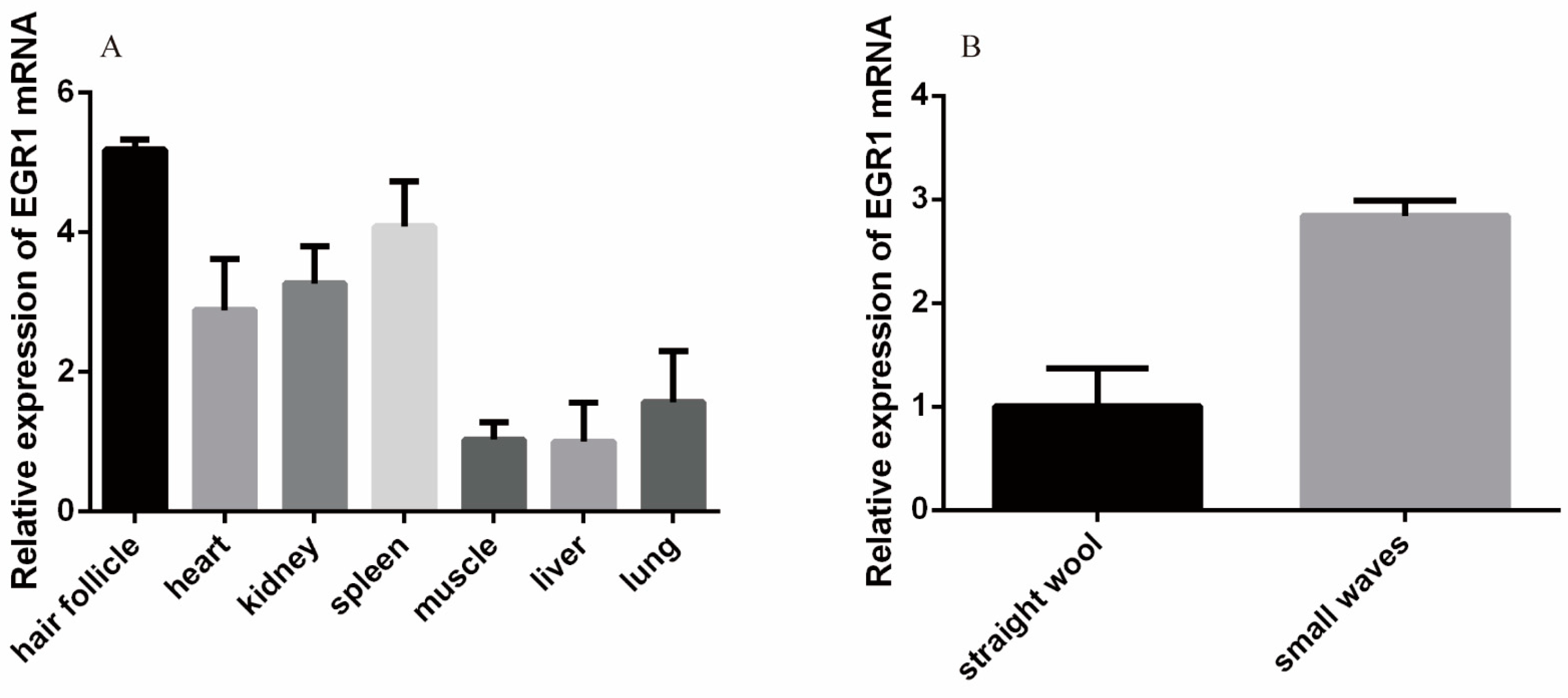
3.3. Analysis of EGR1 Expression Profiles in Different Tissues

According to RT-qPCR results, the EGR1 gene was widely expressed in the heart, liver, spleen, lung, kidney, muscle, and hair follicle tissues of Hu sheep. The highest expression was observed in hair follicle tissue, followed by spleen, and the least in the liver. EGR1 gene expression in different patterns of Hu sheep was higher in small waves than in straight wool (Figure 3).

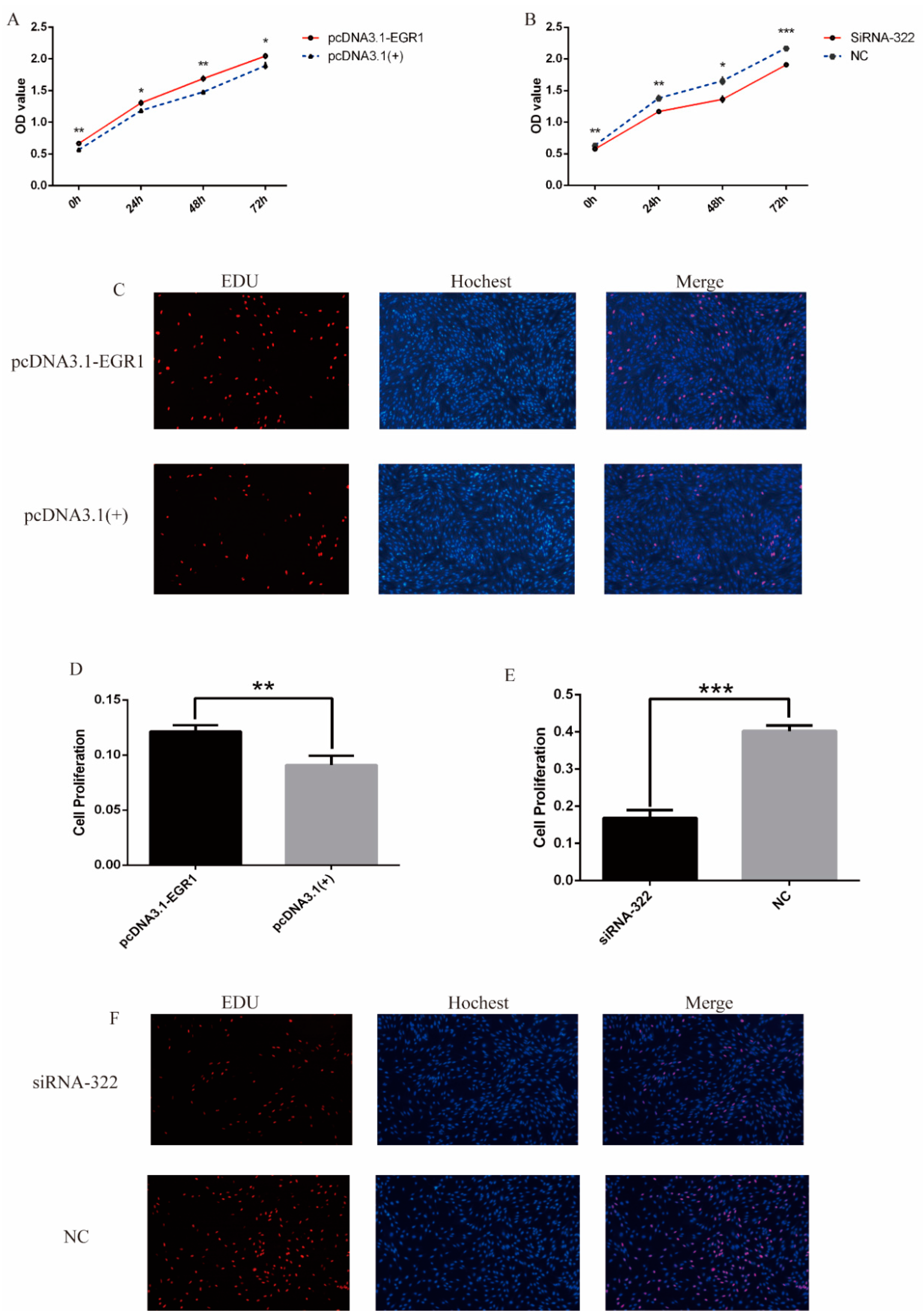
3.4. EGR1 Promoted Proliferation of DPCs

To verify the effect of EGR1 on DPCs, siRNA-322, siRNA632, siRNA1235, NC, pcDNA3.1-EGR1, and pcDNA3.1(+) were introduced into DPCs, cultured in 12-well plates and 6-well plates, respectively. The experiments were grouped as follows: siRNA-322, siRNA-632, siRNA-1235 and NC were transfected with DPCs at the concentration of 50 nM, and each treatment was repeated three times to verify the effect of the three siRNAs. RT-qPCR results showed that pcDNA3.1-EGR1 could up-regulate EGR1 expression at the mRNA expression level (p < 0.01) (Figure 4A). The expression of EGR1 gene was significantly reduced after transfection with siRNA-322 (p < 0.001). EGR1 mRNA expression was significantly reduced after transfection with siRNA-1235 (p < 0.01), while siRNA-632 had no inhibitory effect on EGR1 expression in Hu sheep DPCs (Figure 4B). Therefore, we chose to transfect siRNA-322 into DPCs for the following Western blot, EDU and CCK8 experiments. Western blot results showed that pcDNA3.1-EGR1 up-regulated EGR1 protein expression (Figure 4C,D), whereas siRNA-322 down-regulated EGR1 protein expression (Figure 4E,F). In addition, markers associated with cell proliferation, including PCNA and CDK2, were significantly increased at the mRNA level after pcDNA3.1-EGR1 transfection, whereas they were decreased at the mRNA level after siRNA-322 transfection (Figure 4G,H). Western blot results showed that markers associated with cell proliferation, including PCNA and CDK2, were significantly increased following pcDNA3.1-EGR1 transfection (Figure 4I,J) and decreased under siRNA-322 treatment (Figure 4K,L).
To evaluate the proliferative effect of EGR1 on DPCs, cell proliferation was detected. CCK-8 assay revealed that the proliferation rate of cells in the pcDNA3.1-EGR1 transfected group was significantly higher than that in NC (Figure 5A), while the proliferation rate under siRNA322 treatment was significantly lower than that in NC (Figure 5B). The EDU assay demonstrates similar results, with EGR1 playing a positive effect on the proliferation of DPCs (Figure 5C–F).

4. Discussion

The lambskin quality is majorly determined by the pattern of lambskin; however, in recent years, the lambskin market has been shrinking and its economic value has been decreasing, leading to the neglection of lambskin trait breeding selection and the quality being in decline. However, the lambskin trait, as a unique and excellent trait of Hu sheep, has an important value for germplasm conservation. Therefore, it is of great social value to uncover the molecular mechanism underlying lambskin pattern formation for the future use of molecular breeding approaches to protect and improve the quality of lambskin.
Lambskin pattern formation is closely related to wool bending, which is determined by the cellular activity of the hair follicle cells; precisely, the activity of DPCs and other hair follicle cells affects hair bending by direct or indirect means [5]. DPCs are a group of cells that differentiate from dermal mesenchymal cells and are located in the center of the hair follicle [17]. They play a leading role in the morphogenesis and cyclic regulation of the hair follicle, and are the regulatory center of hair follicle development [18,19]. The activity of the hair follicle depends on DPCs providing it with a number of important nutrients, as well as signal regulators [20,21]. DPCs are considered to be multifunctional stem cells that regulate the growth and development of hair follicles [22]. Some researchers believe that the main cause of hair follicle curvature is the autonomous functioning of multiple papillae centers formed in the dermal papillae, which probably causes asymmetric hair growth and hair curvature [5]. Others insist that curly hairs originate from a hair bulb surrounded by abundant proliferating cells that cause curved hair growth due to the uneven distribution of their proliferation space [23]. As DPCs have certain potential to proliferate and differentiate and are present in the center of the hair bulb, this study investigates the formation of curved wool growth from the perspective of DPCs proliferation.
EGR1 is an important transcription factor that has been widely studied in the areas of oncology, neuropsychiatric disease and diabetic kidney disease (DKD) [24,25,26], but it is not clear whether EGR1 is associated with hair growth. EGR1 encodes a protein with a zinc-binding finger structure, which acts as a DNA-binding domain in several transcriptional regulatory proteins [27]. EGR1 is abundantly expressed during embryogenesis in bone, tendons and skeletal muscle in mice [28]. Interestingly, mice lacking EGR1 could survive despite a reduced body size, suggesting that EGR1 may be associated with organismal development [29,30,31]. Collectively, EGR1 mainly plays a positive regulatory role for cell growth and development, consistent with the basic characteristics of this gene; our results also showed that EGR1 was able to promote the proliferation of dermal papilla cells. Additionally, EGR1 can be regulated at the epigenetic level through microRNAs (miRNAs). Indeed, quite a lot of studies reported direct or indirect targeting of EGR1 by miR-203a, miR-377-3p, miR-125b-2-3p and miR-301b in many cancer cell lines, which slowed down the proliferation of cancer cells to a certain extent by inhibiting the expression of EGR1 [32,33,34,35]. In the investigation of transcription factors important for the BMP7 promoter in Hu sheep, Lv et al. [14] found that EGR1 is located in the core transcriptional region, and that its expression enhances the transcriptional regulation of BMP7, a candidate gene involved in promoting proliferation in Hu dermal papilla cells. In addition, Adly et al. [36] found that BMP7 was highly expressed in hair follicles during the anagen phase and in epidermal hair papillae, and that when follicle cells proliferated at the basal level, these cells immediately expressed BMP7 at high levels; moreover, as follicle cells proliferated into the regressive and quiescent phases, most follicle cells ceased to express BMP7, and expression in epidermal hair papillae diminished accordingly. Combining previous findings with the results of our study, we can speculate that EGR1, as an important transcription factor of BMP7, can enhance the expression of the BMP7 gene and thus promote the proliferation of hair papilla cells. Meanwhile, in our study, we also identified that EGR1 expression was higher in DPCs of Hu sheep small waves than in straight wool. The proliferation rate of DPCs was significantly increased after overexpression of EGR1 in DPCs. The reports confirmed that the proliferation of dermal papilla cells led to curly hair growth; additionally, in our study, the curl was higher in the small waves group, in which the expression level of EGR1 was also higher than that of the straight wool group, which further confirmed the positive correlation between the expression of EGR1 and the proliferation of dermal papilla cells.
Numerous pathways have been proved to be involved in hair follicle growth and development, among which the MAPK/ERK pathway is a classical activating proliferative pathway. One study revealed that EGR1 expression was significantly reduced after treatment with inhibitors of the MAPK/ERK pathway, suggesting that EGR1 was closely related to the MAPK/ERK pathway [37]. In addition to this, it has been shown that attenuating the nuclear fraction of EGR1 apparently inhibits the survival of breast cancer cells by inhibiting MAPK phosphorylation [38]. EGR1 is an activator of the MAPK/ERK pathway, while the activated pathway, in turn, enhances EGR1 expression. Upregulated EGR1 promotes cell proliferation and continues to enhance activation of the MAPK/ERK pathway, which is a key pathway regulating the biological activity of the hair follicle through a positive feedback loop [39]. Therefore, it is reasonable to infer that EGR1 may promote hair follicle growth and development by enhancing the MAPK/ERK pathway, which is also the direction of our subsequent study.

5. Conclusions

We cloned the sequence of CDS region of EGR1 in Hu sheep and found that it can promote the proliferation of dermal papilla cells after cellular analysis, which provides a certain basis for the subsequent study of the mechanism of lambskin pattern formation in Hu sheep.

Author Contributions

W.S. and X.L. designed the experiment; Y.X. and X.L. conducted the experiments; S.W., X.C. and Z.Y. contributed to materials and data collection in this study. Y.X. analyzed the data; Y.X. wrote the manuscript; T.G., J.M.M. and A.H. revised manuscript. All authors have read and agreed to the published version of the manuscript.

Funding

This research was funded by the National Natural Science Foundation of China (31872333, 32172689), the National Natural Science Foundation of China-CGIAR (32061143036), the Major New Varieties of Agricultural Projects in Jiangsu Province (PZCZ201739), the Open Project Program of Joint International Research Laboratory of Agriculture and Agri-Product Safety, the Ministry of Education of China, Yangzhou University (JILAR-KF202205), the Projects of Domesticated Animals Platform of the Ministry of Science, Key Research and Development Plan (modern agriculture) in Jiangsu Province (BE2018354), the Jiangsu Agricultural Science and Technology Innovation Fund (CX(18)2003), the Jiangsu 333 Distinguished Talents Project Foundation [(2022) 2-323], and the Natural Science Foundation of the Jiangsu Higher Education Institutions of China (22KJA230001, 22KJD230003).

Institutional Review Board Statement

The animal study protocol was approved by the Animal Ethics Committee of Yangzhou University (No: NSFC2020-NFY-1. Date: 15 January 2020).

Informed Consent Statement

Not applicable.

Data Availability Statement

The raw data supporting the conclusions of this article will be made available by the authors, without undue reservation.

Conflicts of Interest

All the authors declare no competing interests.

References

  1. Lv, X.; Chen, W.; Sun, W.; Hussain, Z.; Chen, L.; Wang, S.; Wang, J. Expression profile analysis to identify circular RNA expression signatures in hair follicle of Hu sheep lambskin. Genomics 2020, 112, 4454–4462. [Google Scholar] [CrossRef] [PubMed]
  2. Hardy, M.H. The secret life of the hair follicle. Trends Genet. 1992, 8, 55–61. [Google Scholar] [CrossRef]
  3. Botchkarev, V.A. Bone morphogenetic proteins and their antagonists in skin and hair follicle biology. J. Investig. Dermatol. 2003, 120, 36–47. [Google Scholar] [CrossRef] [Green Version]
  4. Reddy, S.; Andl, T.; Bagasra, A.; Lu, M.M.; Epstein, D.J.; Morrisey, E.E.; Millar, S.E. Characterization of Wnt gene expression in developing and postnatal hair follicles and identification of Wnt5a as a target of Sonic hedgehog in hair follicle morphogenesis. Mech. Develop. 2001, 107, 69–82. [Google Scholar] [CrossRef]
  5. Nissimov, J.N.; Das Chaudhuri, A.B. Hair curvature: A natural dialectic and review. Biol. Rev. Camb. Philos. Soc. 2014, 89, 723–766. [Google Scholar] [CrossRef]
  6. Thiel, G.; Cibelli, G. Regulation of life and death by the zinc finger transcription factor Egr-1. J. Cell Physiol. 2002, 193, 287–292. [Google Scholar] [CrossRef]
  7. Unoki, M.; Nakamura, Y. EGR2 induces apoptosis in various cancer cell lines by direct transactivation of BNIP3L and BAK. Oncogene 2003, 22, 2172–2185. [Google Scholar] [CrossRef] [Green Version]
  8. Vaish, V.; Piplani, H.; Rana, C.; Vaiphei, K.; Sanyal, S.N. NSAIDs may regulate EGR-1-mediated induction of reactive oxygen species and non-steroidal anti-inflammatory drug-induced gene (NAG)-1 to initiate intrinsic pathway of apoptosis for the chemoprevention of colorectal cancer. Mol. Cell Biochem. 2013, 378, 47–64. [Google Scholar] [CrossRef] [PubMed]
  9. Kim, J.H.; Jeong, I.Y.; Lim, Y.; Lee, Y.H.; Shin, S.Y. Estrogen receptor beta stimulates Egr-1 transcription via MEK1/Erk/Elk-1 cascade in C6 glioma cells. BMB Rep. 2011, 44, 452–457. [Google Scholar] [CrossRef]
  10. Li, L.; Ameri, A.H.; Wang, S.; Jansson, K.H.; Casey, O.M.; Yang, Q.; Beshiri, M.L.; Fang, L.; Lake, R.G.; Agarwal, S.; et al. EGR1 regulates angiogenic and osteoclastogenic factors in prostate cancer and promotes metastasis. Oncogene 2019, 38, 6241–6255. [Google Scholar] [CrossRef] [PubMed]
  11. Myung, E.; Park, Y.L.; Kim, N.; Chung, C.Y.; Park, H.B.; Park, H.C.; Myung, D.S.; Kim, J.S.; Cho, S.B.; Lee, W.S.; et al. Expression of early growth response-1 in human gastric cancer and its relationship with tumor cell behaviors and prognosis. Pathol. Res. Pract. 2013, 209, 692–699. [Google Scholar] [CrossRef] [PubMed]
  12. Calogero, A.; Arcella, A.; De Gregorio, G.; Porcellini, A.; Mercola, D.; Liu, C.T.; Lombari, V.; Zani, M.; Giannini, G.; Gagliardi, F.M.; et al. The early growth response gene EGR-1 behaves as a suppressor gene that is down-regulated independent of ARF/Mdm2 but not p53 alterations in fresh human gliomas. Clin. Cancer Res. 2001, 7, 2788–2796. [Google Scholar] [PubMed]
  13. Kumbrink, J.; Gerlinger, M.; Johnson, J.P. Egr-1 induces the expression of its corepressor nab2 by activation of the nab2 promoter thereby establishing a negative feedback loop. J. Biol. Chem. 2005, 280, 42785–42793. [Google Scholar] [CrossRef] [PubMed] [Green Version]
  14. Lv, X.; Sun, W.; Zou, S.; Chen, L.; Mwacharo, J.M.; Wang, J. Characteristics of the BMP7 Promoter in Hu Sheep. Animals 2019, 9, 874. [Google Scholar] [CrossRef] [Green Version]
  15. Lv, X.; Gao, W.; Jin, C.; Wang, L.; Wang, Y.; Chen, W.; Zou, S.; Huang, S.; Li, Z.; Wang, J.; et al. Preliminary study on microR-148a and microR-10a in dermal papilla cells of Hu sheep. BMC Genet. 2019, 20, 70. [Google Scholar] [CrossRef] [Green Version]
  16. Schmittgen, T.D.; Livak, K.J. Analyzing real-time PCR data by the comparative C-T method. Nat. Protoc. 2008, 3, 1101–1108. [Google Scholar] [CrossRef]
  17. Reynolds, A.J.; Lawrence, C.; Cserhalmi-Friedman, P.B.; Christiano, A.M.; Jahoda, C.A.B. Trans-gender induction of hair follicles. Nature 1999, 402, 33–34. [Google Scholar] [CrossRef]
  18. Driskell, R.R.; Giangreco, A.; Jensen, K.B.; Mulder, K.W.; Watt, F.M. Sox2-positive dermal papilla cells specify hair follicle type in mammalian epidermis. Development 2009, 136, 2815–2823. [Google Scholar] [CrossRef] [Green Version]
  19. Chi, W.; Wu, E.; Morgan, B.A. Dermal papilla cell number specifies hair size, shape and cycling and its reduction causes follicular decline. Development 2013, 140, 1676–1683. [Google Scholar] [CrossRef] [Green Version]
  20. Alonso, L.; Fuchs, E. The hair cycle. J. Cell Sci. 2006, 119, 391–393. [Google Scholar] [CrossRef]
  21. Jahoda, C.A.B.; Oliver, R.F.; Reynolds, A.J.; Forrester, J.C.; Gillespie, J.W.; Cserhalmi-Friedman, P.B.; Christiano, A.M.; Horne, K.A. Trans-species hair growth induction by human hair follicle dermal papillae. Exp. Dermatol. 2001, 10, 229–237. [Google Scholar] [CrossRef] [PubMed]
  22. Driskell, R.R.; Clavel, C.; Rendl, M.; Watt, F.M. Hair follicle dermal papilla cells at a glance. J. Cell Sci. 2011, 124, 1179–1182. [Google Scholar] [CrossRef] [PubMed] [Green Version]
  23. Pierard-Franchimont, C.; Paquet, P.; Quatresooz, P.; Pierard, G.E. Mechanobiology and cell tensegrity: The root of ethnic hair curling? J. Cosmet. Dermatol. 2011, 10, 163–167. [Google Scholar] [CrossRef] [PubMed]
  24. Saha, S.K.; Islam, S.M.R.; Saha, T.; Nishat, A.; Biswas, P.K.; Gil, M.; Nkenyereye, L.; El-Sappagh, S.; Islam, M.S.; Cho, S.G. Prognostic role of EGR1 in breast cancer: A systematic review. BMB Rep. 2021, 54, 497–504. [Google Scholar] [CrossRef] [PubMed]
  25. Tallafuss, A.; Stednitz, S.J.; Voeun, M.; Levichev, A.; Larsch, J.; Eisen, J.; Washbourne, P. Egr1 Is Necessary for Forebrain Dopaminergic Signaling during Social Behavior. Eneuro 2022, 9, ENEURO.0035-22.2022. [Google Scholar] [CrossRef]
  26. Sheng, S.Y.; Zou, M.N.; Yang, Y.L.; Guan, M.P.; Ren, S.J.; Wang, X.Y.; Wang, L.; Xue, Y.M. miR-23a-3p regulates the inflammatory response and fibrosis in diabetic kidney disease by targeting early growth response 1. In Vitro Cell Dev. Biol.-Anim. 2021, 57, 763–774. [Google Scholar] [CrossRef]
  27. Patwardhan, S.; Gashler, A.; Siegel, M.G.; Chang, L.C.; Joseph, L.J.; Shows, T.B.; Le Beau, M.M.; Sukhatme, V.P. EGR3, a novel member of the Egr family of genes encoding immediate-early transcription factors. Oncogene 1991, 6, 917–928. [Google Scholar]
  28. Gupta, M.P.; Gupta, M.; Zak, R.; Sukhatme, V.P. Egr-1, a serum-inducible zinc finger protein, regulates transcription of the rat cardiac alpha-myosin heavy chain gene. J. Biol. Chem. 1991, 266, 12813–12816. [Google Scholar] [CrossRef]
  29. Lee, S.L.; Tourtellotte, L.C.; Wesselschmidt, R.L.; Milbrandt, J. Growth and differentiation proceeds normally in cells deficient in the immediate early gene NGFI-A. J. Biol. Chem. 1995, 270, 9971–9977. [Google Scholar] [CrossRef] [Green Version]
  30. Beckmann, A.M.; Wilce, P.A. Egr transcription factors in the nervous system. Neurochem. Int. 1997, 31, 477–510. [Google Scholar] [CrossRef]
  31. Topilko, P.; Schneider-Maunoury, S.; Levi, G.; Trembleau, A.; Gourdji, D.; Driancourt, M.A.; Rao, C.V.; Charnay, P. Multiple pituitary and ovarian defects in Krox-24 (NGFI-A, Egr-1)-targeted mice. Mol. Endocrinol. 1998, 12, 107–122. [Google Scholar] [CrossRef] [PubMed]
  32. Wang, L.M.; Sun, H.F.; Wang, X.F.; Hou, N.; Zhao, L.Y.; Tong, D.D.; He, K.; Yang, Y.; Song, T.S.; Yang, J.; et al. EGR1 mediates miR-203a suppress the hepatocellular carcinoma cells progression by targeting HOXD3 through EGFR signaling pathway. Oncotarget 2016, 7, 45302–45316. [Google Scholar] [CrossRef] [PubMed] [Green Version]
  33. Ke, X.; He, L.; Wang, R.; Shen, J.; Wang, Z.; Shen, Y.; Fan, L.; Shao, J.; Qi, H. miR-377-3p-Mediated EGR1 Downregulation Promotes B[a]P-Induced Lung Tumorigenesis by Wnt/Beta-Catenin Transduction. Front. Oncol. 2021, 11, 699004. [Google Scholar] [CrossRef] [PubMed]
  34. Meng, X.Y.; Liu, K.T.; Xiang, Z.F.; Yu, X.; Wang, P.; Ma, Q. MiR-125b-2-3p associates with prognosis of ccRCC through promoting tumor metastasis via targeting EGR1. Am. J. Transl. Res. 2020, 12, 5575–5585. [Google Scholar]
  35. Yan, L.; Wang, Y.; Liang, J.; Liu, Z.; Sun, X.; Cai, K. MiR-301b promotes the proliferation, mobility, and epithelial-to-mesenchymal transition of bladder cancer cells by targeting EGR1. Biochem. Cell Biol. 2017, 95, 571–577. [Google Scholar] [CrossRef]
  36. Adly, M.A.; Assaf, H.A.; Hussein, M.R. Expression of bone morphogenetic protein-7 in human scalp skin and hair follicles. Br. J. Dermatol. 2006, 154, 551–554. [Google Scholar] [CrossRef]
  37. Schwachtgen, J.L.; Houston, P.; Campbell, C.; Sukhatme, V.; Braddock, M. Fluid shear stress activation of egr-1 transcription in cultured human endothelial and epithelial cells is mediated via the extracellular signal-related kinase 1/2 mitogen-activated protein kinase pathway. J. Clin. Investig. 1998, 101, 2540–2549. [Google Scholar] [CrossRef] [Green Version]
  38. Burnatowska-Hledin, M.A.; Kossoris, J.B.; Van Dort, C.J.; Shearer, R.L.; Zhao, P.; Murrey, D.A.; Abbott, J.L.; Kan, C.E.; Barney, C.C. T47D breast cancer cell growth is inhibited by expression of VACM-1, a cul-5 gene. Biochem. Bioph. Res. 2004, 319, 817–825. [Google Scholar] [CrossRef]
  39. Lv, X.Y.; Sun, W.; Yin, J.F.; Ni, R.; Su, R.; Wang, Q.Z.; Gao, W.; Bao, J.J.; Yu, J.R.; Wang, L.H.; et al. An Integrated Analysis of MicroRNA and mRNA Expression Profiles to Identify RNA Expression Signatures in Lambskin Hair Follicles in Hu Sheep. PLoS ONE 2016, 11, e0157463. [Google Scholar] [CrossRef]
Figure 1. The different patterns of lambskin of Hu lambs.
Genes 13 01242 g001
Figure 2. Bioinformatics analysis of EGR1. (A) Phylogenetic tree of EGR1 between different species. (B) The hydropathy profile of EGR1 amino acid. (C) EGR1 protein transmembrane regional analyses. (D) EGR1 signal peptide prediction. (E) EGR1 phosphorylation site analyses. (F) EGR1 glycosyl site analyses. (G) EGR1 protein secondary structure prediction. (H) EGR1 conservative structure domain analyses.
Genes 13 01242 g002
Figure 3. Expression level analysis of EGR1. (A) Expression profiles of EGR1 in different tissues. (B) Expression of the EGR1 gene in different patterns.
Genes 13 01242 g003
Figure 4. The effects of EGR1 on dermal papilla cell proliferation. (A) mRNA relative expression of EGR1 after pcDNA3.1-EGR1 transfection. (B) Relative expression of mRNA for EGR1 in DPCs transfected with siRNA. (C) Relative expression of EGR1 protein after pcDNA3.1-EGR1 transfection. (D) Protein bands after pcDNA3.1-EGR1 transfection. (E) Relative expression of protein for EGR1 gene in DPCs transfected with siRNA-322. (F) Protein bands for EGR1 in DPCs transfected with siRNA-322. (G) mRNA relative expression of PCNA and CDK2 after pcDNA3.1-EGR1 transfection. (H) The mRNA expression level of PCNA and CDK2 in DPCs transfected with siRNA-322. (I) The protein expression level of PCNA and CDK2 after pcDNA3.1-EGR1 transfection. (J) Protein bands for PCNA and CDK2 after pcDNA3.1-EGR1 transfection. (K) Relative expression of protein for PCNA and CDK2 in DPCs transfected with siRNA-322. (L) Protein bands for PCNA and CDK2 in DPCs transfected with siRNA-322. “*” represents a significant difference (p < 0.05), “**” represents a highly significant difference (p < 0.01), “***” represents an extremely significant difference (p < 0.001).
Genes 13 01242 g004
Figure 5. Effect of EGR1 on the proliferation of DPCs. (A) The dermal papilla cell activity after overexpression of EGR1 by CCK-8. (B) The dermal papilla cell activity after EGR1 down-regulation by CCK-8. (C,D) The proliferation rate of DPCs after overexpression of EGR1 by EDU immunofluorescence (100×). (E,F) The rate of proliferating cells after EGR1 down-regulation (100×). “*” represents a significant difference (p < 0.05), “**” represents a highly significant difference (p < 0.01), “***” represents an extremely significant difference (p < 0.001).
Genes 13 01242 g005
Table 1. The sequence information of siRNA-BMP4.
NameSequence (5′→3′)
siRNA-322CCUGACAUCUCUCUGAAUATT
UAUUCAGAGAGAUGUCAGGTT
siRNA-632CCACACCUAACACUGACAUTT
AUGUCAGUGUUAGGUGUGGTT
siRNA-1235GCAAGAGGCAUACCAAGAUTT
AUCUUGGUAUGCCUCUUGCTT
Table 2. The sequence information of genes for RT-qPCR.
Gene IDSequences (5′→30′)Product Length/bpAccession No.
EGR1F: TTCAACCCTCAGGGGGAGG
R: CGCTGACCAGACTGAAGAGG
223EU552504.1
PCNAF: TCTGCAAGTGGAGAACTTGGAA
R: AGGAGACAGTGGAGTGGCTT
162XM_004014340.5
CDK2F: TGGGCCAGGCAGGATTTTAG
R: GTCGAAGGTGAGGTACTGGC
166FJ422550.1
GAPDHF: TCTCAAGGGCATTCTAGGCTAC
R: GCCGAATTCATTGTCGTACCAG
151NM_001190390.1
Table 3. Similarity analysis of nucleotide and amino acid of EGR1.
SpeciesGenBank No.Nucleotide (%)Amino Acid (%)
Bos taurusAY924307.197.0698.53
Homo sapiensMK681487.192.0395.88
Rattus norvegicusAY551092.184.8585.69
Mus musculusBC138615.184.7086.42
Publisher’s Note: MDPI stays neutral with regard to jurisdictional claims in published maps and institutional affiliations.

