Article

Orchid NAC Transcription Factors: A Focused Analysis of CUPULIFORMIS Genes

by
Maria Carmen Valoroso
1,*,
Francesca Lucibelli
2 and
Serena Aceto
2,*
1
Department of Agricultural Sciences, University of Napoli Federico II, 80055 Portici, Italy
2
Department of Biology, University of Naples Federico II, 80126 Napoli, Italy
*
Authors to whom correspondence should be addressed.
Genes 2022, 13(12), 2293; https://doi.org/10.3390/genes13122293
Submission received: 15 November 2022 / Revised: 1 December 2022 / Accepted: 2 December 2022 / Published: 5 December 2022
(This article belongs to the Collection Feature Papers: 'Plant Genetics and Genomics' Section)

Abstract

:
Plant transcription factors are involved in different developmental pathways. NAC transcription factors (No Apical Meristem, Arabidopsis thaliana Activating Factor, Cup-shaped Cotyledon) act in various processes, e.g., plant organ formation, response to stress, and defense mechanisms. In Antirrhinum majus, the NAC transcription factor CUPULIFORMIS (CUP) plays a role in determining organ boundaries and lip formation, and the CUP homologs of Arabidopsis and Petunia are involved in flower organ formation. Orchidaceae is one of the most species-rich families of angiosperms, known for its extraordinary diversification of flower morphology. We conducted a transcriptome and genome-wide analysis of orchid NACs, focusing on the No Apical Meristem (NAM) subfamily and CUP genes. To check whether the CUP homologs could be involved in the perianth formation of orchids, we performed an expression analysis on the flower organs of the orchid Phalaenopsis aphrodite at different developmental stages. The expression patterns of the CUP genes of P. aphrodite suggest their possible role in flower development and symmetry establishment. In addition, as observed in other species, the orchid CUP1 and CUP2 genes seem to be regulated by the microRNA, miR164. Our results represent a preliminary study of NAC transcription factors in orchids to understand the role of these genes during orchid flower formation.

1. Introduction

Plant transcription factors control many developmental processes, regulating the temporal and spatial expression of genes involved in plant growth and development. Among more than 2500 transcription factors present in plant genomes, a large number is involved in the development of the flower [1].
The NAC genes form one of the largest plant-specific families of transcription factors involved in different developmental and physiological processes such as organ formation [2,3,4], root development [5,6,7], organ boundary formation [8], plant defense [9,10,11], and response to abiotic stress [12,13,14].
Their name derives from the NAM gene (No Apical Meristem) in Petunia, ATAF (A. thaliana Activating Factor), and CUC (Cup-shaped Cotyledon) in Arabidopsis. The Petunia nam mutants lack a shoot apical meristem and show defects in organ boundaries [4]. This same phenotype characterizes the Arabidopsis cuc1 and cuc2 mutants and the cupuliformis (cup) mutant of the snapdragon, A. majus [2,8,15].
In the A. thaliana genome, more than 100 genes encode NAC transcription factors, divided into sixteen subfamilies based on amino acid sequence similarity [16,17]. The typical structure of a NAC protein has a conserved NAC domain at the N-terminus, with a twisted antiparallel β-sheet enclosed between two α-helices with five subdomains (A–E) [2,18]. The NAC domain, often found in two tandem repeats, contains positively charged amino acids possibly involved in DNA binding [19]. The NAC domain also enhances the protein homo- and hetero-dimerization, together with the C-terminal end [7,13,20]. This ability can positively or negatively regulate the activity of the NAC proteins.
The C-terminal region of the NAC proteins is poorly conserved; however, it includes a transcriptional activation region (TAR) containing serine, proline, and other simple amino acid repetitions [2,16]. Some atypical NAC transcription factors have a conserved NAC domain at the N-terminus and a conserved or absent C-terminal region [21].
Most studies into NAC transcription factors analyzed their function in root and leaf development and organ boundary formation [6,7,8,22,23,24,25]. However, in A. majus, the role of some NACs in the formation of the lip, the peculiar ventral petal of the snapdragon flower, was also explored. [15]. In this species, the cup mutant displays defects in the vegetative tissues, such as the cuc and nam mutants; it also shows an altered flower development. In particular, its corolla has an open mouth that lacks the folding characteristic of the palate and the lip regions. These mutant flowers lose bilateral symmetry, have similar petals, and their stamens are fused with the corolla tube [15,26]. CUP is a NAC transcription factor, and its involvement in flower formation was also described for Arabidopsis and Petunia [8,15,24].
The CUP gene of A. majus is differentially expressed during flower development. It is transcribed in the basal floral meristem region during the early developmental stages (5-10 days after floral meristem initiation, DAI). Later (11 to 14 DAI), it expands its expression to all the ventral and lateral parts of the flower [15]. During the early stages, the presence of the CUP protein at the organ boundaries is necessary to establish the organ tissue between confines. At the late stages of development, CUP may regulate the formation of the palate and lip in combination with other transcription factors encoded, for example, by the proximodistal genes PALATE and LIP, and the dorsoventral genes DIVARICATA (DIV), CYCLOIDEA (CYC), and DICHOTOMA (DICH) [8,15]. In addition to A. majus, the hypothetical role of the CUP gene in lip development was also proposed for two other Lamiales species, Linaria maroccana and Mimulus guttatus [15].
The activity of the CUP genes is regulated at the post-transcriptional level by the microRNA, miR164, which has a conserved target site on the transcripts of these genes [27,28,29]. In Arabidopsis, miR164 is encoded by three loci: A, B, and C. The isoforms A and B are identical, whereas the isoform C has a substitution of the canonical adenine with guanine at the last nucleotide position [30]. The isoforms miR164 A and B control stem growth, meristem differentiation, leaf shape, and senescence, while the isoform C is involved in floral organ formation [28,29,30,31].
To date, there are few or no studies into the NAC transcription factors in many flowering plant species. For this reason, we decided to analyze these transcription factors in Orchidaceae, one of the most species-rich families of angiosperms, focusing on the genes involved in flower organ formation in other species.
The bilaterally symmetric orchid flowers exhibit an extraordinary differentiation of morphology and adaptations [32,33]. Despite their great morphological diversity, the orchid flowers share a typical structure composed of three outer tepals, two laterals inner tepals, and a highly diversified median inner tepal (lip or labellum) [34]. Male and female reproductive tissues are fused to form the column, with the pollen grains at the apex and the ovary at the base [34].
As in all flowering plants, the genetic program at the basis of flower organ formation in orchids is realized mainly through the action of different MADS-box transcription factors [32,35,36,37,38,39,40,41,42,43]. In addition to this program, known as the ABCE model, an orchid-specific regulatory network involving the class B MADS-box genes DEFICIENS-like (DEF-like) and AGAMOUS-LIKE 6 (AGL6) explains lip formation and symmetry determination [32,35,44].
However, the regulatory pathway underpinning the development of the orchid perianth organs is not restricted to the interaction of different classes of MADS-box genes. Some studies showed a conserved function of the MYB transcription factors DIV, RADIALIS (RAD), and DIV-and-RAD-Interacting-Factor (DRIF) in establishing bilateral symmetry of orchids [44,45] and the possible involvement of TCP and YABBY transcription factors in orchid lip formation [46,47]. Therefore, increasing evidence allows us to hypothesize that the molecular network driving orchid flower formation is more complex than previously thought.
Based on these premises and the role of the CUP genes in the flower organ formation of Antirrhinum and other species, we analyzed the orchid CUP genes to verify their possible involvement in orchid perianth formation.
We identified the orchid genes encoding NAC transcription factors and investigated their genomic organization, starting with the available orchid genomes and transcriptomes [35,42,43,48,49,50]. In addition, we analyzed the expression patterns of the CUP genes and miR164 in the orchid P. aphrodite at the early and late stages of flower development. P. aphrodite is an ornamental epiphytic orchid with many advantageous characteristics for cultivation and manipulation. Its inflorescence has many large flowers, and its genome was sequenced [48]. For these reasons, we used this species to conduct our expression analysis of the CUP genes and miR164.

2. Materials and Methods
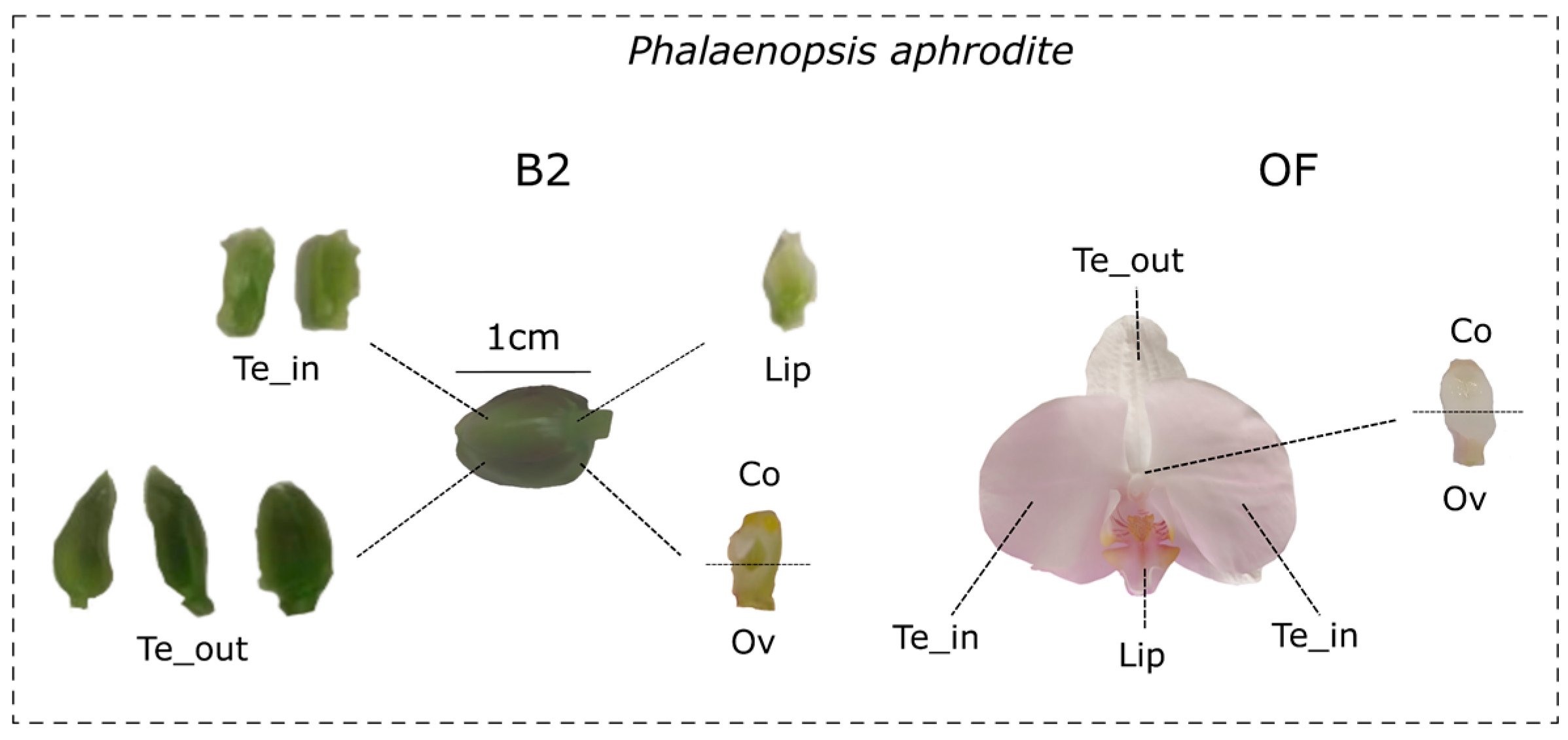
2.1. Plant Material

The plants used in this study were grown in the Department of Biology at the University of Naples Federico II greenhouse (Napoli, Italy) under natural light and temperature. P. aphrodite (subfamily Epidendroideae) is commercially available, while Orchis italica (subfamily Orchidoideae) was collected in Sassano (Salerno, Italy) and is part of the Department of Biology orchid collection.
We sampled P. aphrodite flowers from three different plants at the B2 developmental stage (early, flower bud size 1–1.5 cm) and soon after anthesis (open flower, OF) (Figure 1). We dissected outer tepals, lateral inner tepals, lip, column, and ovary (not pollinated) from each P. aphrodite sample. In addition, we also collected leaf samples of P. aphrodite and O. italica. We stored the collected material in RNA-later (Ambion – Berlin, DE) until RNA extraction.

2.2. Identification, Phylogeny, and Conserved Motif Analysis of the Orchid NAC Genes

Using the conserved NAM domain (PFAM PF02365) as a query, we conducted a TBLASTN search against the available orchid genomes and transcriptomes deposited in the database Orchidstra 2.0 [51], OrchidBase [52], NCBI (https://www.ncbi.nlm.nih.gov/ accessed on 31 July 2022), and the transcriptome of O. italica [49]. When the orchid genome was available, we performed a genome search (Apostasia shenzenica, Dendrobium catenatum, Phalaenopsis equestris–Orchidbase; P. aphrodite–Orchidstra; Vanilla planifolia–NCBI). In the absence of a high-quality genome, we conducted a transcriptome search (Cypripedium fomosanum–Orchidbase; O. italica—reference transcriptome [49]).
We then virtually translated the identified orchid NAC nucleotide sequences to verify the presence of indels and/or stop codons within the coding sequence and evaluate their completeness. We discarded the sequences containing a partial NAM domain and/or stop codons.
In addition, we identified the NAC sequences of selected non-orchid species (A. thaliana, A. majus, Amborella trichopoda, Asparagus officinalis, Zea mays) using BLAST search and the Plaza 4.0 database [53]. The species name and accession number of the sequences used for the analyses are listed in Supplementary Data S1.
We aligned the amino acid sequences of the NAC proteins of Arabidopsis and each orchid species using ClustalW [54]. After manually adjusting the alignments, we constructed the Maximum Likelihood (ML) trees using MEGA7 [55] for each orchid species and Arabidopsis. We classified the orchid NAC sequences based on the ML trees and the subfamily classification of Arabidopsis. We then aligned the NAM amino acid sequences of orchid and non-orchid species and constructed the ML trees with 200 bootstrap replicates using MEGA7.
Using the orchid NAC coding sequences, we conducted a BLASTN search on the genomes of the orchids D. catenatum [43], A. shenzenica [42], P. aphrodite [48], P. equestris [35], and V. planifolia [50]. After reconstructing their genomic organization, we scanned the orchid genomic sequences of the NAC genes using CENSOR v4.2.22 software from Genetic Information Research Institute (GIRI), Cupertino, CA [56] to verify the presence of transposable/repetitive elements within introns.
Using the MEME v5.5 online tool (https://meme-suite.org/meme/) [57], we searched for shared amino acid motifs among the sequences of the orchid NAC proteins, setting the search parameters to a maximum number of 10 motifs and an optimum width from 6 to 50.

2.3. PCR Amplification and Sequencing of the CUP Genes of O. italica

To obtain information about the genomic organization of the highest number of orchid CUP genes, we included O. italica in our analysis. The genome is unavailable for this species; however, we have access to samples of O. italica that are part of the orchid collection of the Department of Biology (University of Naples Federico II). We extracted genomic DNA from leaves of O. italica, PCR-amplified and sequenced the CUP genes, thus reconstructing their exon/intron structure.
Starting with 500 mg of O. italica leaf material, we extracted the genomic DNA following the Doyle and Doyle protocol [58]. We designed specific primer pairs to PCR amplify the CUP genes of O. italica based on the nucleotide sequence of the transcript identified (Table 1). We conducted the amplification reactions in a final volume of 50 µL with the following reagent concentrations: 1X reaction buffer, 0.2 mM of each primer, 0.2 mM dNTP mix Platinum™ GC Enhancer, and 2 U Taq Hot-Start DNA Polymerase Invitrogen™ Platinum™ II (Invitrogen-Waltham, MA). The thermal cycle was the following: 94 °C for 2 min, 35 cycles of 94 °C for 15 sec, 60 °C for 15 sec, and 68 °C for a time ranging from 1:30 sec to 2 min, depending on the amplicon size.
We cloned the amplicons into the pSC-A-amp/kan vector (Agilent, Santa Clara, CA, USA), sequenced them using the T3 and T7 primers (Eurofins Genomics), and deposited the genomic sequences of the CUP genes of O. italica in GenBank with the accession numbers OP752215, OP752217, and OP752219. After reconstructing the genomic organization of the O. italica CUP genes, we verified the presence of transposable/repetitive elements within introns, as described earlier.

2.4. PCR Amplification and Sequencing of P. aphrodite CUP Transcripts

We extracted total RNA from the floral buds of P. aphrodite at the B2 developmental stage using Trizol Reagent (Ambion – Berlin, DE) followed by DNase treatment. We reverse-transcribed 500 ng of total RNA using an Advantage RT-PCR kit (Clontech, Mountain View, CA, USA) with oligo dT and random primers.
We designed CUP-specific primer pairs starting with the sequences of P. aphrodite obtained from the in silico analysis (Table 1). We used 30 ng of first-strand cDNA to PCR amplify the transcripts of the CUP genes. We conducted each amplification reaction in a final volume of 50 µL, with the following reagent concentrations: 1X reaction buffer, 0.5 mM of each primer, and 2.5 U Wonder Taq DNA polymerase (Euroclone–Milan, IT). The thermal cycle was the following: 95 °C for 1 min, 35 cycles of 95 °C for 15 s, 57–59 °C for 15 s, and 72 °C for a time ranging from 30 s to 1:30 min, depending on the amplicon size.
We cloned and sequenced the amplicons as described before.

2.5. Expression Analysis

We extracted the total RNA from the outer and inner tepals, lips, columns, and ovaries collected from the P. aphrodite B2 stage and open flowers, and from the leaf tissue. We extracted, quantified and reverse-transcribed RNA as described in the previous paragraph.
We performed relative expression analysis of the P. aphrodite CUP genes by real-time qPCR on cDNA from flower organs and leaf tissue, using 18S as a reference gene [44,46]. We designed primer pairs specifically for the qPCR analysis (Table 1). We conducted the experiments in biological and technical triplicate using PowerUp™ SYBR™ Green Master Mix (Applied Biosystems™-Foster City, CA) following the manufacturer’s instructions and applying the following thermal conditions: 50 °C for 2 min, 95° for 2 min, 40 cycles of 95 °C for 15 s, 60 °C for 1 min.
We calculated the relative quantity (NRQ) ± SEM for each replicate normalized to the internal control gene [59]. After ANOVA analysis, we applied the Holm–Sidak post hoc test to assess the statistical significance of the differences in NRQ among the different tissues.

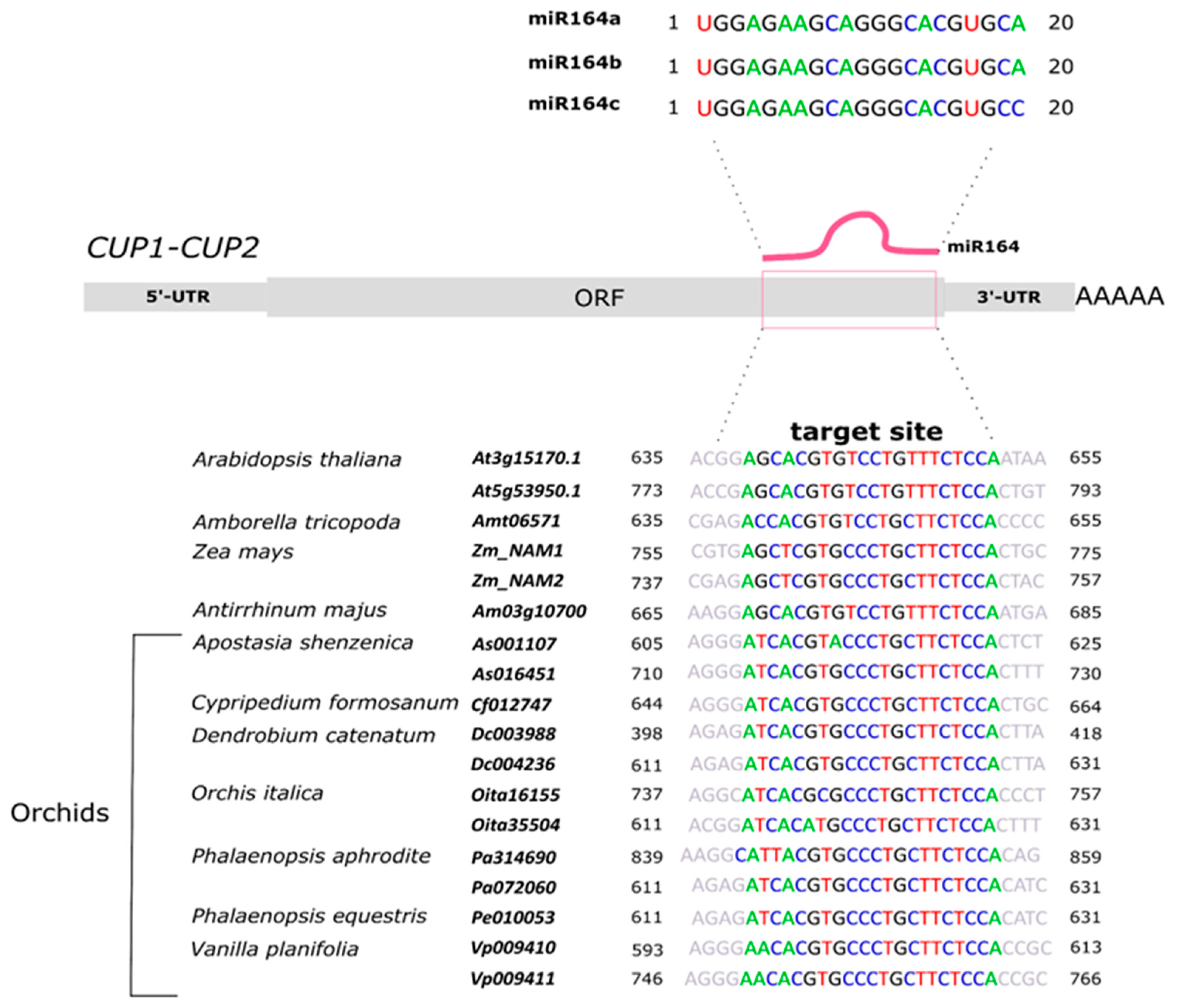
2.6. miR164 Analysis

We used the psRNATarget (2017 Update) online tool from The Zhao Lab© Ardmore, OK [60] with stringent search parameters (maximum expectation 0.0) to search for the presence of the miR164 target site in the orchid CUP sequences.
To identify the eventual specific fragments produced by the cleavage of miR164 on the P. aphrodite CUP transcripts, we performed a modified 5′-RACE experiment. We designed specific reverse primers based on the position of the putative miRNA cleavage site in the P. aphrodite CUP transcripts (Table 1). We ligated the 5′-adaptor in the RLM-RACE GeneRace kit (Invitrogen-Waltham, MA) to the 5′-end of total RNA (500 ng) extracted from B2-stage buds of P. aphrodite without removing the 5′-cap. After reverse transcription, we amplified the cDNA using the reverse primers specific for PaCUP1 and PaCUP2 (Table 1) and the GeneRace 5′ forward primer.
We conducted a stem-loop real-time qPCR experiment to evaluate the expression pattern of the miR164 in P. aphrodite. We reverse-transcribed total RNA (150 ng) from floral organs of B2 floral buds, open flower (OF), and leaf tissue using a specific miR164 or 18S stem-loop primer (Table 1). We conducted the qPCR amplifications in biological and technical triplicate using the specific miR164 and 18S forward primers, and the universal stem-loop reverse primer. Amplification conditions, thermal cycle, NRQ calculation, and statistical analysis were performed as described in the previous paragraph.

2.7. Yeast Two-Hybrid Analysis

To check whether the proteins PaCUP1 and PaCUP2 of P. aphrodite interact with each other and form homodimers, we used the GAL4-based yeast two-hybrid (Y2H) system (Matchmaker two-hybrid system 3, Clontech-Mountain View, CA).
We amplified the full-length coding regions of the PaCUP1/2 transcripts using specific primer pairs (Table 1). We cloned both the amplified coding regions into the pGADT7 (prey) and PGBKT7 (bait) expression vectors containing the activation (AD) and DNA-binding (BD) domains of the yeast transcription factor GAL4, respectively. We transformed the Saccharomyces cerevisiae strain AH109 [61] with all the combinations of pray and bait vectors, conducting each transformation in triplicate. After the yeast transformation, we verified the presence of the recombinant vectors by growing the yeast cells in Synthetic Defined (SD) medium lacking tryptophan and leucine in the presence of different concentrations (from 0 to 70 mM) of 3-aminotriazole (3AT). We transformed the empty vectors pGBKT7 or pGADT7 in combination with the recombinant vectors as negative controls.
In addition, we checked the autoactivation ability of the PaCUP1/2 proteins. To evaluate this, we grew the yeast transformed only with the recombinant PaCUP1 or PaCUP2 vector pGBKT7 (BD) on a selective medium lacking tryptophan and histidine in the presence of different concentrations (from 0 to 70 mM) of 3AT.

3. Results and Discussion

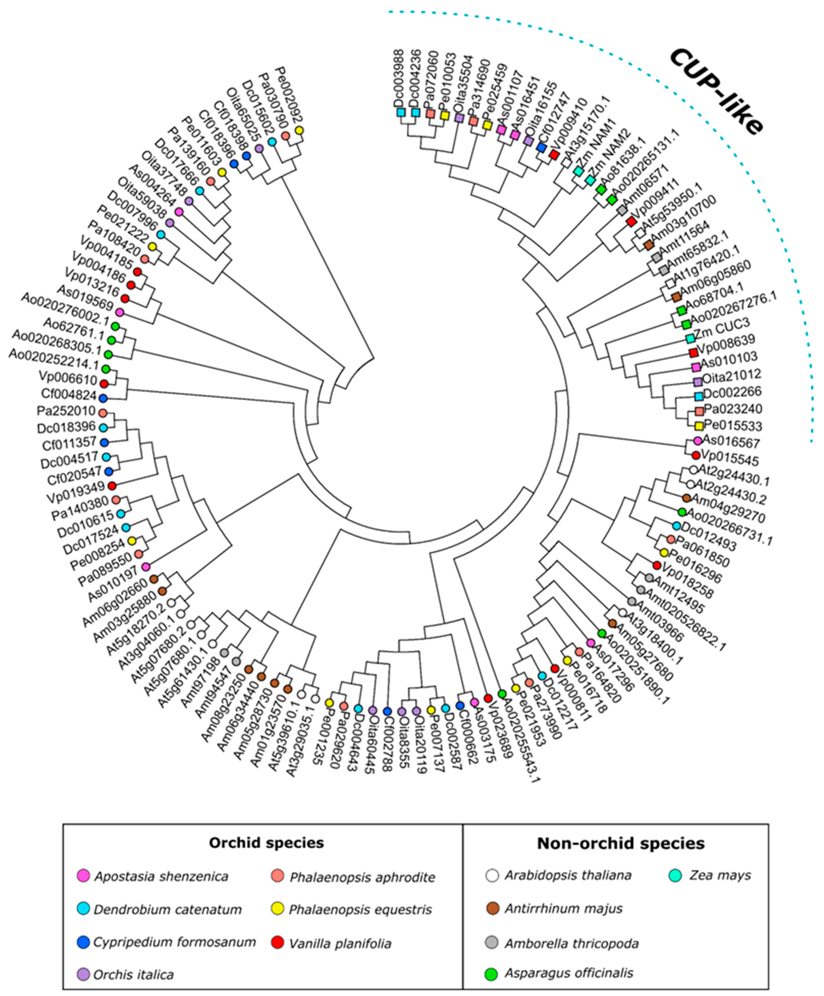
3.1. The NAC Transcription Factors of Orchids

Through BLAST analysis, we identified 423 orchid NAC transcripts. All sequences are listed in Supplementary Data S1 and Supplementary Table S1. Almost all (370) have a complete coding sequence; partial sequences (53) containing the nucleotide region encoding for the NAC and TAR conserved motifs were included in the analysis. In addition, we downloaded the NAC protein sequences of the model species A. thaliana (111) and A. majus (77), the basal angiosperm A. trichopoda (42), and the monocots A. s officinalis (95) and Z. mays (for this latter only the CUP proteins).
The number of NAC transcripts we identified in the different orchid species was variable, from 37 to 74. The species with the smallest number of sequences identified are O. italica (37) and C. formosanum (47). There is no assembled genome available for both species, so we used their transcriptomes to search the NAC sequences. The small number of NACs detected could be due to the incompleteness of their transcriptomes (e.g., obtained from specific tissues where some NAC genes are not expressed).
Based on sequence similarity, the NAC sequences can be classified into the subfamilies previously described in Arabidopsis [16] (Supplementary Material, Table S1). As observed in other species, no orchid NAC belongs to the AtNAC3 and ANAC001 subfamilies, where only Arabidopsis sequences are included [16,62,63]. The analysis of the conserved domains of the NAC proteins reveals the presence of the five conserved subdomains (A-E) and the TAR regions specific to different subfamilies (Supplementary Data S1).
Scanning the available orchid genomes, we reconstructed the genomic organization of the orchid NAC genes. The intron number and size vary among the various orchid NAC subfamilies. Intron number ranges from one to ten, with the highest number found in the NAC2 subfamily; intron size ranges from 52 bp (intron 1 of the A. shenzenica As014187 gene, SENU5 subfamily) to 20,287 bp (intron 1 of the D. catenatum Dc021911 gene, ANAC063 subfamily).
Compared with A. thaliana, the NAC genes of orchids generally have a higher number of introns with a larger intron size in almost all cases (Supplementary Datas S1 and S2). CENSOR analysis revealed traces of transposable/repetitive elements (e.g., DNA transposons, LTR, and Non-LTR Retrotransposons) within the introns of the orchid NAC genes displaying an intron number higher than Arabidopsis (Supplementary Data S3). The presence of repetitive elements and transposons in the NAC introns of orchids reflects a common feature of the orchid genomes. Indeed, transposable and repetitive elements can cover up to 60% of the orchid genomes [64], and orchid genes are often characterized by large introns containing repetitive and transposable elements, as observed in KNOX [65], OitaAP2, OitaAG, OitaSTK [66,67], and MYB genes [44].
The NAM subfamily of the NAC genes includes the highest number of sequences in almost all the species examined. Among them, the CUP transcription factors [68] are involved in the flower development of A. majus and other Lamiales [15]. For this reason, we focused our study on the NAM subfamily, in particular on the CUP genes.
Figure 2 shows the ML tree of the NAM transcription factors of orchids A. thaliana, A. majus, A. officinalis, and Z. mays. The tree topology shows a well-distinguished group that includes CUP-like proteins.
In addition to the NAC domain, NAM proteins have two transcriptional activation regions, TAR1 and TAR2 [16]. The Cf000662 protein in C. formosanum is the only orchid NAM protein containing both the TARs, whereas 34 orchid NAM proteins lack both regions (Supplementary Data S1), as observed in the A. thaliana AT1G76420, AT2G24430, and AT3G18400 sequences. The other orchid NAM sequences show only the TAR1 or TAR2 motif.
As in the other orchid NAC subfamilies, the NAMs have an intron size generally higher than Arabidopsis, as illustrated in Figure 3. Intron 2 shows the highest intron size variability. For example, in D. catenatum, P. aphrodite, P. equestris, and V. planifolia, the size of these introns ranges between 2001 and 9000 bp, values not observed in the Arabidopsis genes (Figure 3). In contrast, the coding region size is generally conserved between Arabidopsis and orchid species (Figure 3).

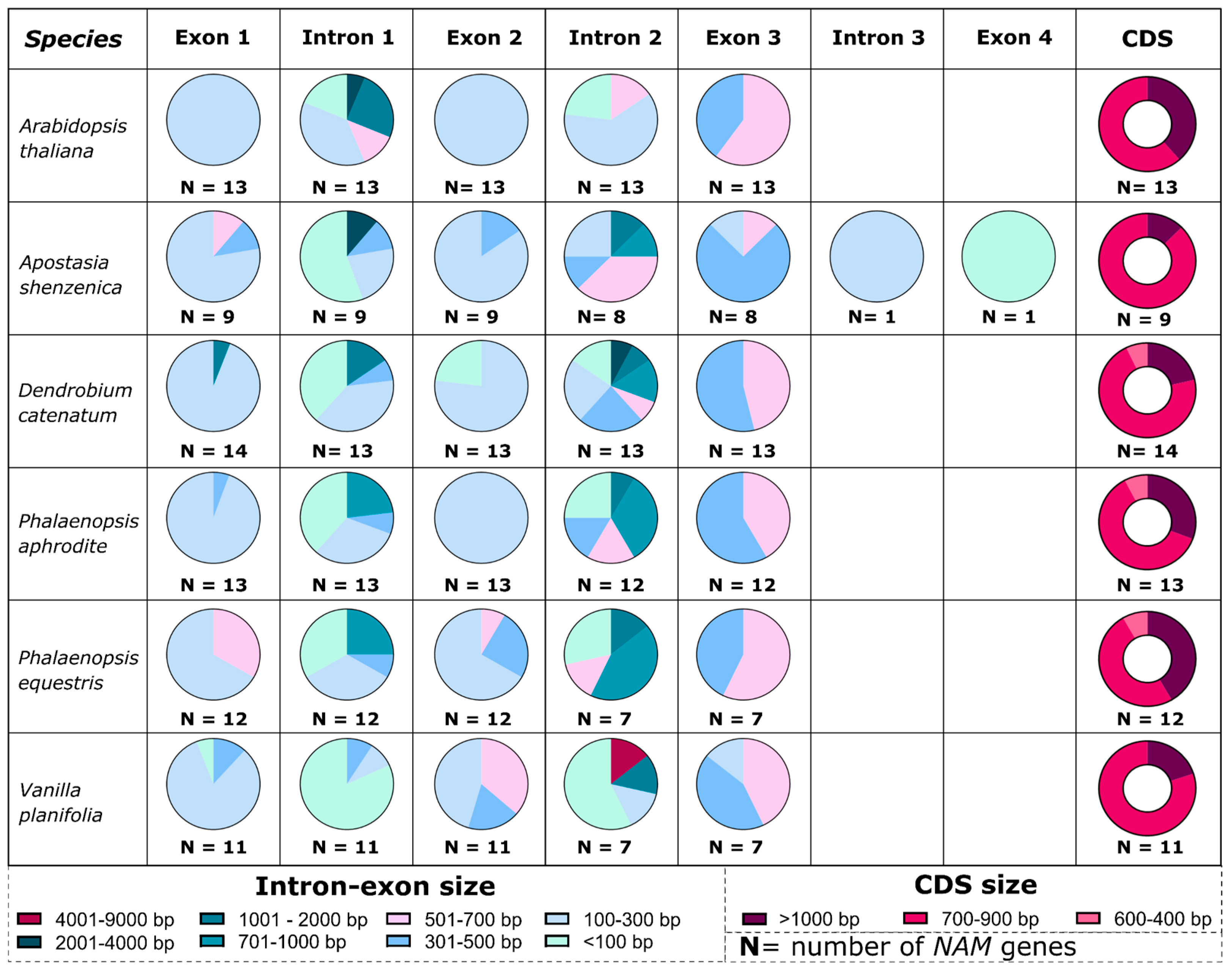
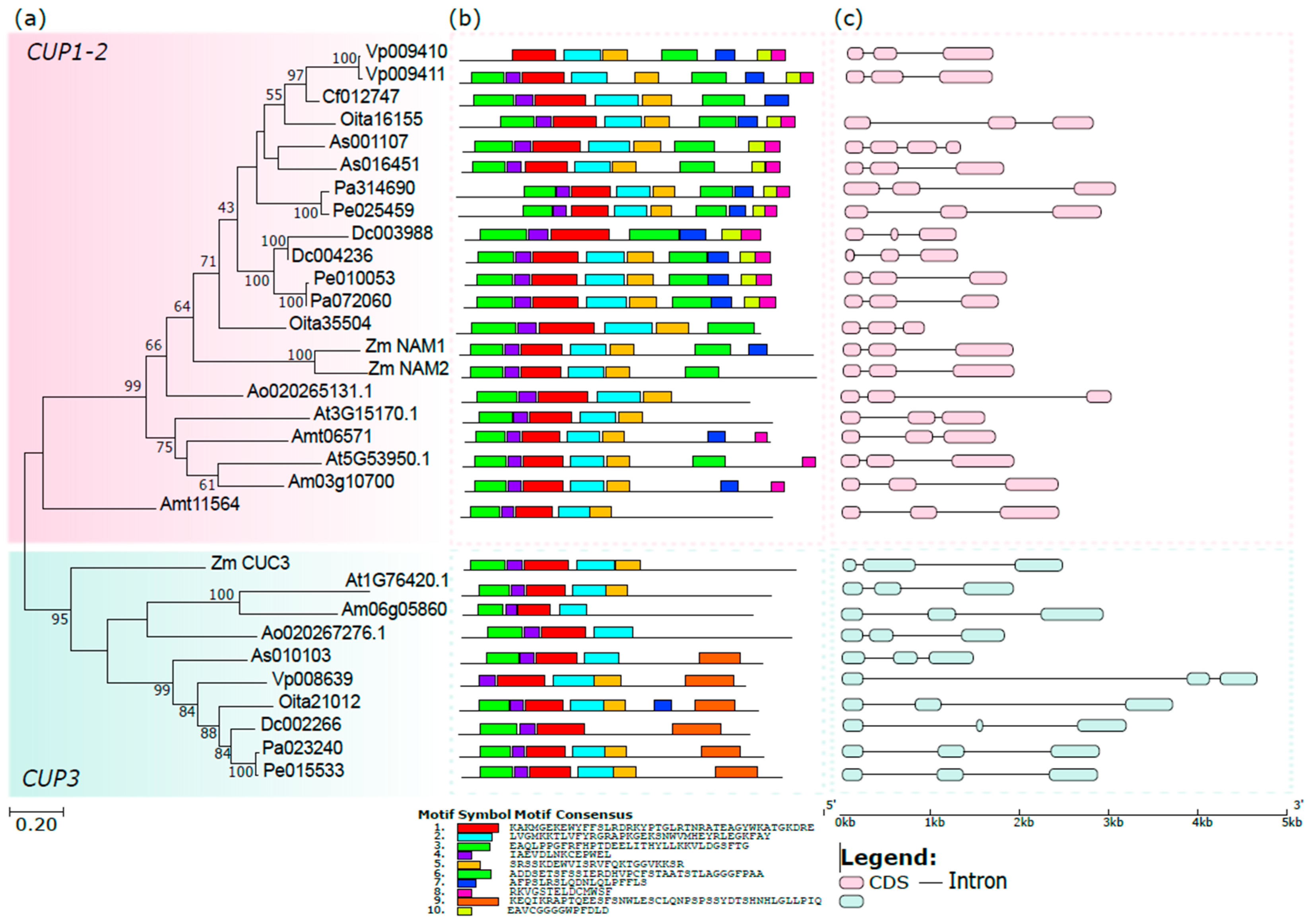
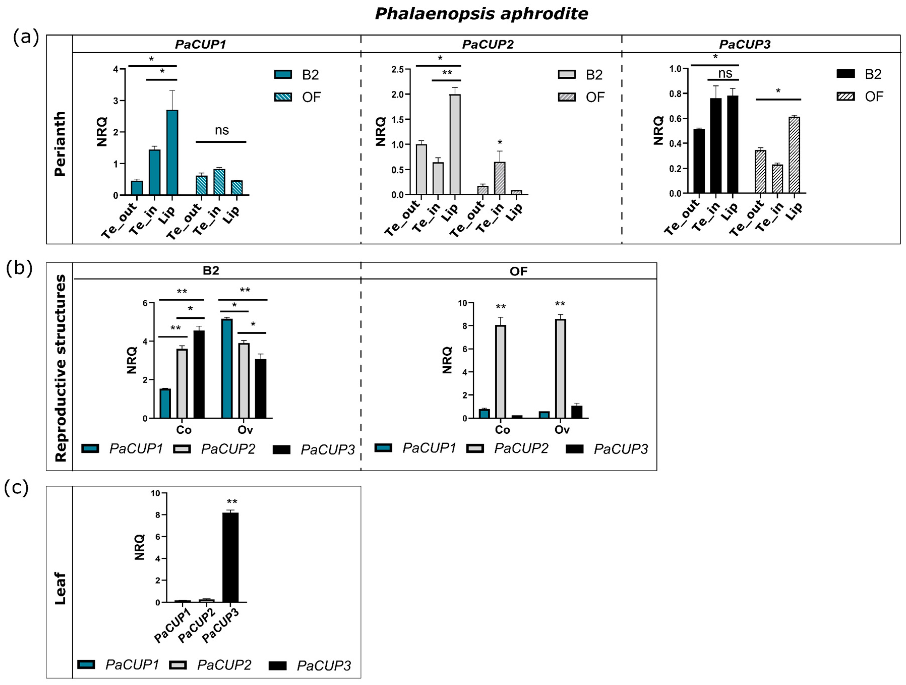
3.2. The Orchid CUP Genes: Phylogeny, Structure, and Expression Pattern

The CUP genes are involved in plant and flower development. In Arabidopsis, there are three CUP gene homologs [2,6]. CUC1 and CUC2 promote apical meristem growth during pre- and post-embryo development [5,31]. All three Arabidopsis CUC genes are involved in the maturation of flower reproductive organs; mutants of these genes reveal a phenotype strongly defective in the gynoecium and ovule structures [6,69]. In addition, the CUC2 and CUC3 genes regulate the formation of the border region of leaf primordia and their subsequent development [6,31]. In A. majus, the CUP gene is expressed, at an early stage of development, at a high level in the bud lateral–ventral region, regulating the boundary for lip development [15].
The ML tree obtained from the amino acid sequences of the orchid CUPs reveals the presence of two clades: the main includes the CUP1 and CUP2 proteins, and the other contains the CUP3 proteins (Figure 4a). This clade separation is not unique to orchids and is observed in other monocots, dicots, and early diverging angiosperm species [27,68,70,71]. The CUP1-2 clade is the most ancient, while duplication events occurring in angiosperms after divergence from gymnosperms generated the CUP3 clade [72].
The analysis of the conserved motifs reveals different patterns between the two clades (Figure 4b). Almost all the sequences have the characteristic NAM domain (motifs from 1 to 5) with few exceptions, possibly due to the incomplete CDS sequences isolated from the available transcriptomes. Motifs 6-7-8-10 are generally conserved within the CUP1-2 clade and absent in CUP3, whereas motif 9 is present only within the CUP3 clade.
Almost all the orchid CUP genes have three exons and two introns of variable size (Figure 4c). The As001107 gene of the basal orchid A. shenzenica is the only orchid CUP gene with four exons and three introns.
To evaluate whether the CUP genes could be involved in orchid flower development, we conducted a preliminary analysis of their expression patterns in the early and late developmental stages of P. aphrodite.
The three PaCUP genes show different expression profiles in the perianth during development. At the early stage, the PaCUP1-2 genes are mainly expressed in the lip, while PaCUP3 is also highly expressed in the inner tepals (Figure 5a). After anthesis, the expression of the three PaCUPs decreases in the perianth (Figure 5a). At this stage, PaCUP2 has higher expression in the inner tepals, while PaCUP3 maintains high expression in the lip (Figure 5a).
All three PaCUP genes are highly expressed in the reproductive organs (column and ovary) at the early stage of development (Figure 5b). After anthesis, PaCUP1 and PaCUP3 decrease their expression level in the column and ovary, whereas the expression of PaCUP2 increases (Figure 5b).
Only the PaCUP3 gene is expressed in leaf tissue, as previously reported in A. thaliana, suggesting its possible role in the leaf [73] (Figure 5c).
At the early stage of flower development, the P. aphrodite CUP genes have an expression pattern similar to that of A. majus [15], being expressed in the lip and inner tepals. However, while in A. majus the CUP genes have a lateral–ventral profile, in the orchid flower the CUP expression is lateral–dorsal due to resupination. In most orchids, including P. aphrodite, resupination occurs before anthesis and is the 180° rotation of the pedicel and ovary, resulting in the reversal position of the dorsal and ventral organs in the mature flower. Consequently, although the lip is a dorsal structure, it has a ventral position [34].
As observed in A. majus, the expression pattern of the CUP genes in the orchid flower suggests their possible role in flower formation. The CUP genes might interact with other transcription factor encoding genes, e.g., MYB and TCP, involved in flower development and symmetry establishment [26,44,45,46,74,75,76]. Similar interactions were demonstrated in A. majus, where the CUPs can interact with the TCP proteins, suggesting their role in the boundary flower formation [8]. In addition, the phenotype of the cup mutant resembles that of the double mutant cyc dich, suggesting a functional correlation between CUP and TCP genes [8]. Furthermore, the expression of the MYB gene, DIV, could positively regulate the CUP expression in the labellum of Antirrhinum [15].
Recently, the orchid YABBY transcription factor, DL2, was proposed as a possible regulator of orchid lip formation [47]. In P. equestris there is a correlation between the expression pattern of DL2 and the class-B DEF-like genes, PeMADS2-PeMADS5, responsible for orchid perianth formation [47]. Curiously, the expression pattern of PaCUP1 in the lip is similar to that of DL2 in Phalaenopsis. Both genes have high expression in the lip during the early developmental stages, followed by a decrease until the mature flower [47].
Our results suggest that, together with MADS-box and other transcription factors, the orchid CUP genes might be involved in the complex molecular pathway of orchid perianth formation.

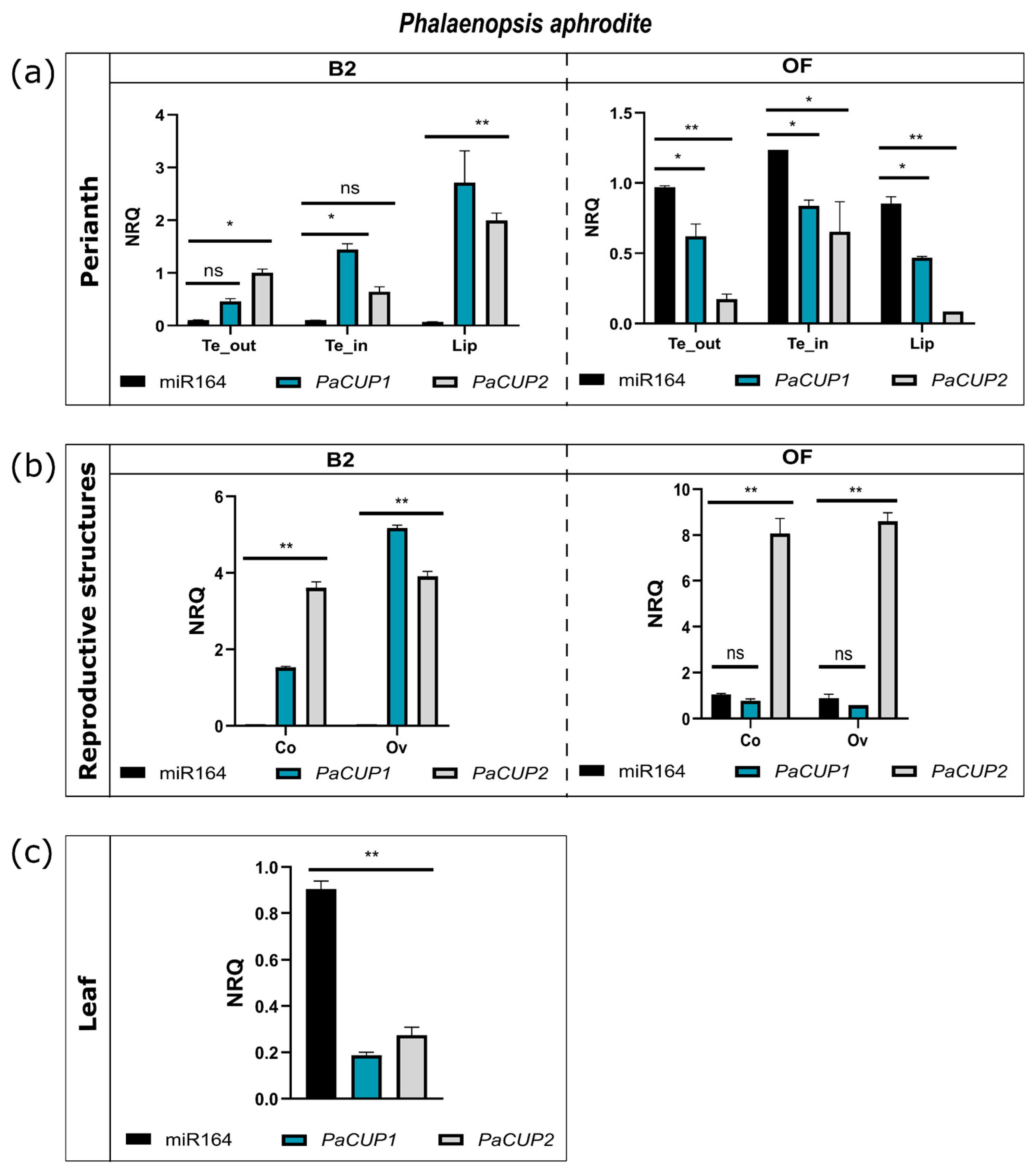
3.3. Expression Pattern of miR164

The transcripts of the CUP1-2 genes have a target site for the microRNA, miR164. This site, located within the region upstream of the 3′UTR, is absent in the CUP3 lineage [71]. In Arabidopsis, microRNAs regulate the CUP1-2 genes in many plant life processes. For example, the miR164c mutant has an altered number of petals, suggesting a role for miR164 in flower structure formation [77]. For these reasons, we investigated the presence of the miR164 target site on the orchid CUP1-2 transcripts and compared the expression profile of this microRNA to that of the PaCUP1-2 genes.
Using the psRNATarget online tool [60], we verified the presence of the putative miR164 target site on the CUP transcripts. Our results show that 12 orchid CUP1-2 transcripts have the miR164 target site (Figure 6). As expected, no CUP3 transcript had the miR164 target site.
We conducted the modified 5′-RACE experiment on the PaCUP transcripts of P. aphrodite in the tissues examined and did not detect any miR164 cleavage fragment of the expected size. The apparent absence of the cleavage product in the selected tissues may be due to the high instability of the mRNA fragment produced after cleavage by miR164.
To compare the expression patterns of miR164 and PaCUP genes, we conducted stem-loop qPCR experiments of miR164 on floral tissues from the B2 and OF stages of P. aphrodite (Figure 7a,b).
In the perianth, miR164 shows an expression trend opposite to that of the PaCUP1-2 genes. At the B2 stage, the miR164 transcripts are almost undetectable, whereas they are expressed in the floral tissues after anthesis (Figure 7a), suggesting that the expression of miR164 reduces the number of the PaCUP1-2 transcripts. These results are in agreement with those obtained in A. majus, where at the first stage of development, the CUP1-2 genes regulate the boundary formation and are expressed in all flower primordia. Their expression then decreases before anthesis, limiting the presence of the CUP transcripts to specific parts of the flower [15].
In the reproductive structures of P. aphrodite, miR164 is not expressed at the B2 stage, allowing a high expression of the PaCUP genes. Later, at the OF stage, mir164 is expressed; however, only the transcript levels of PaCUP1 decrease, in particular in the ovary, whereas PaCUP2 is still highly expressed, suggesting that after anthesis, the levels of PaCUP2 transcripts are not regulated by miR164. The activity of miR164 on the transcript levels of the homologs of the CUP genes was also proposed for A. thaliana, where miR164 is involved in reproductive structure formation and possibly modulates the transcript levels of the CUC1-2 genes (homologs of the CUPs) involved in ovary and column formation [31,69].
Finally, miR164 is expressed in the leaf tissue of P. aphrodite, where the PaCUP1-2 transcript levels are low. The miR164 of A. thaliana regulates the CUC1-2 genes during leaf shape formation and senescence. Mutants of miR164 have an unusual leaf shape, with leaves showing a more pronounced dentition than the wild type [78]. Our results suggest that, also in orchids, miR164 seems to regulate the PaCUP1-2 expression in the leaf.

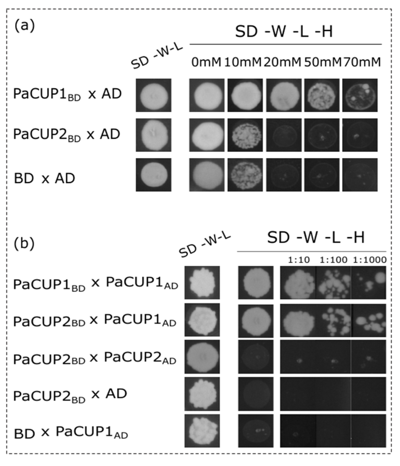
3.4. PaCUP1–PaCUP2 Protein Interaction

The NAC transcription factors of Arabidopsis can form homo- and heterodimers due to their ability to interact through the NAC domains [25]. In addition, CUC1 and CUC2 in Arabidopsis can activate transcription due to the presence of acidic residues within the TAR domain [25]. In a Y2H assay, they activated the transcription of the yeast reporter genes [79]. For these reasons, we checked the autoactivation ability of the P. aphrodite PaCUP1-2 proteins and their ability to form homo- and heterodimers in the Y2H assay.
The yeast clones double-transformed with the recombinant vector pGBKT7 containing the full-length transcripts PaCUP1 or PaCUP2, and the empty vector PGADT7. The clones were grown on a selective medium without tryptophan, lysine, or histidine in the presence of different concentrations of 3AT (from 0 to 70 mM). Only the PaCUP1 protein alone activated the transcription of the yeast GAL4 gene in the absence of histidine and in the presence of up to 20 mM 3AT (Figure 8a). For this reason, the protein–protein interaction experiments were conducted in the presence of 3AT from 50 to 70 mM.
Our results reveal that PaCUP1 forms homodimers (Figure 8a), as observed in Arabidopsis [79], and, as expected, PaCUP1 and PaCUP2 proteins directly interact with each other (Figure 8b), showing that the CUP1-2 proteins have a conserved ability to cross-interact.

4. Conclusions

Orchidaceae represent approximately 10% of flowering plants, with many unique morphological, ecological, and physiological characteristics. Their cultivation and commercialization are a significant part of the worldwide floriculture trade. In this scenario, studies on the molecular basis of orchid flower development are particularly relevant, as they enrich the knowledge of how to develop specific breeding programs to obtain even more new cultivars and varieties.
For the first time in orchids, we analyzed the NAC transcription factor family, focusing on the CUP genes.
Our study reveals that orchid NAC proteins have a conserved domain organization and can be easily classified into different subfamilies based on their sequence.
The expression patterns of the P. aphrodite CUP genes suggest their possible involvement in flower formation and symmetry establishment. In addition, as previously described for different species, the transcript levels of orchid CUP1 and CUP2 genes seem to be regulated by miR164.
The data obtained in this work are preliminary and represent a starting point for understanding the role of these genes. Further investigations, in particular functional analyses, are needed to clarify the role of CUP genes during orchid flower formation.

Supplementary Materials

The following supporting information can be downloaded at: https://www.mdpi.com/article/10.3390/genes13122293/s1, Figure S1: Conserved motif for CUP proteins identified by MEME analysis, Table S1: Number of NAC genes isolated in all species analyzed in this work, Data S1: List of sequences, accession number, conserved motif and genome structures of the genes considered in this work; Data S2: Difference between the size of the intron-exon of the orchid gene species and A. thaliana; Data S3: CENSOR analysis results; Data S4: Statistical analysis results.

Author Contributions

Conceptualization, S.A. and M.C.V.; methodology, M.C.V. and F.L.; validation, M.C.V. and F.L.; investigation, M.C.V. and F.L.; writing—original draft preparation, M.C.V.; writing—review and editing, S.A. and M.C.V.; supervision, S.A. All authors have read and agreed to the published version of the manuscript.

Funding

This research received no external funding.

Institutional Review Board Statement

Not applicable.

Informed Consent Statement

Not applicable.

Data Availability Statement

Conflicts of Interest

The authors declare no conflict of interest.

References

  1. Pérez-Rodríguez, P.; Riaño-Pachón, D.M.; Corrêa, L.G.G.; Rensing, S.A.; Kersten, B.; Mueller-Roeber, B. PlnTFDB: Updated content and new features of the plant transcription factor database. Nucleic Acids Res. 2010, 38, D822–D827. [Google Scholar] [CrossRef] [PubMed] [Green Version]
  2. Aida, M.; Ishida, T.; Fukaki, H.; Fujisawa, H.; Tasaka, M. Genes involved in organ separation in Arabidopsis: An analysis of the cup-shaped cotyledon mutant. Plant Cell 1997, 9, 841–857. [Google Scholar] [CrossRef] [PubMed] [Green Version]
  3. Sablowski, R.W.; Meyerowitz, E.M. A Homolog of NO APICAL MERISTEM Is an Immediate Target of the Floral Homeotic Genes APETALA3/PISTILLATA. Cell 1998, 92, 93–103. [Google Scholar] [CrossRef] [PubMed] [Green Version]
  4. Souer, E.; van Houwelingen, A.; Kloos, D.; Mol, J.; Koes, R. The No Apical Meristem Gene of Petunia Is Required for Pattern Formation in Embryos and Flowers and Is Expressed at Meristem and Primordia Boundaries. Cell 1996, 85, 159–170. [Google Scholar] [CrossRef] [Green Version]
  5. Takada, S.; Hibara, K.; Ishida, T.; Tasaka, M. The CUP-SHAPED COTYLEDON1 gene of Arabidopsis regulates shoot apical meristem formation. Development 2001, 128, 1127–1135. [Google Scholar] [CrossRef]
  6. Vroemen, C.W.; Mordhorst, A.P.; Albrecht, C.; Kwaaitaal, M.A.C.J.; de Vries, S.C. The CUP-SHAPED COTYLEDON3 Gene Is Required for Boundary and Shoot Meristem Formation in Arabidopsis. Plant Cell 2003, 15, 1563–1577. [Google Scholar] [CrossRef]
  7. Xie, Q.; Frugis, G.; Colgan, D.; Chua, N.-H. Arabidopsis NAC1 transduces auxin signal downstream of TIR1 to promote lateral root development. Genes Dev. 2000, 14, 3024–3036. [Google Scholar] [CrossRef] [PubMed] [Green Version]
  8. Weir, I.; Lu, J.; Cook, H.; Causier, B.; Schwarz-Sommer, Z.; Davies, B. CUPULIFORMIS establishes lateral organ boundaries in Antirrhinum. Development 2004, 131, 915–922. [Google Scholar] [CrossRef] [Green Version]
  9. Collinge, M.; Boller, T. Differential induction of two potato genes, Stprx2 and StNAC, in response to infection by Phytophthora infestans and to wounding. Plant Mol. Biol. 2001, 46, 521–529. [Google Scholar] [CrossRef] [PubMed]
  10. Ren, T.; Qu, F.; Morris, T.J. HRT Gene Function Requires Interaction between a NAC Protein and Viral Capsid Protein to Confer Resistance to Turnip Crinkle Virus. Plant Cell 2000, 12, 1917–1926. [Google Scholar] [CrossRef]
  11. Xie, Q.; Sanz-Burgos, A.P.; Guo, H.; García, J.A.; Gutiérrez, C. GRAB proteins, novel members of the NAC domain family, isolated by their interaction with a geminivirus protein. Plant Mol. Biol. 1999, 39, 647–656. [Google Scholar] [CrossRef]
  12. Fujita, M.; Fujita, Y.; Maruyama, K.; Seki, M.; Hiratsu, K.; Ohme-Takagi, M.; Tran, L.-S.P.; Yamaguchi-Shinozaki, K.; Shinozaki, K. A dehydration-induced NAC protein, RD26, is involved in a novel ABA-dependent stress-signaling pathway. Plant J. 2004, 39, 863–876. [Google Scholar] [CrossRef]
  13. Hegedus, D.; Yu, M.; Baldwin, D.; Gruber, M.; Sharpe, A.; Parkin, I.; Whitwill, S.; Lydiate, D. Molecular characterization of Brassica napus NAC domain transcriptional activators induced in response to biotic and abiotic stress. Plant Mol. Biol. 2003, 53, 383–397. [Google Scholar] [CrossRef]
  14. Tran, L.-S.P.; Nakashima, K.; Sakuma, Y.; Simpson, S.D.; Fujita, Y.; Maruyama, K.; Fujita, M.; Seki, M.; Shinozaki, K.; Yamaguchi-Shinozaki, K. Isolation and Functional Analysis of Arabidopsis Stress-Inducible NAC Transcription Factors That Bind to a Drought-Responsive cis-Element in the early responsive to dehydration stress 1 Promoter. Plant Cell 2004, 16, 2481–2498. [Google Scholar] [CrossRef] [PubMed] [Green Version]
  15. Rebocho, A.B.; Kennaway, J.R.; Bangham, J.A.; Coen, E. Formation and Shaping of the Antirrhinum Flower through Modulation of the CUP Boundary Gene. Curr. Biol. 2017, 27, 2610–2622.e3. [Google Scholar] [CrossRef] [Green Version]
  16. Ooka, H.; Satoh, K.; Doi, K.; Nagata, T.; Otomo, Y.; Murakami, K.; Matsubara, K.; Osato, N.; Kawai, J.; Carninci, P.; et al. Comprehensive Analysis of NAC Family Genes in Oryza sativa and Arabidopsis thaliana. DNA Res. 2003, 10, 239–247. [Google Scholar] [CrossRef]
  17. Rushton, P.J.; Bokowiec, M.T.; Han, S.; Zhang, H.; Brannock, J.F.; Chen, X.; Laudeman, T.W.; Timko, M.P. Tobacco Transcription Factors: Novel Insights into Transcriptional Regulation in the Solanaceae. Plant Physiol. 2008, 147, 280–295. [Google Scholar] [CrossRef] [PubMed] [Green Version]
  18. Duval, M.; Hsieh, T.-F.; Kim, S.Y.; Thomas, T.L. Molecular characterization of AtNAM: A member of the Arabidopsis NAC domain superfamily. Plant Mol. Biol. 2002, 50, 237–248. [Google Scholar] [CrossRef]
  19. Ernst, H.A.; Olsen, A.N.; Larsen, S.; Leggio, L.L. Structure of the conserved domain of ANAC, a member of the NAC family of transcription factors. EMBO Rep. 2004, 5, 297–303. [Google Scholar] [CrossRef] [PubMed]
  20. Olsen, A.N.; Ernst, H.A.; Leggio, L.L.; Johansson, E.; Larsen, S.; Skriver, K. Preliminary crystallographic analysis of the NAC domain of ANAC, a member of the plant-specific NAC transcription factor family. Acta Crystallogr. Sect. D Biol. Crystallogr. 2004, 60, 112–115. [Google Scholar] [CrossRef]
  21. Puranik, S.; Sahu, P.P.; Srivastava, P.S.; Prasad, M. NAC proteins: Regulation and role in stress tolerance. Trends Plant Sci. 2012, 17, 369–381. [Google Scholar] [CrossRef] [PubMed]
  22. Byrne, M.E.; Barley, R.; Curtis, M.; Arroyo, J.M.; Dunham, M.; Hudson, A.; Martienssen, R.A. Asymmetric leaves1 mediates leaf patterning and stem cell function in Arabidopsis. Nature 2000, 408, 967–971. [Google Scholar] [CrossRef] [Green Version]
  23. Daimon, Y.; Takabe, K.; Tasaka, M. The CUP-SHAPED COTYLEDON genes promote adventitious shoot formation on calli. Plant Cell Physiol. 2003, 44, 113–121. [Google Scholar] [CrossRef] [PubMed] [Green Version]
  24. Hibara, K.-I.; Takada, S.; Tasaka, M. CUC1 gene activates the expression of SAM-related genes to induce adventitious shoot formation. Plant J. 2003, 36, 687–696. [Google Scholar] [CrossRef] [PubMed]
  25. Olsen, A.N.; Ernst, H.A.; Leggio, L.L.; Skriver, K. NAC transcription factors: Structurally distinct, functionally diverse. Trends Plant Sci. 2005, 10, 79–87. [Google Scholar] [CrossRef]
  26. Luo, D.; Carpenter, R.; Copsey, L.; Vincent, C.; Clark, J.; Coen, E. Control of Organ Asymmetry in Flowers of Antirrhinum. Cell 1999, 99, 367–376. [Google Scholar] [CrossRef] [PubMed] [Green Version]
  27. Adam, H.; Marguerettaz, M.; Qadri, R.; Adroher, B.; Richaud, F.; Collin, M.; Thuillet, A.-C.; Vigouroux, Y.; Laufs, P.; Tregear, J.W.; et al. Divergent Expression Patterns of miR164 and CUP-SHAPED COTYLEDON Genes in Palms and Other Monocots: Implication for the Evolution of Meristem Function in Angiosperms. Mol. Biol. Evol. 2011, 28, 1439–1454. [Google Scholar] [CrossRef] [PubMed] [Green Version]
  28. Laufs, P.; Peaucelle, A.; Morin, H.; Traas, J. MicroRNA regulation of the CUC genes is required for boundary size control in Arabidopsis meristems. Development 2004, 131, 4311–4322. [Google Scholar] [CrossRef] [Green Version]
  29. Mallory, A.C.; Dugas, D.; Bartel, D.P.; Bartel, B. MicroRNA Regulation of NAC-Domain Targets Is Required for Proper Formation and Separation of Adjacent Embryonic, Vegetative, and Floral Organs. Curr. Biol. 2004, 14, 1035–1046. [Google Scholar] [CrossRef] [Green Version]
  30. Zou, H.-W.; Tian, X.-H.; Ma, G.-H.; Li, Z.-X. Isolation and Functional Analysis of ZmLTP3, a Homologue to Arabidopsis LTP. Int. J. Mol. Sci. 2013, 14, 5025–5035. [Google Scholar] [CrossRef]
  31. Larue, C.T.; Wen, J.; Walker, J.C. A microRNA-transcription factor module regulates lateral organ size and patterning in Arabidopsis. Plant J. 2009, 58, 450–463. [Google Scholar] [CrossRef]
  32. Aceto, S.; Gaudio, L. The MADS and the Beauty: Genes Involved in the Development of Orchid Flowers. Curr. Genom. 2011, 12, 342–356. [Google Scholar] [CrossRef] [Green Version]
  33. Cozzolino, S.; Widmer, A. Orchid diversity: An evolutionary consequence of deception? Trends Ecol. Evol. 2005, 20, 487–494. [Google Scholar] [CrossRef] [PubMed]
  34. Rudall, P.J.; Bateman, R.M. Roles of synorganisation, zygomorphy and heterotopy in floral evolution: The gynostemium and labellum of orchids and other lilioid monocots. Biol. Rev. 2002, 77, 403–441. [Google Scholar] [CrossRef] [PubMed]
  35. Cai, J.; Liu, X.; Vanneste, K.; Proost, S.; Tsai, W.-C.; Liu, K.-W.; Chen, L.-J.; He, Y.; Xu, Q.; Bian, C.; et al. The genome sequence of the orchid Phalaenopsis equestris. Nat. Genet. 2015, 47, 65–72. [Google Scholar] [CrossRef] [PubMed] [Green Version]
  36. Dirks-Mulder, A.; Butôt, R.; van Schaik, P.; Wijnands, J.W.P.M.; Berg, R.V.D.; Krol, L.; Doebar, S.; van Kooperen, K.; de Boer, H.; Kramer, E.M.; et al. Exploring the evolutionary origin of floral organs of Erycina pusilla, an emerging orchid model system. BMC Evol. Biol. 2017, 17, 89. [Google Scholar] [CrossRef] [Green Version]
  37. Hsu, H.-F.; Hsu, W.-H.; Lee, Y.-I.; Mao, W.-T.; Yang, J.-Y.; Li, J.-Y.; Yang, C.-H. Model for perianth formation in orchids. Nat. Plants 2015, 1, 15046. [Google Scholar] [CrossRef]
  38. Mondragón-Palomino, M.; Theißen, G. Why are orchid flowers so diverse? Reduction of evolutionary constraints by paralogues of class B floral homeotic genes. Ann. Bot. 2009, 104, 583–594. [Google Scholar] [CrossRef] [Green Version]
  39. Mondragón-Palomino, M.; Theißen, G. Conserved differential expression of paralogous DEFICIENS- and GLOBOSA-like MADS-box genes in the flowers of Orchidaceae: Refining the ‘orchid code’. Plant J. 2011, 66, 1008–1019. [Google Scholar] [CrossRef]
  40. Pan, Z.-J.; Cheng, C.-C.; Tsai, W.-C.; Chung, M.-C.; Chen, W.-H.; Hu, J.-M.; Chen, H.-H. The Duplicated B-class MADS-Box Genes Display Dualistic Characters in Orchid Floral Organ Identity and Growth. Plant Cell Physiol. 2011, 52, 1515–1531. [Google Scholar] [CrossRef]
  41. Valoroso, M.C.; Censullo, M.C.; Aceto, S. The MADS-box genes expressed in the inflorescence of Orchis italica (Orchidaceae). PLoS ONE 2019, 14, e0213185. [Google Scholar] [CrossRef] [PubMed] [Green Version]
  42. Zhang, G.-Q.; Liu, K.-W.; Li, Z.; Lohaus, R.; Hsiao, Y.-Y.; Niu, S.-C.; Wang, J.-Y.; Lin, Y.-C.; Xu, Q.; Chen, L.-J.; et al. The Apostasia genome and the evolution of orchids. Nature 2017, 549, 379–383. [Google Scholar] [CrossRef] [PubMed] [Green Version]
  43. Zhang, G.-Q.; Xu, Q.; Bian, C.; Tsai, W.-C.; Yeh, C.-M.; Liu, K.-W.; Yoshida, K.; Zhang, L.-S.; Chang, S.-B.; Chen, F.; et al. The Dendrobium catenatum Lindl. genome sequence provides insights into polysaccharide synthase, floral development and adaptive evolution. Sci. Rep. 2016, 6, 19029. [Google Scholar] [CrossRef] [Green Version]
  44. Valoroso, M.C.; De Paolo, S.; Iazzetti, G.; Aceto, S. Transcriptome-Wide Identification and Expression Analysis of DIVARICATA- and RADIALIS-Like Genes of the Mediterranean Orchid Orchis italica. Genome Biol. Evol. 2017, 9, 1418–1431. [Google Scholar] [CrossRef] [Green Version]
  45. Valoroso, M.C.; Sobral, R.; Saccone, G.; Salvemini, M.; Costa, M.M.R.; Aceto, S. Evolutionary Conservation of the Orchid MYB Transcription Factors DIV, RAD, and DRIF. Front. Plant Sci. 2019, 10, 1359. [Google Scholar] [CrossRef]
  46. De Paolo, S.; Gaudio, L.; Aceto, S. Analysis of the TCP genes expressed in the inflorescence of the orchid Orchis italica. Sci. Rep. 2015, 5, 16265. [Google Scholar] [CrossRef] [Green Version]
  47. Lucibelli, F.; Valoroso, M.C.; Theißen, G.; Nolden, S.; Mondragon-Palomino, M.; Aceto, S. Extending the Toolkit for Beauty: Differential Co-Expression of DROOPING LEAF-Like and Class B MADS-Box Genes during Phalaenopsis Flower Development. Int. J. Mol. Sci. 2021, 22, 7025. [Google Scholar] [CrossRef] [PubMed]
  48. Chao, Y.-T.; Chen, W.-C.; Chen, C.-Y.; Ho, H.-Y.; Yeh, C.-H.; Kuo, Y.-T.; Su, C.-L.; Yen, S.-H.; Hsueh, H.-Y.; Yeh, J.-H.; et al. Chromosome-level assembly, genetic and physical mapping of Phalaenopsis aphrodite genome provides new insights into species adaptation and resources for orchid breeding. Plant Biotechnol. J. 2018, 16, 2027–2041. [Google Scholar] [CrossRef] [PubMed] [Green Version]
  49. De Paolo, S.; Salvemini, M.; Gaudio, L.; Aceto, S. De Novo Transcriptome Assembly from Inflorescence of Orchis italica: Analysis of Coding and Non-Coding Transcripts. PLoS ONE 2014, 9, e102155. [Google Scholar] [CrossRef] [Green Version]
  50. Hu, Y.; Resende, M.F.R., Jr.; Bombarely, A.; Brym, M.; Bassil, E.; Chambers, A.H. Genomics-based diversity analysis of Vanilla species using a Vanilla planifolia draft genome and Genotyping-By-Sequencing. Sci. Rep. 2019, 9, 3416. [Google Scholar] [CrossRef]
  51. Chao, Y.-T.; Yen, S.-H.; Yeh, J.-H.; Chen, W.-C.; Shih, M.-C. Orchidstra 2.0—A Transcriptomics Resource for the Orchid Family. Plant Cell Physiol. 2017, 58, e9. [Google Scholar] [CrossRef] [Green Version]
  52. Tsai, W.-C.; Fu, C.-H.; Hsiao, Y.-Y.; Huang, Y.-M.; Chen, L.-J.; Wang, M.; Liu, Z.-J.; Chen, H.-H. OrchidBase 2.0: Comprehensive Collection of Orchidaceae Floral Transcriptomes. Plant Cell Physiol. 2013, 54, e7. [Google Scholar] [CrossRef] [PubMed] [Green Version]
  53. Van Bel, M.; Diels, T.; Vancaester, E.; Kreft, Ł.; Botzki, A.; Van De Peer, Y.; Coppens, F.; Vandepoele, K. PLAZA 4.0: An integrative resource for functional, evolutionary and comparative plant genomics. Nucleic Acids Res. 2018, 46, D1190–D1196. [Google Scholar] [CrossRef]
  54. Larkin, M.A.; Blackshields, G.; Brown, N.P.; Chenna, R.; McGettigan, P.A.; McWilliam, H.; Valentin, F.; Wallace, I.M.; Wilm, A.; Lopez, R.; et al. Clustal W and Clustal X version 2. Bioinformatics 2007, 23, 2947–2948. [Google Scholar] [CrossRef] [PubMed] [Green Version]
  55. Kumar, S.; Stecher, G.; Tamura, K. MEGA7: Molecular Evolutionary Genetics Analysis Version 7.0 for Bigger Datasets. Mol. Biol. Evol. 2016, 33, 1870–1874. [Google Scholar] [CrossRef] [PubMed] [Green Version]
  56. Kohany, O.; Gentles, A.J.; Hankus, L.; Jurka, J. Annotation, submission and screening of repetitive elements in Repbase: RepbaseSubmitter and Censor. BMC Bioinform. 2006, 7, 474. [Google Scholar] [CrossRef] [PubMed] [Green Version]
  57. Bailey, T.L.; Johnson, J.; Grant, C.E.; Noble, W.S. The MEME Suite. Nucleic Acids Res. 2015, 43, W39–W49. [Google Scholar] [CrossRef] [Green Version]
  58. Doyle, J.J. Isolation of plant DNA from fresh tissue. Focus 1999, 12, 13–15. [Google Scholar]
  59. Acri-Nunes-Miranda, R.; Mondragón-Palomino, M. Expression of paralogous SEP-, FUL-, AG- and STK-like MADS-box genes in wild-type and peloric Phalaenopsis flowers. Front. Plant Sci. 2014, 5, 76. [Google Scholar] [CrossRef] [Green Version]
  60. Dai, X.; Zhao, P.X. psRNATarget: A plant small RNA target analysis server. Nucleic Acids Res. 2011, 39, W155–W159. [Google Scholar] [CrossRef] [Green Version]
  61. Gietz, R.D.; Schiestl, R.H.; Willems, A.R.; Woods, R.A. Studies on the transformation of intact yeast cells by the LiAc/SS-DNA/PEG procedure. Yeast 1995, 11, 355–360. [Google Scholar] [CrossRef] [PubMed]
  62. Fan, K.; Wang, M.; Miao, Y.; Ni, M.; Bibi, N.; Yuan, S.; Li, F.; Wang, X. Molecular Evolution and Expansion Analysis of the NAC Transcription Factor in Zea mays. PLoS ONE 2014, 9, e111837. [Google Scholar] [CrossRef] [PubMed] [Green Version]
  63. Kikuchi, K.; Ueguchi-Tanaka, M.; Yoshida, K.T.; Nagato, Y.; Matsusoka, M.; Hirano, H.Y. Molecular analysis of the NAC gene family in rice. Mol. Genet. Genom. 2000, 262, 1047–1051. [Google Scholar] [CrossRef] [PubMed]
  64. Albert, V.A.; Carretero-Paulet, L. A genome to unveil the mysteries of orchids. Nat. Genet. 2015, 47, 3–4. [Google Scholar] [CrossRef] [PubMed]
  65. Zhang, D.; Lan, S.; Yin, W.-L.; Liu, Z.-J. Genome-Wide Identification and Expression Pattern Analysis of KNOX Gene Family in Orchidaceae. Front. Plant Sci. 2022, 13, 901089. [Google Scholar] [CrossRef] [PubMed]
  66. Salemme, M.; Sica, M.; Gaudio, L.; Aceto, S. The OitaAG and OitaSTK genes of the orchid Orchis italica: A comparative analysis with other C- and D-class MADS-box genes. Mol. Biol. Rep. 2013, 40, 3523–3535. [Google Scholar] [CrossRef]
  67. Salemme, M.; Sica, M.; Iazzetti, G.; Gaudio, L.; Aceto, S. The AP2-Like Gene OitaAP2 Is Alternatively Spliced and Differentially Expressed in Inflorescence and Vegetative Tissues of the Orchid Orchis italica. PLoS ONE 2013, 8, e77454. [Google Scholar] [CrossRef] [Green Version]
  68. Zimmermann, R.; Werr, W. Pattern Formation in the Monocot Embryo as Revealed by NAMand CUC3 Orthologues from Zea mays L. Plant Mol. Biol. 2005, 58, 669–685. [Google Scholar] [CrossRef] [PubMed]
  69. Kamiuchi, Y.; Yamamoto, K.; Furutani, M.; Tasaka, M.; Aida, M. The CUC1 and CUC2 genes promote carpel margin meristem formation during Arabidopsis gynoecium development. Front. Plant Sci. 2014, 5, 165. [Google Scholar] [CrossRef] [Green Version]
  70. Blein, T.; Pulido, A.; Vialette-Guiraud, A.; Nikovics, K.; Morin, H.; Hay, A.; Johansen, I.E.; Tsiantis, M.; Laufs, P. A Conserved Molecular Framework for Compound Leaf Development. Science 2008, 322, 1835–1839. [Google Scholar] [CrossRef] [Green Version]
  71. Vialette-Guiraud, A.C.M.; Adam, H.; Finet, C.; Jasinski, S.; Jouannic, S.; Scutt, C.P. Insights from ANA-grade angiosperms into the early evolution of CUP-SHAPED COTYLEDON genes. Ann. Bot. 2011, 107, 1511–1519. [Google Scholar] [CrossRef]
  72. Maugarny, A.; Gonçalves, B.; Arnaud, N.; Laufs, P. Chapter 15—CUC Transcription Factors: To the Meristem and Beyond. In Plant Transcription Factors; Academic Press: Cambridge, MA, USA, 2016; pp. 229–247. [Google Scholar] [CrossRef]
  73. Hasson, A.; Plessis, A.; Blein, T.; Adroher, B.; Grigg, S.; Tsiantis, M.; Boudaoud, A.; Damerval, C.; Laufs, P. Evolution and Diverse Roles of the CUP-SHAPED COTYLEDON Genes in Arabidopsis Leaf Development. Plant Cell 2011, 23, 54–68. [Google Scholar] [CrossRef] [Green Version]
  74. Citerne, H.; Jabbour, F.; Nadot, S.; Damerval, C. The Evolution of Floral Symmetry. Adv. Bot. Res. 2010, 10, 85–137. [Google Scholar] [CrossRef]
  75. Corley, S.B.; Carpenter, R.; Copsey, L.; Coen, E. Floral asymmetry involves an interplay between TCP and MYB transcription factors in Antirrhinum. Proc. Natl. Acad. Sci. USA 2005, 102, 5068–5073. [Google Scholar] [CrossRef] [PubMed] [Green Version]
  76. Raimundo, J.; Sobral, R.; Bailey, P.; Azevedo, H.; Galego, L.; Almeida, J.; Coen, E.; Costa, M.M.R. A subcellular tug of war involving three MYB-like proteins underlies a molecular antagonism in Antirrhinum flower asymmetry. Plant J. 2013, 75, 527–538. [Google Scholar] [CrossRef]
  77. Baker, C.C.; Sieber, P.; Wellmer, F.; Meyerowitz, E.M. The early extra petals1 Mutant Uncovers a Role for MicroRNA miR164c in Regulating Petal Number in Arabidopsis. Curr. Biol. 2005, 15, 303–315. [Google Scholar] [CrossRef] [PubMed] [Green Version]
  78. Nikovics, K.; Blein, T.; Peaucelle, A.; Ishida, T.; Morin, H.; Aida, M.; Laufs, P. The Balance between the MIR164A and CUC2 Genes Controls Leaf Margin Serration in Arabidopsis. Plant Cell 2006, 18, 2929–2945. [Google Scholar] [CrossRef] [PubMed] [Green Version]
  79. Gonçalves, B.; Hasson, A.; Belcram, K.; Cortizo, M.; Morin, H.; Nikovics, K.; Vialette-Guiraud, A.; Takeda, S.; Aida, M.; Laufs, P.; et al. A conserved role for CUP-SHAPED COTYLEDON genes during ovule development. Plant J. 2015, 83, 732–742. [Google Scholar] [CrossRef] [PubMed]
Figure 1. Flower and dissected organs of P. aphrodite at the B2 developmental stage (flower bud size 1-1.5 cm) and soon after anthesis (OF). Te_out, outer tepals; Te_in, lateral inner tepals; Co, column; Ov, ovary.
Genes 13 02293 g001
Figure 2. ML tree of the NAM subfamily. The sequences of the CUP and CUP-like proteins are marked with colored squares. The tree includes sequences from all the orchid and non-orchid species examined in this work.
Genes 13 02293 g002
Figure 3. Comparison of NAM gene intron and exon sizes between Arabidopsis and orchids. For each species, the rows represent the intron/exon structure of the NAM genes. The different colors indicate the intron/exon size range. The pie charts show the percentage of genes with a specific size range for each intron/exon (see Supplementary Data S2). The last column shows the size variability in NAM gene CDSs.
Genes 13 02293 g003
Figure 4. Phylogeny of orchid CUP proteins. (a) ML tree of the orchid CUP proteins, A. thaliana, A. majus, A. trichopoda, A. officinalis, and Z. mays; (b) conserved motifs of CUP proteins (see Supplementary Material, Figure S1); (c) genomic organization of orchid CUP genes and other species with available genomes.
Genes 13 02293 g004
Figure 5. Expression patterns in P. aphrodite PaCUP genes. (a) perianth tissue; (b) reproductive tissue; (c) leaf. The expression level is reported as normalized relative quantity (NRQ) (reference gene 18S). The bars represent the SEM of the biological and technical replicates. The asterisks indicate statistically significant differences in the expression level at the B2 (early) and OF (late) stages. p values * <0.0004, ** <0.0001; ns, not significant. All the results are reported in Supplementary Data S4. Te_out, outer tepals; Te_in, inner tepals; Co, column; Ov, ovary.
Genes 13 02293 g005
Figure 6. The miR164 target site on the CUP transcripts of orchid and non-orchid species.
Genes 13 02293 g006
Figure 7. Expression patterns of miR164 in P. aphrodite. Comparison of the expression patterns of miR164 and PaCUP1-2 in perianth structures (a) and reproductive organs (b) of P. aphrodite at the B2 and OF developmental stages. (c) Comparison of the expression pattern of miR164 and PaCUP1-2 in a P. Aphrodite leaf. The expression level is reported as normalized relative quantity (NRQ). The bars represent the SEM of the biological and technical replicates. The asterisks indicate the statistically significant differences between the miR164 and CUP1-2 NRQ values. p values * < 0.0257, ** < 0.0001. The results are reported in Supplementary Data S4. Te_out, outer tepals; Te_in, inner tepals; Co, column; Ov, ovary.
Genes 13 02293 g007
Figure 8. Yeast two-hybrid analysis to verify the interaction between the P. aphrodite PaCUP1 and PaCUP2 proteins. To check for the presence of plasmids, after double transformations, yeasts were grown in absence of tryptophan and leucine (SD-W-L medium); to verify the protein interactions, yeasts were grown in a medium lacking tryptophan, leucine, and histidine (SD-W-L-H medium). (a) Auto-activation experiments using the recombinant vector pGBKT7 containing the coding sequence of PaCUP1 or PaCUP2 and the empty vector pGADT7. Double transformation with the pGADT7 and pGBKT7 empty vectors are the negative control. (b) Interaction of the PaCUP1 and PaCUP2 proteins. 1:10, 1:100, and 1:1000 were the dilution factors applied to the yeast inoculation. AD, GAL4 activation domain; BD, GAL4 DNA-binding domain.
Genes 13 02293 g008
Table 1. List of the primer sequences used for genomic (gDNA) and cDNA sequence isolation, qPCR analysis, miRNA and Yeast two-hybrid analysis.
ApplicationGeneForward (5’-3’)Reverse (5’-3’)
gDNAOita16155TCCTCCTCCTGACAACCCCTGCTGTAGATCTCCCGGTCTT
Oita16155ATGAGTACAGGTTGGAGGGCGCACCGCTTTCCTTCATCTTC
Oita35504CCCTCACTCTTCTGGCATGGGGCATGTGATCCGTCTCGAT
Oita21012ACGTCACCTGTCTTGTTCGGTTGCGGTCTCTTAGGCTGAA
Oita21012AGACTTCTCCTTCCGACACGAGGTGTCTGCAGTTTGGTGG
Oita21012 GCTGTAGATCTCCCGGTCTT
cDNAPa_CUP1ACAAATCCCATACGCTTCGCAACATGCAGTCCAACTCCGT
Pa_CUP2CTTCTAACTATCCCGGCCCCATCTCACACTCTGTCACCGG
Pa_CUP3GGCTTGAATCCTGCTTACAAGGTGCTCCTCCTTGGATTGG
qPCRPa_CUP1ACAAATCCCATACGCTTCGCAACATGCAGTCCAACTCCGT
Pa_CUP2CTTCTAACTATCCCGGCCCCATCTCACACTCTGTCACCGG
Pa_CUP3GGCTTGAATCCTGCTTACAAGGTGCTCCTCCTTGGATTGG
Pa18STTAGGCCACGGAAGTTTGAGACACTTCACCGGACCATTCAA
miRNAmiRNA164_StloopGCGGCGTGGAGAAGCAGGGTCGTATCCAGTGCAGGGTCCGAGGTATTCGCACTGGATACGACCGCACGTGC
Stloop_18S GTCGTATCCAGTGCAGGGTCCGAGGTATTCGCACTGGATACGACCACTTC
Stloop_UNIV GTGCAGGGTCCGAGGT
Y2HPa_CUP1AGGATCCAAATGGAGAACTTCAGCCACCATAACTGCAGGATGATGTCATCCACTTAAAATCGT
Pa_CUP2AGGATCCAAATGGAGAACTTCAGTCAGCATTTCGACAACTGCAGTGGAAGCACAAAACATAATTAGCTC
Publisher’s Note: MDPI stays neutral with regard to jurisdictional claims in published maps and institutional affiliations.

