Review

Overcoming Fish Defences: The Virulence Factors of Yersinia ruckeri

1
Department of Biosciences, University of Oslo, 0316 Oslo, Norway
2
Department of Biosciences, School of Science and Technology, Nottingham Trent University, Nottingham NG1 4FQ, UK
*
Author to whom correspondence should be addressed.
Genes 2019, 10(9), 700; https://doi.org/10.3390/genes10090700
Submission received: 27 July 2019 / Revised: 2 September 2019 / Accepted: 5 September 2019 / Published: 11 September 2019
(This article belongs to the Special Issue Host-Bacterial Associations in Marine Organisms)

Abstract

:
Yersinia ruckeri is the causative agent of enteric redmouth disease, a bacterial infection of marine and freshwater fish. The disease mainly affects salmonids, and outbreaks have significant economic impact on fish farms all over the world. Vaccination routines are in place against the major serotypes of Y. ruckeri but are not effective in all cases. Despite the economic importance of enteric redmouth disease, a detailed molecular understanding of the disease is lacking. A considerable number of mostly omics-based studies have been performed in recent years to identify genes related to Y. ruckeri virulence. This review summarizes the knowledge on Y. ruckeri virulence factors. Understanding the molecular pathogenicity of Y. ruckeri will aid in developing more efficient vaccines and antimicrobial compounds directed against enteric redmouth disease.

1. Introduction

Fish are an important protein source for humans, but the production of fish from capture fisheries is not able to meet the increasing demand for protein-rich food due to stresses on ecosystems (overfishing) as well as the growing human population. Aquaculture, the farming of fish, is considered to be more sustainable, but the breeding and raising of large numbers of fish in relatively restricted artificial habitats comes with new problems, including water pollution and diseases.
Infectious diseases caused by bacterial, viral, parasitic and fungal virulent agents are major challenges in aquaculture and currently cumulate to economic losses of about 6 billion USD per year [1], with new diseases emerging constantly [2].
Bacterial diseases, in contrast to viral diseases, play a minor role in recent fish stock losses, thanks to the success of vaccination programs. In recent years, however, the number of outbreaks caused by the devastating pathogen Yersinia ruckeri, the causative agent of enteric redmouth disease (ERM), has substantially increased [3]. In contrast to older reports, where the main problems with the disease were caused in the freshwater facilities where fish are hatched and raised in the first weeks, there has more recently been a rapid increase in yersiniosis cases in the seawater life stages of Atlantic salmon [3]. Most of the ERM outbreaks are caused by Y. ruckeri serotype O1 biotype 1; however, the incidence of Y. ruckeri serotype O1 biotype 2 outbreaks is increasing worldwide [4,5,6,7,8]. Recent emergence of new Y. ruckeri serotypes (Table 1) has led to reduced efficiency of the existing licensed vaccines, and to an increased interest in research related to the pathogenesis and virulence of the bacterium. This review will summarize Y. ruckeri research in the context of Y. ruckeri pathogenesis.
The Y. ruckeri serotyping scheme is very complex and it has been revised many times by different authors. It is generally based on serological reactions where Y. ruckeri antigenic molecules (lipopolyssachrides, outer membrane proteins, O-antigen) react with antiserum raised against Y. ruckeri. The most commonly used serotyping schemes are those introduced by Romalde [12] and Davies [16] in the early 1990s. Davies proposed a scheme based on heat-stable O-antigens where he distinguished five different serotypes (from O1 to O7). Romalde proposed a scheme where differences in lipopolysaccharide and outer membrane protein profiles were taken into account. Four O-serotypes, from O1 to O4, were distinguished. In recent years, new serotypes are emerging (serotype O8 identified in 2016), which has forced researchers to look for new solutions for Y. ruckeri treatment.

2. Description of Y. ruckeri, a Member of the Yersiniaceae Family

Y. ruckeri is described as a Gram-negative, rod-shaped bacterium of the Yersiniaceae family [16],[17]. The cells of this facultative anaerobe can survive in anaerobic and aerobic environments and are generally uniform in morphology. They are rounded, 0.75 µm in diameter and 1–3 µm in length, although differences in cell sizes and cell morphology have been described [9,17,18]. Y. ruckeri does not form spores or a capsule. Some Y. ruckeri strains are flagellated and consequently exhibit variable motility with peritrichously arranged flagella [19]. Two different biotypes of the bacterium have been described based on the absence or the presence of flagella and their ability to hydrolyze Tween 20/80. Biotype 1 is motile and lipase-positive while biotype 2 is non-motile and lipase-negative. Interestingly, the loss of both motility and lipase activity in biotype 2 is caused by mutations in the flagellar secretion apparatus. Biotype 1 strains isolated in France are longer in size compared to biotype 2 isolates [20]. Y. ruckeri can be recovered from the internal organs of infected fish, and can be cultured on various bacteriological media such as Tryptic Soy Agar [21], Nutrient Agar [22], Brain Heart Infusion Agar [7], Columbia Blood Agar [23] and McConkey Agar [24,25]. After 24-48 h of incubation, the bacterium forms smooth, circular, shiny colonies [26]. The cells grow fairly rapidly in a wide temperature range from 22 °C to 37 °C, but best between 22 °C and 25 °C. Y. ruckeri can be identified using serological and molecular diagnostic tests [20,23,24,25,26,27,28,29,30,31,32,33,34,35,36]. Recently, a multi-locus variable-number tandem-repeat analysis has been developed as a fast and efficient method for high-resolution genotyping of Y. ruckeri isolates [37]. Y. ruckeri strains are biochemically uniform regardless of geographical location. Y. ruckeri ferments fructose, glucose, glycerol, maltose, mannitol and trehalose, contrary to inositol, lactose, rhamnose, raffinose, sorbitol, sucrose, meliobiose, arabinose and salicin. Most Y. ruckeri isolates are Voges-Proskauer-negative and methyl red and citrate-positive. Y. ruckeri strains are positive for β-galactosidase, lysine decarboxylase, ornithine decarboxylase and catalase and negative for indole and hydrogen sulfide production, oxidase, phosphatase, urease and phenylalanine deaminase activity [38].

3. Y. ruckeri as the Causative Agent of Enteric Redmouth Disease

ERM is an acute or chronic bacterial infection in marine and freshwater fish. The mortality rates of ERM are usually low in the initial phase of the disease and then increase rapidly, resulting in severe fish losses. This is especially true when the fish are exposed to stress, for example caused by poor culture conditions [39]. ERM was first documented in the 1950s, when Y. ruckeri was isolated from kidney tissue of rainbow trout in the Hagerman Valley in Idaho, USA [17,40]. In 1975, the Fish Health Section of the American Fisheries Society introduced the universal name ‘enteric redmouth disease’, which, together with yersinosis, continues to be the common name used in the literature. The terminology used to characterize ERM pertains to the clinical signs of the disease: the early clinical signs of ERM usually resemble bacterial septicemia caused by other Gram-negative pathogens such as Aeromonas salmonicida and Pseudomonas fluorescens [41]. The affected fish are darker in color, lose their appetite, become lethargic and swim isolated from the others in regions with slow water flow [19]. Other external signs of the disease include reddening of the mouth (which gave the disease its name), the oral cavity, throat, anus, the base of the fins and the area around the lateral line. This is caused by subcutaneous hemorrhages. Exophthalmia, known as ‘pop-eye’, is another visible sign of the disease. This usually starts unilaterally, but at later stages both eyes can pop out of their sockets. Eventually, exophthalmia causes the eyes to rupture, which results in fish blindness [42]. Internal signs of the infection include petechial hemorrhages in organs such as the liver, pancreas, swim-bladder, stomach and muscles. Hemorrhages and inflammation also occur in the intestine, which is filled with a yellowish, opaque fluid. The kidney and the spleen are often swollen, and the spleen can be black in color [41]. Histological examination of infected rainbow trout tissues typically shows septicemia symptoms in well-vascularized organs such as the kidney, spleen, heart, liver and gills [17,43].

4. Y. ruckeri Has a Broad Host Range and a Broad Geographic Distribution

Y. ruckeri has been isolated from wild and farmed fish as well as from diverse non-fish species [39,44]. Selected non-fish and fish species affected by Y. ruckeri are shown in Table 2 and Table 3. Freshwater fish species, particularly salmonids, are most susceptible to Y. ruckeri infections. The mortality rate can reach 25–75% when the disease is untreated [26]. ERM has a broad geographic distribution. Following the first documented incidences of Y. ruckeri infection in the Hagerman Valley in Idaho, the pathogen was identified in the western states of the USA. From there, the disease has widely spread to other parts of the world, possibly through transport of live farmed fish as shown by the recent multilocus variable-number tandem repeat analysis [26,45]. In 1971, McDaniel documented the presence of the disease in an area of the Rocky Mountains in Wyoming [46]. During the same year, the disease occurred in other states including Alaska, Oregon, Utah and Washington [46]. In 1978, Bullock et al. reported the presence of ERM in 18 states [9]. In 1972, the disease was reported in caged rainbow trout in a southern Saskatchewan lake in Canada [47]. In the following years, the disease spread to Australia [48] and various European countries such as Denmark [49], France [50], Germany [41], Italy [51], Norway [52], Poland [53] and Croatia [54]. ERM was also encountered in Romania [55], New Zealand [56], South Africa [57] and Chile [58]. Today, the disease is spread over the entire globe with the most recent outbreaks in Australia, Norway and Scotland [15].

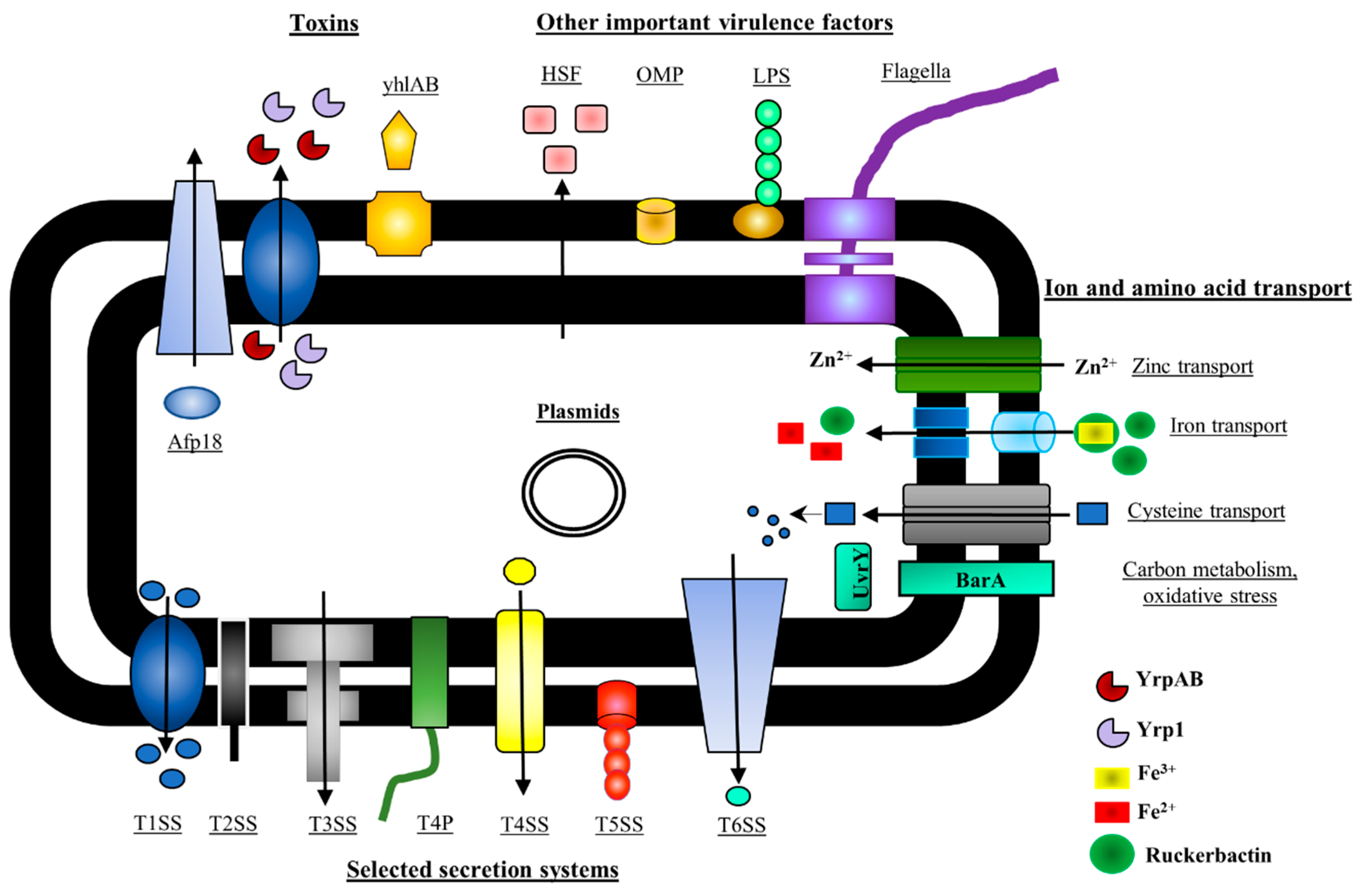
5. Y. ruckeri Pathogenicity Factors

It is well known that Y. ruckeri is responsible for economic losses in rainbow trout and Atlantic salmon farming production. Nevertheless, there are still a limited number of studies examining the virulence mechanisms of this pathogen. This may be the result of the early success of the ERM vaccine in the 1960s, the complex taxonomy of the pathogen, as well as scarce information on Y. ruckeri serotypes [59,83]. The virulence of Y. ruckeri is influenced by several factors, such as iron availability, temperature, pH and osmolarity [83]. Furthermore, it is generally believed that the virulence mechanisms and determinants of Y. ruckeri are similar to those in encountered in other members of the Yersiniaceae family [84]. Indeed, many researchers re-used models and techniques that were already applied to study other members of the Yersiniae. Various approaches have been used to identify and then further study the virulence-related genes of Y. ruckeri [83].
Molecular approaches for identifying virulence factors include the in vivo expression technology (IVET) approach and signature-tagged mutagenesis (STM) [85,86]. The IVET is based on the identification of genes that are expressed in vivo in the host during the infection process. In contrast, STM is issued to screen for Y. ruckeri mutants that survive in vitro, but not necessarily in vivo, meaning that the inactivated gene is essential for survival in the host but not necessarily directly for infection. Other approaches are based on the bioinformatics prediction of the role of virulence factors, and they include proteomic, transcriptomic and genomic analyses [83,87,88]. Although all of these methods have limitations, they have helped to discover many Y. ruckeri virulence factors. A summary of the key factors identified as of today is depicted in Figure 1.

5.1. Y. ruckeri Toxins

5.1.1. Y. ruckeri Protease 1 (Yrp1)

Yrp1, Y. ruckeri protease 1, is a 47-kDa serralysin metalloprotease that was identified by Secades and Guijarro in 1999 [89]. Yrp1 is secreted by an ATP-dependent type 1 secretion system (T1SS), similarly to hemolysin A from uropathogenic Escherichia coli [90]. The secretion system comprises three genes, yrpDEF, and an additional protease inhibitor inh [91]. Yrp1 is produced by some of the most virulent Y. ruckeri strains, named Azo+ strains (where azo is the gene for Yrp1), at the end of the exponential growth phase [89]. Expression of Yrp1 is controlled by osmolarity and temperature: highest expression occurs at 18 °C and under low osmotic pressure [92]. Yrp1 protease has been implicated in virulence [91]. It digests a wide range of matrix and muscle proteins, such as laminin, fibronectin, actin and myosin, causing the typical clinical symptoms of ERM. Finally, inactivated Yrp1 is protective against ERM when delivered as an immunogen [83,92].

5.1.2. Y. ruckeri Peptidases (YrpAB)

yrpAB are two adjacent genes encoding two peptidases from the U32 family. Both genes, identified by IVET, share a high sequence identity and the same genetic arrangement as peptidases present in other members of the Enterobacterales group [86,93]. The yrpAB operon expression is dependent on the presence of peptone in the culture media as well as the presence of oxygen. The expression of the yrpAB operon is highly upregulated under microaerobic conditions, which are encountered in fish gut, indicating that both peptidases may be expressed in the gut tissue. Interestingly, an LD50 experiment showed that at least one of the peptidases, YrpA, is involved in Y. ruckeri virulence. A deletion mutant of yrpA showed attenuated virulence (infective dose 7.1 × 10⁴ colony forming units) compared to the wild-type (WT) strain (1.73 × 10² colony forming units) [93].

5.1.3. Y. ruckeri Pore-Forming Toxin, yhlBA

The yhlBA cluster consists of two adjacent genes termed yhlA and yhlB identified by IVET [86]. yhlA encodes a hemolysin while yhlB is involved in yhlA activation and secretion [94]. Together, they constitute a type Vb secretion system [95]. Both genes have a high sequence similarity to genes encoding type Vb secretion systems with hemolysin activity in Serratia sp. Expression of the operon is regulated by temperature and iron availability and leads to cytolysis and hemolysis, e.g., of erythrocytes and BF-2 fish cells [94]. A high level of yhlBA expression was observed under limited iron availability and at 18 °C. The presence of the yhlBA operon in Y. ruckeri strains from different geographic areas and sources indicates an important role for this cluster in Y. ruckeri virulence [83],[94]. Interestingly, a hemolysin gene cluster is also present in human-pathogenic Yersiniae, but its role in virulence has not been confirmed [94,96].

5.1.4. Y. ruckeri Phospholipase

Phospholipases have the potential to act as exotoxins that disrupt host cell membranes [97], and the phospholipase activity of Y. ruckeri biotype 1 has been implicated in virulence functions. Phospholipase activity is found in the extracellular fraction of Y. ruckeri cultures, and the heat-labile products in the fraction are toxic to fish and show dermatotoxic effects in a rabbit skin model [98],[99]. The secretion of the phospholipase is dependent on the flagellar secretion machinery [100,101,102]. However, as the emerging biotype 2 does not have phospholipase activity, this cannot be considered an essential virulence factor.

5.1.5. Antifeeding Prophage 18 (Afp18)

Afp is a prophage-encoded toxin delivery system that shows similarities to R-pyocins of Pseudomonas aeuroginosa, and the Photohabdus luminescens virulence cassette [103,104]. Afp is present in Gram-negative and Gram-positive bacteria as well as in archaea. It resembles the tail of T4 bacteriophage that is composed of a central tube covered by a contractile sheath and a baseplate with fibers, which together probably constitute a type VI secretion system [105]. Afp encoded by the pADAP plasmid of Serratia entomophila was shown to play a role in delivery of a toxin, Afp18 [106]. In Y. ruckeri, Afp18 contains a glycosyltransferase domain at the C-terminus. After injection of the recombinant glycosyltransferase domain into zebrafish embryos, the development of zebrafish stopped, indicating a direct role in virulence for Afp18 [104].

5.2. Y. ruckeri Secretion Systems

5.2.1. Y. ruckeri Type III Secretion System (T3SS)

T3SSs are used by many Gram-negative pathogens to deliver toxins into host cells. The T3SS, also known as the injectisome, consists of a basal body that spans both inner and outer membrane, and a needle that protrudes out of the cell and makes direct contact between the bacterial cell and a host cell. The T3SS encodes three categories of proteins: structural proteins, chaperones and effector proteins [107].
A complete T3SS, the ysa locus, was recently found in the genome of Y. ruckeri SC09 [108]. Y. ruckeri SC09, isolated from a moribut (Ictalurus punctatus), possesses the largest genome within the species, with a length of 3.9 Mb encoding 3651 predicted protein-coding sequences. The ysa locus is found on the chromosome and shows similarities in gene content, gene sequence and gene arrangement with the Salmonella enterica pathogenicity island 1 and the chromosome-encoded T3SS from Y. enterocolitica biotype 1B. It is thus likely that the T3SS of SC09 is required for intracellular survival in fish macrophages, though this has not been experimentally verified [108].

5.2.2. Y. ruckeri Type IV Secretion Systems (T4SSs)

T4SSs are found in Gram-negative and Gram-positive bacteria, and in some archaea [109]. T4SSs are used by bacteria to transport macromolecules, such as proteins, protein complexes, protein-DNA complexes or DNA molecules across the bacterial cell membrane. Three major groups of T4SSs have been identified based on their function [110]. The first group, called conjugative T4SSs, transfers DNA from one cell to another by direct contact, in a process called conjugation. These T4SSs can be found on self-transmissible plasmids. They can also be a part of a chromosome as part of conjugative transposons. The second group of T4SSs is responsible for DNA uptake and release into the extracellular milieu. Two examples are the ComB system found in Helicobacter pylori (DNA uptake) and Gonococcal Genetic Island found in Neisseria gonorrhoeae (DNA release). The third group of T4SSs transfers virulence proteins and protein complexes into host cells, and thus plays an important role in virulence. This group is exemplified by T4SSs from important human pathogens, such as H. pylori, Bordetella pertussis and Legionella pneumophila [110].
T4SSs have been identified in different Y. ruckeri strains, and interestingly are absent in human-pathogenic Yersiniae. The tra operon of Y. ruckeri 150 was identified by IVET [86]. For Y. ruckeri SC09, it was suggested that the T4SS might be required for pathogen survival in fish macrophages [108]. The tra operon was shown to resemble the tra operon of the pADAP plasmid from S. entomophila, both in sequence and gene organization, and some genes of the tra operon display sequence similarity to the Dot/Icm proteins of L. pneumophila. A mutant of the traI gene of Y. ruckeri 150 was reported to be less virulent than the WT strain [111]. The expression of the tra operon is regulated by temperature and the availability of nutrients. The highest expression of the T4SS genes was reported at 18 °C, with limited nutrients. In contrast to previous reports, where the T4SS genes were reported to be localized on the chromosome, long-read sequencing technologies showed that it is actually encoded on a large plasmid, pYR4, of the Y. ruckeri NVH_3758 strain [112]. Based on this study, the previously reported chromosomal localization of the operon, based on short-read sequencing, should be revised. A recent comparative analysis of genome sequences of different Y. ruckeri O1 serotype strains (150, CSF007-82, ATCC29473) showed 268 O1-specific genes present in all three genomes, and genes of the T4SS were among these shared genes [113].

5.2.3. Y. ruckeri Type V Secretion Systems (T5SSs)

T5SSs or autotransporters are the most widespread secretion systems in Gram-negative bacteria. T5SSs contain a translocator unit, consisting of an outer membrane β-barrel domain and possibly some accessory domains, and an extracellular effector region or passenger [114]. Currently, five subtypes of T5SSs are recognized [95]. The only T5SS subtype present in Y. ruckeri is the type Ve or inverse autotransporter (IAT) subtype, which comprises virulence-associated adhesins that are also present in human-pathogenic Yersiniae [115]. IATs are found in Proteobacteria, Chlamydia, Cyanobacteria and Planctomycetes [116,117]. To date, they have been classified into four different sub-families: intimin, invasin, FdeC-type adhesins and two-partner IATs [117]. The two best-studied examples of the IATs are intimin of E. coli and invasin of Y. enterocolitica and Y. pseudotuberculosis, which play an important role in bacterial attachment to host tissues.
Two IAT genes are present in the genome of Y. ruckeri strains, termed yrInv (for Y. ruckeri invasin) and yrIlm (for Y. ruckeri invasin-like molecule) [118]. These IATs are variably found in Y. ruckeri strains of serotype O1 and serotype O2, isolated from rainbow trout and Atlantic salmon. The presence of the IATs in Y. ruckeri strains from different geographical regions and different sources may indicate a role in virulence, though their effects in vivo are yet to be tested. Both yrInv and yrIlm display a domain organization similar to well-described IATs: an N-terminal signal peptide followed by a periplasmically located LysM minidomain, which mediates oligomer formation and binding to peptidoglycan [119]. The β-barrel domain is between the periplasmic domain and the exported passenger, which contains several immunoglobulin (Ig)-like domains and a C-terminal lectin-like domain. YrInv and YrIlm show, respectively, 53% and 40% sequence similarity with InvA of the human pathogen Y. pseudotuberculosis. YrIlm is significantly longer in sequence compared to YrInv and comprises ~19 practically identical Ig-like domains, with some variation in repeat numbers between different strains. YrInv, by contrast, only contains 3 Ig-like domains with more divergent sequences. Expression of both genes is regulated by environmental factors, including temperature, salt concentration, iron availability and the presence of oxygen [118].

5.2.4. Type IV Pili

Type IV pili are multi-subunit, retractable surface appendages that can bind to surfaces and confer twitching motility. In addition, type IV pili can also have other functions such as DNA uptake or biofilm formation [120]. Type IV pili are secreted by a large complex spanning both membranes of Gram-negative bacteria and are related to T2SSs. Some Y. ruckeri strains contain a type IV pilus gene cluster originally thought to reside on the chromosome, but more recent analysis suggests that this locus (along with a separate T4SS) is located on a plasmid [111,112]. As the pil locus encoding the type IV pilus is adjacent to the T4SS tra locus, these may be functionally coupled. The role of the pil locus in virulence has not been studied, but as it is genetically linked to the tra operon, which does play a role in pathogenicity, a virulence function for pil can also be inferred.

5.3. Operons Involved in Ion and Amino Acids Transport

5.3.1. Iron Acquisition System

Iron acquisition systems aid many bacterial pathogens in colonizing and invading host tissues effectively. Y. ruckeri has such a system and produces the iron-chelating enterobactin-like siderophore ruckerbactin [19]. Siderophores are low-molecular-mass and high-affinity iron-chelating molecules [19]. They are categorized into four major groups: catecholates (enterobactin), hydroxamates (ferrioxamine), carboxylates (rhizobactin) and a mix of different types (pyoverdine) [121]. During bacterial infection, when iron availability is limiting for bacterial growth, the siderophores produced by bacterial cells are excreted into the extracellular environment, where they form a siderophore-Fe3+complex, which is then transported back to the bacterial cell and into the cytoplasm through a specific bacterial outer membrane receptor. Once inside the bacterial cell, the Fe3+ is reduced to Fe2+ in order to release free iron, which can then be utilized in metabolic reactions [19].
The genes involved in production of ruckerbactin are upregulated during the infection process and are highly expressed at 18 °C, but not at 28 °C, which is the optimal temperature for the pathogen’s growth [86]. Most of the ruckerbactin gene cluster has high sequence similarity with the E. coli enterobactin cluster, and their gene organization is similar. There is also a high degree of similarity between the ruckerbactin receptor and the ferrichrysobactin receptor of Dickeya dadantii (formerly known as Erwinia chrysanthemi), but not with the enterobactin receptor of E. coli [86].

5.3.2. The cdsAB Operon

The cdsAB operon was identified using IVET [86]. The operon is involved in the uptake and degradation of L-cysteine. The operon consists of two genes, cdsA and cdsB, that encode a cysteine permease and an L-cysteine desulfidase, respectively. The cdsAB cluster is found in several groups of Gram-negative bacteria, but not in Y. pseudotuberculosis or Y. pestis [122]. However, it is present in Y. ruckeri strains from different geographical locations and origins. In vivo assays and LD50 experiments showed that the cdsAB cluster is required to achieve full virulence. The underlying mechanism for the virulence effects is not known; it has been speculated that the genes are involved in the assembly of virulence-related proteins that contain iron-sulfur clusters, or in the release of H2S to induce host signaling [122].

5.3.3. znuABC and BarA-UvrY

The znuABC operon is composed of three genes, encoding a zinc-binding transporter, a membrane permease, and an ATPase. The genetic organization of the operon is similar to its counterparts found in other Gram-negative bacteria, such as E. coli and Salmonella Typhimurium [123,124]. The znuABC operon is likely to be involved in the transport of zinc, a molecule required for the pathogen’s survival. This is supported by the fact that the growth of E. coli znuABC mutants was restored in a metal-deficient medium when the znuABC operon of Y. ruckeri was supplied on a plasmid. Concomitantly, a znuABC mutant of Y. ruckeri had low virulence and survived poorly in rainbow trout [125]
In uropathogenic E. coli, the BarA-UvrY two-component system has been suggested to regulate switching between glycolysis and gluconeogenesis [126]. The UvrY component of the BarA-UvrY system plays the role of a response regulator, and UvrY was demonstrated to contribute to Y. ruckeri pathogenicity. Mutations in uvrY led to reduced invasion of EPC epithelial fish cells and increased sensitivity to oxidative stress, which in turn impaired the survival of Y. ruckeri in rainbow trout [127].

5.4. Other Virulence Factors

5.4.1. Genes Upregulated at 18 °C

A recent study identified putative virulence genes upregulated at 18 °C, a temperature at which most Y. ruckeri outbreaks occur. A Tn5–based transposon cassette containing the promoterless lux-lac operon was randomly inserted into the genome of Y. ruckeri, causing interruption of genes and using their cognate promotors. As a result of that, 168 clones were identified that had higher β-galactosidase activity at 18 °C than at 28 °C. The interrupted genes included, among others, genes there were involved in the synthesis of legionaminic acid (a component of the lipopolysaccharide (LPS) structure), the yrp1 metalloprotease described above, genes regulated by environmental changes (for example, the diguanylate cyclase, a glycosyltransferase and a S-adenosyl methionedependent methyltransferase), and genes induced under osmotic shock (for example osmY). All these genes were previously implicated in virulence in other organisms [128].

5.4.2. Heat-Sensitive Factor (HSF)

HSF was identified in Y. ruckeri in 1990 by Furones et al. [129]. A comparative study of cell extracts between virulent and avirulent Y. ruckeri strains exhibited a correlation between the production of HSF by virulent strains. Cell extracts of Y. ruckeri strains of serotype I, when injected into fish, caused mortalities only in HSF-positive strains. HSF is likely to play a role in virulence. It was proposed that HSF can mask cell surface antigens, and thus, confer resistance to phagocytic killing. However, a recent study showed that HSF is an alkyl sulphatase, encoded by the yraS gene. The alkyl sulphatase catalyzes the hydrolysis of SDS, but is not directly linked to Y. ruckeri virulence [22].

5.4.3. LPS

LPS is a glycolipid present in the outer leaflet of the outer membranes of Gram-negative bacteria and has been implicated in virulence in numerous species. The lpxD gene is one of the nine genes involved in biosynthesis of the lipid A moiety of LPS [130]. In-frame deletion of the lpxD gene in Y. ruckeri led to a production of an attenuated Y. ruckeri strain. The lpxD mutant, when administered as a vaccine by either immersion or injection to rainbow trout, elicited good protection responses against WT Y. ruckeri. The LPS-deficient mutant was suggested to be a good vaccine candidate against Y. ruckeri infections in rainbow trout [83,130].

5.4.4. Flagella

The flhDC operon of Y. ruckeri is a master regulator of the flagellar secretion apparatus that has been shown to regulate flagella production and phospholipase secretion. Mutation in the flhDC operon prevents motility and phospholipase activity, and additionally, absence of flhD results in transcriptional changes in a variety of genes. In competition assays, the flh∆D mutant of Y. ruckeri was reported to be more virulent than the WT strain. Examination of spleen in rainbow trout revealed an increased cell density of the flh∆D mutant compared to the WT [101]. A very recent study demonstrated that flagella expression is regulated by the Rcs system, which exerts its effect through modulating the expression of flhDC [102]. Flagellar synthesis is repressed within the host, but de-repressed upon host death. The repression of flagella is presumably to avoid detection by the host immune system. Strains with mutations in the rcsB gene produced flagella under conditions where flagellar synthesis is normally repressed and were attenuated in a fish infection model [102].

5.4.5. Plasmids

Plasmids play an important role in the virulence of human-pathogenic of Yersiniae. The pYV-type plasmid (YV stands for Yersinia virulence) is the best-characterized plasmid in the Yersiniae. It is shared among the three human-pathogenic Yersiniae (Y. pseudotuberculosis, Y. enterocolitica and Y. pestis). Removal of this plasmid was shown to result in the complete loss of virulence. The pYV plasmid is a ~70-kb virulence plasmid. In Y. pseudotuberculosis and Y. enterocolitica, it encodes a T3SSs and a T5SS protein named YadA (Yersinia adhesin A) [131,132]. The notorious Y. pestis typically carries two additional plasmids, pPla (encoding the plasminogen activator) and pFra (carrying the genes for murine toxin and the F1 antigen) [133]. The presence of further plasmids in Y. pestis has been reported; however, they are strain-specific and not very well characterized [134,135].
De Grandis and Stevenson examined the presence of plasmids in Y. ruckeri strains representing serotypes I and II [136]. Strains of serotype I contained a large and a small plasmid, while strains of serotype II contained only small plasmids, regardless of the source of the isolate. These results agree with those obtained by others [10,137,138]. The apparent number of plasmids in Y. ruckeri strains can differ depending on the applied plasmid isolation method [138]. The presence of large plasmids was suggested to be related to temperature, as the strains carrying the large plasmids were unable to grow at 37 °C. Y. ruckeri strains that carried the large plasmids exhibited distinct colony morphology and serological reactions, indicating that the large plasmid might encode genes related to these features [137]. Large plasmids have received a lot of attention due to their possible correlation with Yersiniae virulence [139]. Guilvout et al. examined the plasmid profile of 18 Y. ruckeri strains isolated from various ERM outbreaks. Strains of serotype II and V contained four plasmids each with both high and low molecular weights (72 MDa, 62 MDa, 32 MDa, 25 MDa), while strains of serotype I carried only one plasmid (62 MDa) [140]. Southern hybridization experiments between 62-MDa plasmids of Y. ruckeri strains of different origin showed a high level of similarity. However, the 62-MDa plasmid did not show any DNA similarity with the pYV plasmids from other Yersiniae, indicating a different origin and possibly, different functions.
To date, the function of Y. ruckeri plasmids is not clear. Large plasmids present in Y. ruckeri often carry antibiotic resistance genes. De Grandis and Stevenson tested susceptibility of Y. ruckeri strains to 23 antimicrobial agents [141]. All tested Y. ruckeri strains showed a similar antimicrobial susceptibility pattern except for two strains, which carried a 36-MDa plasmid and a 52-MDa plasmid and exhibited resistance to sulfonamides and tetracycline. Conjugation experiments showed that one of these two plasmids encoded antibiotic resistance genes that could be transferred to E. coli and to other Y. ruckeri strains.
Welch et al. showed a comparative DNA sequence analysis between the pIP1202 plasmid of Y. pestis strain IP275 and multi-drug resistance plasmids of Y. ruckeri YR1 (pYR1) and of the S. enterica serotype Newport SL254 (pSN254) [142]. The plasmid backbones were highly similar between those pathogens, indicating that the plasmids had a common ancestor. The common region contained genes involved in plasmid replication and type IV conjugative transfer. A recent study suggested that plasmids can be linked to Y. ruckeri virulence. A plasmid, pYR4, was found and analyzed, that—similarly to pYR3 from earlier studies—encodes both a type IV pilus and a T4SS (see above) that are implicated in virulence [112].

5.4.6. TcpA

TcpA, recently identified by Liu et al., is a virulence protein encoded by integrative and conjugative elements [143]. TcpA is present in Y. ruckeri SC09, but absent in other Y. ruckeri strains. In vivo and in vitro data showed TcpA involvement in virulence. TcpA disrupts Toll like receptor signaling in host cells and thus facilitates the escape from the host immune response, and therefore, enables bacterial survival.

5.4.7. Outer Membrane Proteins (OMPs)

OMPs are partly surface-exposed membrane proteins found in Gram-negative bacteria. They adopt a transmembrane β-barrel fold with eight to 26 strands, and play an essential role in nutrient acquisition, cell survival and host-pathogen interactions [144]. The OMPs of Y. ruckeri are very poorly characterized and there is little knowledge about their involvement in virulence. In recent years, however, OMPs have been discussed as promising vaccine candidates in fish. A recent study explored the protective capabilities of outer membrane porin F (OmpF) in Y. ruckeri [145]. OmpF is one of the major Y. ruckeri OMPs. Phylogenetic and multiple alignment analysis of OmpF demonstrated high sequence identity of OmpF within the Yersiniae (80.8–86.8%), compared to other Enterobacterales (58.8–76.4%), indicating that OmpF is a promising vaccine candidate. Indeed, recombinant OmpF injected into channel catfish (I. punctatus) enhanced the immune response by increasing, among others, lysozyme activity, complement C3 activity and serum antibody levels [145].
A recent study of Y. ruckeri strains isolated from Atlantic salmon and rainbow trout in Scotland demonstrated a high level of diversity between Atlantic salmon and rainbow trout Y. ruckeri isolates [15]. Specifically, the OMPs profile of aerobically grown Y. ruckeri isolates recovered from Atlantic salmon and rainbow trout was clearly different. The OMP profile of Y. ruckeri isolates recovered from Atlantic salmon was diverse: the isolates belonged to three different profile types (2a, 2c and 3a). In contrast, the OMP profiles of Y. ruckeri isolates recovered from rainbow trout were highly homogenous: most of the isolates were assigned to OMP type 3a [15].
A global proteomics analysis of Y. ruckeri strains of biotype 1 and biotype 2 grown under standard culture conditions was recently performed, identifying a wide number of OMPs (OmpA, OmpC, OmpF and OmpW) in addition to other putative virulence factors [88]. In a similar study, protein identification and quantification under standard and iron-limited conditions of motile and non-motile Y. ruckeri strains was performed. Sixty-one proteins were differentially expressed between iron-limiting and normal growth conditions; among the upregulated proteins were multiple OMPs related to iron and copper transport [87]. Similarly, a recently performed comparative bioinformatic and proteomic analysis of the OM proteome of Y. ruckeri strains recovered from Atlantic salmon and rainbow trout identified genes related to host specificity. Eighty-four OMPs were identified that were expressed under in vitro standard culture conditions. Sixteen OMPs were exclusively assigned to Y. ruckeri isolates from rainbow trout, including, e.g., filamentous hemagglutinin, metallopeptidases, a siderophore receptor and components of the T3SS that were predicted to be involved in adhesion to host cells, iron uptake and intracellular survival, respectively. Five proteins were exclusive for Y. ruckeri isolates of Atlantic salmon and these included proteins involved in autotransport, phospholipid-hydrolyzing enzymes and proteins involved in uptake of the ruckerbactin [146].

5.4.8. Biofilm as a Virulence Factor

Y. ruckeri can form biofilms in aquatic environments [147,148,149]. Y. ruckeri adheres to solid supports such as PVC, fiberglass, concrete, wood and other materials commonly found in fish farms. Surface roughness plays an important role in Y. ruckeri adherence to surfaces. Moreover, with longer incubation times, biofilm formation is more pronounced [148]. Finally, it has been suggested that biofilm formation by Y. ruckeri is key to recurrent infections in rainbow trout aquaculture, based on the ability of bacteria in biofilms to persist in circulating water systems [147].
The efficiency of biofilm formation varies from strain to strain and seems to be correlated with the presence of flagella-mediated motility. Environmental Y. ruckeri isolates exhibit a higher ability to adhere to surfaces than the reference strain, and thus, they are more likely to form a biofilm. Surface-associated Y. ruckeri cells are more tolerant to antibiotics than planktonic cells [148]. This can be both a direct effect of biofilm formation, or can in part be explained by differences between the outer membrane pattern of surface-associated Y. ruckeri strains and planktonic cells [147,149].
Y. ruckeri can adhere to and invade into various fish cell lines [150,151]. Application of inhibitors targeting host mechanisms significantly decreased the susceptibility of fish cell lines to be invaded by Y. ruckeri isolates of biotype 1 and biotype 2. Overall, biotype 1 strains were more adhesive and more invasive compared to biotype 2 strains. This finding indicates that Y. ruckeri targets diverse host’s mechanisms to gain entry into host cells. Additionally, the presence of flagella seems to be crucial for bacterial adhesion and biofilm formation and for entry into the host. The role of quorum sensing on the regulation of virulence factors, biofilm formation and swimming motility of Y. ruckeri was recently studied [152]. The supplementation of Y. ruckeri bacterial culture with a signaling molecule (3-oxo-C8-homoserine lactone) significantly increased the swimming motility and biofilm formation of Y. ruckeri isolates in vitro, while quorum quenching ((QQ) = interruption of bacterial communication) bacteria had the opposite effect on biofilm formation and motility of Y. ruckeri strains. This suggests that QQ bacteria can influence the gene expression of Y. ruckeri strains. Interestingly, such QQ bacteria are present on fish gills, in the fish intestine and on fish skin. Because Y. ruckeri enters the host via the secondary lamellae of the gills, it is possible that QQ bacteria might interfere with the pathogen by downregulating the expression of Y. ruckeri virulence genes, and therefore, impede the entry of the pathogen into the fish host [152,153,154].

6. Discussion

The current knowledge on Y. ruckeri virulence factors is incomplete and fragmented. In this review, we have tried to summarize the state of the art. Most studies of Y. ruckeri virulence are either descriptive or rely on genomics and proteomics methods to identify virulence factors based on sequence comparison with better-studied pathogenic species. Future work will have to experimentally address the molecular mechanism of the identified putative virulence factors to verify these findings, and to correlate these findings with the knowledge on the different known serotypes. A deeper understanding of the virulence mechanisms will be the foundation for the development of new antimicrobial strategies against this economically important fish pathogen, to complement existing vaccines and to avoid excessive use of antibiotics.

Author Contributions

A.W. conceptualized and wrote the draft manuscript. D.L. and J.C.L. assisted with reviewing and editing of the draft manuscript. All authors contributed to the final shape of the manuscript. This review is a substantial part of a PhD thesis entitled: “Structural and functional studies of the Yersinia ruckeri inverse autotransporters”, 2018, UiO, Oslo.

Funding

This work was supported by institutional funds by the Department of Biosciences, University of Oslo (COMPI, to D.L.) and by the Research Council of Norway Young Investigator grant 249793 (to J.C.L.).

Conflicts of Interest

The authors declare no conflict of interest.

References

  1. Stentiford, G.D.; Sritunyalucksana, K.; Flegel, T.W.; Williams, B.A.; Withyachumnarnkul, B.; Itsathitphaisarn, O.; Bass, D. New Paradigms to Help Solve the Global Aquaculture Disease Crisis. PLoS Pathog. 2017, 13, e1006160. [Google Scholar] [CrossRef] [PubMed]
  2. Walker, P.; Winton, J. Emerging viral diseases of fish and shrimp. Vet. Res. 2010, 41, 51. [Google Scholar] [CrossRef] [PubMed] [Green Version]
  3. Hjeltnes, B.; Bornø, G.; Jansen, M.; Haukaas, A.; Walde, C. The Health Situation in Norwegian Aquaculture 2016; Norwegian Veterinary Institute: Oslo, Norway, 2017. [Google Scholar]
  4. Austin, D.; Robertson, P.; Austin, B. Recovery of a New Biogroup of Yersinia ruckeri from Diseased Rainbow Trout (Oncorhynchus mykiss, Walbaum). Syst. Appl. Microbiol. 2003, 26, 127–131. [Google Scholar] [CrossRef] [PubMed]
  5. Fouz, B.; Zarza, C.; Amaro, C. First description of non-motile Yersinia ruckeri serovar I strains causing disease in rainbow trout, Oncorhynchus mykiss (Walbaum), cultured in Spain. J. Fish Dis. 2006, 29, 339–346. [Google Scholar] [CrossRef] [PubMed]
  6. Wheeler, R.; Davies, R.L.; Dalsgaard, I.; Garcia, J.; Welch, T.J.; Wagley, S.; Bateman, K.S.; Verner-Jeffreys, D.W. Yersinia ruckeri biotype 2 isolates from mainland Europe and the UK likely represent different clonal groups. Dis. Aquat. Organ. 2009, 84, 25–33. [Google Scholar] [CrossRef] [PubMed]
  7. Arias, C.; Olivares-Fuster, O.; Hayden, K.; Shoemaker, C.; Grizzle, J.; Klesius, P. First Report of Yersinia ruckeri Biotype 2 in the USA. J. Aquat. Anim. Health 2007, 19, 35–40. [Google Scholar] [CrossRef]
  8. Calvez, S.; Gantelet, H.; Blanc, G.; Douet, D.; Daniel, P. Yersinia ruckeri Biotypes 1 and 2 in France: Presence and antibiotic susceptibility. Dis. Aquat. Organ. 2014, 109, 117–126. [Google Scholar] [CrossRef]
  9. Bullock, G.; Stuckey, H.; Shotts, E., Jr. Enteric redmouth bacterium: Comparison of isolates from different geographic areas. J. Fish Dis. 1978, 1, 351–356. [Google Scholar] [CrossRef]
  10. Romalde, J.; MagariÑos, B.; Barja, J.; Toranzo, A. Antigenic and Molecular Characterization of Yersinia ruckeri Proposal for a New Intraspecies Classification. Syst. Appl. Microbiol. 1993, 16, 411–419. [Google Scholar] [CrossRef]
  11. O’Leary, P. Enteric Redmouth Bacterium of Salmonids: A Biochemical and Serological Comparison of Selected Isolates. Master’s Thesis, Oregon State University, Corvallis, OR, USA, 1977; p. 93. [Google Scholar]
  12. Stevenson, R.; Airdrie, R. Serological variation among Yersinia ruckeri strains. J. Fish Dis. 1984, 7, 247–254. [Google Scholar] [CrossRef]
  13. DeGrandis, S.; Krell, P.; Flett, D.; Stevenson, R. Deoxyribonucleic Acid Relatedness of Serovars of Yersinia ruckeri, the Enteric Redmouth Bacterium. J. Syst. Bacteriol. 1988, 38, 49–55. [Google Scholar] [CrossRef]
  14. Davies, R. O-Serotyping of Yersinia ruckeri with Special Emphasis on European Isolates. Vet. Microbiol. 1990, 22, 299–307. [Google Scholar] [CrossRef]
  15. Ormsby, M.; Caws, T.; Burchmore, R.; Wallis, T.; Verner-Jeffreys, D.; Davies, R. Yersinia ruckeri Isolates Recovered from Diseased Atlantic Salmon (Salmo salar) in Scotland Are More Diverse than Those from Rainbow Trout (Oncorhynchus mykiss) and Represent Distinct Subpopulations. Appl. Environ. Microbiol. 2016, 82, 5785–5794. [Google Scholar] [CrossRef] [PubMed]
  16. Adeolu, M.; Alnajar, S.; Naushad, S.; Gupta, R. Genome-based phylogeny and taxonomy of the ‘Enterobacteriales’: Proposal for Enterobacterales ord. nov. divided into the families Enterobacteriaceae, Erwiniaceae fam. nov., Pectobacteriaceae fam. nov., Yersiniaceae fam. nov., Hafniaceae fam. nov., Morganellaceae fam. nov., and Budviciaceae fam. nov. Int. J. Syst. Evol. Microbiol. 2016, 66, 5575–5599. [Google Scholar] [PubMed]
  17. Ross, A.; Rucker, R.; Ewing, W. Description of a bacterium associated with redmouth disease of rainbow trout (Salmo gairdneri). Can. J. Microbiol. 1966, 12, 763–770. [Google Scholar] [CrossRef] [PubMed]
  18. Austin, B.; Green, M.; Rodgers, C. Morphological diversity among strains of Yersinia ruckeri. Aquaculture 1982, 27, 73–78. [Google Scholar] [CrossRef]
  19. Tobback, E.; Decostere, A.; Hermans, K.; Haesebrouck, F.; Chiers, K. Yersinia ruckeri infections in salmonid fish. J. Fish 2007, 30, 257–268. [Google Scholar] [CrossRef] [PubMed]
  20. Moreau, E.; Thomas, T.; Brevet, M.; Thorin, C.; Fournel, C.; Calvez, S. Mutations involved in the emergance of Yersinia ruckeri biotype 2 in France. Transbound. Emerg. Dis. 2019, 66, 1387–1394. [Google Scholar] [CrossRef]
  21. Stevenson, R.M.W.; Daly, J.G. Biochemical and Serological Characteristics of Ontario Isolates of Yersinia ruckeri. Can. J. Fish. Aquat. Sci. 1982, 39, 870–876. [Google Scholar] [CrossRef]
  22. Navais, R.; Méndez, J.; Cascales, D.; Reimundo, P.; Guijarro, J. The heat sensitive factor (HSF) of Yersinia ruckeri is produced by an alkyl sulphatase involved in sodium dodecyl sulphate (SDS) degradation but not in virulence. BMC Microbiol. 2014, 14, 221. [Google Scholar] [CrossRef]
  23. Gibello, A.; Porrero, M.C.; Blanco, M.M.; Vela, A.I.; Liébana, P.; Moreno, M.A.; Fernandez-Garayzabal, J.F.; Domínguez, L. Analysis of the gyrA Gene of Clinical Yersinia ruckeri Isolates with Reduced Susceptibility to Quinolones. Appl. Environ. Microbiol. 2004, 70, 599–602. [Google Scholar] [CrossRef] [PubMed]
  24. Gibello, A.; Blanco, M.M.; Moreno, M.A.; Cutuli, M.T.; Domenech, A.; Domínguez, L.; Fernández-Garayzábal, J.F. Development of a PCR Assay for Detection of Yersinia ruckeri in Tissues of Inoculated and Naturally Infected Trout. Appl. Environ. Microbiol. 1999, 65, 346–350. [Google Scholar] [PubMed]
  25. Akhlaghi, M.; Yazdi, H.S. Detection and identification of virulent Yersinia ruckeri: The causative agent of enteric redmouth disease in rainbow trout (Oncorhynchus mykiss) cultured in Fars province, Iran. Iran. J. Vet. Res. 2008, 9, 347–352. [Google Scholar]
  26. Busch, R. Enteric redmouth disease. Mar. Fish. Rev. 1978, 40, 42–51. [Google Scholar]
  27. Cossarini-Dunier, M. Indirect enzyme-linked immunosorbent assay (ELISA) to titrate rainbow trout serum antibodies against two pathogens: Yersinia ruckeri and Egtved virus. Aquaculture 1985, 49, 197–208. [Google Scholar] [CrossRef]
  28. Ghosh, B.; Crosbie, P.; Nowak, B.; Bridle, A. A highly sensitive, non-invasive qPCR-based strategy for direct quantification of Yersinia ruckeri in fish faeces. J. Fish Dis. 2018, 41, 1421–1428. [Google Scholar] [CrossRef] [PubMed]
  29. Smith, A.; Goldring, O.; Dear, G. The production and methods of use of polyclonal antisera to the pathogenic organisms Aeromonas salmonicida, Yersinia ruckeri and Renibacterium salmoninarum. J. Fish Biol. 1987, 31, 225–226. [Google Scholar] [CrossRef]
  30. Glenn, R.; Taylor, P.; Hanson, K. The use of a real-time PCR primer/probe set to observe infectivity of Yersinia ruckeri in Chinook salmon, Oncorhynchus tshawytscha (Walbaum, and steelhead trout, Oncorhynchus mykiss(Walbaum). J. Fish Dis. 2011, 34, 783–791. [Google Scholar] [CrossRef]
  31. Keeling, S.; Johnston, C.; Wallis, R.; Brosnahan, C.; Gudkovs, N.; Mcdonald, W. Development and validation of real-time PCR for the detection of Yersinia ruckeri. J. Fish Dis. 2012, 35, 119–125. [Google Scholar] [CrossRef]
  32. Saleh, M.; Soliman, H.; El-Matbouli, M. Loop-mediated isothermal amplification as an emerging technology for detection of Yersinia ruckeri the causative agent of enteric red mouth disease in fish. BMC Vet. Res. 2008, 4, 1–10. [Google Scholar] [CrossRef]
  33. Temprano, A.; Yugueros, J.; Hernanz, C.; Sánchez, M.; Berzal, B.; Luengo, J.M.; Naharro, G. Rapid identification of Yersinia ruckeri by PCR amplification of yruI-yruR quorum sensing. J. Fish Dis. 2001, 24, 253–261. [Google Scholar] [CrossRef]
  34. Altinok, I.; Grizzle, J.; Liu, Z. Detection of Yersinia ruckeri in rainbow trout blood by use of the polymerase chain reaction. Dis. Aquat. Organ. 2001, 44, 29–34. [Google Scholar] [CrossRef] [PubMed]
  35. Noga, E.; Levine, J.; Townsend, K.; Bullis, R.; Carlson, C.; Corbett, W. Kidney biopsy: A nonlethal method for diagnosing Yersinia ruckeri infection (enteric redmouth disease) in rainbouw trout (Salmo gairdneri). Am. J. Vet. Res. 1988, 49, 363–365. [Google Scholar] [PubMed]
  36. Bastardo, A.; Ravelo, C.; Romalde, J. Multilocus sequence typing reveals high genetic diversity and epidemic population structure for the fish pathogen Yersinia ruckeri. Environ. Microbiol. 2012, 14, 1888–1897. [Google Scholar] [CrossRef] [PubMed]
  37. Gulla, S.; Mohammad, S.; Colquhoun, D. Multi-locus variable-number tandem-repeat analysis of the fish-pathogenic bacterium Yersinia ruckeri by multiplex PCR and capillary electrophoresis. J. Vis. Exp. 2019, 148, 1–6. [Google Scholar] [CrossRef] [PubMed]
  38. Ewing, W.; Ross, A.; Brenner, D.; Fanning, G. Yersinia ruckeri sp. nov., the Redmouth (RM) Bacterium. Int. J. Syst. Bcteriol. 1978, 28, 37–44. [Google Scholar] [CrossRef]
  39. Kumar, G.; Menanteau-Ledouble, S.; Saleh, M.; El-Matbouli, M. Yersinia ruckeri, the causative agent of enteric redmouth disease in fish. Vet. Res. 2015, 46, 1–10. [Google Scholar] [CrossRef]
  40. Rucker, R. Redmouth disease of rainbow trout (Salmo gairdneri). Bull. Off. Int. Epizoot. 1966, 65, 825–830. [Google Scholar]
  41. Fuhrmann, H.; Böhm, K.; Schlotfeldt, H. An outbreak of enteric redmouth disease in West Germany. J. Fish Dis. 1983, 6, 309–311. [Google Scholar] [CrossRef]
  42. Bullock, G. Enteric Redmouth Disease. Fish Heal. Bull. 1989. Available online: https://apps.dtic.mil/dtic/tr/fulltext/u2/a322943.pdf (accessed on 9 September 2019).
  43. Bruno, D. Enteric Redmouth Disease. Aquac. Inf. Ser. 1990, 10. Available online: https://www2.gov.scot/Topics/marine/science/Publications/FRS-Reports/ais (accessed on 9 September 2019).
  44. Zorriehzahra, M.; Adel, M.; Delshad, S.T. Enteric redmouth disease: Past, present and future: A review. Iran. J. Fish. Sci. 2017, 16, 1135–1156. [Google Scholar]
  45. Gulla, S.; Barnes, A.C.; Welch, T.J.; Romalde, J.L.; Ryder, D.; Ormsby, M.J.; Carson, J.; Lagesen, K.; Verner-Jeffreys, D.W.; Davies, R.L. Multi-Locus Variable number of tandem repeat Analysis (MLVA) of Yersinia ruckeri confirms the existence of host-specificity, geographic endemism and anthropogenic dissemination of virulent clones. Appl. Environ. Microbiol. 2018, 84. [Google Scholar] [CrossRef] [PubMed]
  46. McDaniel, D. Hagerman redmouth. Am. Fish. US Trout News 1971, 15, 14–28. [Google Scholar]
  47. Wobeser, G. An outbreak of redmouth disease in rainbow trout (Salmo gairdneri) in Saskatchewan. J. Fish. Res. Board Can. 1973, 30, 571–575. [Google Scholar] [CrossRef]
  48. Llewellyn, L. A bacterium with similarities to the redmouth bacterium and Serratia liquefaciens (Grimes and Hennerty) causing mortalities in hatchery reared salmonids in Australia. J. Fish Dis. 1980, 3, 29–39. [Google Scholar] [CrossRef]
  49. Dalsgaard, I.; From, J.; Hørlyck, V. First observation of Yersinia ruckeri in Denmark. Bull. Eur. Assoc. Fish Pathol. 1984, 4, 10. [Google Scholar]
  50. Lesel, R.; Lesel, M.; Gavini, F.; VuiIlaume, A. Outbreak of enteric redmouth disease, in rainbow trout, Salmo gairdneri Richardson, in France. J. Fish Dis. 1983, 6, 385–387. [Google Scholar] [CrossRef]
  51. Giorgetti, G.; Ceschia, G.; Bovo, G. First Isolation of Yersinia ruckeri in Farmed Rainbow Trout in Italy; Ellis, A.E., Ed.; Academic Press: London, UK, 1985; pp. 161–166. [Google Scholar]
  52. Sparboe, O.; Koren, C.; Hastein, T.; Poppe, T.; Stenwig, H. The first isolation of Yersinia ruckeri from farmed Norwegian salmon. Bull. Eur. Assoc. Fish Pathol. 1986, 6, 41–42. [Google Scholar]
  53. Grawiński, E. The occurrence of redmouth disease in the rainbow trout (Salmo gairdneri Richardson). Vet. Med. 1990, 46, 183–185. [Google Scholar]
  54. Oraić, D.; Zrnčić, S.; Šoštarić, B.; Bažulić, D.; Lipiej, Z. Occurence of enteric redmouth disease in rainbow trout (Oncorhynchus mykiss) on farms in Croatia. Acta Vet. Hung. 2002, 50, 283–291. [Google Scholar] [CrossRef]
  55. Popescu, A.; Costea, M.; Dăscălescu, P. Characterization of Yersinia ruckeri strains from salmonid populations in Romania. Rev. Rom. Med. 2008, 18. [Google Scholar]
  56. Anderson, C.; Knowles, G.; de Lisle, G. A survey for Yersinia ruckeri and Aeromonas salmonicida in farmed and wild fish. Surveillance 1994, 21, 39–40. [Google Scholar]
  57. Bragg, R.; Henton, M. Isolation of Yersinia ruckeri from rainbow trout in South Africa. Bull. Eur. Assoc. Fish Pathol. 1986, 6, 5–6. [Google Scholar]
  58. Enríquez, R.; Zamora, J. Isolation of Yersinia ruckeri from carp (Cyprinus carpio) in Valdivia. Arch. Med. Vet. 1987, 19, 33–36. [Google Scholar]
  59. Barnes, A. Enteric Redmouth Disease (ERM) (Yersinia ruckeri). In Fish Diseases and Disorders: Viral, Bacterial and Fungal Infections; Woo, P.T.K., Bruno, D.W., Eds.; CABI: Walingford, UK, 2011; Volume 3, pp. 484–511. [Google Scholar]
  60. Collins, R.; Foster, G.; Ross, H. Isolation of Yersinia ruckeri from farmed Atlantic Salmon (Salmo salar), and Arctic Char (Salvelinus alpinus) and a European Otter (Lutra lutra) in several water catchment areas in Scotland. Vet. Rec. 1998, 139, 169. [Google Scholar] [CrossRef]
  61. Gudmundsdottir, B.; Gudmundsdottir, S.; Gudmundsdottir, S.; Magnadottir, B. Yersiniosis in Atlantic cod, Gadus morhua (L.), characterization of the infective strain and host reactions. J. Fish Dis. 2014, 37, 511–519. [Google Scholar] [CrossRef]
  62. Navas, E.; Bohle, H.; Henríquez, P.; Grothusen, H.; Bustamante, F.; Bustos, P.; Mancilla, M. Draft Genome Sequence of the Fish Pathogen Yersinia ruckeri Strain 37551, Serotype O1b, Isolated from Diseased, Vaccinated Atlantic Salmon (Salmo salar) in Chile. Genome Announc. 2014, 2, 10–11. [Google Scholar] [CrossRef]
  63. Xu, B.; Yin, Z.; Chen, Y.; Wu, Y. An outbreak of a new epi- zootic in Silver Carp and Bighead Carp: Yersinia ruckeri, a new pathogen of Silver Carp and Bighead Carp. Chin. Sci. Bull. 1991, 36, 1825. [Google Scholar]
  64. Dwilow, A.; Souter, B.; Knight, K. Isolation of Yersinia ruckeri from burbot, Lota lota (L.) from the Mackenzie River, Canada. J. Fish Dis. 1987, 10, 315–317. [Google Scholar] [CrossRef]
  65. Cipriano, R.; Schill, W.; Pyle, S.; Horner, R. An epizootic in cinook salmon (Oncorhynchus tshawytscha) caused by a sorbitol-positive serovar 2 strain of Yersinia ruckeri. J. Wildl. Dis. 1986, 22, 488–492. [Google Scholar] [CrossRef]
  66. Huang, Y.; Jung, A.; Schäfer, W.; Mock, D.; Michael, G.B.; Runge, M.; Schwarz, S.; Steinhagen, D. Analysis of Yersinia ruckeri strains isolated from trout farms in northwest Germany. Dis. Aquat. Organ. 2015, 116, 243–249. [Google Scholar] [CrossRef]
  67. Valtonen, E.; Rintamaki, P.; Koskivaara, M. Occurence and pathogenicity of Yersinia ruckeri at fish farms in northern and central Finland. J. Fish Dis. 1992, 15, 163–171. [Google Scholar] [CrossRef]
  68. Michel, C.; Fairve, B.; de Kinkelin, P. A clinical case of enteric redmouth in minnows (Pimephales promelas) imported in Europe as bait-fish. Bull. Eur. Assoc. Fish Pathol. 1986, 6, 97–99. [Google Scholar]
  69. Avendaño-Herrer, R.; Tapia-Cammas, D.; Aedo, A.; Saldivia, P.; Ortega, C.; Irgang, R. Disease caused by Yersinia ruckeri serotype O2b found in Chilean-farmed coho salmon, Oncorhynchus kisutch (Walbaum, 1792). J. Fish Dis. 2016, 40, 279–285. [Google Scholar] [CrossRef]
  70. Berc, A.; Petrinec, Z.; Matašin, Ž.; Kozarić, Z. Yersinia ruckeri Septicaemia in Experimentally Infected Carp (Cyprinus Carpio L.) Fingerlings. Acta Vet. Hung. 2015, 47, 161–172. [Google Scholar] [CrossRef]
  71. Daly, J.; Lindvik, B.; Stevenson, R. Serological heterogeneity of recent isolates of Yersinia ruckeri from Ontario and British Columbia. Dis. Aquat. Organ. 1986, 1, 151–153. [Google Scholar] [CrossRef]
  72. McArdle, J.; Dooley-Martyn, C. Isolation of Yersinia ruckeri type I (Hagerman strain) from goldfish Carassius auratus (L.). Bull. Eur. Assoc. Fish Pathol. 1985, 5, 1–2. [Google Scholar]
  73. Rintamäki, P.; Valtonen, E.; Frerichs, G. Occurrence of Yersinia ruckeri infection in farmed whitefish, Coregonus peled Gmelin and Coregonus muksun Pallas, and Atlantic salmon, Salmo salar L., in northern Finland. J. Fish Dis. 1986, 9, 137–140. [Google Scholar] [CrossRef]
  74. Eissa, A.; Moustafa, M.; Abdelaziz, M.; Ezzeldeen, N. Yersinia ruckeri infection in cultured Nile tilapia, Oreochromis niloticus, at a semi-intensive fish farm in lower Egypt. Afr. J. Aquat. Sci. 2008, 33, 283–286. [Google Scholar] [CrossRef]
  75. Duman, M.; Altun, S.; Cengiz, M.; Saticioglu, I.; Buyukekiz, A.; Sahinturk, P. Genotyping and antimicrobial resistance genes of Yersinia ruckeri isolates from rainbow trout farms. Dis. Aquat. Organ. 2017, 125, 31–44. [Google Scholar] [CrossRef]
  76. Popović, N.; Hacmanjek, M.; Teskeredžić, E. Health status of rudd (Scardinius erythrophthalmus hesperidicus H.) in Lake Vrana on the Island of Cres, Croatia. J. Appl. Ichthyol. 2001, 17, 43–45. [Google Scholar] [CrossRef]
  77. Vuillaume, A.; Brun, R.; Chene, P.; Sochon, E.; Lesel, R. First isolation of Yersinia ruckeri from sturgeon, Acipenser baeri Brandt, in South West of France. Bull. Eur. Assoc. Fish Pathol. 1987, 7, 18–19. [Google Scholar]
  78. Korbut, R.; Mehrdana, F.; Kania, P.W.; Larsen, M.H.; Frees, D.; Dalsgaard, I.; Jørgensen, L.V.G. Antigen Uptake during Different Life Stages of Zebrafish (Danio rerio) Using a GFP-Tagged Yersinia ruckeri. PLoS ONE 2016, 11, e0158968. [Google Scholar] [CrossRef]
  79. Willumsen, B. Birds and wild fish as potential vectors of Yersinia ruckeri. J. Fish Dis. Dis. 1987, 40, 275–277. [Google Scholar] [CrossRef]
  80. De Keukeleire, S.; de Bel, A.; Jansen, Y.; Janssens, M.; Wauters, G.; Piérard, D. Yersinia ruckeri, an unusual microorganism isolated from a human wound infection. New Microbes New Infect. 2014, 2, 134–135. [Google Scholar] [CrossRef]
  81. Standish, I.; Erickson, S.; Kleman, E.; McCann, R.; Katona, R.; Leis, E. Yersinia ruckeri Isolated from Common Mudpuppy Necturus maculosus. J. Aquat. Anim. Health 2018, 31, 71–74. [Google Scholar] [CrossRef]
  82. Dudley, D.; Guentzel, M.; Ibarra, M.; Moore, B.; Sagik, B. Enumeration of potentially pathogenic bacteria from sewage sludges. Appl. Environ. Microbiol. 1980, 39, 118–126. [Google Scholar] [Green Version]
  83. Guijarro, J.; García-Torrico, A.; Cascales, D.; Méndez, J. The Infection Process of Yersinia ruckeri: Reviewing the Pieces of the Jigsaw Puzzle. Front. Cell. Infect. Microbiol. 2018, 8, 218. [Google Scholar] [CrossRef]
  84. Fernández, L.; Méndez, J.; Guijarro, J. Molecular virulence mechanisms of the fish pathogen Yersinia ruckeri. Vet. Microbiol. 2007, 125, 1–10. [Google Scholar] [CrossRef]
  85. Dahiya, I.; Stevenson, R. Yersinia ruckeri genes that attenuate survival in rainbow trout (Oncorhynchus mykiss) are identified using signature-tagged mutants. Vet. Microbiol. 2010, 144, 399–404. [Google Scholar] [CrossRef] [PubMed]
  86. Fernández, L.; Marquez, I.; Guijarro, J. Identification of specific In Vivo-induced (ivi) genes in Yersinia ruckeri and analysis of ruckerbactin, a catecholate siderophore iron acquisition system. Appl. Environ. Microbiol. 2004, 70, 5199–5207. [Google Scholar] [CrossRef] [PubMed]
  87. Kumar, G.; Hummel, K.; Ahrens, M.; Menanteau-Ledouble, S.; Welch, T.J.; Eisenacher, M.; Razzazi-Fazeli, E.; El-Matbouli, M. Shotgun proteomic analysis of Yersinia ruckeri strains under normal and iron-limited conditions. Vet. Res. 2016, 47, 100. [Google Scholar] [CrossRef] [PubMed]
  88. Kumar, G.; Hummel, K.; Welch, T.; Razzazi-Fazeli, E.; El-Matbouli, M. Global proteomic profiling of Yersinia ruckeri strains. Vet. Res. 2017, 48, 55. [Google Scholar] [CrossRef] [PubMed]
  89. Secades, P.; Guijarro, J. Purification and Characterization of an Extracellular Protease from the Fish pathogen Yersinia ruckeri and effect of culture condition on production. Appl. Environ. Microbiol. 1999, 65, 3969–3975. [Google Scholar] [PubMed]
  90. Balakrishnan, L.; Hughes, C.; Koronakis, V. Substrate-triggered recruitment of the TolC channel-tunnel during type I export of hemolysin by Escherichia coli. J. Mol. Biol. 2001, 313, 501–510. [Google Scholar] [CrossRef]
  91. Fernandez, L.; Secades, P.; Lopez, J.; Marquez, I.; Guijarro, J. Isolation and analysis of a protease gene with an ABC transport system in the fish pathogen Yersinia ruckeri: Insertional mutagenesis and involvement in virulence. Microbiology 2002, 148, 2233–2243. [Google Scholar] [CrossRef]
  92. Fernandez, L.; Lopez, J.; Secades, P.; Menendez, A.; Marquez, I.; Guijarro, J. In Vitro and In Vivo Studies of the Yrp1 Protease from Yersinia ruckeri and Its Role in Protective Immunity against Enteric Red Mouth Disease of Salmonids. Appl. Environ. Microbiol. 2003, 69, 7328–7335. [Google Scholar] [CrossRef]
  93. Navais, R.; Méndez, J.; Pérez-Pascual, D.; Cascales, D.; Guijarro, J. The yrpAB operon of Yersinia ruckeri encoding two putative U32 peptidases is involved in virulence and induced under microaerobic conditions. Virulence 2014, 5, 619–624. [Google Scholar] [CrossRef]
  94. Fernández, L.; Prieto, M.; Guijaro, J. The iron- and temperature-regulated haemolysin YhlA is a virulence factor of Yersinia ruckeri. Microbiology 2007, 153, 483–489. [Google Scholar] [CrossRef]
  95. Leo, J.; Grin, I.; Linke, D. Type V secretion: Mechanism(s) of autotransport through the bacterial outer membrane. Philos. Trans. R. Soc. Lond. B. Biol. Sci. 2012, 367, 1088–1101. [Google Scholar] [CrossRef] [PubMed]
  96. Chain, P.S.; Carniel, E.; Larimer, F.W.; Lamerdin, J.; Stoutland, P.O.; Regala, W.M.; Georgescu, A.M.; Vergez, L.M.; Land, M.L.; Motin, V.L.; et al. Insights into the evolution of Yersinia pestis through whole-genome comparison with Yersinia pseudotuberculosis. Proc. Natl. Acad. Sci. USA 2004, 101, 13826–13831. [Google Scholar] [CrossRef] [PubMed]
  97. Flores-Diaz, M.; Monturiol-Gross, L.; Naylor, C.; Alape-Giron, A.; Flieger, A. Bacterial Sphingomyelinases and Phospholipases as Virulence Factors. Microbiol. Mol. Biol. Rev. MMBR 2016, 80, 597–628. [Google Scholar] [CrossRef] [PubMed] [Green Version]
  98. Romalde, J.L.; Toranzo, A.E. Pathological activities of Yersinia ruckeri, the enteric redmouth (ERM) bacterium. FEMS Microbiol. Lett. 1993, 112, 291–299. [Google Scholar] [CrossRef] [PubMed]
  99. Romalde, J.; Santos, Y.; Toranzo, A. Presence of skin permeability factors in the extracellular products of Yersinia ruckeri. Curr. Microbiol. 1992, 24, 263–267. [Google Scholar] [CrossRef]
  100. Welch, T.J.; Verner-Jeffreys, D.W.; Dalsgaard, I.; Wiklund, T.; Evenhuis, J.P.; Cabrera, J.A.G.; Hinshaw, J.M.; Drennan, J.D.; LaPatra, S.E. Independent Emergence of Yersinia Ruckeri Biotype 2 in the United States and Europe. Appl. Environ. Microbiol. 2011, 77, 3493–3499. [Google Scholar] [CrossRef] [PubMed]
  101. Jozwick, A.; Graf, J.; Welch, T. The flagellar master operon flhDC is a pleiotropic regulator involved in motility and virulence of the fish pathogen Yersinia ruckeri. J. Appl. Microbiol. 2016, 122, 578–588. [Google Scholar] [CrossRef] [PubMed]
  102. Jozwick, A.; LaPatra, S.; Graf, J.; Welch, T. Flagellar regulation mediated by the Rcs pathway is required for virulence in the fish pathogen Yersinia ruckeri. Fish Shellfish Immunol. 2019, 91, 306–314. [Google Scholar] [CrossRef]
  103. Heymann, J.B.; Bartho, J.D.; Rybakova, D.; Venugopal, H.P.; Winkler, D.C.; Sen, A.; Hurst, M.R.H.; Mitra, A.K. Three-dimensional Structure of the Toxin-delivery Particle Antifeeding Prophage of Serratia entomophila. J. Biol. Chem. 2013, 288, 25276–25284. [Google Scholar] [CrossRef]
  104. Jank, T.; Eckerle, S.; Steinemann, M.; Trillhaase, C.; Schimpl, M.; Wiese, S.; Van Aalten, D.M.F.; Driever, W.; Aktories, K. Tyrosine glycosylation of Rho by Yersinia toxin impairs blastomere cell behaviour in zebrafish embryos. Nat. Commun. 2015, 6, 1–16. [Google Scholar] [CrossRef]
  105. Yang, X.; Pan, J.; Wang, Y.; Shen, X. Type VI Secretion Systems Present New Insights on Pathogenic Yersinia. Front. Cell. Infect. Microbiol. 2018, 8, 260. [Google Scholar] [CrossRef] [PubMed]
  106. Hurst, M.; Beard, S.; Jackson, T.; Jones, S. Isolation and characterization of the Serratia entomophila antifeeding prophage. FEMS Microbiol. Lett. 2007, 270, 42–48. [Google Scholar] [CrossRef] [PubMed]
  107. Puhar, A. Type III secretion system. Curr. Biol. 2014, 24, R784–R791. [Google Scholar] [CrossRef] [PubMed] [Green Version]
  108. Liu, T.; Wang, K.-Y.; Wang, J.; Chen, D.-F.; Huang, X.-L.; Ouyang, P.; Geng, Y.; He, Y.; Zhou, Y.; Min, J. Genome sequence of the fish pathogen Yersinia ruckeri SC09 provides insights into niche adaptation and pathogenic mechanism. Int. J. Mol. Sci. 2016, 17, 557. [Google Scholar] [CrossRef] [PubMed]
  109. Wang, S.; Wang, D.; Du, D.; Li, S.; Yan, W. Advances in the Assembly Model of Bacterial Type IVB Secretion Systems. Appl. Sci. 2018, 8, 2368. [Google Scholar] [CrossRef]
  110. Alvarez-Martinez, C.; Christie, P. Biological diversity of prokaryotic type IV secretion systems. Microbiol Mol Biol Rev. 2009, 73, 775–808. [Google Scholar] [CrossRef] [PubMed]
  111. Méndez, J.; Fernández, L.; Menéndez, A.; Reimundo, P.; Pérez-Pascual, D.; Navais, R.; Guijarro, J.A. A chromosomally located traHIJKCLMN operon encoding a putative type IV secretion system is involved in the virulence of Yersinia ruckeri. Appl. Environ. Microbiol. 2009, 75, 937–945. [Google Scholar] [CrossRef] [PubMed]
  112. Wrobel, A.; Ottoni, C.; Leo, J.; Linke, D. pYR4 From a Norwegian Isolate of Yersinia ruckeri Is a Putative Virulence Plasmid Encoding Both a Type IV Pilus and a Type IV Secretion System. Front. Cell. Infect. Microbiol. 2018, 8, 1–14. [Google Scholar] [CrossRef]
  113. Cascales, D.; Guijarro, J.; Garcia-Torrico, A.; Méndez, J. Comparative genome analysis reveals important genetic differences among serotype O1 and serotype O2 strains of Y. ruckeri and provides insights into host adaptation and virulence. MicrobiologyOpen 2017, 6. [Google Scholar] [CrossRef]
  114. Fan, E.; Chauhan, N.; Udatha, D.; Leo, J.; Linke, D. Type V Secretion Systems in Bacteria. Microbiol. Spectr. 2016, 4, 1–24. [Google Scholar]
  115. Chauhan, N.; Wrobel, A.; Skurnik, M.; Leo, J. Yersinia adhesins: An arsenal for infection. Proteom. Clin. Appl. 2016, 10, 949–963. [Google Scholar] [CrossRef] [PubMed]
  116. Leo, J.; Oberhettinger, P.; Schütz, M.; Linke, D. The inverse autotransporter family: Intimin, invasin and related proteins. Int. J. Med Microbiol. 2015, 305, 276–282. [Google Scholar] [CrossRef] [PubMed]
  117. Heinz, E.; Stubenrauch, C.J.; Grinter, R.; Croft, N.P.; Purcell, A.W.; Strugnell, R.A.; Dougan, G.; Lithgow, T. Conserved features in the structure, mechanism and biogenesis of the inverse autotransporter protein family. Genome Biol. Evol. 2016, 8, 1690–1705. [Google Scholar] [CrossRef] [PubMed]
  118. Wrobel, A.; Ottoni, C.; Leo, J.C.; Gulla, S.; Linke, D. The repeat structure of two paralogous genes, Yersinia ruckeri Invasin (yrInv) and a ‘Y. ruckeri Invasin-like molecule’, (yrIlm) sheds light on the evolution of adhesive capacities of a fish pathogen. J. Struct. Biol. 2017, 201, 171–183. [Google Scholar] [CrossRef] [PubMed]
  119. Leo, J.; Oberhettinger, P.; Chaubey, M.; Schütz, M.; Kühner, D.; Bertsche, U.; Schwarz, H.; Götz, F.; Autenrieth, I.B.; Coles, M.; et al. The Intimin periplasmic domain mediates dimerisation and binding to peptidoglycan. Mol. Microbiol. 2015, 95, 80–100. [Google Scholar] [CrossRef]
  120. Craig, L.; Forest, K.; Maier, B. Type IV pili: Dynamics, biophysics and functional consequences. Nat. Rev. Microbiol. 2019, 17, 429–440. [Google Scholar] [CrossRef]
  121. Ahmed, E.; Holmström, S. Siderophores in environmental research: Roles and applications. Microb. Biotechnol. 2014, 7, 196–208. [Google Scholar] [CrossRef]
  122. Méndez, J.; Reimundo, P.; Pérez-Pascual, D.; Navais, R.; Gómez, E.; Guijarro, A. A Novel cdsAB Operon Is Involved in the Uptake of L-Cysteine and Participates in the Pathogenesis of Yersinia ruckeri. J. Bacteriol. 2011, 193, 944–951. [Google Scholar] [CrossRef]
  123. Patzer, S.; Hantke, K. The ZnuABC high-affinity zinc uptake system and its regulator Zur in Escherichia coli. Mol. Microbiol. 1998, 28, 1199–1210. [Google Scholar] [CrossRef]
  124. Campoy, S.; Jara, M.; Busquets, N.; de Rozas, A.; Badiola, I.; Barbe, J. Role of the high-affinity zinc uptake znuACB system in Salmonella enterica serovar Typhimurium virulence. Infect. Immun. 2002, 70, 4721–4725. [Google Scholar] [CrossRef]
  125. Dahiya, I.; Stevenson, R. The ZnuABC operon is important for Yersinia ruckeri infections of rainbow trout, Oncorhynchus mykiss (Walbaum). J. Fish Dis. 2010, 33, 331–340. [Google Scholar] [CrossRef] [PubMed]
  126. Pernestig, A.; Georgellis, D.; Romeo, T.; Suzuki, K.; Tomenius, H.; Normark, S.; Melefors, O. The Escherichia coli BarA-UvrY two-component system is needed for efficient switching between glycolytic and gluconeogenic carbon sources. J. Bacteriol. 2003, 185, 843–853. [Google Scholar] [CrossRef] [PubMed]
  127. Dahiya, I.; Stevenson, R. The UvrY response regulator of the BarA—UvrY two-component system contributes to Yersinia ruckeri infection of rainbow trout (Oncorhynchus mykiss). Arch. Microbiol. 2010, 192, 541–547. [Google Scholar] [CrossRef] [PubMed]
  128. Mendez, J.; Cascales, D.; Garcia-Torrico, A.; Guijarro, J. Temperature-Dependent Gene Expression in Yersinia ruckeri: Tracking Specific Genes by Bioluminescence during In Vivo Colonization. Front. Microbiol. 2018, 9, 1–15. [Google Scholar] [CrossRef] [PubMed]
  129. Furones, M.; Gilpin, M.; Alderman, D.; Munn, C. Virulence of Yersinia ruckeri serotype I strains is associated with a heat sensitive factor (HSF) in cell extracts. FEMS Microbiol. Lett. 1990, 66, 339–343. [Google Scholar] [CrossRef]
  130. Altinok, I.; Ozturk, R.; Kahraman, U.; Capkin, E. Protection of rainbow trout against yersiniosis by lpxD mutant Yersinia ruckeri. Fish Shellfish Immunol. 2016, 55, 21–27. [Google Scholar] [CrossRef]
  131. Cornelis, G.R.; Boland, A.; Boyd, A.P.; Geuijen, C.; Iriarte, M.; Neyt, C.; Sory, M.-P.; Stainier, I. The Virulence Plasmid of Yersinia, an Antihost Genome. Microbiol. Mol. Biol. Rev. 1998, 62, 1315–1352. [Google Scholar]
  132. Mühlenkamp, M.; Oberhettinger, P.; Leo, J.; Linke, D.; Schütz, M. Yersinia adhesin A (YadA)—Beauty & beast. Int. J. Med. Microbiol. 2015, 305, 252–258. [Google Scholar]
  133. McNally, A.; Thomson, N.; Reuter, S.; Wren, B. ‘Add, stir and reduce’: Yersinia spp. as model bacteria for pathogen evolution. Nat. Rev. Microbiol. 2016, 14, 177–190. [Google Scholar] [CrossRef]
  134. Rajanna, C.; Revazishvili, T.; Rashid, M.H.; Chubinidze, S.; Bakanidze, L.; Tsanava, S.; Imnadze, P.; Bishop-Lilly, K.A.; Sozhamannan, S.; Gibbons, H.S.; et al. Characterization of pPCP1 Plasmids in Yersinia pestis Strains Isolated from the Former Soviet Union. Int. J. Microbiol. 2010, 2010, 1–9. [Google Scholar] [CrossRef]
  135. Eppinger, M.; Radnedge, L.; Andersen, G.; Vietri, N.; Severson, G.; Mou, S.; Ravel, J.; Worsham, P.L. Novel plasmids and resistance phenotypes in Yersinia pestis: Unique plasmid inventory of strain Java 9 mediates high levels of arsenic resistance. PLoS ONE 2012, 7, e32911. [Google Scholar] [CrossRef] [PubMed]
  136. De Grandis, S.; Stevenson, R. Variations in plasmid profiles and growth characteristics of Yersinia ruckeri strains. FEMS Microbiol. Lett. 1982, 15, 199–202. [Google Scholar] [CrossRef]
  137. Stave, J.; Cook, T.; Roberson, B. Chemiluminescent responses of striped bass, Morone saxatilis (Walbaum), phagocytes to strains of Yersinia ruckeri. J. Fish Dis. 1987, 10, 1–10. [Google Scholar] [CrossRef]
  138. Toranzo, A.; Barja, J.; Colwell, R.; Hetrick, F. Characterization of Plasmids in Bacterial Fish Pathogens. Infect. Immun. 1983, 39, 184–192. [Google Scholar] [PubMed]
  139. Garcia, J.; Dominguez, L.; Larsen, J.; Pedersen, K. Ribotyping and plasmid profiling of Yersinia ruckeri. J. Appl. Microbiol. 1998, 85, 949–955. [Google Scholar] [CrossRef] [PubMed]
  140. Guilvout, I.; Quilici, M.; Rabot, S.; Lesel, R.; Mazigh, D. BamHI restriction endonuclease analysis of Yersinia ruckeri plasmids and their relatedness to the genus Yersinia 42- to 47-megadalton plasmid. Appl. Environ. Microbiol. 1988, 54, 2594–2597. [Google Scholar] [PubMed]
  141. De Grandis, S.; Stevenson, R. Antimicrobial susceptibility patterns and R plasmid-mediated resistance of the fish pathogen Yersinia ruckeri. Antimicrob. Agents Chemother. 1985, 27, 938–942. [Google Scholar] [CrossRef]
  142. Welch, T.J.; Fricke, W.F.; McDermott, P.F.; White, D.G.; Rosso, M.-L.; Rasko, D.A.; Mammel, M.K.; Eppinger, M.; Rosovitz, M.; Wagner, D.; et al. Multiple Antimicrobial Resistance in Plague: An Emerging Public Health Risk. PLoS ONE 2007, 2, e309. [Google Scholar] [CrossRef]
  143. Liu, T.; Wang, E.; Wei, W.; Wang, K.; Yang, Q.; Ai, X. TcpA, a novel Yersinia ruckeri TIR-containing virulent protein mediates immune evasion by targeting MyD88 adaptors. Fish Shellfish Immunol. 2019, 94, 58–65. [Google Scholar] [CrossRef]
  144. Rollauer, S.; Sooreshjani, M.; Noinaj, N.; Buchanan, S. Outer membrane protein biogenesis in Gram-negative bacteria. Philos. Trans. R. Soc. Lond. B Biol. Sci. 2015, 370, 20150023. [Google Scholar] [CrossRef]
  145. Wang, E.; Qin, Z.; Yu, Z.; Ai, X.; Wang, K.; Yang, Q.; Liu, T.; Chen, D.; Geng, Y.; Huang, X.; et al. Molecular Characterization, Phylogenetic, Expression, and Protective Immunity Analysis of OmpF, a Promising Candidate Immunogen Against Yersinia ruckeri Infection in Channel Catfish. Front. Immunol. 2018, 9, 1–15. [Google Scholar] [CrossRef] [PubMed]
  146. Ormsby, M.; Grahame, E.; Burchmore, R.; Davies, R. Comparative bioinformatic and proteomic approaches to evaluate the outer membrane proteome of the fish pathogen Yersinia ruckeri. J. Proteomics 2019, 199, 135–147. [Google Scholar] [CrossRef] [PubMed]
  147. Coquet, L.; Cosette, P.; Junter, G.-A.; Beucher, E.; Saiter, J.-M.; Jouenne, T. Adhesion of Yersinia ruckeri to fish farm materials: Influence of cell and material surface properties. Colloids Surfaces B Biointerfaces 2002, 26, 373–378. [Google Scholar] [CrossRef]
  148. Coquet, L.; Cosette, P.; Quillet, L.; Petit, F.; Junter, G.; Jouenne, T. Occurrence and Phenotypic Characterization of Yersinia ruckeri Strains with Biofilm-Forming Capacity in a Rainbow Trout Farm. Appl. Environ. Microbiol. 2002, 68, 470–475. [Google Scholar] [CrossRef] [PubMed]
  149. Coquet, L.; Cosette, P.; Dé, E.; Galas, L.; Vaudry, H.; Rihouey, C.; Lerouge, P.; Junter, G.A.; Jouenne, T. Immobilization induces alterations in the outer membrane protein pattern of Yersinia ruckeri. J. Proteome Res. 2005, 4, 1988–1998. [Google Scholar] [CrossRef]
  150. Menanteau-Ledouble, S.; Lawrence, M.; El-matbouli, M. Invasion and replication of Yersinia ruckeri in fish cell cultures. BMC Vet. Res. 2018, 14, 81. [Google Scholar] [CrossRef]
  151. Tobback, E.; Decostere, A.; Hermans, K.; van den Broeck, W.; Haesebrouck, F.; Chiers, K. In Vitro markers for virulence in Yersinia ruckeri. J. Fish Dis. 2010, 33, 197–209. [Google Scholar] [CrossRef]
  152. Delshad, S.; Soltanian, S.; Sharifiyazdi, H.; Bossier, P. Effect of quorum quenching bacteria on growth, virulence factors and biofilm formation of Yersinia ruckeri in vitro and an in vivo evaluation of their probiotic effect in rainbow trout. J. Fish Dis. 2018, 41, 1429–1438. [Google Scholar] [CrossRef]
  153. Ohtani, M.; Villumsen, K.; Strøm, H.; Raida, M. 3D visualization of the initial Yersinia ruckeri infection route in rainbow trout (Oncorhynchus mykiss) by optical projection tomography. PLoS ONE 2014, 9, e89672. [Google Scholar] [CrossRef]
  154. Ohtani, M.; Kasper, R.; Koppang, E.; Raida, M. Global 3D Imaging of Yersinia ruckeri Bacterin Uptake in Rainbow Trout Fry. PLoS ONE 2014, 10, e0117263. [Google Scholar] [CrossRef]
Figure 1. Schematic representation of the selected virulence factors in Y. ruckeri, modified from [83]. Secretion systems are depicted in different colors: blue (T1SS; type I secretion system), black (T2SS; type II secretion system), grey (T3SS; type III secretion system), dark green (T4P; type IV pili), yellow (T4SS; type IV secretion system), red (T5SS, type V secretion system) and light blue (T6SS; type VI secretion system). A variety of Y. ruckeri toxins are transported via T1SS such as proteases (Yrp1, in light purple) and peptidases (YrpAB, in brown), and via a T5SS a pore forming toxin (yhlBA, in orange). The Antifeeding Prophage 18 (Afp18) secreted via a T6SS is depicted in light blue. Other important virulence factors include OMPs (outer membrane proteins—in dark yellow), LPS (lipopolysaccharides—in green), flagella (in purple) as well as HSF (heat sensitive factor—in light red), and zinc and cysteine transporters. L-cysteine (light blue squares) is taken up by the cell with the help of cysteine permease (cdsA), while L-cysteine breakdown is accomplished inside the cell by L-cysteine desulfidase (cdsB). Ruckerbactin (in dark green) is involved in the transport of iron across the cell membrane. In the iron limiting conditions, ruckerbactin forms a complex with Fe3+ outside a cell. Then, the ruckerbactin- Fe3+ complex is transported across the cell membrane to be reduced to Fe2+, and finally to release free iron that can be used by the cell.
Genes 10 00700 g001
Table 1. Serotyping scheme for Y. ruckeri between 1977 and 2016 [9,10,11,12,13,14,15].
Year
1977197819841988199019932016
Serotype nameIII-O1O1aO1
IIIIIIIIO2O2a,b,cO2
-IIIIIIIIIO1O1bO1
--IV----
--VVO5O3O5
--VIVIO6O4O6
----O7-O7
O8
Table 2. Selected fish species susceptible to Y. ruckeri infections [44,59].
Common Fish NameScientific NameReference
Arctic charSalvelinus alpinus[60]
Atlantic codGadus morhua[61]
Atlantic salmonSalmo salar[15,60,62]
Bighead carpAristichthys nobilis[63]
BurbotLota lota[64]
Brook troutSalvelinus fontinalis[21,65]
Brown troutSalmo trutta[66,67]
CoalfishPollachius virens[68]
Coho salmonOncorhynchus kisutch[69]
Common carpCyprinus carpio[70]
Chinook salmonOncorhynchus tshawytscha[65]
CiscoCoregonus artedii[21]
Cutthroat troutSalmo clarkii[71]
EelAnguilla anguilla[41]
Fathead minnowPimephales promelas[68]
GoldfishCarassius auratus auratus[72]
MuksunCoregonus muksun[73]
Nile tilapiaOreochromis niloticus[74]
PerchPerca fluviatilis[67]
PeledCoregonus peled[73]
Rainbow troutOncorhynchus mykiss[17,38,41,66,67,71,75]
RuddScardinius erythrophthalmus[76]
Sockeye salmonOncorhynchus nerka[38]
SoleSolea solea[68]
Silver carpHypophthalmichthys molitrix[63]
SturgeonAcipenser baeri[77]
TurbotScophthalmus maximus[68]
ZebrafishDanio rerio[78]
Table 3. Selected non-fish species susceptible to Y. ruckeri infections (also includes random water and sewage sampling).
NameScientific NameReference
MuskratOndatra zibethica[21]
Eurasian otterLutra lutra[60]
Sea gulls and other birdsLarus spp.[79]
Falco spp.[39]
Humans (wound infection)Homo sapiens[80]
Common mudpuppyNecturus maculosus[81]
TurtlesCheloniidae[39]
Water
Sewage sludge
-[82]
Aquatic invertebrates-[79]

