Article

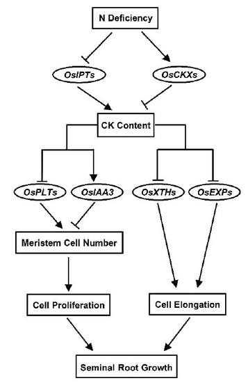
Nitrogen Deficiency-Induced Decrease in Cytokinins Content Promotes Rice Seminal Root Growth by Promoting Root Meristem Cell Proliferation and Cell Elongation

1
College of Plant Science and Technology, Huazhong Agricultural University, Wuhan 430070, China
2
National Key Laboratory of Crop Genetic Improvement, Wuhan 430070, China
3
College of Life Science and Technology, Huazhong Agricultural University, Wuhan 430070, China
*
Author to whom correspondence should be addressed.
Cells 2020, 9(4), 916; https://doi.org/10.3390/cells9040916
Submission received: 1 March 2020 / Revised: 19 March 2020 / Accepted: 7 April 2020 / Published: 9 April 2020
(This article belongs to the Special Issue Hormonal Signaling in Plants and the Changing Environment)

Abstract

:
Rice (Oryza sativa L.) seedlings grown under nitrogen (N) deficiency conditions show a foraging response characterized by increased root length. However, the mechanism underlying this developmental plasticity is still poorly understood. In this study, the mechanism by which N deficiency influences rice seminal root growth was investigated. The results demonstrated that compared with the control (1 mM N) treatment, N deficiency treatments strongly promoted seminal root growth. However, the N deficiency-induced growth was negated by the application of zeatin, which is a type of cytokinin (CK). Moreover, the promotion of rice seminal root growth was correlated with a decrease in CK content, which was due to the N deficiency-mediated inhibition of CK biosynthesis through the down-regulation of CK biosynthesis genes and an enhancement of CK degradation through the up-regulation of CK degradation genes. In addition, the N deficiency-induced decrease in CK content not only enhanced the root meristem cell proliferation rate by increasing the meristem cell number via the down-regulation of OsIAA3 and up-regulation of root-expressed OsPLTs, but also promoted root cell elongation by up-regulating cell elongation-related genes, including root-specific OsXTHs and OsEXPs. Taken together, our data suggest that an N deficiency-induced decrease in CK content promotes the seminal root growth of rice seedlings by promoting root meristem cell proliferation and cell elongation.

Graphical Abstract

1. Introduction

Rice (Oryza sativa L.) is one of the most important food crops in the world [1]. In the past 50 years, rice yield has steadily increased worldwide, partly owing to an increase in nitrogen (N) application. However, at present, the average recovery efficiency of N fertilizer (the percentage of fertilizer N recovered in aboveground plant biomass at the end of the cropping season) is only 33% at the field level [2]. High N input and low N use efficiency not only increase crop production costs but also cause severe environmental pollution [3,4]. Therefore, decreasing N application is an important goal of sustainable agriculture. However, decreasing N application may lead to N deficiency and affect rice root growth, and the underlying mechanism by which N deficiency affects rice root growth is still poorly understood.
Studies of crop responses to N deficiency have focused on the root [5,6], which is the plant organ that is most important for acquiring soil nutrients [7,8]. The developmental plasticity of root architecture is crucial for the acclimation of crops to unfavorable environments, including those that induce N stress. For example, a steeper and deeper root system more efficiently absorbs N in deep soil layers [9]. Root growth is influenced by several external and internal factors, including N availability and phytohormone homeostasis [10,11,12,13]. In general, a supraoptimal N supply inhibits root growth, and the decrease in root size can lead to decreased N uptake [14,15,16,17]. In contrast, N deficiency promotes root growth, and the increase in root size can improve N uptake ability [9,18]. Similarly, supraoptimal levels of the phytohormone cytokinin (CK) inhibit root growth [19], whereas a mild decrease in CK content promotes root growth [19,20,21]. These findings provide evidence that both N and CK are involved in mediating root growth.
CK regulates root growth in a dose-dependent manner [22]. We previously found that a threshold CK content is required for the rapid growth of rice seminal roots, but that supraoptimal CK levels inhibit growth [19]. Usually, the CK contents in roots cultured with high or moderate concentrations of N are supraoptimal for root growth, and thus a mild decrease in CK content promotes root growth. For example, a mild decrease in CK content achieved through overexpression of the CK degradation gene CYTOKININ OXIDASE DEHYDROGENASE (CKX) or mutation of the CK biosynthesis gene ISOPENTENYLTRANSFERASE (IPT) can promote primary root growth in Arabidopsis grown under moderate concentrations of N [20,21]. In contrast, without N application, the endogenous CK content in rice seedlings is optimal for growth of the seminal roots, and thus either a decrease or an increase in CK content leads to growth inhibition of the seminal root [19]. In addition, it has been reported that N treatment can increase CK content in roots [23]. These results suggest that N concentration is closely associated with CK content in the root. However, the mechanism by which the interaction between N and CK mediates rice root growth remains elusive.
Root growth is mainly determined by root meristem cell proliferation and root cell elongation [24,25,26]. The meristem cell proliferation rate is positively correlated with meristem cell number and meristem cell division activity [26]. The root meristem cell number is antagonistically regulated by many regulators, including PLETHORA (PLT) and SHORT HYPOCOTYL2/INDOLE-3-ACETIC ACID3 (SHY2/IAA3) [26,27]; and the meristem cell division activity is positively correlated with the transcription level of cyclin and cyclin-dependent protein kinase genes, such as CycB1;1 and CDKB2;1 [24,28]. PLT genes encode APETALA2 (AP2) transcription factors and are essential for root meristem maintenance [27]. In Arabidopsis, plt1plt2 double mutants show a severe reduction in root meristem cell number, while the ectopic overexpression of PLT2 leads to an increased number of meristematic cells and increased meristem size [27,29]. SHY2/IAA3 controls the root meristem cell number by promoting the mitotic-to-endocycle transition in the root, which in turn decreases the meristematic cell number and reduces the root meristem size [19,26]. Plants with a loss-of-function mutation in SHY2/IAA3 have a larger-than-usual meristem, whereas those with a gain-of-function mutation in SHY2/IAA3 have a smaller meristem than the wild type [25,30]. XYLOGLUCAN ENDOTRANSGLUCOSYLASE/HYDROLASE (XTH) and EXPANSIN (EXP) proteins play important roles in mediating root cell elongation [31,32], and thus mutations in EXP or XTH genes have been found to result in short root cells and short roots. For example, the Arabidopsis atxth31 loss-of-function mutant has shorter root cells and shorter roots than the wild type [33], and the lengths of roots and root cells in OsEXPB2 RNA interference lines were significantly shorter than those in wild-type rice plants [34]. In rice, the transcription levels of OsXTH and OsEXP genes, such as OsXTH1, OsEXP3, and OsEXPB4, which are specifically expressed in the root, are closely correlated with the lengths of mature cells in rice seminal root [19].
CK has been shown to regulate the expression of the genes regulating root meristem cell number and root cell elongation [27,35]. Previous studies have reported that some hormone response elements, including the CK-response element (AGATT), exist in promoter regions of the root-expressed OsPLTs [35,36]. OsPLT1–6 can strongly respond to CK because there are more than seven AGATT elements in each promoter region [35]. In addition, it has been reported that CK can up-regulate OsIAA3 and down-regulate OsXTHs and OsEXPs [19]. However, up to now, it has been unclear whether N and CK collaboratively regulate root meristem cell number and cell elongation by modulating transcription levels of OsPLTs, OsIAA3, OsXTHs, and OsEXPs.
Rice seminal roots are the first roots to emerge from seeds after germination and are responsible for water uptake and nutrient absorption during seedling establishment [37]. In this study, the influence of N deficiency on CK metabolism in rice seminal roots was investigated. Furthermore, the mechanism by which N and CK interact to regulate rice seminal root growth was clarified. We provide evidence that N deficiency reduces CK content by inhibiting CK biosynthesis and promoting CK degradation, which in turn promotes rice seminal root growth. Specifically, the decrease in CK content results in the down-regulation of OsIAA3 and up-regulation of OsPLT genes, leading to an increase in root meristem cell number and cell proliferation rate, and an up-regulation of the OsXTH and OsEXP genes, which promote cell elongation.

2. Materials and Methods

2.1. Plant Material and Growth Conditions

Japonica rice Nipponbare (Oryza sativa L.) was used in this study. Rice seeds were sterilized, soaked, and germinated according to the method of Zou et al. [38]. The germinated rice seeds were grown in an artificial climate incubator (HP 1500 GS, Ruihua Instrument & Equipment Co., Ltd., Wuhan, China) with a 12-h light (29 °C)/12-h dark (26 °C) photoperiod.

2.2. Chemicals and Treatments

Zeatin (Z) (Sigma-Aldrich Trading Co., Ltd., Shanghai, China), a mixture of trans-Z (tZ) and cis-Z (cZ), was dissolved in dimethyl sulfoxide (DMSO) and diluted to the desired concentration with distilled water. NH4NO3 (Shanghai Yuanye Biotechnology Co., Ltd., Shanghai, China), which was used as the source of N, was dissolved directly in distilled water and diluted to the desired concentration. The solution for the control treatment contained 0.5 mM NH4NO3 (1 mM N, 1 N), the solutions for the N deficiency treatments contained 0.125 mM NH4NO3 (0.25 mM N, 1/4 N), 0.03125 mM NH4NO3 (0.0625 mM N, 1/16 N), or 0 mM NH4NO3 (0 mM N, 0 N), and the solution for the N deficiency + Z treatment contained 0 mM N and 4 nM Z (0 N + Z). All solutions also contained 0.3 mM NaH2PO4, 0.15 mM K2SO4, 0.15 mM CaCl2, 0.3 mM MgCl2, 0.08 mM Na2SiO3, 4.6 μM MnSO4, 0.05 μM Na2MoO4, 24.5 μM H3BO3, 0.35 μM ZnSO4, 0.15 μM CuSO4, 32.7 μM Fe (II)-EDTA (Ethylene Diamine Tetraacetic Acid), and 70.8 μM C6H7O8. The pH of each solution was adjusted to 6.5 with 0.1 N HCl or NaOH. Since the Z solutions also contained DMSO, DMSO was added to the solutions without Z to a final concentration of 0.01% (v/v). The germinated rice seeds were incubated in different solutions for one day, two days, three days, or four days. All solutions were refreshed every two days.

2.3. CK Content Analysis

The germinated seeds were incubated in a solution containing 0 N, 1/16 N, 1/4 N, or 1 N (control). After four days of treatment, the seminal roots of the rice seedlings were collected and frozen at −80 °C until use. CKs were extracted, purified, and quantified according to previously described methods [19,39]. Data are reported as means ± standard deviation (SD) of three independent biological replicates.

2.4. RNA Isolation and Quantitative Real-Time PCR (qRT-PCR) Analysis

The germinated seeds were incubated in a solution containing 0 N, 0 N + Z, or 1 N as a control for one day, two days, or four days; then, the seminal roots of the rice seedlings were collected and frozen at −80 °C until use. Total RNA was extracted according to Liu et al. [40]. First-strand cDNA was synthesized using the Fastking RT Kit (Tiangen Biotechnology Co., Ltd., Beijing, China). The relative expression level was analyzed by qRT-PCR according to the method described previously [40]; the gene-specific primers are listed in Supplementary Table S1. The relative expression level was calculated using the comparative threshold method, using OsACTIN as an internal control. Three biological replicates with three technical replicates were used for the statistical analyses and error range analyses, and data are presented as the means ± SD. The relative expression levels of CK metabolism genes, root meristem size-related genes, and root cell elongation-related genes in rice seminal root are presented in Supplementary Figure S1.

2.5. Quantitative Analysis of Root Phenotypes

The seminal roots of the rice seedlings were selected for the quantitative analysis of root phenotypes. Seminal root tips were stained with 4’,6-diamino-2-phenylhydrazine solution to measure root meristem size and meristem cell number, and mature cells in rice seminal roots were stained with propidium iodide solution to measure mature cell length. The staining methods have been reported previously [19]. Images were taken on a confocal microscope (Leica SP8, Leica Corporation, Solms, Germany). Root length was measured with Image J software 1.46 (National Institutes of Health, Bethesda, MD, USA). Root meristem size was determined by measuring the length from the quiescent center to the first elongated epidermal cell. Root meristem cell number was expressed as the number of cortex cells from the quiescent center to the first elongated cell. Root growth rate was calculated as described previously [41]. Cell proliferation rate was calculated using the following equation: cell proliferation rate = root growth rate/mature cell length. Data are presented as means ± SD calculated from 10 biological replicates.

2.6. Statistical Analysis

Statistical analysis was performed using an independent samples t-test or a one-way analysis of variance followed by Duncan’s multiple range test, with at least three biological replicates for each treatment. Values of p < 0.05 were considered statistically significant. All data are presented as means ± SD.

2.7. Accession Numbers

Sequence data from this study can be found in the Chinese national rice data center (http://www.ricedata.cn/gene/index.htm) under the following accession numbers: OsIPT1 (Os03g0358900), OsIPT3 (Os05g0311801), OsIPT4 (Os03g0810100), OsIPT5 (Os07g0211700), OsIPT7 (Os05g0551700), OsIPT8 (Os01g0688300), OsCKX1 (Os01g0187600), OsCKX2 (Os01g0197700), OsCKX3 (Os10g0483500), OsCKX4 (Os01g0940000), OsCKX5 (Os01g0775400), OsCKX6 (Os02g0220000), OsCKX7 (Os06g0572300), OsCKX8 (Os04g0523500), OsCKX9 (Os05g0374200), OsCKX10 (Os06g0572300), OsCKX11 (Os08g0460600), OsPLT1 (Os04g0653600), OsPLT2 (Os06g0657500), OsPLT3 (Os02g0614300), OsPLT4 (Os04g0504500), OsPLT5 (Os01g0899800), OsPLT6 (Os11g0295900), OsCycB1;1 (Os01g0805600), OsCDKB2;1 (Os08g0512600), OsIAA3 (Os01g0231000), OsXTH1 (Os07g0529700), OsXTH2 (Os11g0539200), OsXTH4 (Os08g0240500), OsXTH13 (Os02g0280200), OsXTH15 (Os06g0336001), OsXTH16 (Os04g0604900), OsXTH25 (Os10g0577500), OsEXP3 (Os05g0276500), OsEXP11 (Os01g0274500), OsEXP12 (Os03g0155300), OsEXP13 (Os02g0267200), OsEXP15 (Os03g0155700), OsEXP17 (Os06g0108600), OsEXPB2 (Os10g0555700), OsEXPB8 (Os03g0102500), and OsACTIN (Os03g0718100).

3. Results

3.1. N Deficiency-Induced Growth of Rice Seminal Roots was Negated by Application of CK

In order to investigate the effect of N deficiency on rice seminal root growth, rice seedlings were cultivated hydroponically under control (1 N) and N deficiency (1/4 N, 1/16 N, and 0 N) conditions. After four days of cultivation, the average lengths of rice seminal roots treated with 1 N, 1/4 N, 1/16 N, and 0 N were 4.6 cm, 6.2 cm, 10.4 cm, and 11.5 cm, respectively (Figure 1). The 1/4 N, 1/16 N, and 0 N treatments increased the rice seminal root lengths by 35%, 126%, and 150%, respectively, compared with those of the control. This result demonstrated that N deficiency promoted rice seminal root growth, and that the decrease in N was accompanied by an increase in rice seminal root length. Furthermore, the application of Z (a type of CK) at concentrations of 0.25–4 nM significantly inhibited the N deficiency-induced growth of rice seminal roots, and 4 nM Z completely blocked the N deficiency-induced growth (Figure 1). This result suggested that N deficiency might promote rice seminal root growth by controlling the CK content.

3.2. N Deficiency Reduced CK Content in Rice Seminal Roots

To test whether N deficiency induces rice seminal root growth by controlling the content of endogenous CKs, the effects of N deficiency on the contents of endogenous Z, dihydrozeatin (DZ), and isopentenyladenine (iP), which are the predominant CKs in higher plants [42], were analyzed. Since the previous results clearly demonstrated comparable activities of trans-Z (tZ) and cis-Z (cZ) in the inhibition of rice seminal root elongation [43], thus the total content of Z, including tZ and cZ, was analyzed in this study. As shown in Table 1, the content of Z was significantly higher than that of DZ and iP, indicating that Z is the predominant CK in rice seminal roots. Under control (1 N) treatment, the content of Z in the seminal root was relatively high (198.4 ± 11.1 pmol·g−1 FW) (Table 1). By contrast, under the N deficiency treatments, the content of Z in seminal roots decreased significantly; the contents of Z under the 1/4 N, 1/16 N, and 0 N treatments were only 52%, 36%, and 34% of that of the control, respectively. However, neither DZ nor iP content was affected by N deficiency treatments compared with control (1 N) treatment (Table 1). This result indicated that N deficiency reduced CK content by reducing Z content rather than by controlling DZ or iP content in rice seminal roots.

3.3. N Deficiency Inhibited CK Biosynthesis and Promoted CK Degradation by Affecting the Transcription Levels of CK Metabolic Genes in Rice Seminal Roots

CK homeostasis is mainly controlled by CK biosynthesis and degradation [44,45]. In rice, six OsIPTs (OsIPT1, OsIPT3, OsIPT4, OsIPT5, OsIPT7, and OsIPT8) and 11 OsCKXs (OsCKX1–11) are the key genes modulating CK biosynthesis and degradation, respectively [46,47]. Thus, to investigate the mechanism by which N deficiency regulates CK content, the effects of N deficiency on the transcription levels of these six OsIPTs and 11 OsCKXs in rice seminal roots were investigated. On the one hand, N deficiency could reduce CK content by decreasing CK biosynthesis via the down-regulation of OsIPT genes. As shown in Figure 2a, after one day of treatment, of the six OsIPT genes tested, there were significant differences in the transcription levels between control and N deficiency treatments for four genes; the transcription levels of OsIPT3 and OsIPT4 were significantly down-regulated in response to all the N deficiency treatments, and the transcription levels of OsIPT7 and OsIPT8 were significantly down-regulated in response to the 1/16 N and 0 N treatments. After two days of treatment, the transcription levels of OsIPT3, OsIPT4, and OsIPT8 were significantly down-regulated by the 1/16 N and 0 N treatments (Figure 2b). After four days of treatment, the transcription levels of all six OsIPT genes were significantly down-regulated by treatment with ≤1/4 N (Figure 2c). These results suggest that N deficiency might reduce CK content by inhibiting CK biosynthesis via the down-regulation of OsIPT genes. On the other hand, N deficiency could reduce CK content by promoting CK degradation via the up-regulation of OsCKX genes. As shown in Figure 2d–f, OsCKX10 and OsCKX11 did not respond to N deficiency treatments, but the other eight OsCKX genes were up-regulated to different extents by different N deficiency treatments. Among the nine N deficiency-responsive OsCKX genes, OsCKX4 responded most strongly to N deficiency; the transcription level of OsCKX4 was up-regulated by more than 2-fold after one and two days of N deficiency treatment.

3.4. N Deficiency-Induced Decrease in CK Content Enhanced Root Meristem Cell Proliferation Rate and Promoted Root Cell Elongation

We investigated the effects of control (1 N), N deficiency (0 N), and N deficiency + Z (0 N + Z) treatments on seminal root meristem cell proliferation rate and mature cell length to clarify the mechanism by which the N deficiency-induced decrease in CK content increases seminal root length. The results demonstrated that the meristem sizes and the cell numbers of the seminal roots in the N deficiency treatment group increased significantly compared with those in the control group, but this effect was completely negated by the application of Z (Figure 3a–c). Moreover, the lengths of mature cells in the mature zone of rice seminal roots were also significantly increased by N deficiency treatment; the average root cell length in the mature zone was increased by 45.5% under N deficiency treatment (Figure 3d,e). However, this increase in cell length was completely negated by the addition of Z (Figure 3d,e). Further, the results demonstrate that N deficiency strongly enhanced the root growth rate and root meristem cell proliferation rate, but these effects were negated by the addition of Z (Figure 3f,g). In addition, compared with control treatment, 0 N and 0 N + Z treatments had no influence on the transcription level of root meristem cell division-related genes, such as OsCycB1;1 and OsCDKB2;1 (Figure 3h,i). These results suggest that the N deficiency-induced decrease in CK content enhanced the root meristem cell proliferation rate and promoted root cell elongation, and that the enhanced cell proliferation rate was contributed by the increased meristem cell number rather than by the changed meristem cell division activity.

3.5. Decrease in CK Content Increased Root Meristem Cell Number by Affecting the Transcription Levels of Root Meristem Size-Related Genes

It has been reported that root meristem size is positively regulated by PLT and negatively regulated by IAA3 [25,27]. There are 10 OsPLT genes in rice; among them, only six (OsPLT1–6) are expressed in roots [35]. Therefore, to clarify whether the N deficiency-induced decrease in CK increased meristem cell number by affecting the transcription of meristem size-related genes in rice seminal roots, the effects of control (1 N), N deficiency (0 N), and N deficiency + Z (0 N + Z) treatments on the transcription levels of OsPLT1–6 and OsIAA3 were investigated. After one day of N deficiency treatment, the transcription levels of OsPLT1–6 were significantly increased, while the transcription level of OsIAA3 was significantly decreased, but these effects were antagonized by the application of Z (Figure 4a). After two days of treatment, the transcription levels of most of the root meristem size-related genes changed in response to N deficiency, but these transcriptional changes were smaller in the presence of Z (Figure 4b). After four days of treatment, the transcription levels of the four meristem size-related genes under N deficiency were still slightly but significantly different from those under the control condition, and the transcriptional regulation of these genes was also antagonized by the application of Z (Figure 4c). These results suggest that the N deficiency-induced decrease in CK content led to an increased root meristem cell number through the down-regulation of OsIAA3 and the up-regulation of root-expressed OsPLTs.

3.6. Decrease in CK Content Increased Root Cell Length by Up-Regulating Transcription of Root Cell Elongation-Related Genes

It has been reported that seven OsXTHs and eight OsEXPs are specifically expressed in rice roots [31,32], and that these root-specific genes play important roles in regulating root cell elongation in rice seedlings [31,32]. To clarify whether the N deficiency-induced decrease in CK increased the cell length in rice seminal roots by affecting the transcription levels of cell elongation-related genes, the effects of the control (1 N), N deficiency (0 N), and N deficiency + Z (0 N + Z) treatments on the transcription levels of the root-specific OsXTHs (OsXTH1, OsXTH2, OsXTH4, OsXTH13, OsXTH15, OsXTH16, and OsXTH25) and OsEXPs (OsEXP3, OsEXP11, OsEXP12, OsEXP13, OsEXP15, OsEXP17, OsEXPB2, and OsEXPB8) were investigated. After one day of N deficiency treatment, two-thirds of the root-specific OsXTHs and OsEXPs were up-regulated (Figure 5a). After two days of treatment, all root-specific OsXTHs and OsEXPs except for OsXTH16, OsXTH25, and OsEXP11 were up-regulated by the N deficiency treatment (Figure 5b). After four days of treatment, most of the root-specific OsXTH and OsEXP genes were still up-regulated under the N deficiency treatment (Figure 5c). At every time point, the effects on transcription were reduced in the presence of Z (Figure 5a–c). Taken together, all root-specific OsXTHs and OsEXPs except for OsXTH25 and OsEXP11 were up-regulated to different extents by N deficiency, and the up-regulation of these genes was inhibited by Z treatment. These results indicate that the N deficiency-induced decrease in CK content increased root cell length by up-regulating the transcription levels of cell elongation-related genes, including six OsXTHs and seven OsEXPs, in rice seminal roots.

4. Discussion

4.1. Increasing Root Length is a Strategy for Responding to N Deficiency

Rice seminal roots are the first roots to emerge from seeds after germination and are responsible for water uptake and nutrient absorption during seedling establishment [37]. Here, we provide evidence that N deficiency treatments strongly promote seminal root growth of rice seedlings under a relatively short duration (four days) of N deficiency. Moreover, even under a long duration (30 days or 60 days), rice seminal root growth is still promoted by N deficiency treatments (Supplementary Figure S2). This result is consistent with previous reports that N deficiency treatment promotes root growth in other plants, including wheat [48] and Arabidopsis [49,50]. Root length is an essential parameter allowing crops to increase N use efficiency, as more and deeper roots may improve the uptake of N from deeper soil layers and reduce N losses to the environment [9]. Taken together, these findings suggest that increasing root length is a strategy used by plants to respond to N deficiency. However, because N is an essential element, a long duration of severe N deficiency such as 0 N treatment inhibits adventitious root production and reduces the whole root system volume (Supplementary Figure S2).

4.2. N Deficiency Promotes Rice Seminal Root Growth by Reducing CK Content

We found that the CK contents in rice seminal roots in the N deficiency treatment groups were lower than those of the control, and that a decrease in the concentration of N was accompanied by a decrease in the content of CKs as well as a longer seminal root (Figure 1, Table 1). Moreover, the induction of rice seminal root growth was significantly negated by CK application (Figure 1). Previous work has shown that the application of high or moderate concentrations of N induces CK accumulation in the roots of dicotyledonous plants such as Arabidopsis [51] and monocotyledonous crops, including rice [52], wheat [48], maize [53], and barley [54]; this accumulated CK inhibits root growth [19,55]. Usually, CK content in the roots of plants incubated with high or moderate concentrations of N is supraoptimal for rapid root growth, and thus mild decreases in CK content promote root growth [20,21]. All these findings lead to the conclusion that N deficiency promotes rice seminal root growth by reducing CK content. Moreover, our results indicated that N deficiency reduces CK content by reducing Z content rather than by controlling DZ or iP content in rice seminal roots (Table 1).

4.3. N Deficiency Reduces CK Content by Inhibiting CK Biosynthesis and Promoting CK Degradation

To investigate the regulatory mechanism by which N deficiency affects CK metabolism, the effects of N deficiency on the expression of six OsIPTs and 11 OsCKXs were analyzed in this study. We found that N deficiency inhibited CK biosynthesis and promoted CK degradation through down-regulation of the OsIPT genes and up-regulation of OsCKX genes (Figure 2a–f), respectively, which in turn led to reduced CK content. Our result is supported by studies in Arabidopsis and tobacco that found that expressing CKX genes specifically in the roots results in reduced CK content through the enhancement of root-specific degradation of CK [56]. Moreover, our result is also supported by experiments in rice showing that an increase in endogenous CK content achieved through the knockdown of OsCKX4 or overexpression of OsIPT inhibits the growth of rice seminal roots [47,57]. In addition, our findings are also consistent with a study in rice in which OsIPT4 was found to be responsible for the increased content of CK in roots in response to nitrite (a type of N fertilizer) treatment [23]. Taken together, these results suggest that N deficiency reduces CK content by inhibiting CK biosynthesis and promoting CK degradation.

4.4. N Deficiency-Induced Decrease in CK Content Promotes Rice Seminal Root Growth by Enhancing Meristem Cell Proliferation Rate through Increased Meristem Cell Number

The root meristem cell proliferation rate is positively correlated with meristem cell number and meristem cell division activity [26]. We found that the N deficiency-induced decrease in CK content increases the root meristem cell number by up-regulating OsPLT1–6 and down-regulating OsIAA3 (Figuers 3c,4), which in turn promotes rice seminal root growth by enhancing the root meristem cell proliferation rate (Figure 3g). However, the N deficiency-induced decrease in CK content could not affect meristem cell division activity in rice seminal root (Figure 3h,i). This result is consistent with a previous report that CK had no influence on meristem cell division activity in Arabidopsis primary root [24]. All the evidence suggests that the N deficiency-induced decrease in CK content increases root meristem cell number by up-regulating OsPLT1–6 and down-regulating OsIAA3, which in turn promotes rice seminal root growth by enhancing the root meristem cell proliferation rate through an increased meristem cell number. Our result is supported by previous studies showing that a mild decrease in endogenous CK content promotes root growth [20,21], while CK treatment strongly inhibits root growth [19]. Our result is also consistent with the finding in rice that the transcription of all OsPLT1–6 genes declined to the lowest level after 24 h of CK treatment [35], and that the transcription of the OsIAA3 gene increased significantly within four days of CK treatment [19].

4.5. Decrease in CK Content Promotes Rice Seminal Root Growth by Promoting Root Cell Elongation

Root-specific OsXTHs and OsEXPs have been implicated in the control of rice root cell elongation [19,34]. Here, we provide evidence that all root-specific OsXTHs and OsEXPs except for OsXTH25 and OsEXP11 were up-regulated by N deficiency, but that this up-regulation was blocked by the application of Z (Figure 5a–c). This finding is consistent with our previous finding that the application of exogenous kinetin (a synthetic CK) down-regulated the transcription of OsXTHs and OsEXPs, such as OsXTH1 and OsEXP3, which in turn inhibited rice seminal root growth [19]. These pieces of evidence suggest that the N deficiency-induced decrease in CK content promotes rice seminal root growth by promoting root cell elongation through the up-regulation of root-specific OsXTHs and OsEXPs.

5. Conclusions

In summary, our results demonstrate that N deficiency reduces CK content by inhibiting CK biosynthesis and promoting CK degradation through the down-regulation of CK biosynthesis genes and up-regulation of CK degradation genes, respectively (Figure 6). Moreover, our results indicate that the N deficiency-induced decrease in CK content promotes rice seminal root growth not only by enhancing the root meristem cell proliferation rate through an increased root meristem cell number, but also by promoting root cell elongation through the up-regulation of root-specific OsXTHs and OsEXPs (Figure 6). In addition, our results suggest that the N deficiency-induced decrease in CK content increases the root meristem cell number by up-regulating root-expressed OsPLTs and down-regulating OsIAA3 (Figure 6). Taken together, our data suggest that an N deficiency-induced decrease in CK content promotes rice seminal root growth by promoting root meristem cell proliferation and root cell elongation. Further study is required to characterize the molecular mechanisms involved in the N-regulated transcription of OsIPTs and OsCKXs. Another important issue requiring further in-depth study is the role played by CK signaling in regulating the transcription of OsIAA3, OsPLTs, OsXTHs, and OsEXPs.

Supplementary Materials

The following are available online at https://www.mdpi.com/2073-4409/9/4/916/s1, Figure S1: The relative expression levels of genes investigated in this study, Figure S2: Effects of long-term treatment with N deficiency on rice seminal root growth, Table S1: List of primers used in this research.

Author Contributions

Conceptualization, C.Y. and Y.L.; Data curation, Q.W.; Formal analysis, Q.W.; Funding acquisition, C.Y., Investigation, Q.W.; Y.Z.; X.Z.; J.Z.; Z.K. and X.L.; Methodology, C.Y. and Q.W.; Project administration, C.Y.; Resources, C.Y.; Software, Q.W. and F.L.; Supervision, C.Y.; Validation, C.Y. and Q.W.; Visualization, Q.W.; Writing—original draft, Q.W.; Writing—review and editing, Q.W. and C.Y. All authors have read and agreed to the published version of the manuscript.

Funding

This research was funded by the National Key Research and Development Program of China (No. 2016YFD0300102) and the Fundamental Research Funds for the Central Universities (No. 2662018PY076).

Acknowledgments

We thank Li Liu (Huazhong Agricultural University, China) for technical support during the experiment. We thank Youjia Shen (Nanjing Agricultural University, China) for providing Nipponbare rice seeds.

Conflicts of Interest

The authors declare no conflict of interest.

Abbreviations

CKCytokinin
CKXCytokinin oxidase dehydrogenase
DZdihydrozeatin
EXPExpansin
FWFresh weight
iPIsopentenyladenine
NNitrogen
NSNot significant
PLTPLETHORA
XTHXyloglucan endotransglucosylase/hydrolase
ZZeatin

References

  1. Fageria, N.K. Yield physiology of rice. J. Plant Nutr. 2007, 30, 843–879. [Google Scholar] [CrossRef]
  2. Peng, S.; Buresh, R.J.; Huang, J.; Zhong, X.; Zou, Y.; Yang, J.; Wang, G.; Liu, Y.; Hu, R.; Tang, Q.; et al. Improving nitrogen fertilization in rice by sitespecific N management. Agron. Sustain. Dev. 2010, 30, 649–656. [Google Scholar] [CrossRef]
  3. Ju, X.T.; Xing, G.X.; Chen, X.P.; Zhang, S.L.; Zhang, L.J.; Liu, X.J.; Cui, Z.L.; Yin, B.; Christie, P.; Zhu, Z.L.; et al. Reducing environmental risk by improving n management in intensive chinese agricultural systems. Proc. Natl. Acad. Sci. USA 2009, 106, 3041–3046. [Google Scholar] [CrossRef] [PubMed] [Green Version]
  4. Good, A.G.; Beatty, P.H. Fertilizing nature: A tragedy of excess in the commons. PLoS Biol. 2011, 9, e1001124. [Google Scholar] [CrossRef] [PubMed]
  5. Drew, M.C.; Saker, L.R.; Ashley, T.W. Nutrient supply and the growth of the seminal root system in barley. J. Exp. Bot. 1973, 24, 1189–1202. [Google Scholar] [CrossRef]
  6. Sun, H.W.; Tao, J.Y.; Liu, S.J.; Huang, S.J.; Chen, S.; Xie, X.N.; Yoneyama, K.; Zhang, Y.L.; Xu, G.H. Strigolactones are involved in phosphate- and nitrate-deficiency-induced root development and auxin transport in rice. J. Exp. Bot. 2014, 65, 6735–6746. [Google Scholar] [CrossRef]
  7. Yang, C.; Yang, L.; Yang, Y.; Ouyang, Z. Rice root growth and nutrient uptake as influenced by organic manure in continuously and alternately flooded paddy soils. Agr. Water Manage. 2004, 70, 67–81. [Google Scholar] [CrossRef]
  8. Forde, B.G. Nitrogen signalling pathways shaping root system architecture: An update. Curr. Opin. Plant Biol. 2014, 21, 30–36. [Google Scholar] [CrossRef]
  9. Trachse, S.; Kaeppler, S.M.; Brown, K.M.; Lynch, J.P. Maize root growth angles become steeper under low N conditions. Field Crop. Res. 2013, 140, 18–31. [Google Scholar] [CrossRef]
  10. Sakakibara, H. Nitrate-specific and cytokinin-mediated nitrogen signaling pathways in plants. J. Plant Res. 2003, 116, 253–257. [Google Scholar] [CrossRef]
  11. Sakakibara, H.; Takei, K.; Hirose, N. Interactions between nitrogen and cytokinin in the regulation of metabolism and development. Trends Plant Sci. 2006, 11, 440–448. [Google Scholar] [CrossRef] [PubMed]
  12. Petricka, J.J.; Winter, C.M.; Benfey, P.N. Control of Arabidopsis root development. Annu. Rev. Plant Biol. 2012, 63, 563–590. [Google Scholar] [CrossRef] [PubMed] [Green Version]
  13. Jia, Z.; Giehl, R.F.H.; Meyer, R.C.; Altmann, T.; von Wirén, N. Natural variation of BSK3 tunes brassinosteroid signaling to regulate root foraging under low nitrogen. Nat. Commun. 2019, 10, 2378–2390. [Google Scholar] [CrossRef] [PubMed] [Green Version]
  14. Linkohr, B.I.; Williamson, L.C.; Fitter, A.H.; Leyser, H.M.O. Nitrate and phosphate availability and distribution have different effects on root system architecture of Arabidopsis. Plant J. 2002, 29, 751–760. [Google Scholar] [CrossRef] [PubMed] [Green Version]
  15. Tian, Q.; Chen, F.; Zhang, F.; Mi, G. Possible involvement of cytokinin in nitrate-mediated root growth in maize. Plant Soil 2005, 277, 185–196. [Google Scholar] [CrossRef]
  16. Yu, P.; Li, X.; White, P.J.; Li, C. A large and deep root system underlies high nitrogen-use efficiency in maize production. PLoS ONE 2015, 10, e0126293. [Google Scholar] [CrossRef] [Green Version]
  17. Leskovar, D.; Othman, Y. Low nitrogen fertigation promotes root development and transplant quality in globe artichoke. Hortscience 2016, 51, 567–572. [Google Scholar] [CrossRef] [Green Version]
  18. Mu, X.; Chen, F.; Wu, Q.; Chen, Q.; Wang, J.; Yuan, L.; Mi, G. Genetic improvement of root growth increases maize yield via enhanced post-silking nitrogen uptake. Eur. J. Agron. 2015, 63, 55–61. [Google Scholar] [CrossRef]
  19. Zou, X.; Shao, J.; Wang, Q.; Chen, P.; Zhu, Y.; Yin, C. Supraoptimal cytokinin content inhibits rice seminal root growth by reducing root meristem size and cell length via increased ethylene content. Int. J. Mol. Sci. 2018, 19, 4051. [Google Scholar] [CrossRef] [Green Version]
  20. Werner, T.; Motyka, V.; Laucou, V.; Smets, R.; Onckelen, H.V.; Schmulling, T. Cytokinin-deficient transgenic Arabidopsis plants show multiple developmental alterations indicating opposite functions of cytokinins in the regulation of shoot and root meristem activity. Plant Cell 2003, 15, 2532–2550. [Google Scholar] [CrossRef] [Green Version]
  21. Miyawaki, K.; Tarkowski, P.; Matsumoto-Kitano, M.; Kato, T.; Sato, S.; Tarkowska, D.; Tabata, S.; Sandberg, G.; Kakimoto, T. Roles of Arabidopsis ATP/ADP isopentenyltransferases and tRNA isopentenyltransferases in cytokinin biosynthesis. Proc. Natl. Acad. Sci. USA 2006, 103, 16598–16603. [Google Scholar] [CrossRef] [PubMed] [Green Version]
  22. Thomas, R.G.; Hay, M.J.M. Shoot branching in response to nodal roots is mimicked by application of exogenous cytokinin in Trifolium repens. Funct. Plant Biol. 2015, 42, 115–125. [Google Scholar] [CrossRef]
  23. Mochizuki, S.; Jikumaru, Y.; Nakamura, H.; Koiwai, H.; Sasaki, K.; Kamiya, Y.; Ichikawa, H.; Minami, E.; Nishizawa, Y. Ubiquitin ligase EL5 maintains the viability of root meristems by influencing cytokinin-mediated nitrogen effects in rice. J. Exp. Bot. 2014, 65, 2307–2318. [Google Scholar] [CrossRef] [PubMed]
  24. Ioio, R.D.; Linhares, F.S.; Scacchi, E.; Casamitjana-Martinez, E.; Heidstra, R.; Costantino, P.; Sabatini, S. Cytokinins determine Arabidopsis root-meristem size by controlling cell differentiation. Curr. Biol. 2007, 17, 678–682. [Google Scholar] [CrossRef] [PubMed] [Green Version]
  25. Ioio, R.D.; Nakamura, K.; Moubayidin, L.; Perilli, S.; Taniguchi, M.; Morita, M.T.; Aoyama, T.; Costantino, P.; Sabatini, S. A genetic framework for the control of cell division and differentiation in the root meristem. Science 2008, 322, 1380–1384. [Google Scholar] [CrossRef] [PubMed] [Green Version]
  26. Takatsuka, H.; Umeda, M. Hormonal control of cell division and elongation along differentiation trajectories in roots. J. Exp. Bot. 2014, 65, 2633–2643. [Google Scholar] [CrossRef] [Green Version]
  27. Aida, M.; Beis, D.; Heidstra, R.; Willemsen, V.; Blilou, I.; Galinha, C.; Nussaume, L.; Noh, Y.; Amasino, R.; Scheres, B. The PLETHORA genes mediate patterning of the Arabidopsis root stem cell niche. Cell 2004, 119, 109–120. [Google Scholar] [CrossRef] [Green Version]
  28. Endo, M.; Nakayama, S.; Umeda-Hara, C.; Ohtsuki, N.; Saika, H.; Umeda, M.; Toki, S. CDKB2 is involved in mitosis and DNA damage response in rice. Plant J. 2012, 69, 967–977. [Google Scholar] [CrossRef] [Green Version]
  29. Galinha, C.; Hofhuis, H.; Luijten, M.; Willemsen, V.; Blilou, I.; Heidstra, R.; Scheres, B. PLETHORA proteins as dose-dependent master regulators of Arabidopsis root development. Nature 2007, 449, 1053–1057. [Google Scholar] [CrossRef] [Green Version]
  30. Nakamura, A.; Umemura, I.; Gomi, K.Y.; Kitano, H.; Sazuka, T.; Matsuoka, M. Production and characterization of auxin-insensitive rice by overexpression of a mutagenized rice IAA protein. Plant J. 2006, 46, 297–306. [Google Scholar] [CrossRef]
  31. Lee, Y.; Kende, H. Expression of alpha-expansin and expansin-like genes in deepwater rice. Plant Physiol. 2002, 130, 1396–1405. [Google Scholar] [CrossRef] [PubMed] [Green Version]
  32. Yokoyama, R.; Rose, J.K.C.; Nishitani, K. A surprising diversity and abundance of xyloglucan endotransglucosylase/hydrolases in rice. Classification and expression analysis. Plant Physiol. 2004, 134, 1088–1099. [Google Scholar] [CrossRef] [PubMed] [Green Version]
  33. Zhu, X.F.; Shi, Y.Z.; Lei, G.J.; Fry, S.C.; Zhang, B.C.; Zhou, Y.H.; Braam, J.; Jiang, T.; Xu, X.Y.; Mao, C.Z.; et al. Xth31, encoding an in vitro XEH/XET-active enzyme, regulates aluminum sensitivity by modulating in vivo XET action, cell wall xyloglucan content, and aluminum binding capacity in Arabidopsis. Plant Cell 2012, 24, 4731–4747. [Google Scholar] [CrossRef] [Green Version]
  34. Zou, H.; Wenwen, Y.; Zang, G.; Kang, Z.; Zhang, Z.; Huang, J.; Wang, G. OsEXPB2, a beta-expansin gene, is involved in rice root system architecture. Mol. Breeding 2015, 35, 41–54. [Google Scholar] [CrossRef]
  35. Li, P.; Xue, H. Structural characterization and expression pattern analysis of the rice PLT gene family. Acta Biochim. Biophs. Sin. 2011, 43, 688–697. [Google Scholar] [CrossRef] [PubMed] [Green Version]
  36. Yu, J.; Wang, J.; Lin, W.; Li, S.; Li, H.; Zhou, J.; Ni, P.; Dong, W.; Hu, S.; Zeng, C.; et al. The Genomes of Oryza sativa: A history of duplications. PLoS Biol. 2005, 3. [Google Scholar] [CrossRef] [Green Version]
  37. Wang, S.J.; Ho, C.H.; Chen, H.W. Rice develop wavy seminal roots in response to light stimulus. Plant Cell Rep. 2011, 30, 1747–1758. [Google Scholar] [CrossRef]
  38. Zou, Z.; Zou, X.; Zhao, S.; Xia, C.; Qian, K.; Wang, P.; Yin, C. Fluridone induces leaf bleaching by inhibiting pigment biosynthesis via downregulated transcription levels of pigment biosynthetic genes in rice Oryza sativa L. J. Plant Growth Regul. 2018, 37, 1385–1395. [Google Scholar] [CrossRef]
  39. Liu, H.; Li, X.; Xiao, J.; Wang, S. A convenient method for simultaneous quantification of multiple phytohormones and metabolites: Application in study of rice-bacterium interaction. Plant Methods 2012, 8, 2–13. [Google Scholar] [CrossRef] [Green Version]
  40. Liu, L.; Xia, W.; Li, H.; Zeng, H.; Wei, B.; Han, S.; Yin, C. Salinity inhibits rice seed germination by reducing alpha-amylase activity via decreased bioactive gibberellin content. Front. Plant Sci. 2018, 9, 275–283. [Google Scholar] [CrossRef]
  41. Li, J.; Zhao, Y.; Chu, H.; Wang, L.; Fu, Y.; Liu, P.; Upadhyaya, N.; Chen, C.; Mou, T.; Feng, Y.; et al. SHOEBOX modulates root meristem size in rice through dose-dependent effects of gibberellins on cell elongation and proliferation. PLoS Genet. 2015, 11, e1005464. [Google Scholar] [CrossRef] [PubMed]
  42. Letham, D.S.; Palni, L.M. The biosynthesis and metabolism of cytokinins. Annu. Rev. Plant Physiol. 1983, 34, 163–197. [Google Scholar] [CrossRef]
  43. Kudo, T.; Makita, N.; Kojima, M.; Tokunaga, H.; Sakakibara, H. Cytokinin activity of cis-zeatin and phenotypic alterations induced by overexpression of putative cis-Zeatin-O-glucosyltransferase in rice. Plant Physiol. 2012, 160, 319–331. [Google Scholar] [CrossRef] [PubMed] [Green Version]
  44. Frebort, I.; Kowalska, M.; Hluska, T.; Frebortova, J.; Galuszka, P. Evolution of cytokinin biosynthesis and degradation. J. Exp. Bot. 2011, 62, 2431–2452. [Google Scholar] [CrossRef] [PubMed]
  45. Hwang, I.; Sheen, J.; Müller, B. Cytokinin signaling networks. Annu. Rev. Plant Biol. 2012, 63, 353–380. [Google Scholar] [CrossRef] [PubMed] [Green Version]
  46. Kakimoto, T. Identification of plant cytokinin biosynthetic enzymes as dimethylallyl diphosphate: ATP/ADP isopentenyltransferases. Plant Cell Physiol. 2001, 42, 677–685. [Google Scholar] [CrossRef]
  47. Sakamoto, T.; Sakakibara, H.; Kojima, M.; Yamamoto, Y.; Nagasaki, H.; Inukai, Y.; Sato, Y.; Matsuoka, M. Ectopic expression of KNOTTED1-like homeobox protein induces expression of cytokinin biosynthesis genes in rice. Plant Physiol. 2006, 142, 54–62. [Google Scholar] [CrossRef] [Green Version]
  48. Garnica, M.; Houdusse, F.; Zamarreño, A.M.; Garcia-Mina, J.M. The signal effect of nitrate supply enhances active forms of cytokinins and indole acetic content and reduces abscisic acid in wheat plants grown with ammonium. J. Plant Physiol. 2010, 167, 1264–1272. [Google Scholar] [CrossRef]
  49. Kellermeier, F.; Armengaud, P.; Seditas, T.J.; Danku, J.; Salt, D.E.; Amtmann, A. Analysis of the root system architecture of Arabidopsis provides a quantitative readout of crosstalk between nutritional signals. Plant Cell 2014, 26, 1480–1496. [Google Scholar] [CrossRef] [Green Version]
  50. Ma, W.; Li, J.; Qu, B.; He, X.; Zhao, X.; Li, B.; Fu, X.; Tong, Y. Auxin biosynthetic gene, TAR2 is involved in low nitrogen-mediated reprogramming of root architecture in Arabidopsis. Plant J. 2014, 78, 70–79. [Google Scholar] [CrossRef]
  51. Cerutti, T.; Delatorre, C.A. Nitrogen and phosphorus interaction and cytokinin: Responses of the primary root of Arabidopsis thaliana and the pdr1 mutant. Plant Sci. 2013, 198, 91–97. [Google Scholar] [CrossRef] [PubMed]
  52. Kamada-Nobusada, T.; Makita, N.; Kojima, M.; Sakakibara, H. Nitrogen-dependent regulation of de novo cytokinin biosynthesis in rice: The role of glutamine metabolism as an additional signal. Plant Cell Physiol. 2013, 54, 1881–1893. [Google Scholar] [CrossRef] [PubMed] [Green Version]
  53. Sakakibara, H.; Suzuki, M.; Takei, K.; Deji, A.; Taniguchi, M.; Sugiyama, T. A response-regulator homologue possibly involved in nitrogen signal transduction mediated by cytokinin in maize. Plant J. 1998, 14, 337–344. [Google Scholar] [CrossRef] [PubMed] [Green Version]
  54. Samuelson, M.E.; Larsson, C.M. Nitrate regulation of zeation riboside levels in barley roots: Effects of inhibitors of N assimilation and comparison with ammonium. Plant Sci. 1993, 93, 77–84. [Google Scholar] [CrossRef]
  55. Werner, T.; Motyka, V.; Strnad, M.; Schmülling, T. Regulation of plant growth by cytokinin. Proc. Natl. Acad. Sci. USA 2001, 98, 10487–10492. [Google Scholar] [CrossRef] [PubMed] [Green Version]
  56. Werner, T.; Nehnevajova, E.; Kollmer, I.; Novak, O.; Strnad, M.; Kramer, U.; Schmulling, T. Root-specific reduction of cytokinin causes enhanced root growth, drought tolerance, and leaf mineral enrichment in Arabidopsis and tobacco. Plant Cell 2010, 22, 3905–3920. [Google Scholar] [CrossRef] [Green Version]
  57. Gao, S.; Fang, J.; Xu, F.; Wang, W.; Sun, X.; Chu, J.; Cai, B.; Feng, Y.; Chu, C. Cytokinin oxidase/dehydrogenase4 integrates cytokinin and auxin signaling to control rice crown root formation. Plant Physiol. 2014, 165, 1035–1046. [Google Scholar] [CrossRef] [Green Version]
Figure 1. Effects of the N and Z treatments on rice seedling seminal root growth. (a) Phenotypic comparison of seminal roots of rice seedlings grown under different treatments. Scale bar is 5 cm. (b) Seminal root lengths under different treatments. In this experiment, germinated rice seeds were incubated in different solutions for four days, then photographs were taken, and the lengths of the seminal roots were measured. The data are the means ± SD calculated from 10 biological replicates. Significant differences (p < 0.05) are indicated by different letters (a, b, c, d, e, f). N, nitrogen; Z, zeatin.
Cells 09 00916 g001
Figure 2. Comparison of the relative expression levels of CK metabolic genes between control and N deficiency treatments using qRT-PCR. (ac) Relative expression levels of CK biosynthesis genes after one day, two days, and four days of treatment. (df) Relative expression levels of CK degradation genes after one day, two days, and four days of treatment. Germinated rice seeds were incubated in control (1 N) or N deficiency (1/4 N, 1/16 N, and 0 N) solution. After one day, two days, and four days of treatment, the seminal roots of rice seedlings were sampled for qRT-PCR analyses. Data are presented as the means ± SD. Three biological replicates with three technical replicates were used for the statistical and error range analyses. The relative expression levels of CK metabolic genes in the control were set as 1. Significant differences (p < 0.05) in the relative expression level of the same gene between control and N deficiency treatments are indicated by different letters (a, b, c, d). N, nitrogen; 1 N, 1 mM N; 1/4 N, 1/4 mM N; 1/16 N, 1/16 mM N; 0 N, 0 mM N.
Cells 09 00916 g002
Figure 3. Effects of 1 N, 0 N, and 0 N + Z treatments on root phenotype, root growth rate, and meristem cell proliferation rate in rice seminal roots. Germinated rice seeds were incubated with a solution containing 0 N, 0 N + Z, or 1 N as a control for one day, two days, three days, or four days. (ac) Comparison of root phenotype, meristem size, and meristem cell number between different treatments in rice seminal roots. The meristems of the rice seminal roots were visualized using 4′,6-diamidino-2-phenylindole staining and measured. Root meristem size was determined by measuring the length from the quiescent center to the first elongated epidermal cell. Meristem cell number in rice seminal root was expressed as the number of cortex cells from the quiescent center to the first elongated cell. (d,e) Comparison of mature cell phenotype and mature cell length between different treatments in rice seminal roots. The lengths of cells in the mature zones of the seminal roots were visualized using propidium iodide staining and measured. (f,g) Comparison of root growth rate and root cell proliferation rate between different treatments in rice seminal roots. Cell proliferation rate = root growth rate / mature cell length. (h,i) Comparison of the relative expression levels of root meristem cell division-related genes between different treatments in rice seminal roots. The relative expression levels of OsCycB1;1 and OsCDKB2;1 were used to indicate the meristem cell division activities in rice seminal roots. Bars = 100 μm for (a,d). Data are presented as means ± SD calculated from 10 biological replicates, and significant differences (p < 0.05) are indicated by asterisk or different letters (a, b). N, nitrogen; 1 N, 1 mM N; 0 N, 0 mM N; 0 N + Z, 0 mM N + 4 nM Z; Z, zeatin.
Cells 09 00916 g003
Figure 4. Transcriptional regulation of root meristem size-related genes in rice seminal roots under control, N deficiency, and N deficiency + Z treatments. Germinated rice seeds were incubated in a solution containing 0 N, 0 N + Z, or 1 N as a control. Rice seminal roots were collected after one day (a), two days (b), and four days (c) of incubation, and qRT-PCR was used to compare the transcription levels of root meristem size-related genes, including six OsPLTs and OsIAA3, under each treatment at each time point. Data are presented as means ± SD. Three biological replicates with three technical replicates each were included in statistical and error range analyses. The relative expression levels of root meristem size-related genes in control roots were set as 1. Asterisk indicates a significant difference (p < 0.05) in the relative expression level of the same gene in different treatments. N, nitrogen; NS, not significant; 1 N, 1 mM N; 0 N, 0 mM N; 0 N + Z, 0 mM N + 4 nM Z; Z, zeatin.
Cells 09 00916 g004
Figure 5. Transcriptional regulation of cell elongation-related genes in rice seminal roots under control, N deficiency, and N deficiency + Z treatments. Germinated rice seeds were incubated in a solution containing 0 N, 0 N + Z, or 1 N as a control. Rice seminal roots were collected after one day (a), two days (b), and four days (c) of incubation, and qRT-PCR was used to compare the transcription levels of root cell elongation-related genes, including seven OsXTHs and eight OsEXPs, under each treatment at each time point. Data are presented as means ± SD. Three biological replicates with three technical replicates each were included in statistical and error range analyses. The relative expression levels of root cell elongation-related genes in control roots were set as 1. Asterisk indicates a significant difference (p < 0.05) in the relative expression level of the same gene in different treatments. N, nitrogen; NS, not significant; 1 N, 1 mM N; 0 N, 0 mM N; 0 N + Z, 0 mM N + 4 nM Z; Z, zeatin.
Cells 09 00916 g005
Figure 6. Suggested model for the regulation of seminal root responses to N deficiency in rice seedlings. N, nitrogen; CK, cytokinin.
Cells 09 00916 g006
Table 1. Contents of CKs in rice seminal roots under different N treatments. Germinated rice seeds were cultured in solutions containing different concentrations of N for four days, and then the contents of the CKs, including Z, DZ, and iP in rice seminal roots, were measured. Data are presented as means ± SD of three independent biological replicates. Significant differences (p < 0.05) in the content of the same type of CK between different treatments are indicated by different letters (a, b, c, d). CKs, cytokinins; DZ, dihydrozeatin; FW, fresh weight; iP, isopentenyladenine; N, nitrogen; 1 N, 1 mM N; 1/4 N, 1/4 mM N; 1/16 N, 1/16 mM N; 0 N, 0 mM N. Z, zeatin.
TreatmentsZ
(pmol g−1 FW)
DZ
(pmol·g−1 FW)
iP
(pmol·g−1 FW)
1 N198.4 ± 11.1 a1.12 ± 0.06 a6.36 ± 0.16 a
1/4 N102.3 ± 2.6 b1.09 ± 0.07 a6.32 ± 0.21 a
1/16 N71.3 ± 2.8 c1.13 ± 0.06 a6.40 ± 0.17 a
0 N68.2 ± 0.8 d1.09 ± 0.04 a6.30 ± 0.33 a

