Article

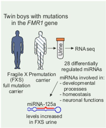
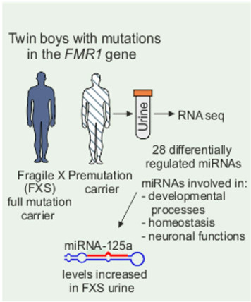
Urine microRNA Profiling Displays miR-125a Dysregulation in Children with Fragile X Syndrome

1
Faculty of Medicine, Physiology, University of Helsinki, FI-00014 Helsinki, Finland
2
Turku Centre for Biotechnology, University of Turku and Åbo Akademi University, Tykistökatu 6A, FI-20520 Turku, Finland
3
Leucadia Therapeutics Inc., Riverside, CA 92506, USA
4
The Joint Clinical Biochemistry Laboratory of University of Turku, University Central Hospital and Wallac Oy, FI-20520 Turku, Finland
5
Department of Clinical Genetics, University Hospital of Helsinki, 160, FI-00290 Helsinki, Finland
6
Institute of Biomedical Research of Malaga (IBIMA) and Mental Health Unit, Regional University Hospital of Malaga, University of Malaga, Research lab. Hospital Civil, Pab-6 sot. 29009 Malaga, Spain
7
Rinnekoti Foundation, FIN-02980 Espoo, Finland
8
Division of Biomedical Sciences, School of Medicine, University of California, Riverside, CA 92521, USA
*
Authors to whom correspondence should be addressed.
Cells 2020, 9(2), 289; https://doi.org/10.3390/cells9020289
Submission received: 5 November 2019 / Revised: 6 January 2020 / Accepted: 22 January 2020 / Published: 24 January 2020
(This article belongs to the Special Issue microRNA as Biomarker)

Abstract

:
A triplet repeat expansion leading to transcriptional silencing of the FMR1 gene results in fragile X syndrome (FXS), which is a common cause of inherited intellectual disability and autism. Phenotypic variation requires personalized treatment approaches and hampers clinical trials in FXS. We searched for microRNA (miRNA) biomarkers for FXS using deep sequencing of urine and identified 28 differentially regulated miRNAs when 219 reliably identified miRNAs were compared in dizygotic twin boys who shared the same environment, but one had an FXS full mutation, and the other carried a premutation allele. The largest increase was found in miR-125a in the FXS sample, and the miR-125a levels were increased in two independent sets of urine samples from a total of 19 FXS children. Urine miR-125a levels appeared to increase with age in control subjects, but varied widely in FXS subjects. Should the results be generalized, it could suggest that two FXS subgroups existed. Predicted gene targets of the differentially regulated miRNAs are involved in molecular pathways that regulate developmental processes, homeostasis, and neuronal function. Regulation of miR-125a has been associated with type I metabotropic glutamate receptor signaling (mGluR), which has been explored as a treatment target for FXS, reinforcing the possibility that urine miR-125a may provide a novel biomarker for FXS.

Graphical Abstract

1. Introduction

MicroRNAs (miRNAs) are short (19–24 nucleotides in length) noncoding RNAs that regulate translation by promoting mRNA degradation and attenuating protein translation [1,2]. Active trafficking of miRNAs contributes to intercellular communication by local control of miRNA-mediated regulation of target genes and signaling events. Several miRNAs reach the circulation using microvesicle-dependent or RNA-binding-protein-associated active secretion or by passive leakage from cells [3]. Measurable levels of miRNAs can be isolated from bodily fluids, including urine [4]. Mounting evidence indicates that miRNAs are involved in the pathophysiology of several disorders, and disease-specific extracellular miRNA profiles have been identified [5]. Changes in circulating miRNAs have been implicated in metabolic crosstalk between organs as well as in neurodegeneration [6,7]. The accessibility and stability of circulating miRNAs support their use as biomarkers for patient stratification to improve the efficacy of targeted treatments [3].
Fragile X syndrome (FXS) is the most common cause of inherited intellectual disability and the best-known single-gene cause of autism spectrum disorder, with a prevalence of 1/4000 males and ~1/6000 females. The behavioral phenotype of FXS includes hyperactivity, attention disorders, social anxiety and mood instability, and abnormalities in sensory stimuli [8,9]. The severity of these features, as well as responses to current pharmacological treatments, varies individually. Epilepsy associates with FXS in about 15%–20% of FXS males [10]. In most cases, expansion of a triplet CGG repeat in the untranslated region of the FMR1 gene from the normal 5–55 repeat range to >200 leads to transcriptional silencing and lack of FMR1 protein (FMRP), which results in FXS [11]. A CGG repeat between 55 and 200 triplets is a fragile X premutation that can be inherited from the mother as an expanded full mutation [12]. Premutation carriers do not show early-childhood-onset intellectual disability syndrome, and paradoxically, they have abnormally high levels of FMR1 mRNA, which predisposes them to a late-onset neurodegenerative disorder called fragile X-associated tremor/ataxia syndrome (FXTAS) [13].
FMRP expression is widespread with high abundant expression in neurons and testes [14]. The absence of functional FMRP impairs normal synaptic formation and plasticity in the central nervous system (CNS) and results in macro-orchidism due to the overproduction of Sertoli cells in testes [9,15]. FMRP is a messenger RNA (mRNA)-binding protein and controls protein translation by interacting with specific mRNAs. FMRP also associates with miRNAs and proteins incorporated into a multiprotein complex called an RNA-induced silencing complex (RISC). There is evidence that disruption of miRNA pathways contributes to the abnormal synaptogenesis in FXS [16,17,18]. A RISC consisting of miR-125a and FMRP on postsynaptic density-95 (PSD-95) mRNA has been identified as a selective mechanism that controls PSD-95 mRNA translation for signaling of group 1 metabotropic glutamate receptors (mGluR) at synapses [19]. Expression of miR-125a is decreased in synaptoneurosomes in the brain of Fmr1 knockout (KO) mice, indicating that dysregulation of miR-125a is involved in impaired synapse function in FXS. DeMarco et al. showed very recently that phosphorylation status of FMRP regulates the stability of the miR-125a-guided RISC and PSD-95 mRNA complex [18]. Since mGluR signaling alters FMRP phosphorylation, augmented mGluR signaling in FXS may affect turnover and/or cellular cycling [20] of miR-125a.
We compared urine miRNA profiles of a boy with FXS and his twin brother, a premutation carrier, and found 28 differentially expressed miRNAs. The most pronounced increase was found in levels of miR-125a in urine of the FXS twin as well as a larger group of FXS donors. A detailed analysis of miR-125a levels revealed an age-dependent increase in control urine, whereas the scattered distribution of FXS samples suggested that two subgroups of FXS subjects might exist. Our results implicate miR-125a dysregulation in the pathophysiology of FXS, consistent with previous studies of FXS mice, and provide evidence that urine miR-125a could be a potential novel biomarker in FXS clinical trials.

2. Materials and Methods

2.1. Human Urine Samples

The research using human urine was approved by the Ethics Committee of Helsinki and Uusimaa Hospital District and by the Ethics Committee of the Regional University Hospital of Málaga. Written informed consent was signed by parents and donors to obtain human samples. One set of urine samples was collected from males and a female with FXS (n = 9), a premutation carrier (n = 1; the twin boy), and healthy controls (n = 8) recruited under auspices of Rinnekoti Foundation. Another independent set of urine samples obtained from control (n = 8) and FXS donors (n = 10) was analyzed at the Regional University Hospital of Malaga (Spain) for replication purposes. The APHS Biobank has coordinated the collection, processing, management and assignment of the biological samples used in this study, according to the standard procedures established for this purpose. Morning urine was collected in preservation tubes (Norgen Biotek Corp, Thorold, ON, Canada) and stored at 4 °C until miRNA extraction.
FXS molecular diagnostics was performed on blood samples and the detection of CGG repeats in the FMR1 gene was made using the Fragile X PCR test (Abbott Laboratories, IL, USA). The triplet repeat primed PCR assay provided accurate sizing of CGG repeats; size accuracy on 5–70 CGG repeat alleles + 1 CGG repeat and on 71–230 repeat alleles 3 CGG repeats by capillary electrophoresis, alleles > 231 resolved by agarose gel with low accuracy for sizing, according to the kit information provided by the manufacturer.

2.2. RNA Extraction and Quantitative Real-Time PCR

Urine miRNAs were extracted with Urine microRNA Purification Kit (Norgen Biotek Corp., Ontario, Canada) according to the manufacturer’s instructions. Isolated miRNA samples were eluted in 40 μl of RNase-free water. RNA concentrations were measured with Nanodrop Spectrophotometer (Thermo Scientific, Wilmington, DE, USA) and equal amounts of miRNA (10 ng) were used in reverse transcription reactions. The levels of mature miRNAs were analyzed using individual TaqMan microRNA Assays (Applied Biosystems, Life Technologies Ltd., Carlsbad, CA, USA) according to the manufacturer’s instructions. Specific miRNA primers were used to reverse-transcribe 10 ng of total miRNA with the TaqMan MicroRNA Reverse Transcription Kit (Applied Biosystems, Life Technologies Ltd., Carlsbad, CA, USA) with specific primers (Table S1). The expression levels of miRNAs were analyzed by quantitative real-time PCR (RT-qPCR) in 96-well plates on Light Cycler 480 II Real-Time PCR System (Roche Diagnostics GmbH, Mannheim, Germany). The TaqMan probes (FAM) were detected with 465–510 nm. The miRNAs were normalized to mean Ct values of all studied miRNAs in a urine sample, as previously reported [21,22].

2.3. Deep Sequencing

Fragment libraries were prepared for each sample with TruSeq Small RNA Library Prep Kit (Illumina) and sequenced using the HiSeq 2500 system (Illumina, Inc., San Diego, CA, USA) as instructed by the manufacturer. Read lengths were 36 bases.

2.4. Analysis of Data

Raw sequence data were imported in CLC Genomics Workbench (version 7.0.3; https://www.qiagenbioinformatics.com/) for read quality inspection, adapter trimming, and miRNA annotation based on miRBase (http://www.mirbase.org/). The count data grouped on mature miRNAs were then exported to R/Bioconductor v. 3.1.0 (www.bioconductor.org) (R Core Team, 2015), where the normalization and statistical testing were carried out with the DESeq2 package [23]. The sequencing dataset was deposited in the GEO database (accession #GSE143347).
Differentially expressed miRNAs were filtered requiring absolute fold-change > 2 and Wald test p-value < 0.05. Target gene and pathway analysis was carried out using miRSystem [24] and DAVID gene annotation tool [25]. In miRNA system, queried miRNAs were first converted to the last miRBase annotation (version 17) and significantly enriched signaling pathways were identified. In addition to the hypergeometric p-values, empirical p-values of each function/pathway were determined by ranking the enriched hypergeometric probability as compared with null baseline probabilities [24]. The weight of a miRNA was determined by dividing the absolute expression value of the miRNA by the absolute sum of the expression values of all input miRNAs. Thereafter, the ranking score was obtained by summation of the weight of miRNA times its enrichment –log (p-value) from the predicted target genes. Linear regression analysis of urine miR-125a levels was performed using GraphPad Prism 5.0. Data are expressed as the mean + SEM and were analyzed for statistical significance using Student’s t test. A p-value < 0.05 was considered significant.

3. Results

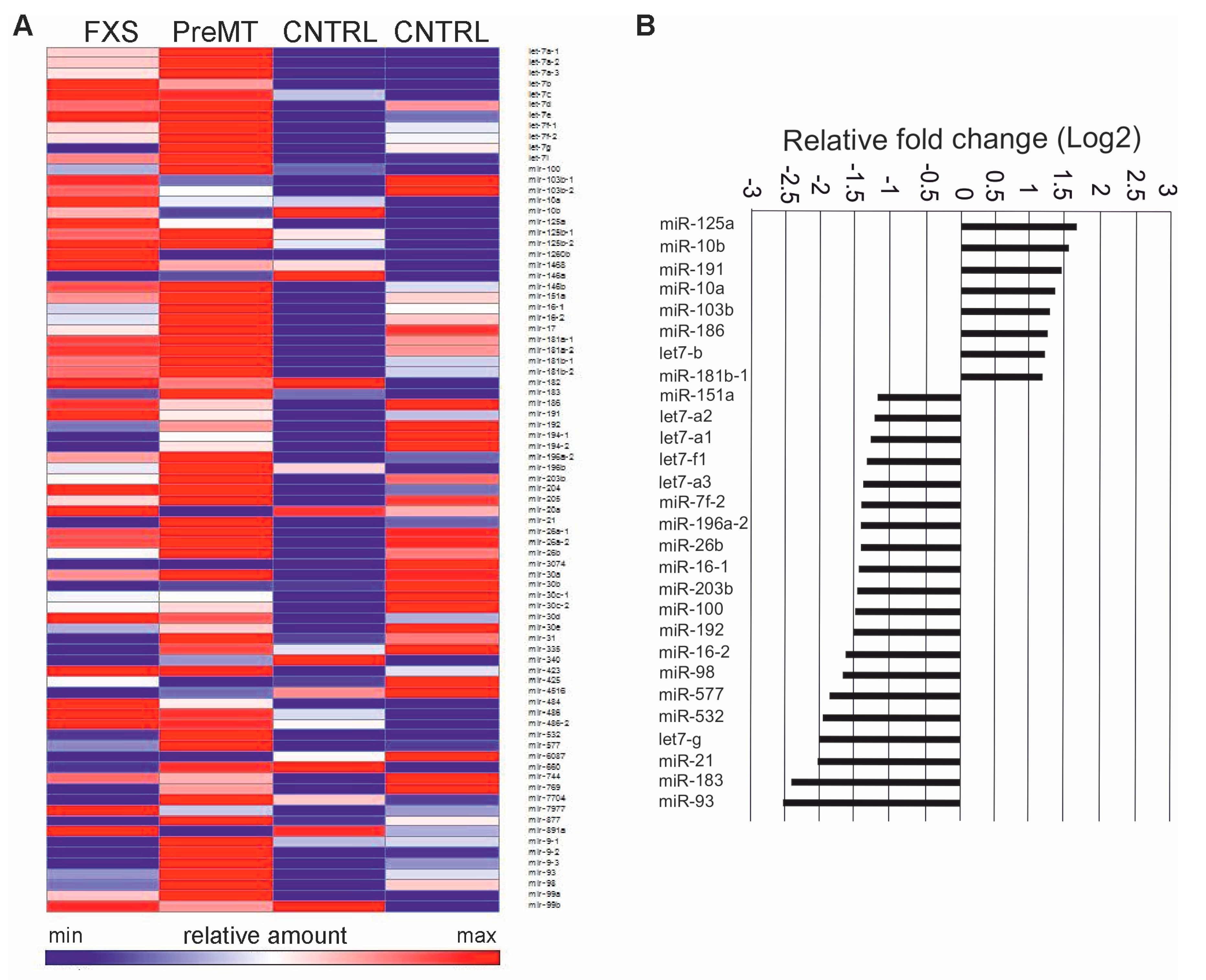
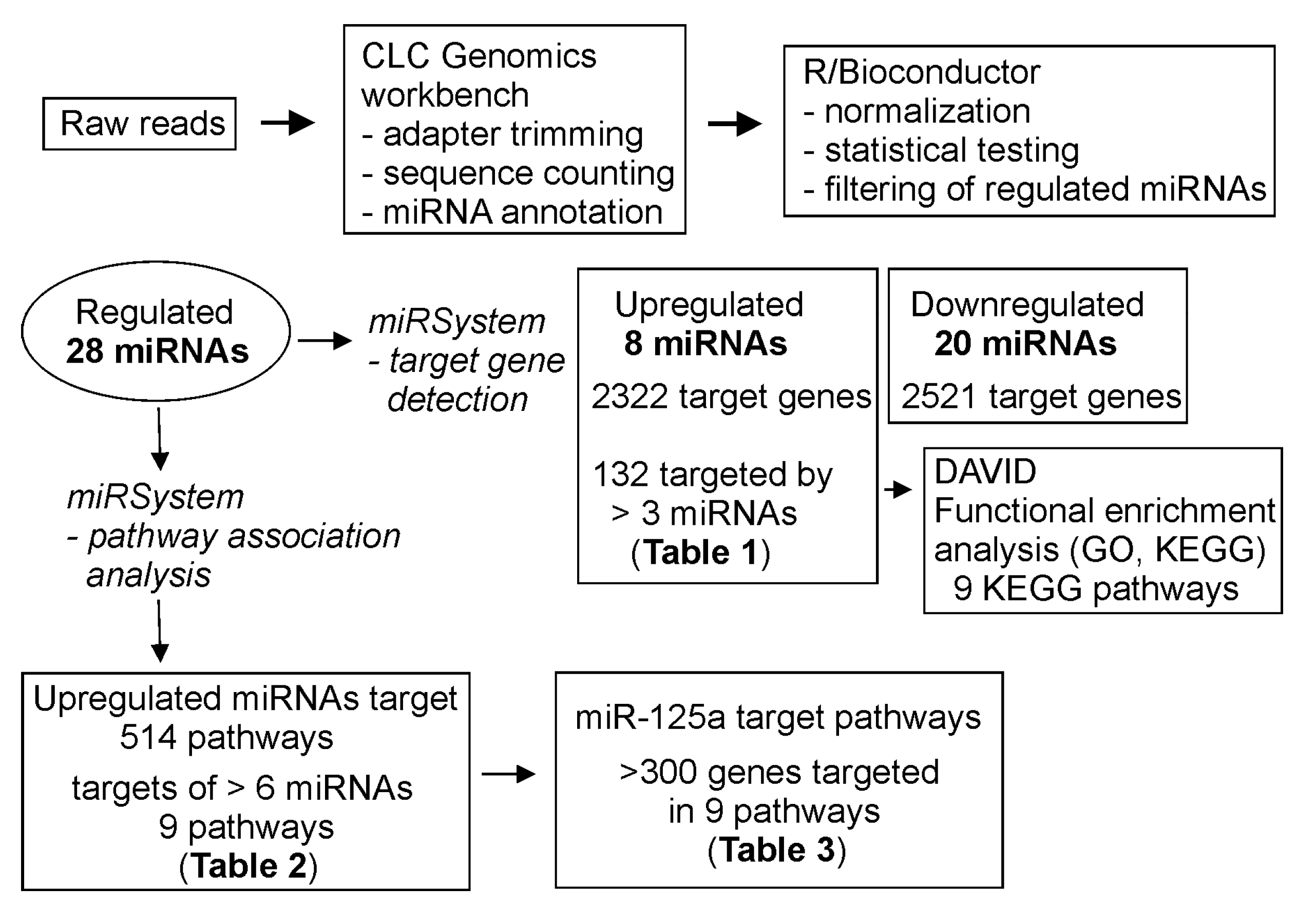
3.1. Profiling of FXS Urine miRNAs by Deep Sequencing

To uncover FXS-specific alterations in urine miRNAs, miRNA-sequencing of urine samples obtained from individuals with and without FXS was performed by massive parallel sequencing using a HiSeq 2500 system. The initial analysis provided evidence of age-dependent clustering of FXS samples, although the sequencing read counts were highly variable across subjects. The donors included seven-year-old twin boys whose total urine miRNA reads were similar, suggesting less variation in metabolic and epigenetic measures affecting miRNA levels. One of the twin boys was diagnosed with FXS by molecular diagnostics of >200 CGG repeats (300 repeats) in the FMR1 gene, while the other twin boy without clinical FXS diagnosis carried CGG repeats in the range of the FMR1 gene premutation (175 repeats). The comparison of urine of the twin boys revealed 28 differentially regulated miRNAs in a total of 219 miRNAs analyzed with >50 read counts each (Figure 1A). Of those 28 notable miRNAs, 8 were upregulated and 20 were downregulated in FXS urine compared to non-FXS urine (Figure 1B). The largest increase was found in the levels of hsa-miR-125a-5p.

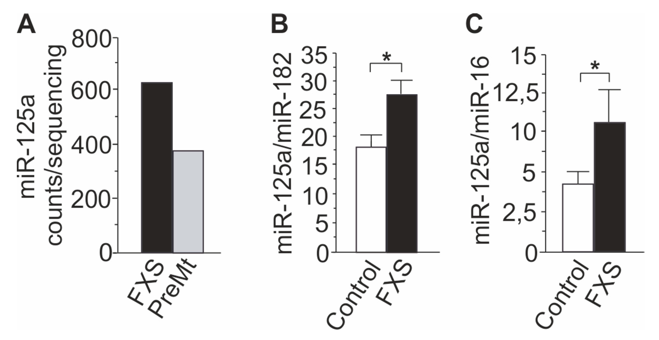
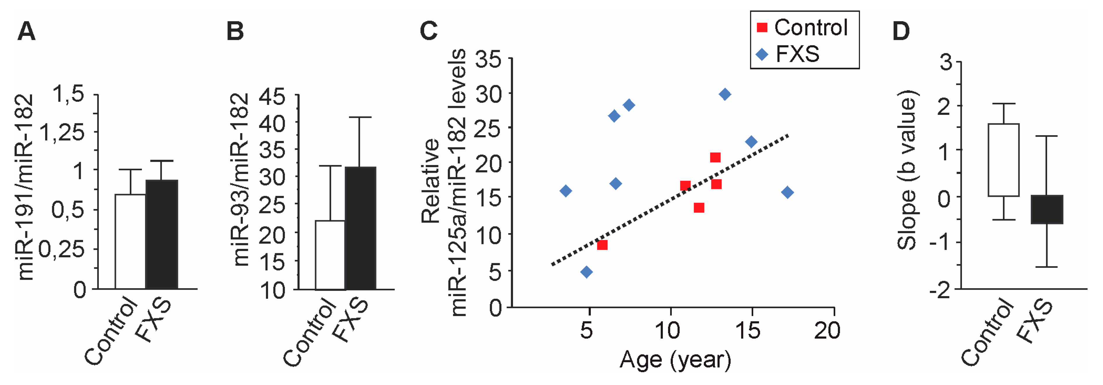
3.2. Increased miR-125a in Urine of Children with FXS

The RT-qPCR analysis confirmed the abnormal increase in miR-125a levels in urine of the FXS twin boy found by deep sequencing (Figure 2A). The miR-125a levels were 1.6-fold higher in urine of the FXS twin boy than those of his twin brother, and an identical increase was seen in replicate samples taken one year after the first sampling. The miR-125a levels were also found to be increased in a larger number of FXS samples from donors with a homogenous Finnish genetic heritage (Table 1) compared to healthy controls (Figure 2B). The data were normalized to miR-182 that was not regulated in the sequencing analysis, and the increase was more pronounced when miR-16 was used for normalization consistent with the reduction of miR-16 in FXS urine in the sequencing analysis (Figure 2C).
The miR-125a urine analysis was replicated in an independent pool of FXS urine samples collected by a Spanish lab at the Regional University Hospital of Malaga-IBIMA. The comparison of urine samples of Spanish FXS children (n = 10) aged 2–7 years and healthy controls (n = 8) aged 2–6 years confirmed the 1.6-fold increase (p = 0,01) in miR-125a levels in urine: control 13.9 (SD = 3.7, SEM = 1.51) and FXS 22.5 (SD = 2.9, SEM = 1.02). The levels of miR-182 did not differ between FXS and control samples. Altogether, the data suggested that urine miR-125a levels were differentially regulated in FXS urine compared to those of healthy controls, consistent with the previous studies that have shown FMRP-dependent regulation of miR-125a.

3.3. Age-Dependent Dysregulation of miR-125a in Urine of Children with FXS

The relative levels of miR-191 and miR-93 did not differ between FXS and control urine in the expanded pool of samples (Figure 3A), suggesting that the miRNA differences identified in urine of twins were small and that miRNA levels may show high variability. The difference of urine miR-125a levels between FXS and control urine was confirmed in two sets of samples, increasing the reliability of the results, and larger studies are needed to explore the impact of the other miRNAs identified to be dysregulated individually. Furthermore, since urine miRNAs of premutation carrier and FXS twin boys were initially compared, the assessment of the role of each miRNA has to be done in a set of samples which includes both premutation carriers and FXS individuals.
The distribution of miR-125a in urine of healthy children aged 4–18 years (n = 5) showed an age-dependent linear increase (1.559*X – 2.147; R2 = 0.779; p = 0.0473) whereas the miR-125 levels in FXS samples were scattered (Y = −0.648*X + 29.18; R2 = 0.074; p = 0.478), and the samples did not correlate (Pearson’s correlation r = −0.209) (Figure 3B). Urine miR-125a levels of two FXS boys were lower than the levels of controls and other 5 FXS donors, while all other FXS donors had higher urine miR-125a levels than the age-matched controls. The small sample size in the study associated with a high variation of the slope of the line in the analysis of FXS samples, leading to significant limitations in the interpretation of the results. However, the distribution of urine miR-125a samples indicated a possibility that the FXS samples could be divided into two groups, but larger studies are needed to investigate and possibly confirm the potential of miR-125a as a tool for stratification of FXS males.
In our set of samples, low levels of urine miR-125a did not associate with genetic mosaicism of the FMR1 full mutation, and there was no correlation between urine miR-125a levels and specific pharmacological intervention in the sample pool (Table 1). Levels of miR-125a were low in urine of a 17-year-old FXS boy with no medications as well as in urine of a 5-year-old FXS boy treated with risperidone for aggressive behavior combined with methylphenidate for hyperactivity. On the other hand, urine miR-125a levels were abnormally increased in two FXS donors with methylphenidate treatment for hyperactivity. The controls did not have any medications. Only one FXS boy (FXS6) had epilepsy, and his miR-125a levels were in the range of the high FXS values.

3.4. Target Gene and Pathway Analysis of miRNAs Regulated Differentially in FXS Urine

We analyzed predicted target genes and pathways associated with the miRNAs that were differentially regulated in urine of the FXS twin boys using miRSystem and DAVID gene annotation tool as shown in the summary of the sequencing data analysis in Figure 4 (http://mirsystem.cgm.ntu.edu miRNAs) [24,25].
Target gene analysis of increased miRNAs (all but miR-103b-1) identified 2322 possible targets. Genes targeted by five miRNAs included gamma-aminobutyric acid (GABA) A receptor, beta 2 (GABRB2) and StAR-related lipid transfer (START) domain containing 13 (STARD13), and genes targeted by 4 miRNAs included BCL2L2, BDNF, GRIN3A, among others (Table 2). Metabotropic glutamate receptor 3 (GRM3) was targeted by two miRNAs.
We chose 132 genes targeted by three or more miRNAs that were increased in FXS urine for initial pathway analysis using DAVID gene annotation tool24, GO term, and KEGG pathway annotations [26]. We found nine KEGG pathway terms enriched with these genes: MAPK signaling pathway, pathways in cancer, Wnt signaling pathway, focal adhesion, glioma, chronic myeloid leukemia, neurotrophic signaling pathway, pancreatic cancer, and TGF-beta signaling pathway. The miRsystem tool found 514 pathways where the miRNAs were involved. A functional annotation summary of pathways of six enriched miRNAs is shown in Table 3.
The pathway ranking summary revealed that all six miRNAs were involved in nine pathways: signaling by insulin receptor, focal adhesion, MAPK signaling pathway, neurotrophin signaling pathway, signaling to ERKs, pathways in cancer, signaling by NGF, NGF signaling via TrkA from the plasma membrane, and hemostasis (Table 4).
Pathway analysis of miR-125a showed similarities to the predicted pathways of the group of increased miRNAs, including pathways of developmental biology, adaptive immune system, hemostasis, transmembrane transport of small molecules, GPCR ligand binding, cell cycle mitotic, cancer, neuroactive ligand-receptor activation, and class A1 of rhodopsin-like receptors (Table 5). Furthermore, the predicted pathways for increased and reduced miRNAs showed similarities. MAPK signaling pathway and pathways in cancer and developmental biology had the highest scores in the pathway ranking summary for the reduced miRNAs.

4. Discussion

The present study identified FXS-specific changes in urine miR-125a. The levels of miR-125a were found to be abnormally increased in FXS urine when urine samples of twin boys and an expanded pool of donors collected in two independent labs were compared. Previous studies have demonstrated that regulation of miR-125a is affected in the absence of FMRP, which stressed the potential importance of urine dysregulation of miR-125a among 28 differentially regulated miRNAs identified by deep sequencing. Levels of miR-125a are shown to be reduced in synaptoneurosomes isolated from the FXS mouse brain [19], and the phosphorylation state of FMRP regulates the stability of miR-125a-guided RISC-PSD 95 mRNA complex, which is critical for synapse function [18]. Increased urine miR-125a levels may reflect increased production and/or secretion of miR-125a, but it is not possible to make any direct correlations between human urine and mouse brain miRNA levels. Furthermore, a detailed analysis of miR-125a levels in children’s urine revealed an age-dependent regulation, whereas FXS samples did not show linear correlation indicating higher individual variability. Urine miR-125a levels of two FXS males were lower than those of the healthy controls and of five other FXS males. There was no clear correlation between the miR-125a levels and the length of the repeat expansion in the FMR1 gene, genetic mosaicism, or pharmacological interventions within the small set of samples. The FXS children with low levels of miR-125a in urine may present a subgroup whose cellular homeostasis differs from that of the subgroup with higher levels of miR-125a. A well-described association between miR-125a- and mGluR5 signaling [19] suggests that urine miR-125a levels may provide a novel tool to subgroup FXS children based on individual differences linked particularly to mGluR5 signaling, which is considered to be the most critically dysregulated signaling pathway in FXS.
Expression of miR-125a is high in the ovary, epididymis, spleen, and in some endocrine organs and regions of the brain (www.microRNA.org). There is evidence that members of the miR-125 family can have disease-suppressing properties, implying that it could predict disease onset or have prognostic value during disease progression [27,28]. Recently ectopic expression of miR-125a was found to promote granulocyte differentiation, and improved understanding of miR-125a function may assist in the development of novel miR-125a-targeted therapies [29]. A role for miR-125a-5p has been identified in the regulation of endothelial tightness, supporting the potential of miR-125a as a disease biomarker in circulating biofluids [28,30]. There is evidence that miR-125a reduces endothelin-1 expression and immune cell efflux in inflammation. It has been shown that miR-125a regulates the secretion of some inflammatory cytokines (interleukin (IL)2, IL6, TNF-alpha, and TNF-beta) [31]. In mouse in vivo and in vitro models of thyroiditis, an increased miR-125a expression reduces autophagy and cell proliferation and increases the apoptotic rate and the expression of proinflammatory factors tumor necrosis factor-α, IL-1β, IL-6, and IL-18 via downregulation of the phosphoinositide 3-kinase/protein kinase B/mammalian target of rapamycin (PI3K/Akt/mTOR)signaling pathway [32]. Low levels of miR-125a-5p are found in different types of tumors [33]. Levels of miR-125a-5p are also found to be decreased in the hippocampus of rats with pentylenetetrazol (PTZ)-induced epilepsy, whereas miR-125a-5p overexpression can attenuate seizures and decrease inflammatory factors in these rats [34].
In the present study, a total of 219 miRNAs with the expression of more than 50 read counts were identified in human urine by RNA seq. At least a 1.5-fold expression difference cutoff is suggested by several miRNA profiling studies that have explored the impact of changes of miRNA levels on cellular biology [35]. We observed 28 differentially expressed miRNAs in urine of an FXS boy compared to urine of his twin brother. The most increased miRNA in FXS urine was miR-125a, and its increase was confirmed by RT-qPCR. The smaller increase of miR-191 levels and reduction of miR-93 levels in FXS urine were not confirmed in the expanded pool of samples, suggesting that urine miRNA levels are low and show high individual variability affecting their analysis. Therefore, dysregulation of the miRNAs other than miR-125a remains to be investigated in a larger set of samples which include both premutation carriers and FXS individuals. A panel of circulating miRNAs may have more potential to show efficacy than single miRNAs in drug response monitoring as observed in biomarker studies of cancer patients [19]. Many predicted target genes of the differentially regulated miRNAs were shown to be involved in pathways that regulate molecular and cellular processes known to be disrupted in FXS, including axon guidance and neurotrophin signaling in the nervous systems. The data suggest that urine miRNA levels may reflect common pathological miRNA-dependent processes caused by the absence of FMRP in multiple tissues [36].
Although most miRNAs are intracellular, significant levels of miRNAs appear outside cells and circulate in human body fluids, including urine [4,20,37]. Each body fluid has its own miRNA composition, but the origin of circulating miRNAs is not well understood, although correlations between circulating and tissue miRNAs exist [38]. There is evidence that miRNAs can reach the circulation from active secretion or passive leakage from broken cells [3]. Certain miRNAs are targeted for export and actively secreted to extracellular fluids [39]. Active miRNA secretion can be mediated via microvesicles or a microvesicle-free, RNA-binding protein-dependent pathway. The majority of urinary miRNAs originate from renal and urethral cells, but other tissues can also actively release circulating extracellular miRNAs packaged in exosomes (lipid vesicles) [40] or bound to RNA-binding proteins [35,41] into urine via renal epithelial cells. Interestingly, the profiles of miRNAs in urine and cerebrospinal fluid show many similarities, such as low miRNA abundance [4]. Both cell-free and exosomal preparations are found in urine samples [3], and the small number of miRNAs may indicate that only distinct miRNAs are stabilized by microvesicles or associated with RNA-binding protein and high-density lipoprotein (HDL) as a carrier and protected from degradation by ribonucleases in the circulation.
The present study did not examine correlations of miR-125a levels in urine and other bodily fluids. Previously slight but not significant reduction of miR-125a-5p was observed in FXTAS patients’ blood by deep sequencing [42]. The miRNA profiling of serum in children with autism spectrum disorder (ASD) identified thirteen differentially expressed miRNAs in individuals with ASD compared to the controls, and miR-125a was not among the dysregulated miRNAs [43].
There are 12 brain miRNAs identified to interact with FMRP [16], and these miRNAs include miR-125a. Involvement of miR-125a in fate determination of neuronal lineages [44] and synaptic plasticity [45] links dysregulation of miR-125a to FXS but potentially also to several other neurodevelopmental disorders. Differential expression of miR-125a in the male and female frontal lobe region during normal development has been reported [46], and many similar sexually dimorphic miRNAs are associated with autism-related diseases and processes [47]. Gender effects on the analysis of FXS urine miR-125a levels were reduced in the current study by using only male controls. Only one female FXS subject was included in the study, and interestingly, her urine miR-125a levels were just slightly above the age-matched control levels.

5. Conclusions

Our study is the first to examine and show disease-specific changes in the urinary miRNA profile in a neurodevelopmental disorder. Levels of miR-125a were shown to be increased in FXS urine, consistent with previous observations showing the involvement of FMRP in the regulation of miR-125a expression. Altered peripheral miRNA levels have been detected in several neuropsychiatric disorders, including depression, schizophrenia, and ASD [48,49], and increased plasma miR-125a levels were very recently shown in patients with bipolar disorder and particularly in bipolar manic patients [50]. The results of the present study demonstrate the potential of urine miRNA profiling for miRNA biomarker development in FXS. Larger studies are necessary to confirm the abnormalities of urine miR-125a levels and to explore the impact of the other dysregulated miRNAs observed in urine profiling in FXS.

Supplementary Materials

The following are available online at https://www.mdpi.com/2073-4409/9/2/289/s1, Table S1 showing primers (Life Technologies Ltd) used in the RT-qPCR.

Author Contributions

N.P. collected and assembled the data, A.L. analyzed and interpreted the data, D.E. interpreted the data and contributed to the writing of the manuscript, J.P. (Juha Pursiheimo) and A.M.G. analyzed the data, A.-K.A. contributed to data interpretation, J.P. (Juho Pitkonen) contributed to the editing, Y.d.D.-O. provided financial support, study material and analyzed the data, and M.L.C. designed experiments, provided financial support and study material, analyzed and interpreted the data, and wrote the manuscript. All authors reviewed the manuscript. All authors have read and agreed to the published version of the manuscript.

Funding

This research was funded by the Arvo and Lea Ylppö Foundation, the Sakari and Orvokki Sohlberg Foundation, the Finnish Brain Foundation, Finnish Foundation for Pediatric Research, and the Academy of Finland. Y.D.O. is a recipient of a Nicolás Monardes Appointment, Consejería de Salud, Andalusian Ministry of Health, and Funds from Economy and Innovation Regional Ministry, Andalusian Government Grant PI10-CTS-05704, and the Fundación Alicia Koplowitz-2016 (Madrid).

Acknowledgments

We thank the Finnish Fragile X Family Association, Frax ry, and all the families that participated in the study. We also thank Daniel Ozer for guidance in the statistical analysis and David W.E. Ramsden for revising the English language in the manuscript. Open access funding provided by University of Helsinki.

Conflicts of Interest

The authors declare no conflict of interest.

References

  1. Bartel, D. Micrornas: Genomics, biogenesis, mechanism, and function. Cell 2004, 116, 281–297. [Google Scholar] [CrossRef] [Green Version]
  2. Lee, R.; Feinbaum, R.; Ambros, V. The c. elegans heterochronic gene lin-4 encodes small RNAs with antisense complementarity to lin-14. Cell 1993, 75, 843–854. [Google Scholar] [CrossRef]
  3. Cheng, L.; Sun, X.; Scicluna, B.; Coleman, B.; Hill, A. Characterization and deep sequencing analysis of exosomal and non-exosomal miRNA in human urine. Kidney Int. 2014, 86, 433–444. [Google Scholar] [CrossRef] [PubMed] [Green Version]
  4. Weber, J.; Baxter, D.; Zhang, S.; Huang, D.; Huang, K.; Lee, M.; Galas, D.; Wang, K. The microRNA spectrum in 12 body fluids. Clin. Chem. 2010, 56, 1733–1741. [Google Scholar] [CrossRef] [PubMed]
  5. Im, H.; Kenny, P. MicroRNAs in neuronal function and dysfunction. Trends Neurosci. 2012, 35, 325–334. [Google Scholar] [CrossRef] [Green Version]
  6. Lehmann, S. An unconventional role for miRNAs: Let-7 activates Toll-like receptor 7 and causes neurodegeneration. Nat. Neurosci. 2012, 15, 827–835. [Google Scholar] [CrossRef]
  7. Xihua, L. Circulating miR-143-3p inhibition protects against insulin resistance in Metabolic Syndrome via targeting of the insulin-like growth factor 2 receptor. Transl. Res. 2018, 205, 33–43. [Google Scholar] [CrossRef] [Green Version]
  8. Hagerman, R.; Berry-Kravis, E.; Kaufmann, W.; Ono, M.; Tartaglia, N.; Lachiewicz, A.; Kronk, R.; Delahunty, C.; Hessl, D.; Visootsak, J.; et al. Advances in the treatment of fragile X syndrome. Pediatrics 2009, 123, 378–390. [Google Scholar] [CrossRef] [Green Version]
  9. Terraciano, A.; Chiurazza, P.; Neri, G. Fragile X syndrome. Am. J. Med. Genet. C Semin. Med. Genet. 2005, 137, 32–37. [Google Scholar] [CrossRef]
  10. Berry-Kravis, E. Epilepsy in fragile X syndrome. Dev. Med. Child. Neurol. 2002, 44, 724–728. [Google Scholar] [CrossRef]
  11. Pieretti, M.; Zhang, F.; Fu, Y.-H.; Warren, S.T.; Oostra, B.A.; Caskey, C.T.; Nelson, D.L. Absence of expression of the FMR-1 gene in fragile X syndrome. Cell 1991, 66, 817–822. [Google Scholar] [CrossRef]
  12. Hagerman, R.J.; Hagerman, P.J. The fragile X premutation: Into the phenotypic fold. Curr. Opin Genet. Dev. 2002, 12, 278–283. [Google Scholar] [CrossRef]
  13. Hagerman, P.; Hagerman, R. Fragile X-associated tremor/ataxia syndrome (FXTAS). Mental Ret. Dev. Disabil. Res. Rev. 2004, 10, 25–30. [Google Scholar] [CrossRef] [PubMed]
  14. Khandjian, E.W.; Fortin, A.; Thibodeau, A.; Tremblay, S.; Cote, F.; Devys, D.; Mandel, J.L.; Rousseau, F. A heterogeneous set of FMR1 proteins is widely distributed in mouse tissues and is modulated in cell culture. Hum. Mol. Genet. 1995, 4, 783–789. [Google Scholar] [CrossRef] [PubMed]
  15. Slegtenhorst-Eegdeman, K.; de Rooij, D.; Verhoef-Post, M.; van de Kant, H.; Bakker, C.; Oostra, B.; Grootegoed, J.; Themmen, A. Macroorchidism in fmr1 knockout mice is caused by increased Sertoli cell proliferation during testicular development. Endocrinology 1998, 39, 156–162. [Google Scholar] [CrossRef] [PubMed]
  16. Edbauer, D.; Neilson, J.; Foster, K.; Wang, C.; Seeburg, D.; Batterton, M.; Tada, T.; Dolan, B.; Sharp, P.; Sheng, M. Regulation of synaptic structure and function by FMRP-associated microRNAs miR-125b and miR-132. Neuron 2010, 65, 373–384. [Google Scholar] [CrossRef] [Green Version]
  17. Gong, X.; Wang, Y.; Zeng, J.; Li, S.; Luo, Y. Computational identification and experimental validation of microRNAs binding to the fragile X syndrome gene Fmr1. Neurochem. Res. 2015, 40, 109–117. [Google Scholar] [CrossRef]
  18. DeMarco, B.; Stefanovic, S.; Williams, A.; Moss, K.; Anderson, B.; Bassell, G.; Mihailescu, M. FMRP-G-quadruplex mRNA-miR-125a interactions: Implications for miR-125a mediated translation regulation of PSD-95 mRNA. PLoS ONE 2019, 14, e0217275. [Google Scholar] [CrossRef]
  19. Muddashetty, R.; Nalavadi, V.; Gross, C.; Yao, X.; Xing, L.; Laur, O.; Warren, S.; Bassell, G. Reversible inhibition of PSD-95 mRNA translation by miR-125a, FMRP phosphorylation, and mGluR signaling. Mol. Cell 2011, 42, 673–688. [Google Scholar] [CrossRef] [Green Version]
  20. Jin, X.; Wu, N.; Wang, L.; Li, J. Circulating microRNAs: A novel class of potential biomarkers for diagnosing and prognosing central nervous system diseases. Cell Mol. Neurobiol. 2013, 33, 601–613. [Google Scholar] [CrossRef]
  21. Mestdagh, P.; Van Vlierberghe, P.; De Weer, A.; Muth, D.; Westermann, F.; Speleman, F.; Vandesompele, J. A novel and universal method for microRNA RT-qPCR data normalization. Genome Biol. 2009, 10, R64. [Google Scholar] [CrossRef] [PubMed] [Green Version]
  22. Qureshi, R.; Sacan, A. A novel method for the normalization of microRNA RT-PCR data. BMC Med. Genomics 2013, 6 (Suppl. 1), 14. [Google Scholar] [CrossRef] [Green Version]
  23. Love, M.; Huber, W.; Anders, S. Moderated estimation of fold change and dispersion for rna-seq data with DESeq2. Genome Biol. 2014, 15, 550. [Google Scholar] [CrossRef] [PubMed] [Green Version]
  24. Lu, T.-P.; Lee, C.-Y.; Tsai, M.-H.; Chiu, Y.-C.; Hsiao, C.; Lai, L.-C.; Chuang, E. miRSystem: An integrated system for characterizing enriched functions and pathways of microRNA targets. PLoS ONE 2012, 7, e42390. [Google Scholar] [CrossRef] [PubMed] [Green Version]
  25. Huang, D.; Sherman, B.; Lempicki, R. Systematic and integrative analysis of large gene lists using DAVID Bioinformatics Resources. Nat. Protoc. 2009, 4, 44–57. [Google Scholar] [CrossRef] [PubMed]
  26. Kanehisa, M.; Sato, Y.; Kawashima, M.; Furumichi, M.; Tanabe, M. KEGG as a reference resource for gene and protein annotation. Nucleic Acids Res. 2016, 44, 457–462. [Google Scholar] [CrossRef] [Green Version]
  27. Sun, Y.; Lin, K.; Chen, Y. Diverse functions of miRNA-125a family in different cell contexts. Hematol. Oncol. 2013, 6, 6. [Google Scholar] [CrossRef] [Green Version]
  28. Reijerkerk, A.; Lopez-Ramirez, M.; van Het Hof, B.; Drexhage, J.; Kamphuis, W.; Kooij, G.; Vos, J.; van der Pouw Kraan, T.; van Zonneveld, A.; Horrevoets, A.; et al. MicroRNAs regulate human brain endothelial cell-barrier function in inflammation: Implications for multiple sclerosis. J. Neurosci 2013, 33, 6857–6863. [Google Scholar] [CrossRef] [Green Version]
  29. Dakir, E.-H.; Mollinedo, F. Genome-wide miRNA profiling and pivotal roles of miRs 125a-5p and 17-92 cluster in human neutrophil maturation and differentiation of acute myeloid leukemia cells. Oncotarget 2019, 10, 5313–5331. [Google Scholar] [CrossRef] [Green Version]
  30. Li, D.; Yang, P.; Xiong, Q.; Song, X.; Yang, X.; Liu, L.; Yuan, W.; Rui, Y. MicroRNA-125a/b-5p inhibits endothelin-1 expression in vascular endothelial cells. J. Hypertens. 2010, 8, 1646–1654. [Google Scholar] [CrossRef]
  31. Niu, W.; Sun, B.; Li, M.; Cui, J.; Huang, J.; Zhang, L. TLR-4/microRNA-125a/NF-κB signaling modulates the immune response to Mycobacterium tuberculosis infection. Cell Cycle 2018, 17, 1931–1945. [Google Scholar] [CrossRef] [PubMed] [Green Version]
  32. Chen, D.; Huang, X.; Lu, S.; Deng, H.; Gan, H.; Huang, R.; Zhang, B. miRNA-125a modulates autophagy of thyroiditis through PI3K/Akt/mTOR signaling pathway. Exp. Ther. Med. 2019, 17, 2465–2472. [Google Scholar] [CrossRef] [PubMed] [Green Version]
  33. Ye, H.; Zhu, W.; Mei, L.; Lu, Z. Prognostic and clinicopathologic significance of MicroRNA-125a-5p in cancers: A meta-analysis. Medicine 2019, 98, e16685. [Google Scholar] [CrossRef] [PubMed]
  34. Liu, Q.; Wang, L.; Yan, G.; Zhang, W.; Huan, Z.; Li, J. miR-125a-5p alleviates dysfunction and inflammation of pentylenetetrazol-induced epilepsy through targeting calmodulin-dependent protein kinase IV (CAMK4). Curr. Neurovasc. Res. 2019. [Google Scholar] [CrossRef] [PubMed]
  35. Vickers, K.; Remaley, A. Lipid-based carriers of microRNAs and intercellular communication. Curr. Opin. Lipidol. 2012, 23, 91–97. [Google Scholar] [CrossRef] [Green Version]
  36. Wang, T.; Bray, S.M.; Warren, S.T. New perspectives on the biology of fragile X syndrome. Curr. Opin. Genet. Dev. 2012, 22, 256–263. [Google Scholar] [CrossRef] [Green Version]
  37. Zen, K.; Zhang, C. Circulating microRNAs: A novel class of biomarkers to diagnose and monitor human cancers. Med. Res. Rev. 2012, 32, 326–348. [Google Scholar] [CrossRef]
  38. Liang, H.; Gong, F.; Zhang, S.; Zhang, C.; Zen, K.; Chen, X. The origin, function, and diagnostic potential of extracellular microRNAs in human body fluids. Wiley Interdiscip. Rev. 2013, 5, 285–300. [Google Scholar] [CrossRef]
  39. Chen, X.; Liang, H.; Zhang, J.; Zen, K.; Zhang, C. Secreted microRNAs: A new form of intercellular communication. Trends Cell Biol. 2012, 22, 125–132. [Google Scholar] [CrossRef]
  40. Valadi, H.; Ekstrom, K.; Bossios, A.; Sjöstrand, M.; Lee, J.; Lötvall, J. Exosome-mediated transfer of mRNAs and microRNAs is a novel mechanism of genetic exchange between cells. Nat. Cell Biol. 2007, 9, 654–659. [Google Scholar] [CrossRef] [Green Version]
  41. Arroyo, J.; Chevillet, J.; Kroh, E.; Ruf, I.; Pritchard, C.; Gibson, D.; Mitchell, P.; Bennett, C.; Pogosova-Agadjanyan, E.; Stirewalt, D.; et al. Argonaute2 complexes carry a population of circulating microRNAs independent of vesicles in human plasma. Proc. Natl. Acad. Sci. USA 2011, 108, 5003–5008. [Google Scholar] [CrossRef] [PubMed] [Green Version]
  42. Alvarez-Mora, M.; Rodriguez-Revenga, L.; Madrigal, I.; Torres-Silva, F.; Mateu-Huertas, E.; Lizano, E.; Friedländer, M.; Martí, E.; Estivill, X.; Milà, M. MicroRNA expression profiling in blood from fragile X-associated tremor/ataxia syndrome patients. Genes Brain Behav. 2013, 12, 595–603. [Google Scholar] [CrossRef] [PubMed] [Green Version]
  43. Mundalil Vasu, M.; Anitha, A.; Thanseem, I.; Suzuki, K.; Yamada, K.; Takahashi, T.; Wakuda, T.; Iwata, K.; Tsujii, M.; Sugiyama, T.; et al. Serum microRNA profiles in children with autism. Mol. Autism 2014, 5, 40. [Google Scholar] [CrossRef] [PubMed] [Green Version]
  44. Boissart, C.; Nissan, X.; Giraud-Triboult, K.; Peschanski, M.; Benchoua, A. miR-125 potentiates early neural specification of human embryonic stem cells. Development 2012, 139, 1247–1257. [Google Scholar] [CrossRef] [Green Version]
  45. Lin, S.-L. microRNAs and Fragile X Syndrome. Adv. Exp. Med. Biol. 2015, 888, 107–121. [Google Scholar] [CrossRef]
  46. Ziats, M.; Rennert, O. Identification of differentially expressed microRNAs across the developing human brain. Mol. Psychiatry 2014, 19, 848–852. [Google Scholar] [CrossRef] [Green Version]
  47. Schumann, C.; Sharp, F.; Ander, B.; Stamova, B. Possible sexually dimorphic role of miRNA and other sncRNA in ASD brain. Mol. Autism 2017, 8, 4. [Google Scholar] [CrossRef] [Green Version]
  48. Abu-Elneel, K.; Liu, T.; Gazzaniga, F.; Nishimura, Y.; Wall, D.; Geschwind, D.; Lao, K.; Kosik, K. Heterogeneous dysregulation of microRNAs across the autism spectrum. Neurogenetics 2008, 9, 153–161. [Google Scholar] [CrossRef]
  49. Gurwitz, D. Genomics and the future of psychopharmacology: MicroRNAs offer novel therapeutics. Dialogues Clin. Neurosci. 2019, 21, 131–148. [Google Scholar]
  50. Camkurt, M.; Karababa, İ.; Erdal, M.; Kandemir, S.; Fries, G.; Bayazıt, H.; Ay, M.; Kandemir, H.; Ay, Ö.; Coşkun, S.; et al. MicroRNA dysregulation in manic and euthymic patients with bipolar disorder. J. Affect. Disord 2019, 12, 284–290. [Google Scholar] [CrossRef]
Figure 1. Profiling of fragile X syndrome (FXS) urine miRNAs by deep sequencing. (A) Comparison of identified urine miRNAs in urine of FXS full mutation (FXS) male and premutation (PreMt) carrier to controls (CNTRL). (B) Differentially expressed miRNAs in urine of the FXS boy compared to that of his twin brother.
Cells 09 00289 g001
Figure 2. Increase in miRNA-125a levels in FXS urine. (A) Comparison of miR-125a levels in urine of the FXS boy vs. his twin brother by deep sequencing. (B) A bar graph showing the increase in the levels of miR-125a in urine of FXS subjects compared to controls normalized by miR-182 and (C) after normalization with miR-16. Control (n) = 8; FXS (n) = 9. Data are expressed as the means SEM. Asterisks indicate a statistically significant difference (p < 0.05) with Student’s t test.
Cells 09 00289 g002
Figure 3. Distribution of miRNA levels. (A) Levels of miR-191 and (B) miR-93 did not statistically differ between control and FXS urine. Control (n) = 8; FXS (n) = 9. (C) Age-dependent relative levels of miR-125a in urine of healthy donors (Control) and FXS cases. (D) The variation of the slope of the lines (95 confident band). Data are expressed as means ± SEM.
Cells 09 00289 g003
Figure 4. Schematic presentation of the analysis of urine sequencing data.
Cells 09 00289 g004
Table 1. Summary of the FMR1 mutations and medications of the nine Finnish FXS donors.
Subject/phenotypeAge; YearsGenderRepeat LengthMedication
FXS14malemosaic
FXS25male>200risperidone, methyphenidate
FXS36male86/520methylphenidate
FXS46male>200
FXS58male>200
FXS68male300oxcarbatzepine
FXS713male>200methylphenidate
FXS814female34/500
FXS917male300
Table 2. Target gene analysis of increased miRNAs.
Target GeneGene DescriptionObserved Number of miRNA
STARD13StAR-related lipid transfer (START) domain containing 135
GABRB2gamma-aminobutyric acid (GABA) A receptor, beta 25
BDNFbrain-derived neurotrophic factor4
CECR6cat eye syndrome chromosome region, candidate 64
CNOT6CCR4-NOT transcription complex, subunit 64
CRKv-crk avian sarcoma virus CT10 oncogene homolog4
DPF2D4, zinc and double PHD fingers family 24
DVL3dishevelled segment polarity protein 34
ELOVL6ELOVL fatty acid elongase 64
EPHA4EPH receptor A44
FIGNfidgetin4
BCL2L2BCL2-like 24
FOXP1forkhead box P14
GRIN3Aglutamate receptor, ionotropic, N-methyl-D-aspartate 3A4
IGF2BP2insulin-like growth factor 2 mRNA binding protein 24
IL1RAPL1interleukin 1 receptor accessory protein-like 14
KCNA1potassium voltage-gated channel, shaker-related subfamily, member 1 (episodic ataxia with myokymia)4
MTF1metal-regulatory transcription factor 14
MYCBPMYC binding protein4
ONECUT2one cut homeobox 24
RASL10BRAS-like, family 10, member B4
SORT1sortilin 14
Table 3. Functional annotation summary (miRSystem) of pathways for 6 upregulated miRNAs in FXS urine.
DatabasePathwayTarget genesScore
REACTOMEDevelopmental Biology1053.663
REACTOMEAxon guidance723.588
KEGGMAPK signaling pathway673.212
REACTOMEL1CAM interactions323.050
KEGGPathways in cancer732.827
BIOCARTABiocarta MAPK pathways312.802
REACTOMEInteraction between L1 and ankyrins132.585
REACTOMEFatty acid triacylglycerol and ketone body metabolism222.563
KEGGWNT signaling pathway362.559
REACTOMENeuronal system592.365
PATHWAY INTERACTION DATABASENeurotrophic factor-mediated Trk receptor signaling162.331
KEGGFocal adhesion482.328
KEGGGlioma202.323
KEGGChronic myeloid leukemia202.307
KEGGNeurotrophin signaling pathway292.241
PATHWAY INTERACTION DATABASEC-myb transcription factor network262.237
REACTOMESignaling by NGF492.194
KEGGPancreatic cancer212.138
KEGGTGF-beta signaling pathway282.129
PATHWAY INTERACTION DATABASESignaling events regulated by Ret tyrosine kinase112.103
REACTOMENGF signaling via TrkA from Plasma membrane372.057
Table 4. Pathway ranking summary (miRSystem) of pathways targeted by upregulated miRNAs identified in FXS urine.
DatabasePathwayGenesRawEmpirical
REACTOMESignaling by insulin receptor41.41372e-25.07712e-2
KEGGFocal adhesion67.34030e-33.22732e-2
KEGGMAPK signaling pathway78.68277e-38.62286e-2
KEGGNeurotrophin signaling pathway62.23557e-21.32203e-1
REACTOMESignaling to ERKs33.57835e-33.10591e-2
KEGGPathways in cancer59.69353e-23.33703e-1
REACTOMESignaling by NGF61.14105e-25.61302e-2
REACTOMENGF signaling via TrkA from plasma membrane56.46094e-31.29899e-2
REACTOMEHemostasis51.66621e-13.67254e-1
Table 5. Pathway analysis (miRSystem) of miR-125a.
DatabasePathwayGenesRawEmpirical
REACTOMEDevelopmental Biology4948.78e-43.89749e-1
REACTOMEAdaptive immune system4822.519e-22.90419e-1
REACTOMEHemostasis4676.955e-33.12984e-1
REACTOMETransmembrane transport of small molecules4271.07902e-15.3854e-1
REACTOMEGPCR ligand binding4101.5103e-25.7303e-2
REACTOMECell cycle mitotic3301.06856e-14.15975e-1
KEGGPathways in cancer3253.1018e-25.8577e-1
KEGGNeuroactive ligand receptor activation3189.2561e-22.12222e-1
REACTOMEClass A1 (Rhodopsin-Like receptors)3056.7227e-22.74648e-1

