
Sarcopenia in Children with Solid Organ Tumors: An Instrumental Era


Abstract
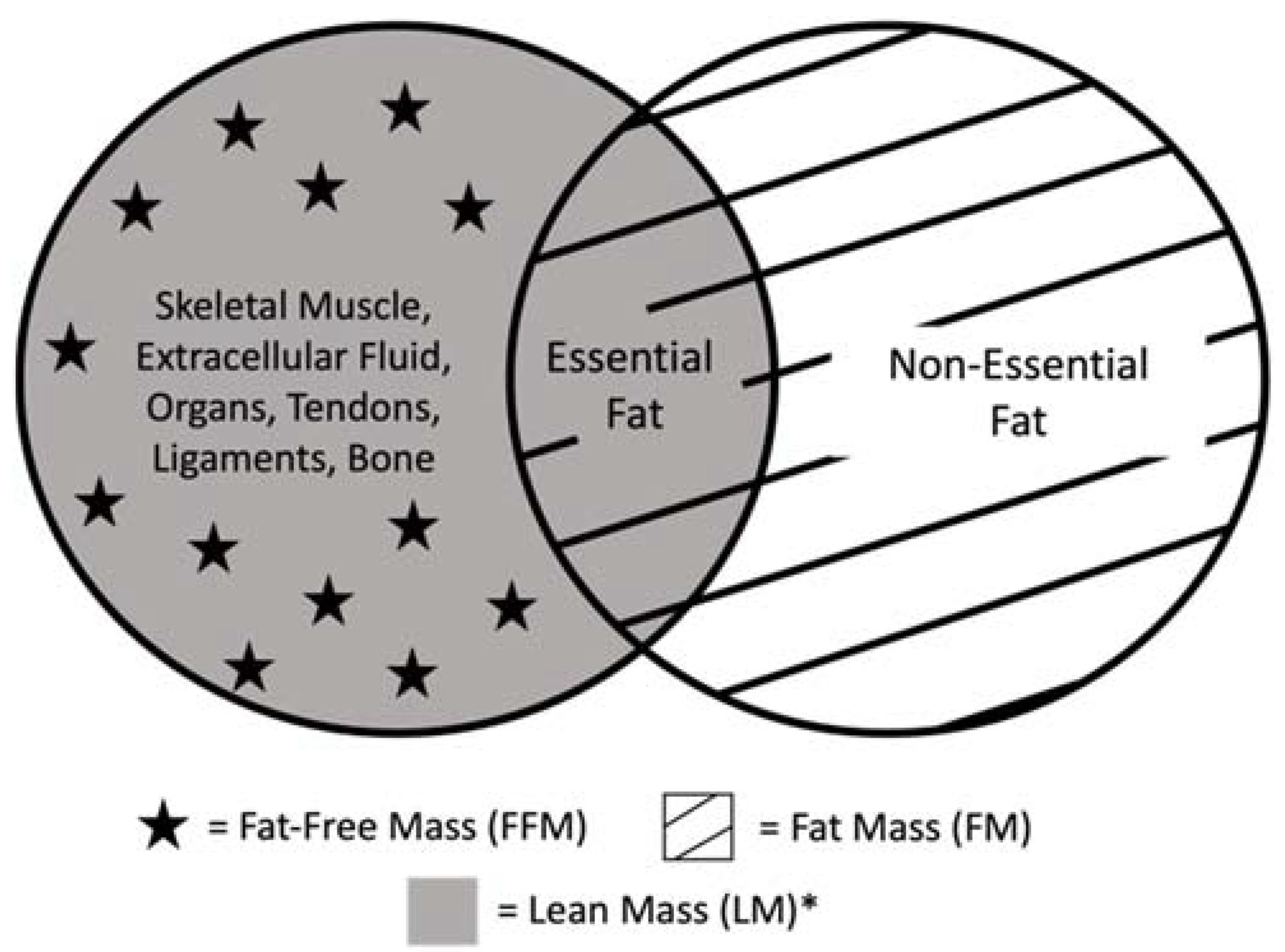
:1. Definitions of Sarcopenia
2. Changes in Muscle and Body Composition during Childhood
3. Causes of Sarcopenia in Children with Solid Organ Tumors
3.1. Disuse
3.2. Medications and Cancer Treatment
3.3. Endocrine Processes
3.4. Malnutrition and Cachexia
4. Sarcopenic Obesity
5. Frailty
6. Diagnosing Sarcopenia
6.1. Modalities Used to Measure Muscle Mass
6.2. Measuring Muscle Mass on CT and MRI
6.3. Measuring Muscle Strength and Function
7. Current Research on Sarcopenia
Sarcopenia in Children with Solid Organ Tumors
8. Conclusions and Future Outlook
Author Contributions
Funding
Conflicts of Interest
References
- Cederholm, T.; Barazzoni, R.; Austin, P.; Ballmer, P.; Biolo, G.; Bischoff, S.C.; Compher, C.; Correia, I.; Higashiguchi, T.; Holst, M.; et al. ESPEN guidelines on definitions and terminology of clinical nutrition. Clin. Nutr. 2017, 36, 49–64. [Google Scholar] [CrossRef] [PubMed]
- Cruz-Jentoft, A.J.; Bahat, G.; Bauer, J.; Boirie, Y.; Bruyère, O.; Cederholm, T.; Cooper, C.; Landi, F.; Rolland, Y.; Sayer, A.A.; et al. Sarcopenia: Revised European consensus on definition and diagnosis. Age Ageing 2019, 48, 16–31. [Google Scholar] [CrossRef] [PubMed] [Green Version]
- Argilés, J.M.; Busquets, S.; Stemmler, B.; López-Soriano, F.J. Cancer cachexia: Understanding the molecular basis. Nat. Rev. Cancer 2014, 14, 754–762. [Google Scholar] [CrossRef] [PubMed]
- Glass, D.J. Skeletal muscle hypertrophy and atrophy signaling pathways. Int. J. Biochem. Cell Biol. 2005, 37, 1974–1984. [Google Scholar] [CrossRef]
- Sandri, M. Protein breakdown in cancer cachexia. Semin. Cell Dev. Biol. 2016, 54, 11–19. [Google Scholar] [CrossRef]
- Millward, D.J.; Garlick, P.J.; Stewart, R.J.; Nnanyelugo, D.O.; Waterlow, J.C. Skeletal-muscle growth and protein turnover. Biochem. J. 1975, 150, 235–243. [Google Scholar] [CrossRef] [Green Version]
- Butte, N.F.; Hopkinson, J.M.; Wong, W.W.; Smith, E.O.; Ellis, K.J. Body composition during the first 2 years of life: An updated reference. Pediatr. Res. 2000, 47, 578–585. [Google Scholar] [CrossRef] [Green Version]
- Olhager, E.; Flinke, E.; Hannerstad, U.; Forsum, E. Studies on human body composition during the first 4 months of life using magnetic resonance imaging and isotope dilution. Pediatr. Res. 2003, 54, 906–912. [Google Scholar] [CrossRef] [Green Version]
- Mourtzakis, M.; Prado, C.M.M.; Lieffers, J.R.; Reiman, T.; McCargar, L.J.; Baracos, V.E. A practical and precise approach to quantification of body composition in cancer patients using computed tomography images acquired during routine care. Appl. Physiol. Nutr. Metab. 2008, 33, 997–1006. [Google Scholar] [CrossRef]
- Scafoglieri, A.; Clarys, J.P. Dual energy X-ray absorptiometry: Gold standard for muscle mass? J. Cachexia Sarcopenia Muscle 2018, 9, 786–787. [Google Scholar] [CrossRef]
- Prado, C.M.M.; Baracos, V.E.; McCargar, L.J.; Reiman, T.; Mourtzakis, M.; Tonkin, K.; Mackey, J.R.; Koski, S.; Pituskin, E.; Sawyer, M.B. Sarcopenia as a determinant of chemotherapy toxicity and time to tumor progression in metastatic breast cancer patients receiving capecitabine treatment. Clin. Cancer Res. 2009, 15, 2920–2926. [Google Scholar] [CrossRef] [PubMed] [Green Version]
- Verdijk, L.B.; Snijders, T.; Drost, M.; Delhaas, T.; Kadi, F.; van Loon, L.J.C. Satellite cells in human skeletal muscle; from birth to old age. Age 2014, 36, 545–547. [Google Scholar] [CrossRef] [PubMed] [Green Version]
- Rogol, A.D.; Clark, P.A.; Roemmich, J.N. Growth and pubertal development in children and adolescents: Effects of diet and physical activity. Am. J. Clin. Nutr. 2000, 72 (Suppl. S2), 521S–528S. [Google Scholar] [CrossRef]
- Arslanian, S.A.; Kalhan, S.C. Protein turnover during puberty in normal children. Am. J. Physiol. 1996, 270 Pt 1, E79–E84. [Google Scholar] [CrossRef]
- Beckett, P.R.; Jahoor, F.; Copeland, K.C. The efficiency of dietary protein utilization is increased during puberty. J. Clin. Endocrinol. Metab. 1997, 82, 2445–2449. [Google Scholar] [CrossRef] [PubMed]
- Leger, J.; Carel, C.; Legrand, I.; Paulsen, A.; Hassan, M.; Czernichow, P. Magnetic resonance imaging evaluation of adipose tissue and muscle tissue mass in children with growth hormone (GH) deficiency, Turner’s syndrome, and intrauterine growth retardation during the first year of treatment with GH. J. Clin. Endocrinol. Metab. 1994, 78, 904–909. [Google Scholar]
- Venken, K.; Movérare-Skrtic, S.; Kopchick, J.J.; Coschigano, K.T.; Ohlsson, C.; Boonen, S.; Bouillon, R.; Vanderschueren, D. Impact of androgens, growth hormone, and IGF-I on bone and muscle in male mice during puberty. J. Bone Min. Res. 2007, 22, 72–82. [Google Scholar] [CrossRef]
- Toth, M.J.; Poehlman, E.T.; Matthews, D.E.; Tchernof, A.; MacCoss, M.J. Effects of estradiol and progesterone on body composition, protein synthesis, and lipoprotein lipase in rats. Am. J. Physiol. Endocrinol. Metab. 2001, 280, E496–E501. [Google Scholar] [CrossRef]
- Weber, D.R.; Leonard, M.B.; Zemel, B.S. Body composition analysis in the pediatric population. Pediatr. Endocrinol. Rev. 2012, 10, 130–139. [Google Scholar]
- Kline, N.E.; Sevier, N. Solid tumors in children. J. Pediatr. Nurs. 2003, 18, 96–102. [Google Scholar] [CrossRef]
- Tomlinson, D.; Zupanec, S.; Jones, H.; O’Sullivan, C.; Hesser, T.; Sung, L. The lived experience of fatigue in children and adolescents with cancer: A systematic review. Support. Care Cancer 2016, 24, 3623–3631. [Google Scholar] [CrossRef] [PubMed]
- Hockenberry-Eaton, M.; Hinds, P.S. Fatigue in children and adolescents with cancer: Evolution of a program of study. Semin. Oncol. Nurs. 2000, 16, 261–272. [Google Scholar] [CrossRef] [PubMed]
- Van Deuren, S.; Boonstra, A.; van Dulmen-den Broeder, E.; Blijlevens, N.; Knoop, H.; Loonen, J. Severe fatigue after treatment for childhood cancer. Cochrane Database Syst. Rev. 2020, 3, CD012681. [Google Scholar] [CrossRef] [PubMed]
- Wilson, C.L.; Stratton, K.; Leisenring, W.L.; Oeffinger, K.C.; Nathan, P.C.; Wasilewski-Masker, K.; Hudson, M.; Castellino, S.M.; Stovall, M.; Armstrong, G.T.; et al. Decline in physical activity level in the Childhood Cancer Survivor Study cohort. Cancer Epidemiol. Biomark. Prev. 2014, 23, 1619–1627. [Google Scholar] [CrossRef] [Green Version]
- Van Dijk-Lokkart, E.M.; Steur, L.M.H.; Braam, K.I.; Veening, M.A.; Huisman, J.; Takken, T.; Bierings, M.; Merks, J.H.; Van den Heuvel-Eibrink, M.M.; Kaspers, G.J.L.; et al. Longitudinal development of cancer-related fatigue and physical activity in childhood cancer patients. Pediatr. Blood Cancer 2019, 66, e27949. [Google Scholar] [CrossRef]
- Bloomfield, S.A. Changes in musculoskeletal structure and function with prolonged bed rest. Med. Sci. Sports Exerc. 1997, 29, 197–206. [Google Scholar] [CrossRef]
- Topp, R.; Ditmyer, M.; King, K.; Doherty, K.; Hornyak, J. The effect of bed rest and potential of prehabilitation on patients in the intensive care unit. AACN Clin. Issues 2002, 13, 263–276. [Google Scholar] [CrossRef] [Green Version]
- Kortebein, P.; Symons, T.B.; Ferrando, A.; Paddon-Jones, D.; Ronsen, O.; Protas, E.; Conger, S.; Lombeida, J.; Wolfe, R.; Evans, W.J. Functional impact of 10 days of bed rest in healthy older adults. J. Gerontol. A Biol. Sci. Med. Sci. 2008, 63, 1076–1081. [Google Scholar] [CrossRef] [Green Version]
- Sakai, H.; Sagara, A.; Arakawa, K.; Sugiyama, R.; Hirosaki, A.; Takase, K.; Jo, A.; Sato, K.; Chiba, Y.; Yamazaki, M.; et al. Mechanisms of cisplatin-induced muscle atrophy. Toxicol. Appl. Pharmacol. 2014, 278, 190–199. [Google Scholar] [CrossRef]
- Damrauer, J.S.; Stadler, M.E.; Acharyya, S.; Baldwin, A.S.; Couch, M.E.; Guttridge, D.C. Chemotherapy-induced muscle wasting: Association with NF-κB and cancer cachexia. Eur. J. Transl. Myol. 2018, 28, 7590. [Google Scholar] [CrossRef] [Green Version]
- Smuder, A.J.; Kavazis, A.N.; Min, K.; Powers, S.K. Exercise protects against doxorubicin-induced oxidative stress and proteolysis in skeletal muscle. J. Appl. Physiol. 2011, 110, 935–942. [Google Scholar] [CrossRef] [PubMed] [Green Version]
- Gilliam, L.A.A.; St Clair, D.K. Chemotherapy-induced weakness and fatigue in skeletal muscle: The role of oxidative stress. Antioxid. Redox Signal. 2011, 15, 2543–2563. [Google Scholar] [CrossRef] [PubMed] [Green Version]
- Chen, J.L.; Colgan, T.D.; Walton, K.L.; Gregorevic, P.; Harrison, C.A. The TGF-β Signalling Network in Muscle Development, Adaptation and Disease. Adv. Exp. Med. Biol. 2016, 900, 97–131. [Google Scholar] [PubMed]
- Ramesh, G.; Reeves, W.B. TNF-alpha mediates chemokine and cytokine expression and renal injury in cisplatin nephrotoxicity. J. Clin. Investig. 2002, 110, 835–842. [Google Scholar] [CrossRef]
- Sultani, M.; Stringer, A.M.; Bowen, J.M.; Gibson, R.J. Anti-inflammatory cytokines: Important immunoregulatory factors contributing to chemotherapy-induced gastrointestinal mucositis. Chemother. Res. Pract. 2012, 2012, 490804. [Google Scholar] [CrossRef] [Green Version]
- Tah, P.C.; Nik Shanita, S.; Poh, B.K. Nutritional status among pediatric cancer patients: A comparison between hematological malignancies and solid tumors. J. Spec. Pediatr. Nurs. 2012, 17, 301–311. [Google Scholar] [CrossRef]
- Bauer, J.; Jürgens, H.; Frühwald, M.C. Important aspects of nutrition in children with cancer. Adv. Nutr. 2011, 2, 67–77. [Google Scholar] [CrossRef] [Green Version]
- Davis, M.P.; Panikkar, R. Sarcopenia associated with chemotherapy and targeted agents for cancer therapy. Ann. Palliat Med. 2019, 8, 86–101. [Google Scholar] [CrossRef]
- Nakamura, N.; Kishimoto, K.; Ishida, T.; Nakamura, S.; Tamura, A.; Kozaki, A.; Saito, A.; Hasegawa, D.; Kosaka, Y. Muscle mass change during chemotherapy in children with high-risk neuroblastoma: A retrospective case series of 24 patients. Eur. J. Pediatr. 2021, 180, 3265–3271. [Google Scholar] [CrossRef]
- Caiozzo, V.J.; Giedzinski, E.; Baker, M.; Suarez, T.; Izadi, A.; Lan, M.; Cho-Lim, J.; Tseng, B.P.; Limoli, C.L. The radiosensitivity of satellite cells: Cell cycle regulation, apoptosis and oxidative stress. Radiat. Res. 2010, 174, 582–589. [Google Scholar] [CrossRef] [Green Version]
- Rosenblatt, J.D.; Yong, D.; Parry, D.J. Satellite cell activity is required for hypertrophy of overloaded adult rat muscle. Muscle Nerve 1994, 17, 608–613. [Google Scholar] [CrossRef] [PubMed]
- Jurdana, M. Radiation effects on skeletal muscle. Radiol. Oncol. 2008, 42, 15–22. [Google Scholar] [CrossRef] [Green Version]
- Paulino, A.C. Late effects of radiotherapy for pediatric extremity sarcomas. Int. J. Radiat. Oncol. Biol. Phys. 2004, 60, 265–274. [Google Scholar] [CrossRef]
- Ryken, T.C.; McDermott, M.; Robinson, P.D.; Ammirati, M.; Andrews, D.W.; Asher, A.L.; Burri, S.H.; Cobbs, C.S.; Gaspar, L.E.; Kondziolka, D.; et al. The role of steroids in the management of brain metastases: A systematic review and evidence-based clinical practice guideline. J. Neurooncol. 2010, 96, 103–114. [Google Scholar] [CrossRef] [PubMed] [Green Version]
- Rostásy, K.; Wilken, B.; Baumann, M.; Müller-Deile, K.; Bieber, I.; Gärtner, J.; Möller, P.; Angelini, P.; Hero, B. High dose pulsatile dexamethasone therapy in children with opsoclonus-myoclonus syndrome. Neuropediatrics 2006, 37, 291–295. [Google Scholar] [CrossRef] [Green Version]
- De Alarcon, P.A.; Matthay, K.K.; London, W.B.; Naranjo, A.; Tenney, S.C.; Panzer, J.A.; Hogarty, M.D.; Park, J.R.; Maris, J.M.; Cohn, S.L. Intravenous immunoglobulin with prednisone and risk-adapted chemotherapy for children with opsoclonus myoclonus ataxia syndrome associated with neuroblastoma (ANBL00P3): A randomised, open-label, phase 3 trial. Lancet Child. Adolesc. Health 2018, 2, 25–34. [Google Scholar] [CrossRef]
- Braun, T.P.; Marks, D.L. The regulation of muscle mass by endogenous glucocorticoids. Front. Physiol. 2015, 6, 12. [Google Scholar] [CrossRef] [Green Version]
- Schakman, O.; Gilson, H.; Thissen, J.P. Mechanisms of glucocorticoid-induced myopathy. J. Endocrinol. 2008, 197, 1–10. [Google Scholar] [CrossRef] [PubMed] [Green Version]
- Odedra, B.R.; Bates, P.C.; Millward, D.J. Time course of the effect of catabolic doses of corticosterone on protein turnover in rat skeletal muscle and liver. Biochem. J. 1983, 214, 617–627. [Google Scholar] [CrossRef] [Green Version]
- Steiner, S.J.; Noe, J.D.; Denne, S.C. Corticosteroids increase protein breakdown and loss in newly diagnosed pediatric Crohn disease. Pediatr. Res. 2011, 70, 484–488. [Google Scholar] [CrossRef] [Green Version]
- Quattrocelli, M.; Barefield, D.Y.; Warner, J.L.; Vo, A.H.; Hadhazy, M.; Earley, J.U.; Demonbreun, A.; McNally, E.M. Intermittent glucocorticoid steroid dosing enhances muscle repair without eliciting muscle atrophy. J. Clin. Investig. 2017, 127, 2418–2432. [Google Scholar] [CrossRef] [PubMed] [Green Version]
- Mantovani, G.; Madeddu, C. Proinflammatory Cytokines: Their Role in Multifactorial Cancer Cachexia. In Cachexia and Wasting: A Modern Approach; Springer: Milano, Italy, 2006; pp. 477–482. [Google Scholar]
- Valente, V.B.; Verza, F.A.; Lopes, F.Y.K.; Ferreira, J.Z.; dos Santos, P.S.P.; Sundefeld, M.L.M.M.; Biasoli, É.R.; Miyahara, G.I.; Soubhia, A.M.P.; de Andrade, M.; et al. Stress hormones concentrations in the normal microenvironment predict risk for chemically induced cancer in rats. Psychoneuroendocrinology 2018, 89, 229–238. [Google Scholar] [CrossRef] [PubMed] [Green Version]
- Baracos, V.E.; Mazurak, V.C.; Bhullar, A.S. Cancer cachexia is defined by an ongoing loss of skeletal muscle mass. Ann. Palliat Med. 2019, 8, 3–12. [Google Scholar] [CrossRef] [PubMed]
- Wang, H.S.; Oh, D.S.; Ohning, G.V.; Pisegna, J.R. Elevated serum ghrelin exerts an orexigenic effect that may maintain body mass index in patients with metastatic neuroendocrine tumors. J. Mol. Neurosci. 2007, 33, 225–231. [Google Scholar] [CrossRef]
- Kerem, M.; Ferahkose, Z.; Yilmaz, U.T.; Pasaoglu, H.; Ofluoglu, E.; Bedirli, A.; Salman, B.; Sahin, T.T.; Akin, M. Adipokines and ghrelin in gastric cancer cachexia. World J. Gastroenterol. 2008, 14, 3633–3641. [Google Scholar] [CrossRef]
- Takahashi, M.; Terashima, M.; Takagane, A.; Oyama, K.; Fujiwara, H.; Wakabayashi, G. Ghrelin and leptin levels in cachectic patients with cancer of the digestive organs. Int. J. Clin. Oncol. 2009, 14, 315–320. [Google Scholar] [CrossRef]
- Karapanagiotou, E.M.; Polyzos, A.; Dilana, K.D.; Gratsias, I.; Boura, P.; Gkiozos, I.; Syrigos, K.N. Increased serum levels of ghrelin at diagnosis mediate body weight loss in non-small cell lung cancer (NSCLC) patients. Lung Cancer 2009, 66, 393–398. [Google Scholar] [CrossRef]
- Reano, S.; Graziani, A.; Filigheddu, N. Acylated and unacylated ghrelin administration to blunt muscle wasting. Curr. Opin. Clin. Nutr. Metab. Care 2014, 17, 236–240. [Google Scholar] [CrossRef]
- Sheriff, S.; Kadeer, N.; Joshi, R.; Friend, L.A.; James, J.H.; Balasubramaniam, A. Des-acyl ghrelin exhibits pro-anabolic and anti-catabolic effects on C2C12 myotubes exposed to cytokines and reduces burn-induced muscle proteolysis in rats. Mol. Cell Endocrinol. 2012, 351, 286–295. [Google Scholar] [CrossRef]
- Yu, A.P.; Pei, X.M.; Sin, T.K.; Yip, S.P.; Yung, B.Y.; Chan, L.W.; Wong, C.S.; Siu, P.M. Acylated and unacylated ghrelin inhibit doxorubicin-induced apoptosis in skeletal muscle. Acta Physiol. 2014, 211, 201–213. [Google Scholar] [CrossRef]
- Chance, W.T.; Xiao, C.; Dayal, R.; Sheriff, S. Alteration of NPY and Y1 receptor in dorsomedial and ventromedial areas of hypothalamus in anorectic tumor-bearing rats. Peptides 2007, 28, 295–301. [Google Scholar] [CrossRef] [PubMed]
- Chance, W.T.; Balasubramaniam, A.; Borchers, M.; Fischer, J.E. Refractory hypothalamic adenylate cyclase in anorectic tumor-bearing rats: Implications for NPY-induced feeding. Brain Res. 1995, 691, 180–184. [Google Scholar] [CrossRef]
- Moschovi, M.; Trimis, G.; Vounatsou, M.; Katsibardi, K.; Margeli, A.; Dimitriadi, F.; Papassotiriou, I.; Chrousos, G.; Tzortzatou-Stathopoulou, F. Serial plasma concentrations of PYY and ghrelin during chemotherapy in children with acute lymphoblastic leukemia. J. Pediatr. Hematol. Oncol. 2008, 30, 733–737. [Google Scholar] [CrossRef] [PubMed]
- Garcia, M.; Seelaender, M.; Sotiropoulos, A.; Coletti, D.; Lancha, A.H. Vitamin D, muscle recovery, sarcopenia, cachexia, and muscle atrophy. Nutrition 2019, 60, 66–69. [Google Scholar] [CrossRef]
- Mohr, S.B.; Gorham, E.D.; Kim, J.; Hofflich, H.; Cuomo, R.E.; Garland, C.F. Could vitamin D sufficiency improve the survival of colorectal cancer patients? J. Steroid Biochem. Mol. Biol. 2015, 148, 239–244. [Google Scholar] [CrossRef]
- Juhász, O.; Jakab, Z.; Szabó, A.; Garami, M. Examining the Vitamin D Status of Children with Solid Tumors. J. Am. Coll. Nutr. 2020, 39, 128–134. [Google Scholar] [CrossRef]
- Murphy, A.J.; White, M.; Davies, P.S.W. Body composition of children with cancer. Am. J. Clin. Nutr. 2010, 92, 55–60. [Google Scholar] [CrossRef] [Green Version]
- Fearon, K.; Strasser, F.; Anker, S.D.; Bosaeus, I.; Bruera, E.; Fainsinger, R.L.; Jatoi, A.; Loprinzi, C.; MacDonald, N.; Mantovani, G.; et al. Definition and classification of cancer cachexia: An international consensus. Lancet Oncol. 2011, 12, 489–495. [Google Scholar] [CrossRef]
- Evans, W.J.; Morley, J.E.; Argilés, J.; Bales, C.; Baracos, V.; Guttridge, D.; Jatoi, A.; Kalantar-Zadeh, K.; Lochs, H.; Mantovani, G.; et al. Cachexia: A new definition. Clin. Nutr. 2008, 27, 793–799. [Google Scholar] [CrossRef]
- Langhands, W.; Hrupka, B.J. Cytokines and Appetite. In Cytokines and Mental Health; Springer: Boston, MA, USA, 2003; Volume 7, 43p. [Google Scholar]
- Feng, P.; Jyotaki, M.; Kim, A.; Chai, J.; Simon, N.; Zhou, M.; Bachmanov, A.A.; Huang, L.; Wang, H. Regulation of bitter taste responses by tumor necrosis factor. Brain Behav. Immun. 2015, 49, 32–42. [Google Scholar] [CrossRef] [Green Version]
- Argilés, J.M.; Busquets, S.; Moore-Carrasco, R.; Figueras, M.; Almendro, V.; López-Soriano, F.J. Targets in clinical oncology: The metabolic environment of the patient. Front. Biosci. 2007, 12, 3024–3051. [Google Scholar] [CrossRef] [Green Version]
- Holland-Fischer, P.; Greisen, J.; Grøfte, T.; Jensen, T.S.; Hansen, P.O.; Vilstrup, H. Increased energy expenditure and glucose oxidation during acute nontraumatic skin pain in humans. Eur. J. Anaesthesiol. 2009, 26, 311–317. [Google Scholar] [CrossRef] [PubMed]
- Harris, R.B.S. Chronic and acute effects of stress on energy balance: Are there appropriate animal models? Am. J. Physiol. Regul. Integr. Comp. Physiol. 2015, 308, R250–R265. [Google Scholar] [CrossRef] [PubMed] [Green Version]
- Holroyde, C.P.; Skutches, C.L.; Boden, G.; Reichard, G.A. Glucose metabolism in cachectic patients with colorectal cancer. Cancer Res. 1984, 44 Pt 1, 5910–5913. [Google Scholar] [PubMed]
- Holroyde, C.P.; Gabuzda, T.G.; Putnam, R.C.; Paul, P.; Reichard, G.A. Altered glucose metabolism in metastatic carcinoma. Cancer Res. 1975, 35, 3710–3714. [Google Scholar] [PubMed]
- Antunes, D.; Padrão, A.I.; Maciel, E.; Santinha, D.; Oliveira, P.; Vitorino, R.; Moreira-Gonçalves, D.; Colaço, B.; Pires, M.J.; Nunes, C.; et al. Molecular insights into mitochondrial dysfunction in cancer-related muscle wasting. Biochim. Biophys. Acta 2014, 1841, 896–905. [Google Scholar] [CrossRef] [PubMed]
- Shum, A.M.Y.; Mahendradatta, T.; Taylor, R.J.; Painter, A.B.; Moore, M.M.; Tsoli, M.; Tan, T.C.; Clarke, S.J.; Robertson, G.R.; Polly, P. Disruption of MEF2C signaling and loss of sarcomeric and mitochondrial integrity in cancer-induced skeletal muscle wasting. Aging 2012, 4, 133–143. [Google Scholar] [CrossRef] [Green Version]
- Padrão, A.I.; Oliveira, P.; Vitorino, R.; Colaço, B.; Pires, M.J.; Márquez, M.; Castellanos, E.; Neuparth, M.J.; Teixeira, C.; Costa, C.; et al. Bladder cancer-induced skeletal muscle wasting: Disclosing the role of mitochondria plasticity. Int. J. Biochem. Cell Biol. 2013, 45, 1399–1409. [Google Scholar] [CrossRef]
- Dumas, J.-F.; Goupille, C.; Julienne, C.M.; Pinault, M.; Chevalier, S.; Bougnoux, P.; Servais, S.; Couet, C. Efficiency of oxidative phosphorylation in liver mitochondria is decreased in a rat model of peritoneal carcinosis. J. Hepatol. 2011, 54, 320–327. [Google Scholar] [CrossRef]
- Proctor, M.J.; Morrison, D.S.; Talwar, D.; Balmer, S.M.; O’Reilly, D.S.J.; Foulis, A.K.; Horgan, P.G.; McMillan, D.C. An inflammation-based prognostic score (mGPS) predicts cancer survival independent of tumour site: A Glasgow Inflammation Outcome Study. Br. J. Cancer 2011, 104, 726–734. [Google Scholar] [CrossRef] [Green Version]
- Sunny, N.E.; Parks, E.J.; Browning, J.D.; Burgess, S.C. Excessive hepatic mitochondrial TCA cycle and gluconeogenesis in humans with nonalcoholic fatty liver disease. Cell Metab. 2011, 14, 804–810. [Google Scholar] [CrossRef] [PubMed] [Green Version]
- Porporato, P.E. Understanding cachexia as a cancer metabolism syndrome. Oncogenesis 2016, 5, e200. [Google Scholar] [CrossRef] [PubMed] [Green Version]
- Tian, M.; Nishijima, Y.; Asp, M.L.; Stout, M.B.; Reiser, P.J.; Belury, M.A. Cardiac alterations in cancer-induced cachexia in mice. Int. J. Oncol. 2010, 37, 347–353. [Google Scholar] [PubMed] [Green Version]
- Eschenhagen, T.; Force, T.; Ewer, M.S.; de Keulenaer, G.W.; Suter, T.M.; Anker, S.D.; Avkiran, M.; de Azambuja, E.; Balligand, J.-L.; Brutsaert, D.L.; et al. Cardiovascular side effects of cancer therapies: A position statement from the Heart Failure Association of the European Society of Cardiology. Eur. J. Heart Fail. 2011, 13, 1–10. [Google Scholar] [CrossRef]
- Sjöström, M.; Wretling, M.L.; Karlberg, I.; Edén, E.; Lundholm, K. Ultrastructural changes and enzyme activities for energy production in hearts concomitant with tumor-associated malnutrition. J. Surg. Res. 1987, 42, 304–313. [Google Scholar] [CrossRef]
- Palus, S.; von Haehling, S.; Flach, V.C.; Tschirner, A.; Doehner, W.; Anker, S.D.; Springer, A. Simvastatin reduces wasting and improves cardiac function as well as outcome in experimental cancer cachexia. Int. J. Cardiol. 2013, 168, 3412–3418. [Google Scholar] [CrossRef]
- Zhou, X.; Wang, J.L.; Lu, J.; Song, Y.; Kwak, K.S.; Jiao, Q.; Rosenfeld, R.; Chen, Q.; Boone, T.; Simonet, W.S.; et al. Reversal of cancer cachexia and muscle wasting by ActRIIB antagonism leads to prolonged survival. Cell 2010, 142, 531–543. [Google Scholar] [CrossRef] [Green Version]
- Bernaba, B.N.; Chan, J.B.; Lai, C.K.; Fishbein, M.C. Pathology of late-onset anthracycline cardiomyopathy. Cardiovasc. Pathol. 2010, 19, 308–311. [Google Scholar] [CrossRef]
- Argilés, J.M.; Busquets, S.; Stemmler, B.; López-Soriano, F.J. Cachexia and sarcopenia: Mechanisms and potential targets for intervention. Curr. Opin. Pharmacol. 2015, 22, 100–106. [Google Scholar] [CrossRef]
- Bindels, L.B.; Beck, R.; Schakman, O.; Martin, J.C.; De Backer, F.; Sohet, F.M.; Dewulf, E.M.; Pachikian, B.D.; Neyrinck, A.M.; Thissen, J.-P.; et al. Restoring specific lactobacilli levels decreases inflammation and muscle atrophy markers in an acute leukemia mouse model. PLoS ONE 2012, 7, e37971. [Google Scholar] [CrossRef] [Green Version]
- Bindels, L.B.; Neyrinck, A.M.; Claus, S.P.; Le Roy, C.I.; Grangette, C.; Pot, B.; Martinez, I.; Walter, J.; Cani, P.D.; Delzenne, N.M. Synbiotic approach restores intestinal homeostasis and prolongs survival in leukaemic mice with cachexia. ISME J. 2016, 10, 1456–1470. [Google Scholar] [CrossRef] [PubMed]
- Pötgens, S.A.; Brossel, H.; Sboarina, M.; Catry, E.; Cani, P.D.; Neyrinck, A.M.; Delzenne, N.M.; Bindels, L.B. Klebsiella oxytoca expands in cancer cachexia and acts as a gut pathobiont contributing to intestinal dysfunction. Sci. Rep. 2018, 8, 12321. [Google Scholar] [CrossRef] [PubMed] [Green Version]
- Varian, B.J.; Gourishetti, S.; Poutahidis, T.; Lakritz, J.R.; Levkovich, T.; Kwok, C.; Teliousis, K.; Ibrahim, Y.M.; Mirabal, S.; Erdman, S.E. Beneficial bacteria inhibit cachexia. Oncotarget 2016, 7, 11803–11816. [Google Scholar] [CrossRef] [PubMed] [Green Version]
- Scales, B.S.; Dickson, R.P.; Huffnagle, G.B. A tale of two sites: How inflammation can reshape the microbiomes of the gut and lungs. J. Leukoc. Biol. 2016, 100, 943–950. [Google Scholar] [CrossRef] [Green Version]
- Larsen, J.M.; Musavian, H.S.; Butt, T.M.; Ingvorsen, C.; Thysen, A.H.; Brix, S. Chronic obstructive pulmonary disease and asthma-associated Proteobacteria, but not commensal Prevotella spp., promote Toll-like receptor 2-independent lung inflammation and pathology. Immunology 2015, 144, 333–342. [Google Scholar] [CrossRef] [Green Version]
- Ghosh, S.S.; Wang, J.; Yannie, P.J.; Ghosh, S. Intestinal Barrier Dysfunction, LPS Translocation, and Disease Development. J. Endocr. Soc. 2020, 4, bvz039. [Google Scholar] [CrossRef] [Green Version]
- Bindels, L.B.; Neyrinck, A.M.; Loumaye, A.; Catry, E.; Walgrave, H.; Cherbuy, C.; Leclercq, S.; Van Hul, M.; Plovier, H.; Pachikian, B.; et al. Increased gut permeability in cancer cachexia: Mechanisms and clinical relevance. Oncotarget 2018, 9, 18224–18238. [Google Scholar] [CrossRef] [Green Version]
- Sundström, G.M.; Wahlin, A.; Nordin-Andersson, I.; Suhr, O.B. Intestinal permeability in patients with acute myeloid leukemia. Eur. J. Haematol. 1998, 61, 250–254. [Google Scholar] [CrossRef]
- Prado, C.M.M.; Wells, J.C.K.; Smith, S.R.; Stephan, B.C.M.; Siervo, M. Sarcopenic obesity: A Critical appraisal of the current evidence. Clin. Nutr. 2012, 31, 583–601. [Google Scholar] [CrossRef]
- Anandavadivelan, P.; Brismar, T.B.; Nilsson, M.; Johar, A.M.; Martin, L. Sarcopenic obesity: A probable risk factor for dose limiting toxicity during neo-adjuvant chemotherapy in oesophageal cancer patients. Clin. Nutr. 2016, 35, 724–730. [Google Scholar] [CrossRef]
- Zhang, W.-T.; Lin, J.; Chen, W.-S.; Huang, Y.-S.; Wu, R.-S.; Chen, X.-D.; Lou, N.; Chi, C.-H.; Hu, C.-Y.; Shen, X. Sarcopenic Obesity Is Associated with Severe Postoperative Complications in Gastric Cancer Patients Undergoing Gastrectomy: A Prospective Study. J. Gastrointest. Surg. 2018, 22, 1861–1869. [Google Scholar] [CrossRef]
- Mintziras, I.; Miligkos, M.; Wächter, S.; Manoharan, J.; Maurer, E.; Bartsch, D.K. Sarcopenia and sarcopenic obesity are significantly associated with poorer overall survival in patients with pancreatic cancer: Systematic review and meta-analysis. Int. J. Surg. 2018, 59, 19–26. [Google Scholar] [CrossRef] [PubMed]
- Orgel, E.; Mueske, N.M.; Sposto, R.; Gilsanz, V.; Freyer, D.R.; Mittelman, S.D. Limitations of body mass index to assess body composition due to sarcopenic obesity during leukemia therapy. Leuk. Lymphoma 2018, 59, 138–145. [Google Scholar] [CrossRef] [PubMed]
- Marriott, C.J.C.; Beaumont, L.F.; Farncombe, T.H.; Cranston, A.N.; Athale, U.H.; Yakemchuk, V.N.; Webber, C.E.; Barr, R.D. Body composition in long-term survivors of acute lymphoblastic leukemia diagnosed in childhood and adolescence: A focus on sarcopenic obesity. Cancer 2018, 124, 1225–1231. [Google Scholar] [CrossRef] [PubMed] [Green Version]
- Malhotra, P.; Kapoor, G.; Jain, S.; Jain, S.; Sharma, A. Obesity and Sarcopenia in Survivors of Childhood Acute Lymphoblastic Leukemia. Indian Pediatr. 2021, 58, 436–440. [Google Scholar] [CrossRef] [PubMed]
- Nakayama, H.; Noguchi, M.; Fukano, R.; Ueda, T.; Taguchi, S.; Yoshimaru, K.; Namie, M.; Shimokawa, M.; Okamura, J. Sarcopenia and obesity in long-term survivors of childhood leukemia/lymphoma: A report from a single institution. Jpn. J. Clin. Oncol. 2021, 51, 1100–1106. [Google Scholar] [CrossRef] [PubMed]
- Murphy, A.J.; White, M.; Elliott, S.A.; Lockwood, L.; Hallahan, A.; Davies, P.S. Body composition of children with cancer during treatment and in survivorship. Am. J. Clin. Nutr. 2015, 102, 891–896. [Google Scholar] [CrossRef] [PubMed] [Green Version]
- Fried, L.P.; Tangen, C.M.; Walston, J.; Newman, A.B.; Hirsch, C.; Gottdiener, J.; Seeman, T.; Tracy, R.; Kop, W.J.; Burke, G.; et al. Frailty in older adults: Evidence for a phenotype. J. Gerontol. A Biol. Sci. Med. Sci. 2001, 56, M146–M156. [Google Scholar] [CrossRef]
- Ethun, C.G.; Bilen, M.A.; Jani, A.B.; Maithel, S.K.; Ogan, K.; Master, V.A. Frailty and cancer: Implications for oncology surgery, medical oncology, and radiation oncology. CA Cancer J. Clin. 2017, 67, 362–377. [Google Scholar] [CrossRef] [Green Version]
- Lurz, E.; Quammie, C.; Englesbe, M.; Alonso, E.M.; Lin, H.C.; Hsu, E.K.; Furuya, K.N.; Gupta, N.A.; Venkat, V.L.; Daniel, J.F.; et al. Frailty in Children with Liver Disease: A Prospective Multicenter Study. J. Pediatr. 2018, 194, 109–115.e4. [Google Scholar] [CrossRef]
- Panchangam, C.; White, D.A.; Goudar, S.; Birnbaum, B.; Malloy-Walton, L.; Gross-Toalson, J.; Reid, K.J.; Shirali, G.; Parthiban, A. Translation of the Frailty Paradigm from Older Adults to Children with Cardiac Disease. Pediatr. Cardiol. 2020, 41, 1031–1041. [Google Scholar] [CrossRef] [PubMed]
- Trowbridge, F.L.; Hiner, C.D.; Robertson, A.D. Arm muscle indicators and creatinine excretion in children. Am. J. Clin. Nutr. 1982, 36, 691–696. [Google Scholar] [CrossRef] [PubMed] [Green Version]
- Jacobs, J.; Jansen, M.; Janssen, H.; Raijmann, W.; van Alfen, N.; Pillen, S. Quantitative muscle ultrasound and muscle force in healthy children: A 4-year follow-up study. Muscle Nerve 2013, 47, 856–863. [Google Scholar] [CrossRef] [PubMed]
- McCarthy, H.D.; Samani-Radia, D.; Jebb, S.A.; Prentice, A.M. Skeletal muscle mass reference curves for children and adolescents. Pediatr. Obes. 2014, 9, 249–259. [Google Scholar] [CrossRef]
- Guo, M.; Zemel, B.S.; Hawkes, C.P.; Long, J.; Kelly, A.; Leonard, M.B.; Jaramillo, D.; Mostoufi-Moab, S. Sarcopenia and preserved bone mineral density in paediatric survivors of high-risk neuroblastoma with growth failure. J. Cachexia Sarcopenia Muscle 2021, 12, 1024–1033. [Google Scholar] [CrossRef]
- Lurz, E.; Patel, H.; Frimpong, R.G.; Ricciuto, A.; Kehar, M.; Wales, P.W.; Towbin, A.J.; Chavhan, G.B.; Kamath, B.M.; Ng, V.L. Sarcopenia in Children with End-Stage Liver Disease. J. Pediatr. Gastroenterol. Nutr. 2018, 66, 222–226. [Google Scholar] [CrossRef]
- Ritz, A.; Kolorz, J.; Hubertus, J.; Ley-Zaporozhan, J.; von Schweinitz, D.; Koletzko, S.; Häberle, B.; Schmid, I.; Kappler, R.; Berger, M.; et al. Sarcopenia is a prognostic outcome marker in children with high-risk hepatoblastoma. Pediatr. Blood Cancer 2021, 4, e28862. [Google Scholar] [CrossRef]
- Ritz, A.; Froeba-Pohl, A.; Kolorz, J.; Vigodski, V.; Hubertus, J.; Ley-Zaporozhan, J.; von Schweinitz, D.; Häberle, B.; Schmid, I.; Kappler, R.; et al. Total Psoas Muscle Area as a Marker for Sarcopenia Is Related to Outcome in Children with Neuroblastoma. Front. Surg. 2021, 8, 333. [Google Scholar] [CrossRef]
- Mazahery, H.; von Hurst, P.R.; McKinlay, C.J.D.; Cormack, B.E.; Conlon, C.A. Air displacement plethysmography (pea pod) in full-term and pre-term infants: A comprehensive review of accuracy, reproducibility, and practical challenges. Matern. Health Neonatol. Perinatol. 2018, 4, 12. [Google Scholar] [CrossRef]
- Barbosa-Cortés, L.; Tapia-Rojas, M.; López-Aguilar, E.; Mejía-Aranguré, J.M.; Rivera-Márquez, H. Body composition by dilution of deuterium oxide in Mexican children with lymphoma and solid tumors. Nutrition 2007, 23, 739–744. [Google Scholar] [CrossRef]
- Clark, R.V.; Walker, A.C.; Miller, R.R.; O’Connor-Semmes, R.L.; Ravussin, E.; Cefalu, W.T. Creatine(methyl-d3) dilution in urine for estimation of total body skeletal muscle mass: Accuracy and variability vs. MRI and DXA. J. Appl. Physiol. 2018, 124, 1–9. [Google Scholar] [CrossRef] [Green Version]
- Wang, Z.; Heshka, S.; Pietrobelli, A.; Chen, Z.; Silva, A.M.; Sardinha, L.B.; Wang, J.; Gallager, D.; Heymsfield, S.B. A new total body potassium method to estimate total body skeletal muscle mass in children. J. Nutr. 2007, 137, 1988–1991. [Google Scholar] [CrossRef] [PubMed]
- Arrowsmith, F.E.; Allen, J.R.; Gaskin, K.J.; Gruca, M.A.; Clarke, S.L.; Briody, J.N.; Howman-Giles, R.B.; Somerville, H.; O’Loughlin, E.V. Reduced body protein in children with spastic quadriplegic cerebral palsy. Am. J. Clin. Nutr. 2006, 83, 613–618. [Google Scholar] [CrossRef]
- Vatanen, A.; Hou, M.; Huang, T.; Söder, O.; Jahnukainen, T.; Kurimo, M.; Ojala, T.H.; Sarkola, T.; Turanlahti, M.; Saarinen-Pihkala, U.M.; et al. Clinical and biological markers of premature aging after autologous SCT in childhood cancer. Bone Marrow Transplant. 2017, 52, 600–605. [Google Scholar] [CrossRef]
- Kawakubo, N.; Kinoshita, Y.; Souzaki, R.; Koga, Y.; Oba, U.; Ohga, S.; Taguchi, T. The Influence of Sarcopenia on High-Risk Neuroblastoma. J. Surg. Res. 2019, 236, 101–105. [Google Scholar] [CrossRef] [PubMed]
- Joffe, L.; Shen, W.; Shadid, G.; Jin, Z.; Ladas, E.J. Skeletal muscle and adipose tissue changes in the first phase of treatment of pediatric solid tumors. Cancer Med. 2021, 10, 15–22. [Google Scholar] [CrossRef] [PubMed]
- IJpma, I.; Lequin, M.H.; Nievelstein, R.A.J.; Fiocco, M.; Tissing, W.J.E. Body composition of patients with neuroblastoma using computed tomography. Pediatr. Blood Cancer 2021, 68, e29337. [Google Scholar] [CrossRef]
- Romano, A.; Triarico, S.; Rinninella, E.; Natale, L.; Brizi, M.G.; Cintoni, M.; Raoul, P.; Maurizi, P.; Attinà, G.; Mastrangelo, S.; et al. Clinical Impact of Nutritional Status and Sarcopenia in Pediatric Patients with Bone and Soft Tissue Sarcomas: A Pilot Retrospective Study (SarcoPed). Nutrients 2022, 14, 383. [Google Scholar] [CrossRef]
- Behling, E.B.; Camelo Júnior, J.S.; Ferriolli, E.; Pfrimer, K.; Monteiro, J.P. Nutritional status in children with cancer: Comparison of deuterium oxide dilution with bioelectric impedance analysis and anthropometry. Rev. Paul. Pediatr. 2021, 39, e2019209. [Google Scholar] [CrossRef]
- Webber, C.; Halton, J.; Walker, S.; Young, A.; Barr, R.D. The prediction of lean body mass and fat mass from arm anthropometry at diagnosis in children with cancer. J. Pediatr. Hematol. Oncol. 2013, 35, 530–533. [Google Scholar] [CrossRef]
- Kuriyan, R.; Thomas, T.; Kurpad, A.V. Total body muscle mass estimation from bioelectrical impedance analysis & simple anthropometric measurements in Indian men. Indian J. Med. Res. 2008, 127, 441–446. [Google Scholar] [PubMed]
- Chomtho, S.; Fewtrell, M.S.; Jaffe, A.; Williams, J.E.; Wells, J.C.K. Evaluation of Arm Anthropometry for Assessing Pediatric Body Composition: Evidence from Healthy and Sick Children. Pediatr. Res. 2006, 59, 860–865. [Google Scholar] [CrossRef] [PubMed]
- Collins, L.; Beaumont, L.; Cranston, A.; Savoie, S.; Nayiager, T.; Barr, R. Anthropometry in Long-Term Survivors of Acute Lymphoblastic Leukemia in Childhood and Adolescence. J. Adolesc. Young Adult Oncol. 2017, 6, 294–298. [Google Scholar] [CrossRef] [PubMed]
- Bakshi, N.; Singh, K. Nutrition assessment and its effect on various clinical variables among patients undergoing liver transplant. Hepatobiliary Surg. Nutr. 2016, 5, 358–371. [Google Scholar] [CrossRef] [Green Version]
- Verhagen, M.V.; Levolger, S.; Hulshoff, J.B.; Werner, M.J.M.; van der Doef, H.P.J.; Viddeleer, A.R.; de Kleine, R.H.; de Haas, R.J. Utility of Preoperative Computed Tomography-Based Body Metrics in Relation to Postoperative Complications in Pediatric Liver Transplantation Recipients. Liver Transpl. 2021, 27, 1779–1787. [Google Scholar] [CrossRef]
- Tillquist, M.; Kutsogiannis, D.J.; Wischmeyer, P.E.; Kummerlen, C.; Leung, R.; Stollery, D.; Karvellas, C.J.; Preiser, J.-C.; Bird, N.; Kozar, R.; et al. Bedside ultrasound is a practical and reliable measurement tool for assessing quadriceps muscle layer thickness. JPEN J. Parenter. Enter. Nutr. 2014, 38, 886–890. [Google Scholar] [CrossRef] [Green Version]
- Mayans, D.; Cartwright, M.S.; Walker, F.O. Neuromuscular ultrasonography: Quantifying muscle and nerve measurements. Phys. Med. Rehabil. Clin. N. Am. 2012, 23, 133–148. [Google Scholar] [CrossRef] [Green Version]
- Pillen, S.; Verrips, A.; van Alfen, N.; Arts, I.M.P.; Sie, L.T.L.; Zwarts, M.J. Quantitative skeletal muscle ultrasound: Diagnostic value in childhood neuromuscular disease. Neuromuscul. Disord. 2007, 17, 509–516. [Google Scholar] [CrossRef]
- Walter-Kroker, A.; Kroker, A.; Mattiucci-Guehlke, M.; Glaab, T. A practical guide to bioelectrical impedance analysis using the example of chronic obstructive pulmonary disease. Nutr. J. 2011, 10, 35–38. [Google Scholar] [CrossRef] [Green Version]
- Buckinx, F.; Landi, F.; Cesari, M.; Fielding, R.A.; Visser, M.; Engelke, K.; Maggi, S.; Dennison, E.; Al-Daghri, N.M.; Allepaerts, S.; et al. Pitfalls in the measurement of muscle mass: A need for a reference standard. J. Cachexia Sarcopenia Muscle 2018, 9, 269–278. [Google Scholar] [CrossRef]
- Heymsfield, S.B.; McManus, C.; Stevens, V.; Smith, J. Muscle mass: Reliable indicator of protein-energy malnutrition severity and outcome. Am. J. Clin. Nutr. 1982, 35 (Suppl. S5), 1192–1199. [Google Scholar] [CrossRef] [PubMed] [Green Version]
- Selberg, O.; Selberg, D. Norms and correlates of bioimpedance phase angle in healthy human subjects, hospitalized patients, and patients with liver cirrhosis. Eur. J. Appl. Physiol. 2002, 86, 509–516. [Google Scholar] [CrossRef] [Green Version]
- Belarmino, G.; Gonzalez, M.C.; Torrinhas, R.S.; Sala, P.; Andraus, W.; D’Albuquerque, L.A.C.; Pereira, R.M.R.; Caparbo, V.F.; Ravacci, G.R.; Damiani, L.; et al. Phase angle obtained by bioelectrical impedance analysis independently predicts mortality in patients with cirrhosis. World J. Hepatol. 2017, 9, 401–408. [Google Scholar] [CrossRef] [PubMed]
- Fields, D.A.; Higgins, P.B.; Hunter, G.R. Assessment of body composition by air-displacement plethysmography: Influence of body temperature and moisture. Dyn. Med. 2004, 3, 3–7. [Google Scholar] [CrossRef] [Green Version]
- Murphy-Alford, A.J.; White, M.; Lockwood, L.; Hallahan, A.; Davies, P.S.W. Body composition, dietary intake and physical activity of young survivors of childhood cancer. Clin. Nutr. 2019, 38, 842–847. [Google Scholar] [CrossRef] [PubMed] [Green Version]
- Proctor, D.N.; O’Brien, P.C.; Atkinson, E.J.; Nair, K.S. Comparison of techniques to estimate total body skeletal muscle mass in people of different age groups. Am. J. Physiol. 1999, 277, E489–E495. [Google Scholar] [CrossRef] [PubMed]
- Bredella, M.A.; Ghomi, R.H.; Thomas, B.J.; Torriani, M.; Brick, D.J.; Gerweck, A.V.; Misra, M.; Klibanski, A.; Miller, K.K. Comparison of DXA and CT in the assessment of body composition in premenopausal women with obesity and anorexia nervosa. Obesity 2010, 18, 2227–2233. [Google Scholar] [CrossRef] [Green Version]
- Kim, J.; Shen, W.; Gallagher, D.; Jones, A.; Wang, Z.; Wang, J.; Heshka, S.; Heymsfield, S.B. Total-body skeletal muscle mass: Estimation by dual-energy X-ray absorptiometry in children and adolescents. Am. J. Clin. Nutr. 2006, 84, 1014–1020. [Google Scholar] [CrossRef]
- Webber, C.E.; Barr, R.D. Age- and gender-dependent values of skeletal muscle mass in healthy children and adolescents. J. Cachexia Sarcopenia Muscle 2012, 3, 25–29. [Google Scholar] [CrossRef] [Green Version]
- Kim, K.; Hong, S.; Kim, E.Y. Reference Values of Skeletal Muscle Mass for Korean Children and Adolescents Using Data from the Korean National Health and Nutrition Examination Survey 2009–2011. PLoS ONE 2016, 11, e0153383. [Google Scholar] [CrossRef]
- Liu, J.; Yan, Y.; Xi, B.; Huang, G.; Mi, J. China Child and Adolescent Cardiovascular Health (CCACH) Study Group. Skeletal muscle reference for Chinese children and adolescents. J. Cachexia Sarcopenia Muscle 2019, 10, 155–164. [Google Scholar] [CrossRef] [PubMed]
- Ofenheimer, A.; Breyer-Kohansal, R.; Hartl, S.; Burghuber, O.C.; Krach, F.; Schrott, A.; Franssen, F.M.E.; Wouters, E.F.M.; Breyer, M.-K. Reference charts for body composition parameters by dual-energy X-ray absorptiometry in European children and adolescents aged 6 to 18 years-Results from the Austrian LEAD (Lung, hEart, sociAl, boDy) cohort. Pediatr. Obes. 2021, 16, e12695. [Google Scholar] [CrossRef]
- Clark, P.; Denova-Gutiérrez, E.; Ambrosi, R.; Szulc, P.; Rivas-Ruiz, R.; Salmerón, J. Reference Values of Total Lean Mass, Appendicular Lean Mass, and Fat Mass Measured with Dual-Energy X-ray Absorptiometry in a Healthy Mexican Population. Calcif. Tissue Int. 2016, 99, 462–471. [Google Scholar] [CrossRef] [PubMed]
- Heymsfield, S.B.; Gonzalez, M.C.; Lu, J.; Jia, G.; Zheng, J. Skeletal muscle mass and quality: Evolution of modern measurement concepts in the context of sarcopenia. Proc. Nutr. Soc. 2015, 74, 355–366. [Google Scholar] [CrossRef] [PubMed] [Green Version]
- Chianca, V.; Albano, D.; Messina, C.; Gitto, S.; Ruffo, G.; Guarino, S.; Del Grande, F.; Sconfienza, L.M. Sarcopenia: Imaging assessment and clinical application. Abdom. Radiol. 2021. online ahead of print. [Google Scholar]
- Faron, A.; Sprinkart, A.M.; Kuetting, D.L.R.; Feisst, A.; Isaak, A.; Endler, C.; Chang, J.; Nowak, S.; Block, W.; Thomas, D.; et al. Body composition analysis using CT and MRI: Intra-individual intermodal comparison of muscle mass and myosteatosis. Sci. Rep. 2020, 10, 11765. [Google Scholar] [CrossRef] [PubMed]
- Klopfenstein, B.J.; Kim, M.S.; Krisky, C.M.; Szumowski, J.; Rooney, W.D.; Purnell, J.Q. Comparison of 3 T MRI and CT for the measurement of visceral and subcutaneous adipose tissue in humans. Br. J. Radiol. 2012, 85, e826–e830. [Google Scholar] [CrossRef] [PubMed] [Green Version]
- Eloi, J.C.; Epifanio, M.; de Gonçalves, M.M.; Pellicioli, A.; Vieira, P.F.G.; Dias, H.B.; Bruscato, N.; Soder, R.B.; Santana, J.C.B.; Mouzaki, M.; et al. Quantification of Abdominal Fat in Obese and Healthy Adolescents Using 3 Tesla Magnetic Resonance Imaging and Free Software for Image Analysis. PLoS ONE 2017, 12, e0167625. [Google Scholar] [CrossRef]
- Marunowski, K.; Świętoń, D.; Bzyl, W.; Grzywińska, M.; Kaszubowski, M.; Bandosz, P.; Khrichenko, D.; Piskunowicz, M. MRI-Derived Subcutaneous and Visceral Adipose Tissue Reference Values for Children Aged 6 to Under 18 Years. Front. Nutr. 2021, 8, 757274. [Google Scholar] [CrossRef]
- Baker, S.T.; Strauss, B.J.; Prendergast, L.A.; Panagiotopoulos, S.; Thomas, G.E.; Vu, T.; Proietto, J.; Jerums, G. Estimating dual-energy X-ray absorptiometry-derived total body skeletal muscle mass using single-slice abdominal magnetic resonance imaging in obese subjects with and without diabetes: A pilot study. Eur. J. Clin. Nutr. 2012, 66, 628–632. [Google Scholar] [CrossRef] [Green Version]
- Shen, W.; Punyanitya, M.; Wang, Z.; Gallagher, D.; St-Onge, M.-P.; Albu, J.; Heymsfield, S.B.; Heshka, S. Total body skeletal muscle and adipose tissue volumes: Estimation from a single abdominal cross-sectional image. J. Appl. Physiol. 2004, 97, 2333–2338. [Google Scholar] [CrossRef] [Green Version]
- Schweitzer, L.; Geisler, C.; Pourhassan, M.; Braun, W.; Glüer, C.-C.; Bosy-Westphal, A.; Müller, M.J. What is the best reference site for a single MRI slice to assess whole-body skeletal muscle and adipose tissue volumes in healthy adults? Am. J. Clin. Nutr. 2015, 102, 58–65. [Google Scholar] [CrossRef] [PubMed] [Green Version]
- Van Vugt, J.L.A.; Levolger, S.; de Bruin, R.W.F.; van Rosmalen, J.; Metselaar, H.J.; IJzermans, J.N.M. Systematic Review and Meta-Analysis of the Impact of Computed Tomography-Assessed Skeletal Muscle Mass on Outcome in Patients Awaiting or Undergoing Liver Transplantation. Am. J. Transplant. 2016, 16, 2277–2292. [Google Scholar] [CrossRef] [PubMed]
- Cao, Q.; Xiong, Y.; Zhong, Z.; Ye, Q. Computed Tomography-Assessed Sarcopenia Indexes Predict Major Complications following Surgery for Hepatopancreatobiliary Malignancy: A Meta-Analysis. Ann. Nutr. Metab. 2019, 74, 24–34. [Google Scholar] [CrossRef] [PubMed]
- Peterson, C.M.; Su, H.; Thomas, D.M.; Heo, M.; Golnabi, A.H.; Pietrobelli, A.; Heymsfield, S.B. Tri-Ponderal Mass Index vs Body Mass Index in Estimating Body Fat During Adolescence. JAMA Pediatr. 2017, 171, 629–636. [Google Scholar] [CrossRef]
- Cole, T.J. Weight/heightp compared to weight/height2 for assessing adiposity in childhood: Influence of age and bone age on p during puberty. Ann. Hum. Biol. 1986, 13, 433–451. [Google Scholar] [CrossRef] [PubMed]
- Hawkins, R.B.; Mehaffey, J.H.; Charles, E.J.; Kern, J.A.; Lim, D.S.; Teman, N.R.; Ailawadi, G. Psoas Muscle Size Predicts Risk-Adjusted Outcomes After Surgical Aortic Valve Replacement. Ann. Thorac. Surg. 2018, 106, 39–45. [Google Scholar] [CrossRef] [Green Version]
- Zuckerman, J.; Ades, M.; Mullie, L.; Trnkus, A.; Morin, J.-F.; Langlois, Y.; Ma, F.; Levental, M.; Morais, J.A.; Afilalo, J. Psoas Muscle Area and Length of Stay in Older Adults Undergoing Cardiac Operations. Ann. Thorac. Surg. 2017, 103, 1498–1504. [Google Scholar] [CrossRef] [Green Version]
- Jones, K.I.; Doleman, B.; Scott, S.; Lund, J.N.; Williams, J.P. Simple psoas cross-sectional area measurement is a quick and easy method to assess sarcopenia and predicts major surgical complications. Colorectal Dis. 2015, 17, O20–O26. [Google Scholar] [CrossRef] [Green Version]
- López, J.J.; Cooper, J.N.; Albert, B.; Adler, B.; King, D.; Minneci, P.C. Sarcopenia in children with perforated appendicitis. J. Surg. Res. 2017, 220, 1–5. [Google Scholar] [CrossRef]
- Park, S.Y.; Yoon, J.-K.; Lee, S.J.; Haam, S.; Jung, J. Postoperative change of the psoas muscle area as a predictor of survival in surgically treated esophageal cancer patients. J. Thorac. Dis. 2017, 9, 355–361. [Google Scholar] [CrossRef] [Green Version]
- Peng, P.; Hyder, O.; Firoozmand, A.; Kneuertz, P.; Schulick, R.D.; Huang, D.; Makary, M.; Hirose, K.; Edil, B.; Choti, M.A.; et al. Impact of Sarcopenia on Outcomes Following Resection of Pancreatic Adenocarcinoma. J. Gastrointest. Surg. 2012, 16, 1478–1486. [Google Scholar] [CrossRef] [Green Version]
- Lieffers, J.R.; Bathe, O.F.; Fassbender, K.; Winget, M.; Baracos, V.E. Sarcopenia is associated with postoperative infection and delayed recovery from colorectal cancer resection surgery. Br. J. Cancer 2012, 107, 931–936. [Google Scholar] [CrossRef] [PubMed] [Green Version]
- Hua, X.; Liu, S.; Liao, J.-F.; Wen, W.; Long, Z.-Q.; Lu, Z.-J.; Guo, L.; Lin, H.-X. When the Loss Costs Too Much: A Systematic Review and Meta-Analysis of Sarcopenia in Head and Neck Cancer. Front. Oncol. 2019, 9, 1561. [Google Scholar] [CrossRef] [PubMed]
- Wong, A.; Zhu, D.; Kraus, D.; Tham, T. Radiologically Defined Sarcopenia Affects Survival in Head and Neck Cancer: A Meta-Analysis. Laryngoscope 2021, 131, 333–341. [Google Scholar] [CrossRef]
- Trejo-Avila, M.; Bozada-Gutiérrez, K.; Valenzuela-Salazar, C.; Herrera-Esquivel, J.; Moreno-Portillo, M. Sarcopenia predicts worse postoperative outcomes and decreased survival rates in patients with colorectal cancer: A systematic review and meta-analysis. Int. J. Colorectal Dis. 2021, 36, 1077–1096. [Google Scholar] [CrossRef] [PubMed]
- Harbaugh, C.M.; Zhang, P.; Henderson, B.; Derstine, B.A.; Holcombe, S.A.; Wang, S.C.; Kohoyda-Inglis, C.; Ehrlich, P.F. Personalized medicine: Enhancing our understanding of pediatric growth with analytic morphomics. J. Pediatr. Surg. 2017, 52, 837–842. [Google Scholar] [CrossRef] [PubMed]
- Lurz, E.; Patel, H.; Lebovic, G.; Quammie, C.; Woolfson, J.P.; Perez, M.; Ricciuto, A.; Wales, P.W.; Kamath, B.M.; Chavhan, G.B.; et al. Paediatric reference values for total psoas muscle area. J. Cachexia Sarcopenia Muscle 2020, 11, 405–414. [Google Scholar] [CrossRef]
- Derstine, B.A.; Holcombe, S.A.; Goulson, R.L.; Ross, B.E.; Wang, N.C.; Sullivan, J.A.; Su, G.L.; Wang, S.C. Quantifying Sarcopenia Reference Values Using Lumbar and Thoracic Muscle Areas in a Healthy Population. J. Nutr. Health Aging 2017, 21, 180–185. [Google Scholar] [CrossRef]
- Metzger, G.A.; Sebastião, Y.V.; Carsel, A.C.; Nishimura, L.; Fisher, J.G.; Deans, K.J.; Minneci, P.C. Establishing Reference Values for Lean Muscle Mass in the Pediatric Patient. J. Pediatr. Gastroenterol. Nutr. 2020, 72, 316–323. [Google Scholar] [CrossRef]
- Rutten, I.J.G.; Ubachs, J.; Kruitwagen, R.F.P.M.; Beets-Tan, R.G.H.; Olde Damink, S.W.M.; van Gorp, T. Psoas muscle area is not representative of total skeletal muscle area in the assessment of sarcopenia in ovarian cancer. J. Cachexia Sarcopenia Muscle 2017, 8, 630–638. [Google Scholar] [CrossRef] [Green Version]
- Baracos, V.E. Psoas as a sentinel muscle for sarcopenia: A flawed premise. J. Cachexia Sarcopenia Muscle 2017, 8, 527–528. [Google Scholar] [CrossRef] [PubMed]
- Schneider, C.A.; Rasband, W.S.; Eliceiri, K.W. NIH Image to ImageJ: 25 years of image analysis. Nat. Methods 2012, 9, 671–675. [Google Scholar] [CrossRef] [PubMed]
- Van Vugt, J.L.A.; Levolger, S.; Gharbharan, A.; Koek, M.; Niessen, W.J.; Burger, J.W.A.; Willemsen, S.P.; de Bruin, R.W.F.; IJzermans, J.N.M. A comparative study of software programmes for cross-sectional skeletal muscle and adipose tissue measurements on abdominal computed tomography scans of rectal cancer patients. J. Cachexia Sarcopenia Muscle 2017, 8, 285–297. [Google Scholar] [CrossRef] [PubMed]
- Rollins, K.E.; Awwad, A.; Macdonald, I.A.; Lobo, D.N. A comparison of two different software packages for analysis of body composition using computed tomography images. Nutrition 2019, 57, 92–96. [Google Scholar] [CrossRef] [PubMed]
- Joffe, L.; Dwyer, S.; Glade Bender, J.L.; Frazier, A.L.; Ladas, E.J. Nutritional status and clinical outcomes in pediatric patients with solid tumors: A systematic review of the literature. Semin. Oncol. 2019, 46, 48–56. [Google Scholar] [CrossRef] [PubMed]
- Ha, J.; Park, T.; Kim, H.-K.; Shin, Y.; Ko, Y.; Kim, D.W.; Sung, Y.S.; Lee, J.; Ham, S.J.; Khang, S.; et al. Development of a fully automatic deep learning system for L3 selection and body composition assessment on computed tomography. Sci. Rep. 2021, 11, 21656. [Google Scholar] [CrossRef]
- Lenchik, L.; Barnard, R.; Boutin, R.D.; Kritchevsky, S.B.; Chen, H.; Tan, J.; Cawthon, P.M.; Weaver, A.A.; Hsu, F.-C. Automated Muscle Measurement on Chest CT Predicts All-Cause Mortality in Older Adults From the National Lung Screening Trial. J. Gerontol. A Biol. Sci. Med. Sci. 2021, 76, 277–285. [Google Scholar] [CrossRef]
- Barnard, R.; Tan, J.; Roller, B.; Chiles, C.; Weaver, A.A.; Boutin, R.D.; Kritchevsky, S.B.; Lenchik, L. Machine Learning for Automatic Paraspinous Muscle Area and Attenuation Measures on Low-Dose Chest CT Scans. Acad. Radiol. 2019, 26, 1686–1694. [Google Scholar] [CrossRef]
- Kim, Y.J. Machine Learning Models for Sarcopenia Identification Based on Radiomic Features of Muscles in Computed Tomography. Int. J. Environ. Res. Public Health 2021, 18, 8710. [Google Scholar] [CrossRef]
- Häger-Ross, C.; Rösblad, B. Norms for grip strength in children aged 4–16 years. Acta Paediatr. 2002, 91, 617–625. [Google Scholar] [CrossRef]
- Mckirdy, S.; Nichols, B.; Williamson, S.; Gerasimidis, K. Handgrip strength as a surrogate marker of lean mass and risk of malnutrition in paediatric patients. Clin. Nutr. 2021, 40, 5189–5195. [Google Scholar] [CrossRef] [PubMed]
- Ploegmakers, J.J.W.; Hepping, A.M.; Geertzen, J.H.B.; Bulstra, S.K.; Stevens, M. Grip strength is strongly associated with height, weight and gender in childhood: A cross sectional study of 2241 children and adolescents providing reference values. J. Physiother. 2013, 59, 255–261. [Google Scholar] [CrossRef] [Green Version]
- Geiger, R.; Strasak, A.; Treml, B.; Gasser, K.; Kleinsasser, A.; Fischer, V.; Geiger, H.; Loeckinger, A.; Stein, J.I. Six-minute walk test in children and adolescents. J. Pediatr. 2007, 150, 395–399.e2. [Google Scholar] [CrossRef]
- Ekelund, U. Cardiorespiratory fitness, exercise capacity and physical activity in children: Are we measuring the right thing? Arch. Dis. Child. 2008, 93, 455–456. [Google Scholar] [CrossRef] [PubMed]
- Pitcher, C.A.; Elliott, C.M.; Williams, S.A.; Licari, M.K.; Kuenzel, A.; Shipman, P.J.; Valentine, J.P.; Reid, S.L. Childhood muscle morphology and strength: Alterations over six months of growth. Muscle Nerve 2012, 46, 360–366. [Google Scholar] [CrossRef] [PubMed]
- Ozaki, H.; Abe, T.; Dankel, S.J.; Loenneke, J.P.; Natsume, T.; Deng, P.; Naito, H. The Measurement of Strength in Children: Is the Peak Value Truly Maximal? Children 2020, 8, 9. [Google Scholar] [CrossRef] [PubMed]
- Wind, A.E.; Takken, T.; Helders, P.J.M.; Engelbert, R.H.H. Is grip strength a predictor for total muscle strength in healthy children, adolescents, and young adults? Eur. J. Pediatr. 2010, 169, 281–287. [Google Scholar] [CrossRef]
- Steffl, M.; Chrudimsky, J.; Tufano, J.J. Using relative handgrip strength to identify children at risk of sarcopenic obesity. PLoS ONE 2017, 15, e0177006. [Google Scholar] [CrossRef]
- Gajdosik, C.G. Ability of very young children to produce reliable isometric force measurements. Pediatr. Phys. Ther. 2005, 17, 251–257. [Google Scholar] [CrossRef]
- Laurson, K.R.; Saint-Maurice, P.F.; Welk, G.J.; Eisenmann, J.C. Reference Curves for Field Tests of Musculoskeletal Fitness in U.S. Children and Adolescents: The 2012 NHANES National Youth Fitness Survey. J. Strength Cond. Res. 2017, 31, 2075–2082. [Google Scholar] [CrossRef]
- Ramos-Sepúlveda, J.A.; Ramírez-Vélez, R.; Correa-Bautista, J.E.; Izquierdo, M.; García-Hermoso, A. Physical fitness and anthropometric normative values among Colombian-Indian schoolchildren. BMC Public Health 2016, 16, 962. [Google Scholar] [CrossRef] [PubMed] [Green Version]
- Tomkinson, G.R.; Carver, K.D.; Atkinson, F.; Daniell, N.D.; Lewis, L.K.; Fitzgerald, J.S.; Lang, J.J.; Ortega, F.B. European normative values for physical fitness in children and adolescents aged 9–17 years: Results from 2,779,165 Eurofit performances representing 30 countries. Br. J. Sports Med. 2018, 52, 1445–1563. [Google Scholar] [CrossRef] [PubMed] [Green Version]
- Mylius, C.F.; Paap, D.; Takken, T. Reference value for the 6-minute walk test in children and adolescents: A systematic review. Expert Rev. Respir. Med. 2016, 10, 1335–1352. [Google Scholar] [CrossRef] [PubMed]
- De Assis Pereira Cacau, L.; de Santana-Filho, V.J.; Maynard, L.G.; Gomes, M.; Fernandes, M.; Carvalho, V.O. Reference Values for the Six-Minute Walk Test in Healthy Children and Adolescents: A Systematic Review. Braz. J. Cardiovasc. Surg. 2016, 31, 381–388. [Google Scholar]
- Zaino, C.A.; Marchese, V.G.; Westcott, S.L. Timed up and down stairs test: Preliminary reliability and validity of a new measure of functional mobility. Pediatr. Phys. Ther. 2004, 16, 90–98. [Google Scholar] [CrossRef] [PubMed]
- Bodensteiner, J.B. The evaluation of the hypotonic infant. Semin. Pediatr. Neurol. 2008, 15, 10–20. [Google Scholar] [CrossRef] [PubMed]
- Reus, L.; van Vlimmeren, L.A.; Staal, J.B.; Janssen, A.J.W.M.; Otten, B.J.; Pelzer, B.J.; Nijhuis-van der Sanden, M.W.G. Objective evaluation of muscle strength in infants with hypotonia and muscle weakness. Res. Dev. Disabil. 2013, 34, 1160–1169. [Google Scholar] [CrossRef]
- Oba, H.; Matsui, Y.; Arai, H.; Watanabe, T.; Iida, H.; Mizuno, T.; Yamashita, S.; Ishizuka, S.; Suzuki, Y.; Hiraiwa, H.; et al. Evaluation of muscle quality and quantity for the assessment of sarcopenia using mid-thigh computed tomography: A cohort study. BMC Geriatr. 2021, 21, 239. [Google Scholar] [CrossRef]
- Kamarajah, S.K.; Bundred, J.; Tan, B.H.L. Body composition assessment and sarcopenia in patients with gastric cancer: A systematic review and meta-analysis. Gastric Cancer 2019, 22, 10–22. [Google Scholar] [CrossRef] [Green Version]
- Zhang, X.-M.; Dou, Q.-L.; Zeng, Y.; Yang, Y.; Cheng, A.S.K.; Zhang, W.-W. Sarcopenia as a predictor of mortality in women with breast cancer: A meta-analysis and systematic review. BMC Cancer 2020, 20, 172. [Google Scholar] [CrossRef]
- Yang, M.; Shen, Y.; Tan, L.; Li, W. Prognostic Value of Sarcopenia in Lung Cancer: A Systematic Review and Meta-analysis. Chest 2019, 156, 101–111. [Google Scholar] [CrossRef] [PubMed]
- Boshier, P.R.; Heneghan, R.; Markar, S.R.; Baracos, V.E.; Low, D.E. Assessment of body composition and sarcopenia in patients with esophageal cancer: A systematic review and meta-analysis. Dis. Esophagus 2018, 31, doy047. [Google Scholar]
- Bundred, J.; Kamarajah, S.K.; Roberts, K.J. Body composition assessment and sarcopenia in patients with pancreatic cancer: A systematic review and meta-analysis. HPB 2019, 21, 1603–1612. [Google Scholar] [CrossRef] [PubMed]
- Simonsen, C.; de Heer, P.; Bjerre, E.D.; Suetta, C.; Hojman, P.; Pedersen, B.K.; Svendsen, L.B.; Christensen, J.F. Sarcopenia and Postoperative Complication Risk in Gastrointestinal Surgical Oncology: A Meta-analysis. Ann. Surg. 2018, 268, 58–69. [Google Scholar] [CrossRef]
- Ubachs, J.; Ziemons, J.; Minis-Rutten, I.J.G.; Kruitwagen, R.F.P.M.; Kleijnen, J.; Lambrechts, S.; Olde Damink, S.W.M.; Rensen, S.S.; Van Gorp, T. Sarcopenia and ovarian cancer survival: A systematic review and meta-analysis. J. Cachexia Sarcopenia Muscle 2019, 10, 1165–1174. [Google Scholar] [CrossRef] [Green Version]
- Shachar, S.S.; Williams, G.R.; Muss, H.B.; Nishijima, T.F. Prognostic value of sarcopenia in adults with solid tumours: A meta-analysis and systematic review. Eur. J. Cancer 2016, 57, 58–67. [Google Scholar] [CrossRef]
- Sun, G.; Li, Y.; Peng, Y.; Lu, D.; Zhang, F.; Cui, X.; Zhang, Q.; Li, Z. Can sarcopenia be a predictor of prognosis for patients with non-metastatic colorectal cancer? A systematic review and meta-analysis. Int. J. Colorectal Dis. 2018, 33, 1419–1427. [Google Scholar] [CrossRef]
- Mangus, R.S.; Bush, W.J.; Miller, C.; Kubal, C.A. Severe Sarcopenia and Increased Fat Stores in Pediatric Patients With Liver, Kidney, or Intestine Failure. J. Pediatr. Gastroenterol. Nutr. 2017, 65, 579–583. [Google Scholar] [CrossRef]
- Faron, A.; Luetkens, J.A.; Schmeel, F.C.; Kuetting, D.L.R.; Thomas, D.; Sprinkart, A.M. Quantification of fat and skeletal muscle tissue at abdominal computed tomography: Associations between single-slice measurements and total compartment volumes. Abdom. Radiol. 2019, 44, 1907–1916. [Google Scholar] [CrossRef]
- Takeda, M.; Sakamoto, S.; Uchida, H.; Shimizu, S.; Yanagi, Y.; Fukuda, A.; Nosaka, S.; Kasahara, M. Impact of sarcopenia in infants with liver transplantation for biliary atresia. Pediatr. Transplant. 2021, 25, e13950. [Google Scholar] [CrossRef]
- Boster, J.M.; Browne, L.P.; Pan, Z.; Zhou, W.; Ehrlich, P.F.; Sundaram, S.S. Higher Mortality in Pediatric Liver Transplant Candidates With Sarcopenia. Liver Transpl. 2021, 27, 808–817. [Google Scholar] [CrossRef] [PubMed]
- Woolfson, J.P.; Perez, M.; Chavhan, G.B.; Johara, F.T.; Lurz, E.; Kamath, B.M.; Ng, V.L. Sarcopenia in Children With End-Stage Liver Disease on the Transplant Waiting List. Liver Transplant. 2021, 27, 641–651. [Google Scholar] [CrossRef] [PubMed]
- Mager, D.R.; Hager, A.; Ooi, P.H.; Siminoski, K.; Gilmour, S.M.; Yap, J.Y.K. Persistence of Sarcopenia After Pediatric Liver Transplantation Is Associated With Poorer Growth and Recurrent Hospital Admissions. JPEN J. Parenter. Enter. Nutr. 2019, 43, 271–280. [Google Scholar] [CrossRef] [PubMed]
- Mager, D.R.; Carroll, M.W.; Wine, E.; Siminoski, K.; MacDonald, K.; Kluthe, C.L.; Medvedev, P.; Chen, M.; Wu, J.; Turner, J.M.; et al. Vitamin D status and risk for sarcopenia in youth with inflammatory bowel diseases. Eur. J. Clin. Nutr. 2018, 72, 623–626. [Google Scholar] [CrossRef] [PubMed]
- Amevor, A.A.; Yodoshi, T.; Trout, A.T.; Dillman, J.R.; Singh, R.; Jarvis, R.; Fei, L.; Liu, C.; Taylor, A.; Miethke, A.; et al. Sarcopenia is highly prevalent in children with autoimmune liver diseases and is linked to visceral fat and parent-perceived general health. Liver Int. 2021, 42, 394–401. [Google Scholar] [CrossRef] [PubMed]
- Atlan, L.; Cohen, S.; Shiran, S.; Sira, L.B.; Pratt, L.-T.; Yerushalmy-Feler, A. Sarcopenia is a Predictor for Adverse Clinical Outcome in Pediatric Inflammatory Bowel Disease. J. Pediatr. Gastroenterol. Nutr. 2021, 72, 883–888. [Google Scholar] [CrossRef]
- Dedhia, P.H.; White, Y.; Dillman, J.R.; Adler, J.; Jarboe, M.D.; Teitelbaum, D.H.; Hirschl, R.B.; Gadepalli, S.K. Reduced paraspinous muscle area is associated with post-colectomy complications in children with ulcerative colitis. J. Pediatr. Surg. 2018, 53, 477–482. [Google Scholar] [CrossRef]
- Rayar, M.; Webber, C.E.; Nayiager, T.; Sala, A.; Barr, R.D. Sarcopenia in children with acute lymphoblastic leukemia. J. Pediatr. Hematol. Oncol. 2013, 35, 98–102. [Google Scholar] [CrossRef]
- Suzuki, D.; Kobayashi, R.; Sano, H.; Hori, D.; Kobayashi, K. Sarcopenia after induction therapy in childhood acute lymphoblastic leukemia: Its clinical significance. Int. J. Hematol. 2018, 107, 486–489. [Google Scholar] [CrossRef]
- Hishiki, T.; Matsumoto, K.; Ohira, M.; Kamijo, T.; Shichino, H.; Kuroda, T.; Yoneda, A.; Soejima, T.; Nakazawa, A.; Takimoto, T.; et al. Results of a phase II trial for high-risk neuroblastoma treatment protocol JN-H-07: A report from the Japan Childhood Cancer Group Neuroblastoma Committee (JNBSG). Int. J. Clin. Oncol. 2018, 23, 965–973. [Google Scholar] [CrossRef]
- Dutch Childhood Oncology Group (DCOG). DCOG NBL 2009 Treatment Protocol for Risk Adapted Treatment of Children with Neuroblastoma Amendment 1; Dutch Childhood Oncology Group (DCOG): Den Haag, The Netherlands, 2012. [Google Scholar]
- Montalvo, R.N.; Counts, B.R.; Carson, J.A. Understanding sex differences in the regulation of cancer-induced muscle wasting. Curr. Opin. Support. Palliat Care 2018, 12, 394–403. [Google Scholar] [CrossRef] [PubMed]
- Abe, T.; Bemben, M.G.; Kondo, M.; Kawakami, Y.; Fukunaga, T. Comparison of skeletal muscle mass to fat-free mass ratios among different ethnic groups. J. Nutr. Health Aging 2012, 16, 534–538. [Google Scholar] [CrossRef] [PubMed]
- Jensen, B.; Braun, W.; Geisler, C.; Both, M.; Klückmann, K.; Müller, M.J.; Bosy-Westphal, A. Limitations of Fat-Free Mass for the Assessment of Muscle Mass in Obesity. Obes. Facts 2019, 12, 307–315. [Google Scholar] [CrossRef] [PubMed]
- Brinksma, A.; Roodbol, P.F.; Sulkers, E.; Kamps, W.A.; de Bont, E.S.J.M.; Boot, A.M.; Burgerhof, J.G.M.; Tamminga, K.Y.J.; Tissing, W.J.E. Changes in nutritional status in childhood cancer patients: A prospective cohort study. Clin. Nutr. 2015, 34, 66–73. [Google Scholar] [CrossRef] [PubMed]


| Direct Factors | Indirect Factors |
|---|---|
|
|
| Author, Year | Cohort Size | Tumor Type | Age (Years) | Measurement Modality | Markers for Muscle Mass | Markers for Muscle Strength or Function | Summary |
|---|---|---|---|---|---|---|---|
| Vatanen et al., 2017 [126] | n = 19 | High-risk NB | median 22 (range 16–30) | DXA | Whole body LMI * | Strength: Sit-up test Function: Shuttle-run test § | Survivors have a low LM and higher risk of frail health |
| Kawakubo et al., 2019 [127] | n = 13 | High-risk NB | PFS: mean 2 (range 0–5) R/D: mean 3.1 (range 2–5) | CT | tPMA at L3 | During standard treatment, the rate of change increased in the progression-free survival group and decreased in the relapse and death group | |
| Joffe et al., 2020 [128] | N = 39 (n = 8, 7, 16, 8, respectively) | Ewing sarcoma, Osteosarcoma, Rhabdomyosarcoma, Wilms tumor | median 11 (range 1.33–20) | CT | SMM at T12-L1 (n = 39), L3 (n = 22) | After 6–12 weeks, skeletal muscle and residual lean tissue decreased and visceral adipose tissue increased | |
| Nakamura et al., 2021 [39] | n = 24 | High-risk NB | median 2 (range 0–6) | CT | PMI † at L4 | During induction chemotherapy, the strongest reduction in PMI occurred at the beginning of chemotherapy and younger children and boys tended to show a recovery in PMI between the second and last measurements | |
| Guo et al., 2021 [117] | n = 20 | High-risk NB | mean 12.4 ± SD 1.6 | DXA | Leg LM, appendicular LM, total body LM (excluding bone mass) | Strength: isometric ankle dynamometry | Survivors have a low leg LM and muscle strength |
| Ritz et al., 2021 [120] | n = 101 | NB | median 3 (IQR 2.25–5) | CT, MRI | tPMA at L3–4, 4–5 | Before surgery, the majority had tPMA z < −2 and pre-operative tPMA z < −2 risk factor for 5-year mortality, and girls have lower tPMA z-scores | |
| Ijpma et al., 2021 [129] | n = 29 | High-risk NB | median 3.0 (IQR 2.0–4.5) | CT | SMI ‡, skeletal muscle density at L3 | During treatment, skeletal muscle mass, skeletal muscle density, and intermuscular adipose tissue increased minimally and visceral and subcutaneous adipose tissue increased | |
| Ritz et al., 2021 [119] | n = 33 | Hepatoblastoma | median 2.15 (IQR 1.47, 3.24) | CT, MRI | tPMA at L3–4, 4–5 | Before surgery, majority had tPMA z < −2; in high-risk HB, pre-operative tPMA z < −2 was a risk factor for relapse; and girls have lower tPMA z-scores | |
| Romano et al., 2022 [130] | n = 21 | Ewing Sarcoma, Rhabdomyosarcoma, Desmoplastic tumor | median 10.5 (IQR 6.6, 15.1) | CT | tPMA at L4–5 | At diagnosis and after 1 year, majority had tPMA z < −1 and tPMA z decreased significantly after 1 year |
Publisher’s Note: MDPI stays neutral with regard to jurisdictional claims in published maps and institutional affiliations. |
© 2022 by the authors. Licensee MDPI, Basel, Switzerland. This article is an open access article distributed under the terms and conditions of the Creative Commons Attribution (CC BY) license (https://creativecommons.org/licenses/by/4.0/).
Share and Cite
Ritz, A.; Lurz, E.; Berger, M. Sarcopenia in Children with Solid Organ Tumors: An Instrumental Era. Cells 2022, 11, 1278. https://doi.org/10.3390/cells11081278
Ritz A, Lurz E, Berger M. Sarcopenia in Children with Solid Organ Tumors: An Instrumental Era. Cells. 2022; 11(8):1278. https://doi.org/10.3390/cells11081278
Chicago/Turabian StyleRitz, Annika, Eberhard Lurz, and Michael Berger. 2022. "Sarcopenia in Children with Solid Organ Tumors: An Instrumental Era" Cells 11, no. 8: 1278. https://doi.org/10.3390/cells11081278