Article

CRISPR/Cas9-Mediated Mutagenesis of Sex-Specific Doublesex Splicing Variants Leads to Sterility in Spodoptera frugiperda, a Global Invasive Pest

1
College of Plant Protection, Shenyang Agricultural University, Shenyang 110866, China
2
State Key Laboratory of Cotton Biology, School of Life Sciences, College of Agriculture, Henan University, Kaifeng 475004, China
3
Department of Entomology, University of Kentucky, Lexington, KY 40546, USA
*
Authors to whom correspondence should be addressed.
These authors contributed equally to this work.
Cells 2022, 11(22), 3557; https://doi.org/10.3390/cells11223557
Submission received: 14 October 2022 / Revised: 6 November 2022 / Accepted: 8 November 2022 / Published: 10 November 2022
(This article belongs to the Special Issue CRISPR Genome Editing: Principle, Method, Tool and Application)

Abstract

:
Spodoptera frugiperda (J. E. Smith), an emerging invasive pest worldwide, has posed a serious agricultural threat to the newly invaded areas. Although somatic sex differentiation is fundamentally conserved among insects, the sex determination cascade in S. frugiperda is largely unknown. In this study, we cloned and functionally characterized Doublesex (dsx), a “molecular switch” modulating sexual dimorphism in S. frugiperda using male- and female-specific isoforms. Given that Lepidoptera is recalcitrant to RNAi, CRISPR/Cas9-mediated mutagenesis was employed to construct S. frugiperda mutants. Specifically, we designed target sites on exons 2, 4, and 5 to eliminate the common, female-specific, and male-specific regions of S. frugiperda dsx (Sfdsx), respectively. As expected, abnormal development of both the external and internal genitalia was observed during the pupal and adult stages. Interestingly, knocking out sex-specific dsx variants in S. frugiperda led to significantly reduced fecundity and fertility in adults of corresponding sex. Our combined results not only confirm the conserved function of dsx in S. frugiperda sex differentiation but also provide empirical evidence for dsx as a potential target for the Sterile Insect Technique (SIT) to combat this globally invasive pest in a sustainable and environmentally friendly way.

1. Introduction

Spodoptera frugiperda (J. E. Smith) (Lepidoptera: Noctuidae), commonly known as fall armyworm (FAW), is an important agricultural pest native to the United States that has spread and become a serious threat to a variety of other countries [1,2]. The species has been divided into a corn strain (CS) and rice strain (RS) according to host preference and genetic difference [3,4]. In China, S. frugiperda was first reported in the Yangtze River Valley, bordering Burma, in 2019 [5]. This invasive pest can cause significant losses when infesting crops such as sorghum, cotton, and rice [6,7]. Control of S. frugiperda becomes imperative to preventing dramatic loss of crops, and conventional management of S. frugiperda relies on the use of chemical agents [8,9,10,11] or transgenic plants harboring Bacillus thuringiensis toxins [12,13]. Considering the occurrence of pest resistance to chemical and biological pesticides [14,15], alternative strategies incorporating genetic approaches, such as the Sterile Insect Technique (SIT), are urgently needed for long-term pest control.
Doublesex (dsx) acts as a regulatory element that controls sexual dimorphism in insects [16]. Dsx was first found to produce male- and female-specific proteins via alternative splicing that regulate sex differentiation in Drosophila melanogaster [16]. Later, dsx homologues were reported to contribute to sexual dimorphism and polymorphism in a number of different insect species, including honey bees, wasps [17,18], flies, mosquitoes [19,20,21,22,23], beetles [24,25,26], and butterflies and moths [27,28]. Sexually dimorphic traits in insects include body patterning, body size, abdominal genitalia, and sex-specific physiology [29]. Expression of dsx is initiated in the early embryonic stage and persists until maturity to regulate the development of sexual dimorphic traits [30,31]. Although previous studies show that splicing isoforms of dsxF and dsxM dictate sex determination in multiple insects, i.e., somatic sexual differentiation of dsx is fundamentally conserved among insects, the sex determination cascade in S. frugiperda is still not fully understood.
Functional studies of dsx through genetic approaches have recently been reviewed [32]. Silencing of dsx by RNA interference led to female-like genitalia development in Nasonia vitripennis males [17] and disrupted olfaction and reproduction in Aedes aegypti [33]. Most recently, the CRISPR/Cas9-mediated mutagenesis system was applied to address the functionality of dsx in Lepidoptera. Knocking out Plutella xylostella dsx resulted in malformation of external genitalia and decreased hatchability of eggs [34]. In Spodoptera litura, adult male dsx mutants showed smaller testes and an inability to mate with wildtype females [35]. A recent study in Ostrinia furnacalis also verified the vital role of dsx in sexual dimorphism [36]. CRISPR/Cas9-mediated gene editing of dsx reduced the size of reproductive organs and fertility in Apis melifera [37]. In addition, dsx knockout combined with a gene drive system has rapidly spread the disrupted dsx allele to elimination of Anopheles gambiae population after 7–11 generations in laboratory settings [38].
Factors involved in the sex determination cascade include primary signals, executors, and transducer master regulators [32]. Dsx is a highly conserved downstream gene dictating sex differentiation at the bottom of the sex determination cascade [39]. The primary signal of sexual dimorphism in Bombyx mori comes from the female-specific factor, PIWI-interacting RNA (piRNA), located on the W chromosome [40]. In the female sex determination cascade, piRNA is controlled by the primary signal Fem to target and cleave the downstream gene Masculinizer (Masc), which encodes the zinc-finger protein masculinizer (MASC) [32]. Masc is transcribed from the Z chromosome and plays an important role in masculinization and dosage compensation and regulates the formation of dsxM in the male sex determination cascade [41,42]. The sex-specifically expressed genes, Olfactory Receptor (OR) and Pheromone Binding Protein (PBP), are associated with sexual dimorphism in O. furnacalis [43]. Research on identification of OR and PBP also confirmed that knockout of dsx interferes with expression of sexually dimorphic genes [34,36,44].
SIT is a classical genetic pest control strategy used to suppress pest populations in the field by releasing insects carrying sex-specific lethal genes or sex-specific sterilized genes. In insects, dsx determines sex differentiation at the bottom of the pathway [39]. In B. mori, the functional importance of dsx was verified, and its potential application in SIT was proposed [45]. Novel population genetic control methods, gene drives, targeting female-specific lethal genes have been reported in A. gambiae, such as a sex distorter targeting Fle and a gene drive targeting dsx, which caused progressive decreased ratio of female and eventually collapsed population [38,46,47]. Experiments that address alternative splicing to dsx would help to test the feasibility of using dsx as a target gene for SIT.
In this study, we hypothesized that alternative splicing of Sfdsx regulates sex determination and is involved in fecundity and fertility of S. frugiperda. The objective of this study was to investigate the functional role of dsx in S. frugiperda. Through the CRISPR/Cas9-mediated gene-editing system, exons encoding common female- and male-specific transcript of dsx were designed for specific knockouts. Impacts on both morphology and physiology were observed.

2. Materials and Methods

2.1. Insect Rearing and Sexing

The S. frugiperda strain used in this study was collected from Dongyang, Zhejiang Province, China. Larvae were kept in the lab within acrylic boxes and fed with artificial diet. The main components of artificial diet were yeast extract, wheat bran, vitamin C, sucrose, and agar as described previously [36]. The strain was maintained under conditions of 25 °C, 70% relative humidity, and a photoperiod of 16:8 h L:D. After pupation, pupae were sexed based on external abdominal characters and kept separated by sex to prepare for pairing. After eclosion, adults were maintained in a plastic bag provisioned with cotton balls soaked with 10% honey water for reproduction.

2.2. Molecular Cloning of Sfdsx

Total RNA of day one third-instar S. frugiperda was extracted with Trizol reagent (Invitrogen, Carlsbad, CA, USA), and cDNA was synthesized using a GoScriptTM reverse transcription kit (Promega, Madison, WI, USA) according to the manufacturers’ instructions. The dsx of S. frugiperda was identified through blast against the amino acid sequences of D. melanogaster (GenBank accession number NP_001287220.1) and B. mori (GenBank accession number NP_001036871.1) against S. frugiperda in NCBI. Primers were designed using the Primer 3 website (https://primer3.ut.ee, accessed on 1 May 2000) to amplify exons 2 to 5 flanking the coding regions of female- and male-specific regions of Sfdsx (Table S1).

2.3. Synthesis of Sfdsx sgRNAs In Vitro

Specific target sites on exons harboring alternative splicing of dsx were selected, specifically, sgRNA targeting exon 2 was designed to target the common region, exon 4 for the female-specific region, and exon 5 for the male-specific region. All the gRNAs were designed using an online tool, CRISPRdirect (http://crispr.dbcls.jp/, accessed on 20 November 2014) [48]. We designed gRNAs targeting SfdsxC, SfdsxF, and SfdsxM sites following the rule of 5′-GG-(N)18-NGG-3′ on dsx. The SfdsxC, SfdsxF, and SfdsxM gRNA sequences were designed with a length of 20 bp and aligned with S. frugiperda genome sequence (ASM1297921v2) to determine their specificity. The sgRNAs were sub-cloned and ligated to the pJET1.2 vector (ThermoFisher Scientific, Waltham, MA, USA). sgRNAs were synthesized using MEGAScript T7 (Ambion, Austin, TX, USA) in vitro following the manufacturer’s instructions. The TrueCutTM Cas9 Protein (Invitrogen, Carlsbad, CA, USA) was purchased commercially and stored at −80 °C for experimental use.

2.4. Embryo Microinjection

To collect eggs for microinjection, five pairs of S. frugiperda adults were sexed and paired in a transparent plastic bag. Collected eggs were injected with 300 ng/μL Cas9 protein mixed with 300 ng/μL sgRNA within 1 h of oviposition under microscope (Olympus ZSX16, Tokyo, Japan). After injection, the eggs were incubated at 25 °C for 4 days until hatching and transferred to containers with artificial diet.

2.5. Genomic DNA Extraction and Mutagenesis Analysis

Genomic DNA of the pupal shell was extracted using phenolic chloroform and precipitated with isopropanol sodium acetate by incubation with protease K (ThermoFisher Scientific, Waltham, MA, USA). Primers were designed to amplify the spanning region of the three knockout sites using Hieff Canace Gold High Fidelity DNA Polymerase (Yeasen, Shanghai, China), following these reaction conditions: 98 °C 3 min pre-denaturation; 98 °C 10 s, 55 °C 20 s, 72 °C 30 s, altogether 35 cycles. After 72 °C 30 s of final extension, the product was connected to the pJET1.2 vector (ThermoFisher Scientific, Waltham, MA, USA) and sent to Sangon Biological Company for sequencing. The phenotype of the mutant was photographed using a micro-imaging system (KEYENCE VHX7000, Osaka, Japan).

2.6. Phenotypic Impacts of Mutagenesis

The phenotypic impact on morphology of pupae and adults, including external genitalia, testes, and ovaries, was imaged under a stereo microscope (KEYENCE VHX7000, Osaka, Japan). The number of injected eggs used for gene knockout, hatching rate, pupation rate, and sex ratio of molted adults were recorded and summarized in Table 1.
To investigate the impact of dsx mutagenesis on adult fecundity and sterility, two-day-old dsx mutants were paired with opposite-sex adults. SfdsxC, SfdsxF, and SfdsxM mutants were paired with opposite-sex WT virgin females or males, while WT pairings were used as a control. Each pair was maintained in a single plastic bag supplemented with 10% honey water. Eggs were collected, and their number was recorded each day for ten days post-pairing. Afterward, egg hatching rate was recorded for each pair. Experiments were performed three times with three to five pairs for each treatment.

2.7. Real Time Quantitative PCR (RT-qPCR)

After sex-specific mutagenesis, total RNA was extracted from the whole body of adults using Trizol reagent (Invitrogen, Carlsbad, CA, USA), including wildtype females and males, SfdsxC males and females, SfdsxF females, and SfdsxM males. cDNAs was synthesized using 1 μg total RNA as template using a GoScriptTM reverse transcription kit (Promega, Madison, WI, USA) according to the manufacturers’ instructions. To investigate the effects of Sfdsx mutagenesis on the OR and PBP genes, RT-qPCR was performed to examine the relative expression of OR1, PBP1, and PBP2 after Sfdsx was mutated. ß-actin was the internal reference [49]. The reaction conditions were pre-denaturation at 98 °C for 3 min; 98 °C 10 s, 55 °C 20 s, 72 °C 30 s, 40 cycles in total; 72 °C for 5 min using a Hieff canal gold high-fidelity DNA polymerase kit (Yeasen, Shanghai, China) for gene amplification.
To investigate the temporal expression of Sfdsx among different developmental stages, total RNA from S. frugiperda eggs, day one first to sixth instar larvae, wandering stage larvae, pupae, female adults, and male adults was extracted using Trizol reagent (Invitrogen, Carlsbad, CA, USA). A total of 5 μg RNA was used as a template, and cDNA was synthesized with a GoScriptTM reverse transcription kit (Promega, Madison, WI, USA). Quantitative real time PCR was performed to examine the temporal expression of Sfdsx, and ß-actin was the internal reference. Relative gene expression was calculated following the 2−ΔΔCT method [50]. Statistical analysis was performed using IBM SPSS statistics 22 software using a two-tailed t-test. Column and error bars stand for mean ± SEM in all cases. p < 0.05 was considered a significant difference.

3. Results

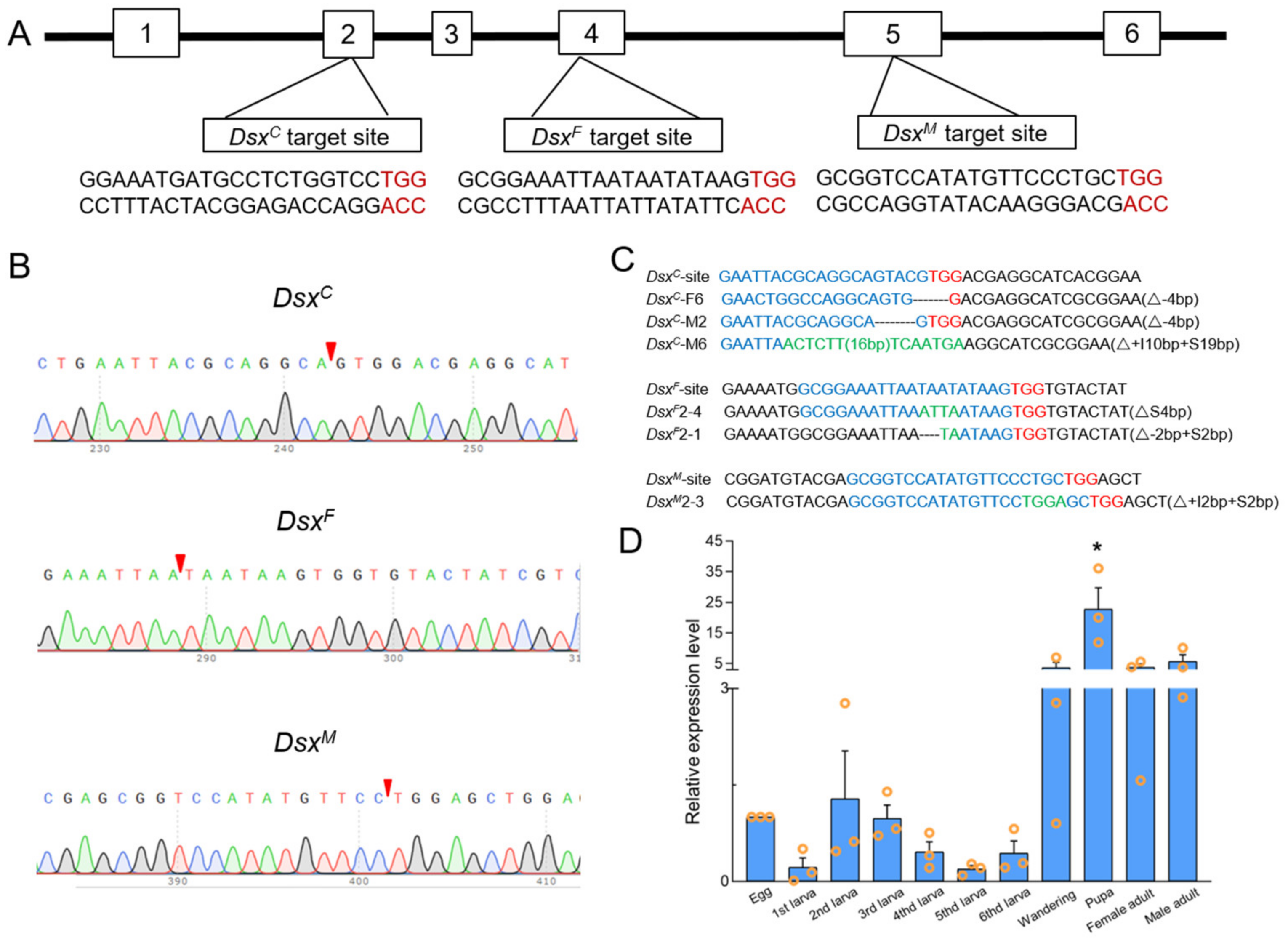
3.1. Identification and Cloning of Sfdsx

The genomic sequence length of Sfdsx was 196,795 bp, consisting of six exons with two sex-specific splicing patterns. The length of Sfdsx female-specific ORFs was 780 bp, encoding 259 amino acids. The length of male-specific ORFs was 801 bp, encoding 266 amino acids. The predicted alternative splicing patterns are shown in Figure S1. We used the NCBI BLAST program to identify Sfdsx against homologous amino acid sequences in D. melanogaster (GenBank accession number NP_001287220.1) and B. mori (GenBank Accession number NP_001036871.1).

3.2. CRISPR/Cas9-Mediated Sfdsx Mutagenesis

We selected one target site for mutagenesis within the common, female- and male-specific regions on exon 2, 4, and 5, respectively. Exon 2 was predicted to encode a common region of male and female transcripts, exon 4 a female-specific isoform, and exon 5 a male-specific isoform (Figure 1A). We injected 815, 702, and 1325 embryos within 1.5 h post-oviposition with Cas9 protein mixed with pre-synthesized SfdsxC, SfdsxF, and SfdsxM sgRNAs, respectively. The hatching and mutant ratios for each treatment are recorded in Table 1. We obtained pupal mutant ratios of 38.5%, 35.4%, and 35.1% for SfdsxC, SfdsxF, and SfdsxM treatments, respectively (Table 1). After eclosion, we found a significantly male-biased sex ratio in SfdsxC mutants in comparison to wildtype adults. In SfdsxF and SfdsxM mutants, morphological changes were observed exclusively in the corresponding sex of sex-specific knockouts (Table 1). To identify the mutated alleles, we extracted genomic DNA from pupa shell, and the results of genomic sequencing showed successful deletion of nucleotides within the target sites in SfdsxC, SfdsxF, and SfdsxM isoforms (Figure 1C). To investigate the temporal expression of Sfdsx in different developmental stages, relative expression levels of Sfdsx in eggs, first to sixth instar larvae, wandering stage larvae, pupae, and adults were examined. Results showed that the relative expression level of Sfdsx was significantly increased at the pupal stage but showed no significant change among the other stages (Figure 1D).

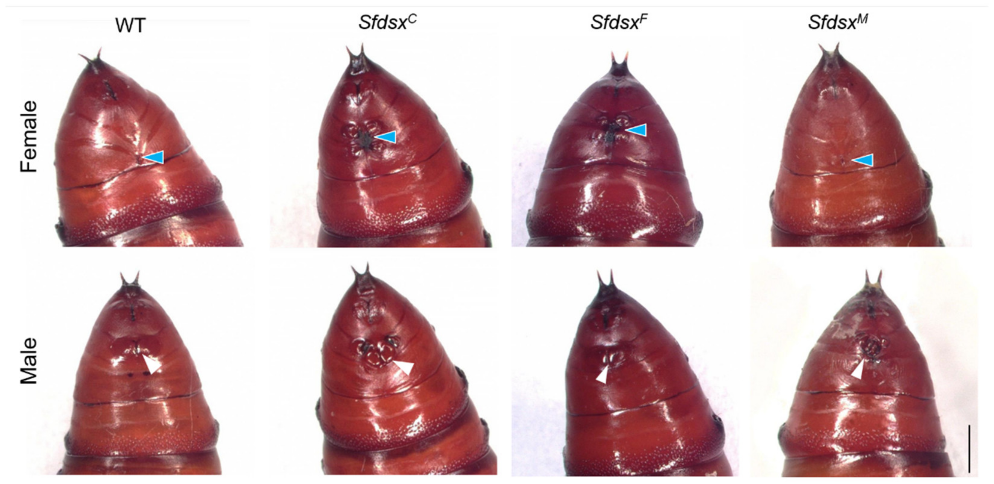
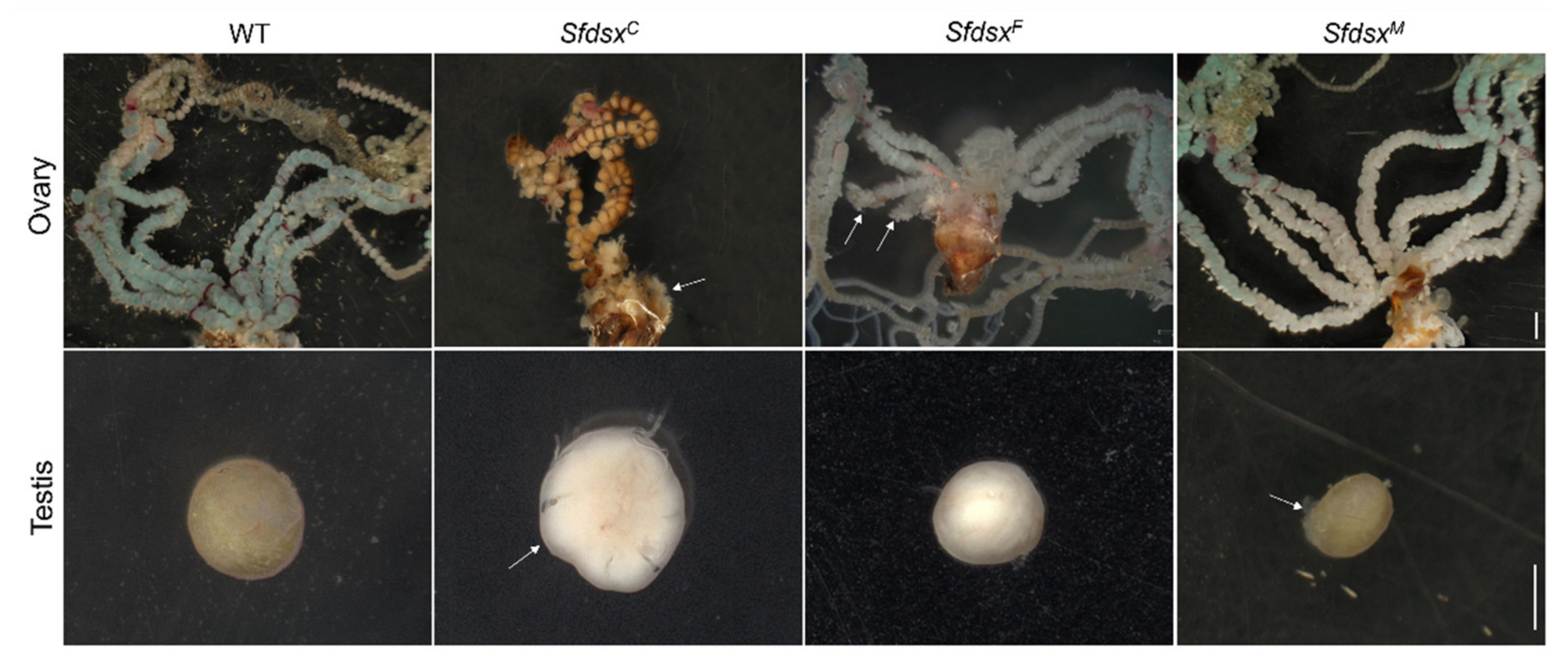
3.3. Phenotypic Impacts of Sfdsx Mutagenesis

3.3.1. External Genitalia

Phenotypic changes brought by gene knockout were recorded. We distinguished the sex of pupae based on external morphological characteristics, specifically, male pupae exhibit two noticeable points in the middle of the ninth abdominal segment, while female pupa exhibit two “^”-shaped lines across the eighth abdominal segment (Figure 2). In SfdsxC injections, all female and male pupal mutants exhibited abnormal external genitalia on the abdomen (Figure 2). All male pupae in the SfdsxF knockout group and all female pupae in the SfdsxM knockout group were externally similar to the wildtype (Figure 2). The corresponding phenotypic changes of SfdsxF and SfdsxM knockout groups were similar with the phenotypic changes found in SfdsxC mutants. In almost all SfdsxF mutants, the two “^”-shaped lines became absent with other irregular structural changes located across the eighth and ninth segments, while SfdsxF-injected males were identical to the wildtype. For SfdsxM mutants, abnormally shaped and numbered masses were found on both sides of the gonopore on the eighth to ninth abdominal segment in males, but females were not affected (Figure 2). The figure representing the gonopore in males is also obfuscated after SfdsxM mutagenesis (Figure 2).
Female and male pupae and adults of S. frugiperda are morphologically different and can be distinguished based on the structure of the gonads on the eighth segment of the abdomen (Figure 3). In S. frugiperda, the external genitalia of wildtype male adults consist of a pair of harpago, an aedeagus, and an uncus, while the ventral plate and genital papilla are female-specific (Figure 3). In the SfdsxC mutants, there were both male and female mutants, and the mutant phenotypes were similar to those of SfdsxF and SfdsxM mutants. Male SfdsxC mutants exhibited a deformed aedeagus, and female SfdsxC mutants showed disrupted genital papillae and a deformed ventral plate (Figure 3). SfdsxF mutants showed a severely disrupted reproductive ovipositor. In SfdsxM mutants, the harpago was severely deformed or developed incompletely, or even completely disappeared, and nodular growth appeared around the external genitalia of some mutants (Figure 3). We did not observe any female-specific external structures in SfdsxM mutants or male-specific external structures in SfdsxF mutants. This observation suggests that the sex-specific structural development of external genitalia is regulated by Sfdsx.

3.3.2. Internal Genitalia

Sfdsx mutants with phenotypic changes in the adult stage were dissected to observe internal genital structures. Three days after eclosion, the ovaries of females and testes of males from wildtype and Sfdsx adult mutant groups were dissected. As shown in Figure 4, wildtype females have a pair of ovaries, each of which is supplemented with four symmetrical lateral oviducts. Wildtype males have a single fused, rounded testis (Figure 4).
In SfdsxC mutants, both females and males exhibited severe malformation of the ovaries and testes (Figure 4). In SfdsxC female mutants, the oviduct was severely deformed, possessing four asymmetrical oviducts (Figure 4). The number of premature eggs was highly decreased in the oviducts of SfdsxC female mutants. The testes of SfdsxC male mutants appeared as irregular spheres with smooth surfaces, having less adhesion to the trachea than those wildtype males, suggesting incomplete development of testes (Figure 4).
In SfdsxF mutants, the ovaries of females exhibited obvious oviduct malformation. Two oviducts harboring from one side of the ovary were dramatically shortened in comparison to the other oviducts and those from wildtype ovaries (Figure 4). The SfdsxF male mutants showed normal testes compared with wildtype males (Figure 4).
In SfdsxM mutants, females showed a normal phenotype. However, the testes in male mutants were smaller in size and exhibited a disrupted structure, indicating failed fusion of the testes during development. Among these mutant phenotypes, gonadal abnormalities were found in male SfdsxM mutants and SfdsxC mutants of both sexes.

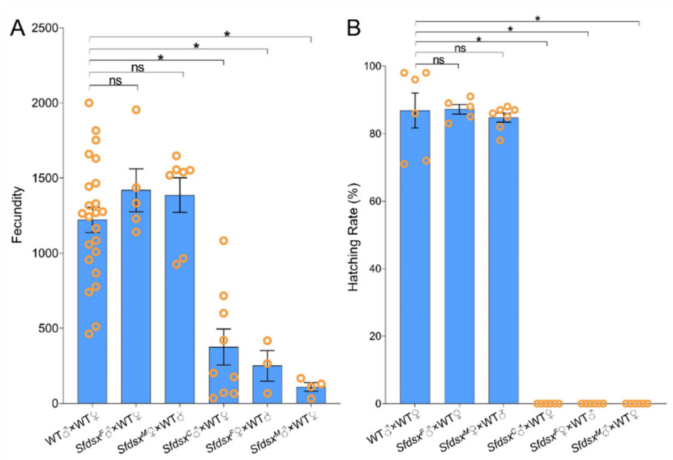
3.4. Physiological Impacts

Sex-specific genitalia are important for successful copulation between female and male adults. To investigate if sex-specific Sfdsx mutagenesis induces adult sterility, fecundity and hatching rate were recorded continuously for ten days after pairing individuals from different combinations of treatment groups. In wildtype pairings, females lay an average of 1221 ± 84 eggs across the ten days (Figure 5). Pairing SfdsxF males and SfdsxM females showed no significant differences in fecundity compared to wildtype pairs, demonstrating that the sex-specific exon mutant did not affect the fertility of SfdsxF males or SfdsxM females. However, fecundity by SfdsxC male mutants paired with wildtype female was decreased by approximately 72% to 374 ± 120 eggs on average (Figure 5A), and the hatching rate of these eggs was almost zero (Figure 5B). In SfdsxF female mutants, fecundity was decreased by approximately 80% to 249 ± 101 eggs on average (Figure 5A), and the eggs were unable to hatch normally (Figure 5B). SfdsxM males paired with wildtype female showed an approximately 91% decrease in fecundity, producing an average of 109 ± 29 eggs (Figure 5A), none of which hatched (Figure 5B).

3.5. Pleiotropic Impacts

OR1, PBP1, and PBP2 are pheromone receptors and are involved in mating and reproductive behavior in adults. The expression of the OR1 was significantly downregulated in SfdsxC mutant males and upregulated in SfdsxF mutant females (Figure 6A). PBP1 and PBP2 were also significantly downregulated in SfdsxC mutant males, but expression of PBP1 and PBP2 showed no significant difference among SfdsxM mutant males (Figure 6A, B). All three genes exhibited decreased transcript level in SfdsxC male mutants relative to the wildtype control group, and the transcript levels of OR1 and PBP1 were dramatically upregulated in SfdsxF female mutants compared to wildtype females (Figure 6). The relative transcript level of PBP1 was also significantly downregulated in SfdsxC female and male mutants, indicating its vital role for both male and female pheromone recognition (Figure 6B).

3.6. Sex-Specific Expression of Sfdsx

Primers amplifying the Sfdsx female-specific isoform and male-specific isoform show alternative splicing to Sfdsx (Figure S1). In wildtype adults, the female-specific isoform is longer (481 bp) than the male-specific band (232 bp), suggesting alternative splicing of the Sfdsx (Figure S1A). Although the bands of wildtype females and males appear in one lane, transcript levels of SfdsxF female-specific and SfdsxM male-specific isoforms decreased to a dramatically low level, and the faint bands with a fairly small size suggest successful gene knockout in both SfdsxF female and SfdsxM male mutants (Figure S1A).

4. Discussion

Dsx has been identified as a downstream gene of the sex determination pathway, playing an important role in the development of sexually dimorphic traits in a variety of insects [51,52]. The functional role of dsx has been studied in Hyphantria cunea [44], P. xylostella [34], A. gambiae [38], Bicyclus anynana [53], B. mori [28], Agrotis ipsilon [54], A. mellifera [37,55], and S. litura [35]. Given that lepidopterans are recalcitrant to RNAi, CRISPR/Cas9-mediated mutagenesis was employed to decipher the functionality of dsx in S. frugiperda. Sfdsx consists of six exons, of which exons 3 and 4 are female-specific, exon 5 is male-specific, and exon 6 does not participate in transcription (Figure 1A). The nucleic acid sequences of dsx from D. melanogaster (GenBank accession number NP_001287220.1) and B. mori (GenBank Accession number NP_001036871.1) were used as query to identify the putative coding sequence of Sfdsx. We cloned female- and male-specific regions of Sfdsx and identified one male-specific and three female-specific dsx transcripts in S. frugiperda (Figure S1). The alternative splicing is similar to that in S. litura, of which exons 1 and 2 encode the common region, exons 3 and 4 encode the female-specific region, and exon 5 encodes the male-specific region [35]. Similar splicing patterns were also found in P. xylostella, in which one male-specific and three female-specific transcripts were identified [34]. Temporal expression of Sfdsx was examined in S. frugiperda, and results showed that Sfdsx was highly expressed in the pupal stage relative to the other developmental stages (Figure 1D).
Successful gene knockout through the CRISPR/Cas9 gene-editing system was achieved by targeting the common, male-, and female-specific regions of Sfdsx (Figure 1B,C). Sex-specific mutagenesis disrupted the development of genitalia in the pupal and adult stages (Figure 2 and Figure 4). In SfdsxC mutants, male pupae showed abnormal phenotypes at the eighth abdominal segment, which is consistent with what was observed in SfdsxM mutants (Figure 3). The same phenomenon was found in O. furnacalis, in which dsxC mutants displayed an ectopic ventral plate and malformed genital structure [36]. Knockout of dsxM and dsxF results in an abnormal abdominal structure at the pupal stage (Figure 2) and malformed genitalia at the adult stage (Figure 3). These phenotypic observations of external genitalia are consistent with the results of dsx manipulations in A. gambiae, in which disruption of exon 5 led to an intersex phenotype and sterility in females but did not affect development or fertility in males [38]. A male biased sex ratio among SfdsxC mutants was observed in comparison to the wildtype adults; this bias might be caused by the lethal effect of SfdsxC sgRNAs against females during the embryonic stage [56] (Table 1). In SfdsxC and SfdsxF female mutants, the oviducts were dramatically shortened or malformed in comparison to those of wildtype females, while in SfdsxM mutants, the testes in male mutants were smaller in size and possessed an irregular spherical shape (Figure 4). These observations are consistent with what has been observed in P. xylostella, in which smaller testes and disrupted structure of the ovaries were detected in dsx mutants [34]. We found that mutagenesis of either the common, female-, or male-specific regions of dsx in S. frugiperda resulted in reduced fecundity (Figure 5). Similarly, mutagenesis of dsx targeting the common region also caused decreased fecundity and fertility of adults in O. furnacalis [36]. Reduced fertility after dsx mutagenesis was also observed in P. xylostella [34]. Our result is in accordance with previous publications showing that dsx is associated with sex determination, fecundity, and fertility in insects.
Dsx is located downstream of the insect sex determination pathway, plays an important role in morphological development of insect sexual dimorphism, and has a profound impact on insect physiology and behavior [57,58]. As is known in A. aegypti, dsx not only affects female genital development but also shortens the length of females’ antennae and antennal receptors [33]. In H. cunea, dsx also affects the development of adult somites [44]. Our result concerning transcript changes of OR1, PBP1, and PBP2 in Sfdsx mutants showed that all three genes exhibited decreased transcript levels in SfdsxC male mutants and dramatically increased levels in SfdsxF female mutants compared to the wildtype (Figure 6). Alteration of the expression of pheromone genes may also be a factor affecting mating and oviposition of S. frugiperda adults, resulting in the infertility of S. frugiperda mutant adults (Figure 5). In D. melanogaster, male adults produce courtship songs by vibrating their wings, a behavior that is regulated by dsx [59]. At the same time, dsx regulates abdominal pigment deposition [51] and Drop transcription to affect the normal development of male genital discs [60].
SIT is an environmentally friendly pest management strategy that hinders pest propagation by releasing large numbers of sterile insects into the wild [61,62]. Severe mutations in the external genitalia as a result of dsx manipulation have been reported in multiple Lepidopterans [34,36,44]. In our study, knocking out sex-specific dsx splicing variants in S. frugiperda led to sterility, with the number of eggs produced reduced by 72% in pairings including SfdsxC male mutants, 80% in those including SfdsxF female mutants, 91% in those including SfdsxM male mutants (Figure 5A). Eggs produced by pairings involving Sfdsx mutants did not hatch (Figure 5B). Normally, wildtype males display courtship behavior and approach females actively; however, in SfdsxC and SfdsxM males, the aedeagus structure was shortened, distorted, or even completely absent, which prevented copulation. In SfdsxF females, the genital papillae and ventral plate were deformed, also preventing copulation. Eggs laid by Sfdsx mutants were dried and ceased development after one to two days, resulting in no hatching (Figure 5). Similarly, a recent study of O. furnacalis and P. xylostella have shown that eggs laid by dsx mutants fail to hatch, suggesting the role of dsx as a potential target for genetic control [34,36]. In recent years, complete collapse of laboratory populations of A. gambiae through sex-specific sterility has been achieved [46], and complete control of laboratory populations of A. gambiae was enabled by targeting dsx [38]. We identified loci that cause genital malformations in sex-specific regions of Sfdsx. Our study provides a genetic basis for CRISPR gene drive-mediated population suppression and suggests the potential of targeting Sfdsx for genetic control of S. frugiperda.

5. Conclusions

In this study, our combined results support our hypothesis that alternative splicing of Sfdsx regulates sex determination and is involved in the fecundity and fertility of S. frugiperda. Mutants exhibited substantial external genital abnormalities and distortion of internal genitalia. The fecundity and hatching rate of eggs produced by corresponding mutants was significantly compromised in comparison to wildtype controls. Our results provide empirical evidence of a genetic basis for potentially targeting dsx as a part of the Sterile Insect Technique (SIT) to combat this global invasive pest in a sustainable and environmentally friendly way.

Supplementary Materials

The following supporting information can be downloaded at: https://www.mdpi.com/article/10.3390/cells11223557/s1; Figure S1: Sex-specific expression of Sfdsx in S. frugiperda. Table S1. Primers used in this study.

Author Contributions

Conceptualization, Q.Z.; methodology, Q.Z. and J.G.; software, J.G. and J.W.; validation, H.B., X.L., Q.Z. and J.W.; formal analysis, X.L. and P.Z.; investigation, J.G.; resources, Q.Z.; data curation, Q.Z. and J.W.; writing—original draft preparation, J.G. and A.M.; writing—review and editing, Q.Z. and H.B.; visualization, Q.Z. and H.B.; supervision, Q.Z.; project administration, Q.Z.; funding acquisition, Q.Z. and H.B. X.Z. oversaw the entire project and revised and edited manuscript. All authors have read and agreed to the published version of the manuscript.

Funding

This study was supported by grants from Natural Science Foundation of China (32072482) and Young Scholars’ Fund by Department of Education of Liaoning Province (LSNQN202017) to Z.Q., and Scientific and Technological Project of Henan Province (222102110108) to B.H.L.

Institutional Review Board Statement

Not applicable.

Informed Consent Statement

Not applicable.

Data Availability Statement

All the data and resources generated for this study are included in the article and the Supplemental Materials.

Acknowledgments

We are grateful for Yongping Huang’s guidance on the experimental design and Jizhe Shi’s assistance with data presentation.

Conflicts of Interest

The authors declare no conflict of interest.

References

  1. Nagoshi, R.N.; Nagoshi, B.Y.; Canarte, E.; Navarrete, B.; Solorzano, R.; Garces-Carrera, S. Genetic characterization of fall armyworm (Spodoptera frugiperda) in Ecuador and comparisons with regional populations identify likely migratory relationships. PLoS ONE 2019, 14, e0222332. [Google Scholar] [CrossRef] [PubMed]
  2. Goergen, G.; Kumar, P.L.; Sankung, S.B.; Togola, A.; Tamo, M. First Report of Outbreaks of the Fall Armyworm Spodoptera frugiperda (J E Smith) (Lepidoptera, Noctuidae), a New Alien Invasive Pest in West and Central Africa. PLoS ONE 2016, 11, e0165632. [Google Scholar] [CrossRef] [PubMed] [Green Version]
  3. Dumas, P.; Legeai, F.; Lemaitre, C.; Scaon, E.; Orsucci, M.; Labadie, K.; Gimenez, S.; Clamens, A.L.; Henri, H.; Vavre, F.; et al. Spodoptera frugiperda (Lepidoptera: Noctuidae) host-plant variants: Two host strains or two distinct species? Genetica 2015, 143, 305–316. [Google Scholar] [CrossRef] [Green Version]
  4. Pashley, D.P.J.E. Quantitative genetics, development, and physiological adaptation in host strains of fall armyworm. Evolution 1988, 42, 93–102. [Google Scholar] [CrossRef] [PubMed]
  5. Wu, Q.L.; He, L.M.; Shen, X.J.; Jiang, Y.Y.; Liu, J.; Hu, G.; Wu, K.M. Estimation of the Potential Infestation Area of Newly-invaded Fall Armyworm Spodoptera frugiperda in the Yangtze River Valley of China. Insects 2019, 10, 298. [Google Scholar] [CrossRef] [PubMed] [Green Version]
  6. Montezano, D.G.; Specht, A.; Sosa-Gomez, D.R.; Roque-Specht, V.F.; Sousa-Silva, J.C.; Paula-Moraes, S.V.; Peterson, J.A.; Hunt, T.E. Host plants of Spodoptera frugiperda (Lepidoptera: Noctuidae) in the Americas. Afr. Entomol. 2018, 26, 286–300. [Google Scholar] [CrossRef] [Green Version]
  7. Xiao, H.; Ye, X.; Xu, H.; Mei, Y.; Yang, Y.; Chen, X.; Yang, Y.; Liu, T.; Yu, Y.; Yang, W.; et al. The genetic adaptations of fall armyworm Spodoptera frugiperda facilitated its rapid global dispersal and invasion. Mol. Ecol. Res. 2020, 20, 1050–1068. [Google Scholar] [CrossRef] [PubMed]
  8. Bolzan, A.; Padovez, F.E.; Nascimento, A.R.; Kaiser, I.S.; Lira, E.C.; Amaral, F.S.; Kanno, R.H.; Malaquias, J.B.; Omoto, C. Selection and characterization of the inheritance of resistance of Spodoptera frugiperda (Lepidoptera: Noctuidae) to chlorantraniliprole and cross-resistance to other diamide insecticides. Pest Manag. Sci. 2019, 75, 2682–2689. [Google Scholar] [CrossRef] [PubMed]
  9. Lira, E.C.; Bolzan, A.; Nascimento, A.R.; Amaral, F.S.; Kanno, R.H.; Kaiser, I.S.; Omoto, C. Resistance of Spodoptera frugiperda (Lepidoptera: Noctuidae) to spinetoram: Inheritance and cross-resistance to spinosad. Pest Manag. Sci. 2020, 76, 2674–2680. [Google Scholar] [CrossRef] [PubMed]
  10. Wu, J.; Li, X.; Hou, R.; Zhao, K.; Wang, Y.; Huang, S.; Cheng, D.; Zhang, Z. Examination of acephate absorption, transport, and accumulation in maize after root irrigation for Spodoptera frugiperda control. Environ. Sci. Pollut. Res. Int. 2021, 28, 57361–57371. [Google Scholar] [CrossRef]
  11. Feng, B.; Zhi, H.; Chen, H.; Cui, B.; Zhao, X.; Sun, C.; Wang, Y.; Cui, H.; Zeng, Z. Development of Chlorantraniliprole and Lambda Cyhalothrin Double-Loaded Nano-Microcapsules for Synergistical Pest Control. Nanomaterials 2021, 11, 2730. [Google Scholar] [CrossRef] [PubMed]
  12. Van den Berg, J.; Prasanna, B.M.; Midega, C.A.O.; Ronald, P.C.; Carriere, Y.; Tabashnik, B.E. Managing Fall Armyworm in Africa: Can Bt Maize Sustainably Improve Control? J. Econ. Entomol. 2021, 114, 1934–1949. [Google Scholar] [CrossRef] [PubMed]
  13. Yu, H.; Yang, C.J.; Li, N.; Zhao, Y.; Chen, Z.M.; Yi, S.J.; Li, Z.Q.; Adang, M.J.; Huang, G.H. Novel strategies for the biocontrol of noctuid pests (Lepidoptera) based on improving ascovirus infectivity using Bacillus thuringiensis. Insect Sci. 2021, 28, 1452–1467. [Google Scholar] [CrossRef] [PubMed]
  14. Zhang, L.; Liu, B.; Zheng, W.; Liu, C.; Zhang, D.; Zhao, S.; Li, Z.; Xu, P.; Wilson, K.; Withers, A.; et al. Genetic structure and insecticide resistance characteristics of fall armyworm populations invading China. Mol. Ecol. Res. 2020, 20, 1682–1696. [Google Scholar] [CrossRef] [PubMed]
  15. Chandrasena, D.I.; Signorini, A.M.; Abratti, G.; Storer, N.P.; Olaciregui, M.L.; Alves, A.P.; Pilcher, C.D. Characterization of field-evolved resistance to Bacillus thuringiensis-derived Cry1F delta-endotoxin in Spodoptera frugiperda populations from Argentina. Pest Manag. Sci. 2018, 74, 746–754. [Google Scholar] [CrossRef] [Green Version]
  16. Burtis, K.C.; Baker, B.S.J.C. Drosophila doublesex gene controls somatic sexual differentiation by producing alternatively spliced mRNAs encoding related sex-specific polypeptides. Cell 1989, 56, 997–1010. [Google Scholar] [CrossRef]
  17. Wang, Y.; Rensink, A.H.; Fricke, U.; Riddle, M.C.; Trent, C.; van de Zande, L.; Verhulst, E.C. Doublesex regulates male-specific differentiation during distinct developmental time windows in a parasitoid wasp. Insect Biochem. Mol. Biol. 2022, 142, 103724. [Google Scholar] [CrossRef]
  18. Cho, S.; Huang, Z.Y.; Zhang, J. Sex-specific splicing of the honeybee doublesex gene reveals 300 million years of evolution at the bottom of the insect sex-determination pathway. Genetics 2007, 177, 1733–1741. [Google Scholar] [CrossRef] [Green Version]
  19. Scali, C.; Catteruccia, F.; Li, Q.; Crisanti, A. Identification of sex-specific transcripts of the Anopheles gambiae doublesex gene. J. Exp. Biol. 2005, 208, 3701–3709. [Google Scholar] [CrossRef] [Green Version]
  20. Salvemini, M.; Mauro, U.; Lombardo, F.; Milano, A.; Zazzaro, V.; Arca, B.; Polito, L.C.; Saccone, G. Genomic organization and splicing evolution of the doublesex gene, a Drosophila regulator of sexual differentiation, in the dengue and yellow fever mosquito Aedes aegypti. BMC Evol. Biol. 2011, 11, 41. [Google Scholar] [CrossRef]
  21. Price, D.C.; Egizi, A.; Fonseca, D.M. Characterization of the doublesex gene within the Culex pipiens complex suggests regulatory plasticity at the base of the mosquito sex determination cascade. BMC Evol. Biol. 2015, 15, 108. [Google Scholar] [CrossRef] [PubMed] [Green Version]
  22. Concha, C.; Li, F.; Scott, M.J. Conservation and sex-specific splicing of the doublesex gene in the economically important pest species Lucilia cuprina. J. Genet. 2010, 89, 279–285. [Google Scholar] [CrossRef] [PubMed]
  23. Hediger, M.; Burghardt, G.; Siegenthaler, C.; Buser, N.; Hilfiker-Kleiner, D.; Dubendorfer, A.; Bopp, D. Sex determination in Drosophila melanogaster and Musca domestica converges at the level of the terminal regulator doublesex. Dev. Genes Evol. 2004, 214, 29–42. [Google Scholar] [CrossRef] [PubMed] [Green Version]
  24. Shukla, J.N.; Palli, S.R. Doublesex target genes in the red flour beetle, Tribolium castaneum. Sci. Rep. 2012, 2, 948. [Google Scholar] [CrossRef] [Green Version]
  25. Gotoh, H.; Miyakawa, H.; Ishikawa, A.; Ishikawa, Y.; Sugime, Y.; Emlen, D.J.; Lavine, L.C.; Miura, T. Developmental Link between Sex and Nutrition; doublesex Regulates Sex-Specific Mandible Growth via Juvenile Hormone Signaling in Stag Beetles. PLoS Genet. 2014, 10, e1004098. [Google Scholar] [CrossRef] [Green Version]
  26. Ito, Y.; Harigai, A.; Nakata, M.; Hosoya, T.; Araya, K.; Oba, Y.; Ito, A.; Ohde, T.; Yaginuma, T.; Niimi, T. The role of doublesex in the evolution of exaggerated horns in the Japanese rhinoceros beetle. EMBO Rep. 2013, 14, 561–567. [Google Scholar] [CrossRef] [Green Version]
  27. Kunte, K.; Zhang, W.; Tenger-Trolander, A.; Palmer, D.H.; Martin, A.; Reed, R.D.; Mullen, S.P.; Kronforst, M.R. doublesex is a mimicry supergene. Nature 2014, 507, 229–232. [Google Scholar] [CrossRef]
  28. Xu, J.; Wang, Y.; Li, Z.; Ling, L.; Zeng, B.; James, A.A.; Tan, A.; Huang, Y. Transcription activator-like effector nuclease (TALEN)-mediated female-specific sterility in the silkworm, Bombyx mori. Insect Mol. Biol. 2014, 23, 800–807. [Google Scholar] [CrossRef] [Green Version]
  29. Prakash, A.; Monteiro, A. Molecular mechanisms of secondary sexual trait development in insects. Curr. Opin. Insect Sci. 2016, 17, 40–48. [Google Scholar] [CrossRef]
  30. Morrow, J.L.; Riegler, M.; Frommer, M.; Shearman, D.C. Expression patterns of sex-determination genes in single male and female embryos of two Bactrocera fruit fly species during early development. Insect Mol. Biol. 2014, 23, 754–767. [Google Scholar] [CrossRef]
  31. Robinett, C.C.; Vaughan, A.G.; Knapp, J.M.; Baker, B.S. Sex and the single cell. II. There is a time and place for sex. PLoS Biol. 2010, 8, e1000365. [Google Scholar] [CrossRef] [PubMed] [Green Version]
  32. Yang, X.; Chen, K.; Wang, Y.; Yang, D.; Huang, Y. The Sex Determination Cascade in the Silkworm. Genes 2021, 12, 315. [Google Scholar] [CrossRef] [PubMed]
  33. Mysore, K.; Sun, L.; Tomchaney, M.; Sullivan, G.; Adams, H.; Piscoya, A.S.; Severson, D.W.; Syed, Z.; Duman-Scheel, M. siRNA-Mediated Silencing of doublesex during Female Development of the Dengue Vector Mosquito Aedes aegypti. PLoS Negl. Trop. Dis. 2015, 9, e0004213. [Google Scholar] [CrossRef] [PubMed] [Green Version]
  34. Wang, Y.; Chen, X.; Liu, Z.; Xu, J.; Li, X.; Bi, H.; Andongma, A.A.; Niu, C.; Huang, Y. Mutation of doublesex induces sex-specific sterility of the diamondback moth Plutella xylostella. Insect Biochem. Mol. Biol. 2019, 112, 103180. [Google Scholar] [CrossRef]
  35. Du, Q.; Wen, L.; Zheng, S.C.; Bi, H.L.; Huang, Y.P.; Feng, Q.L.; Liu, L. Identification and functional characterization of doublesex gene in the testis of Spodoptera litura. Insect Sci. 2019, 26, 1000–1010. [Google Scholar] [CrossRef]
  36. Bi, H.; Li, X.; Xu, X.; Wang, Y.; Zhou, S.; Huang, Y. Masculinizer and Doublesex as Key Factors Regulate Sexual Dimorphism in Ostrinia furnacalis. Cells 2022, 11, 2161. [Google Scholar] [CrossRef]
  37. Roth, A.; Vleurinck, C.; Netschitailo, O.; Bauer, V.; Otte, M.; Kaftanoglu, O.; Page, R.E.; Beye, M. A genetic switch for worker nutrition-mediated traits in honeybees. PLoS Biol. 2019, 17, e3000171. [Google Scholar] [CrossRef] [Green Version]
  38. Kyrou, K.; Hammond, A.M.; Galizi, R.; Kranjc, N.; Burt, A.; Beaghton, A.K.; Nolan, T.; Crisanti, A. A CRISPR-Cas9 gene drive targeting doublesex causes complete population suppression in caged Anopheles gambiae mosquitoes. Nat. Biotechnol. 2018, 36, 1062–1066. [Google Scholar] [CrossRef] [Green Version]
  39. Shukla, J.; Nagaraju, J.J.J.O.G. Doublesex: A conserved downstream gene controlled by diverse upstream regulators. J. Genet. 2010, 89, 341–356. [Google Scholar] [CrossRef]
  40. Kiuchi, T.; Koga, H.; Kawamoto, M.; Shoji, K.; Sakai, H.; Arai, Y.; Ishihara, G.; Kawaoka, S.; Sugano, S.; Shimada, T.; et al. A single female-specific piRNA is the primary determiner of sex in the silkworm. Nature 2014, 509, 633–636. [Google Scholar] [CrossRef]
  41. Katsuma, S.; Sugano, Y.; Kiuchi, T.; Shimada, T. Two Conserved Cysteine Residues Are Required for the Masculinizing Activity of the Silkworm Masc Protein. J. Biol. Chem. 2015, 290, 26114–26124. [Google Scholar] [CrossRef] [PubMed] [Green Version]
  42. Sakai, H.; Sumitani, M.; Chikami, Y.; Yahata, K.; Uchino, K.; Kiuchi, T.; Katsuma, S.; Aoki, F.; Sezutsu, H.; Suzuki, M.G. Transgenic Expression of the piRNA-Resistant Masculinizer Gene Induces Female-Specific Lethality and Partial Female-to-Male Sex Reversal in the Silkworm, Bombyx mori. PLoS Genet. 2016, 12, e1006203. [Google Scholar] [CrossRef] [PubMed] [Green Version]
  43. Yang, B.; Ozaki, K.; Ishikawa, Y.; Matsuo, T. Identification of candidate odorant receptors in Asian corn borer Ostrinia furnacalis. PLoS ONE 2015, 10, e0121261. [Google Scholar] [CrossRef] [PubMed] [Green Version]
  44. Li, X.; Liu, Q.; Liu, H.; Bi, H.; Wang, Y.; Chen, X.; Wu, N.; Xu, J.; Zhang, Z.; Huang, Y.; et al. Mutation of doublesex in Hyphantria cunea results in sex-specific sterility. Pest Manag. Sci. 2020, 76, 1673–1682. [Google Scholar] [CrossRef]
  45. Xu, J.; Zhan, S.; Chen, S.; Zeng, B.; Li, Z.; James, A.A.; Tan, A.; Huang, Y. Sexually dimorphic traits in the silkworm, Bombyx mori, are regulated by doublesex. Insect Biochem. Mol. Biol. 2017, 80, 42–51. [Google Scholar] [CrossRef] [Green Version]
  46. Simoni, A.; Hammond, A.M.; Beaghton, A.K.; Galizi, R.; Taxiarchi, C.; Kyrou, K.; Meacci, D.; Gribble, M.; Morselli, G.; Burt, A.; et al. A male-biased sex-distorter gene drive for the human malaria vector Anopheles gambiae. Nat. Biotechnol. 2020, 38, 1054–1060. [Google Scholar] [CrossRef]
  47. Krzywinska, E.; Ferretti, L.; Li, J.; Li, J.C.; Chen, C.H.; Krzywinski, J. femaleless Controls Sex Determination and Dosage Compensation Pathways in Females of Anopheles Mosquitoes. Curr. Biol. 2021, 31, 1084–1091.e4. [Google Scholar] [CrossRef]
  48. Naito, Y.; Hino, K.; Bono, H.; Ui-Tei, K. CRISPRdirect: Software for designing CRISPR/Cas guide RNA with reduced off-target sites. Bioinformatics 2015, 31, 1120–1123. [Google Scholar] [CrossRef] [Green Version]
  49. Rodriguez-de la Noval, C.; Rodriguez-Cabrera, L.; Izquierdo, L.; Espinosa, L.A.; Hernandez, D.; Ponce, M.; Moran-Bertot, I.; Tellez-Rodriguez, P.; Borras-Hidalgo, O.; Huang, S.; et al. Functional expression of a peritrophin A-like SfPER protein is required for larval development in Spodoptera frugiperda (Lepidoptera: Noctuidae). Sci. Rep. 2019, 9, 2630. [Google Scholar] [CrossRef] [Green Version]
  50. Livak, K.J.; Schmittgen, T.D. Analysis of relative gene expression data using real-time quantitative PCR and the 2(-Delta Delta C(T)) Method. Methods 2001, 25, 402–408. [Google Scholar] [CrossRef]
  51. Williams, T.M.; Selegue, J.E.; Werner, T.; Gompel, N.; Kopp, A.; Carroll, S.B. The regulation and evolution of a genetic switch controlling sexually dimorphic traits in Drosophila. Cell 2008, 134, 610–623. [Google Scholar] [CrossRef] [PubMed] [Green Version]
  52. Williams, T.M.; Carroll, S.B. Genetic and molecular insights into the development and evolution of sexual dimorphism. Nat. Rev. Genet. 2009, 10, 797–804. [Google Scholar] [CrossRef] [PubMed]
  53. Prakash, A.; Monteiro, A. Doublesex Mediates the Development of Sex-Specific Pheromone Organs in Bicyclus Butterflies via Multiple Mechanisms. Mol. Biol. Evol. 2020, 37, 1694–1707. [Google Scholar] [CrossRef] [PubMed] [Green Version]
  54. Chen, X.; Cao, Y.; Zhan, S.; Tan, A.; Palli, S.R.; Huang, Y. Disruption of sex-specific doublesex exons results in male- and female-specific defects in the black cutworm, Agrotis ipsilon. Pest Manag. Sci. 2019, 75, 1697–1706. [Google Scholar] [CrossRef] [PubMed]
  55. McAfee, A.; Pettis, J.S.; Tarpy, D.R.; Foster, L.J. Feminizer and doublesex knock-outs cause honey bees to switch sexes. PLoS Biol. 2019, 17, e3000256. [Google Scholar] [CrossRef] [Green Version]
  56. Zhang, Z.J.; Niu, B.L.; Ji, D.F.; Li, M.W.; Li, K.; James, A.A.; Tan, A.J.; Huang, Y.P. Silkworm genetic sexing through W chromosome-linked, targeted gene integration. Proc. Natl. Acad. Sci. USA 2018, 115, 8752–8756. [Google Scholar] [CrossRef] [Green Version]
  57. Rideout, E.J.; Dornan, A.J.; Neville, M.C.; Eadie, S.; Goodwin, S.F. Control of sexual differentiation and behavior by the doublesex gene in Drosophila melanogaster. Nat. Neurosci. 2010, 13, 458–466. [Google Scholar] [CrossRef] [Green Version]
  58. Matsushima, D.; Kasahara, R.; Matsuno, K.; Aoki, F.; Suzuki, M.G. Involvement of Ecdysone Signaling in the Expression of the doublesex Gene during Embryonic Development in the Silkworm, Bombyx mori. Sex. Dev. 2019, 13, 151–163. [Google Scholar] [CrossRef]
  59. Shirangi, T.R.; Wong, A.M.; Truman, J.W.; Stern, D.L. Doublesex Regulates the Connectivity of a Neural Circuit Controlling Drosophila Male Courtship Song. Dev. Cell 2016, 37, 533–544. [Google Scholar] [CrossRef] [Green Version]
  60. Chatterjee, S.S.; Uppendahl, L.D.; Chowdhury, M.A.; Ip, P.L.; Siegal, M.L. The female-specific doublesex isoform regulates pleiotropic transcription factors to pattern genital development in Drosophila. Development 2011, 138, 1099–1109. [Google Scholar] [CrossRef]
  61. Vreysen, M.J.B.; Abd-Alla, A.M.M.; Bourtzis, K.; Bouyer, J.; Caceres, C.; de Beer, C.; Oliveira Carvalho, D.; Maiga, H.; Mamai, W.; Nikolouli, K.; et al. The Insect Pest Control Laboratory of the Joint FAO/IAEA Programme: Ten Years (2010–2020) of Research and Development, Achievements and Challenges in Support of the Sterile Insect Technique. Insects 2021, 12, 346. [Google Scholar] [CrossRef] [PubMed]
  62. Bourtzis, K.; Vreysen, M.J.B. Sterile Insect Technique (SIT) and Its Applications. Insects 2021, 12, 638. [Google Scholar] [CrossRef] [PubMed]
Figure 1. CRISPR/Cas9-mediated mutation within Sfdsx target sites and temporal expression of Sfdsx gene. (A) Target sites on exons 2, 4, and 5 to eliminate the common, female-specific, and male-specific regions of the Sfdsx gene. Red represents PAM sequences, and black represents substitutive sequences. (B) Sequencing chromatogram of the SfdsxC, SfdsxF, and SfdsxM mutants. The red arrow refers to the position of PCR and sequencing on common, female-specific, and male-specific regions of Sfdsx by the CRISPR/Cas9 gene-editing system. (C) Common, female-specific, and male-specific mutants of Sfdsx detected by sequencing. The dashed lines “–” indicate deleted nucleotides relative to wildtype. The guide RNA target sequences are marked in blue. (D) Temporal expression of Sfdsx across different developmental stages of S. frugiperda. The internal reference is ß-actin. The asterisks (*) indicate significant differences (p < 0. 05). Data were analyzed using Tukey’s test (one-way ANOVA).
Cells 11 03557 g001
Figure 2. Malformed genital structure after SfdsxC, SfdsxF, and SfdsxM mutagenesis. Photographs of typical wildtype and mutant pupae. Oviposition holes are present on female pupa on the 8th abdominal segment. A longitudinal fissure representing the gonopore is present in the middle of the 9th abdominal segment of the WT male pupa. SfdsxC-F, SfdsxC-M, SfdsxF-F, and SfdsxM-M showed abnormal morphology of the gonopore compared with that in WT. Blue arrows depict oviposition holes in females, while white arrows indicate gonopores in males. Scale bars: 1 mm.
Cells 11 03557 g002
Figure 3. Deformation of external genitalia in SfdsxC, SfdsxF, and SfdsxM mutants at adult stage. The WT male-specific adult external structure consists of a pair of harpago, an aedeagus, and an uncus, whereas the female-specific external genitals include genital papillae and a ventral plate. Compared with WT, the external genitalia of SfdsxC, SfdsxF, and SfdsxM mutants were abnormal. Different color of triangles represents sex-specific external genital structures. Scale bars: 1 mm.
Cells 11 03557 g003
Figure 4. Malformation of ovaries and testes in adult Sfdsx mutants. The ovaries and testes of wildtype, SfdsxC, SfdsxF, and SfdsxF adults were dissected on the third day post-eclosion (PAE3). White arrows indicate mutations on ovaries or testes. Scale bars: 1 mm.
Cells 11 03557 g004
Figure 5. Compromised fecundity and fertility after Sfdsx mutagenesis. (A) Adult mutants of Sfdsx injected with common, female-, and male-specific sgRNAs and Cas9 showed decreased fecundity compared to the wildtype. (B) The hatching rates of eggs laid by SfdsxC males, SfdsxF females, and SfdsxM males are shown. The asterisks (*) indicate significant differences (p < 0.05) between mutants and wildtypes. “ns” stands for “not significant” (p > 0.05). Data were analyzed using a two-tailed t-test.
Cells 11 03557 g005
Figure 6. Relative expressions of OR1, PBP1, and PBP2 after SfdsxC, SfdsxF, and SfdsxM mutagenesis. The relative transcript levels of OR1 (A), PBP1 (B), and PBP2 (C) were examined in WT male and female, SfdsxC male, SfdsxF female, and SfdsxM males. ß-actin was the internal reference. The asterisks (*) indicate significant differences (p < 0.05) between mutants and wildtypes. “ns” stands for “not significant” (p > 0.05). Data were analyzed using a two-tailed t-test.
Cells 11 03557 g006
Table 1. Mutagenesis of Sfdsx induced by CRISPR/Cas9 system.
sgRNAInjected 1Hatched 2Pupate 3P Mutant 4Adult (F/M) 5A Mutant (F/M) 6
dsxC815585 (71.8%)130 (22.2%)50 (38.5%)117 (39/78)48 (1/47)
dsxF702449 (64.0%)79 (17.6%)28 (35.4%)73 (31/42)22 (22/0)
dsxM1325461 (34.8%)37 (6.9%)13 (35.1%)36 (17/19)12 (0/12)
WT776665 (85.7%)469 (70.5%)-366 (177/189)-
1 Number of injected individuals, 2 Number and percent (%) of hatched individuals, 3 Number and percent (%) of larvae that entered pupal stage, 4 Number and percent (%) of pupal mutants based on morphological change, 5 Number and percent (%) of pupae and sex ratio that entered adult stage, 6 Number and percent (%) of adult mutants and sex ratio based on morphological change.
Publisher’s Note: MDPI stays neutral with regard to jurisdictional claims in published maps and institutional affiliations.

