Review

Enhancing an Oxidative “Trojan Horse” Action of Vitamin C with Arsenic Trioxide for Effective Suppression of KRAS-Mutant Cancers: A Promising Path at the Bedside

1
Department of Biology, Nazarbayev University, Astana 010000, Kazakhstan
2
National Laboratory Astana, Nazarbayev University, Astana 010000, Kazakhstan
*
Author to whom correspondence should be addressed.
Cells 2022, 11(21), 3454; https://doi.org/10.3390/cells11213454
Submission received: 16 September 2022 / Revised: 19 October 2022 / Accepted: 24 October 2022 / Published: 1 November 2022

Abstract

:
The turn-on mutations of the KRAS gene, coding a small GTPase coupling growth factor signaling, are contributing to nearly 25% of all human cancers, leading to highly malignant tumors with poor outcomes. Targeting of oncogenic KRAS remains a most challenging task in oncology. Recently, the specific G12C mutant KRAS inhibitors have been developed but with a limited clinical outcome because they acquire drug resistance. Alternatively, exploiting a metabolic breach of KRAS-mutant cancer cells related to a glucose-dependent sensitivity to oxidative stress is becoming a promising indirect cancer targeting approach. Here, we discuss the use of a vitamin C (VC) acting in high dose as an oxidative “Trojan horse” agent for KRAS-mutant cancer cells that can be potentiated with another oxidizing drug arsenic trioxide (ATO) to obtain a potent and selective cytotoxic impact. Moreover, we outline the advantages of VC’s non-natural enantiomer, D-VC, because of its distinctive pharmacokinetics and lower toxicity. Thus, the D-VC and ATO combination shows a promising path to treat KRAS-mutant cancers in clinical settings.

1. Introduction

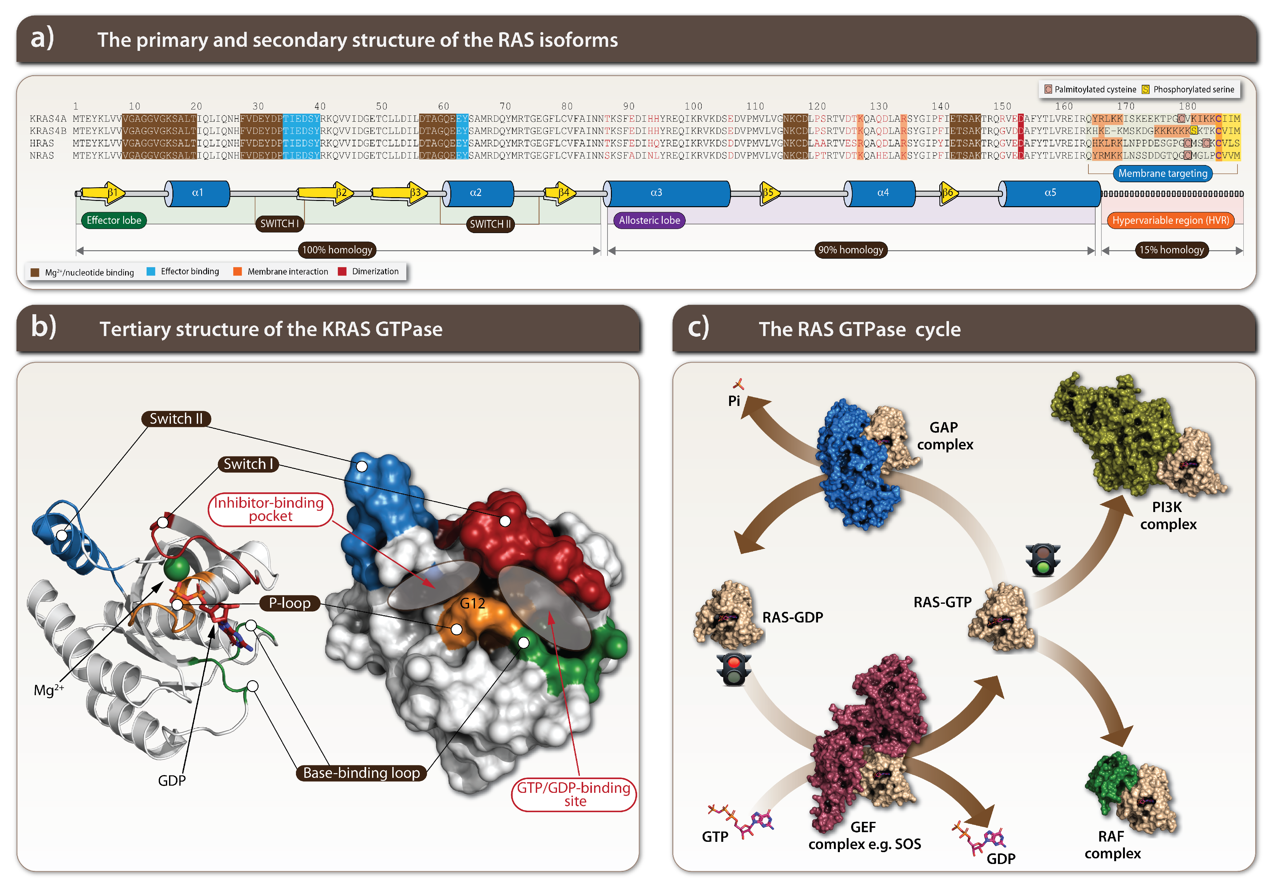
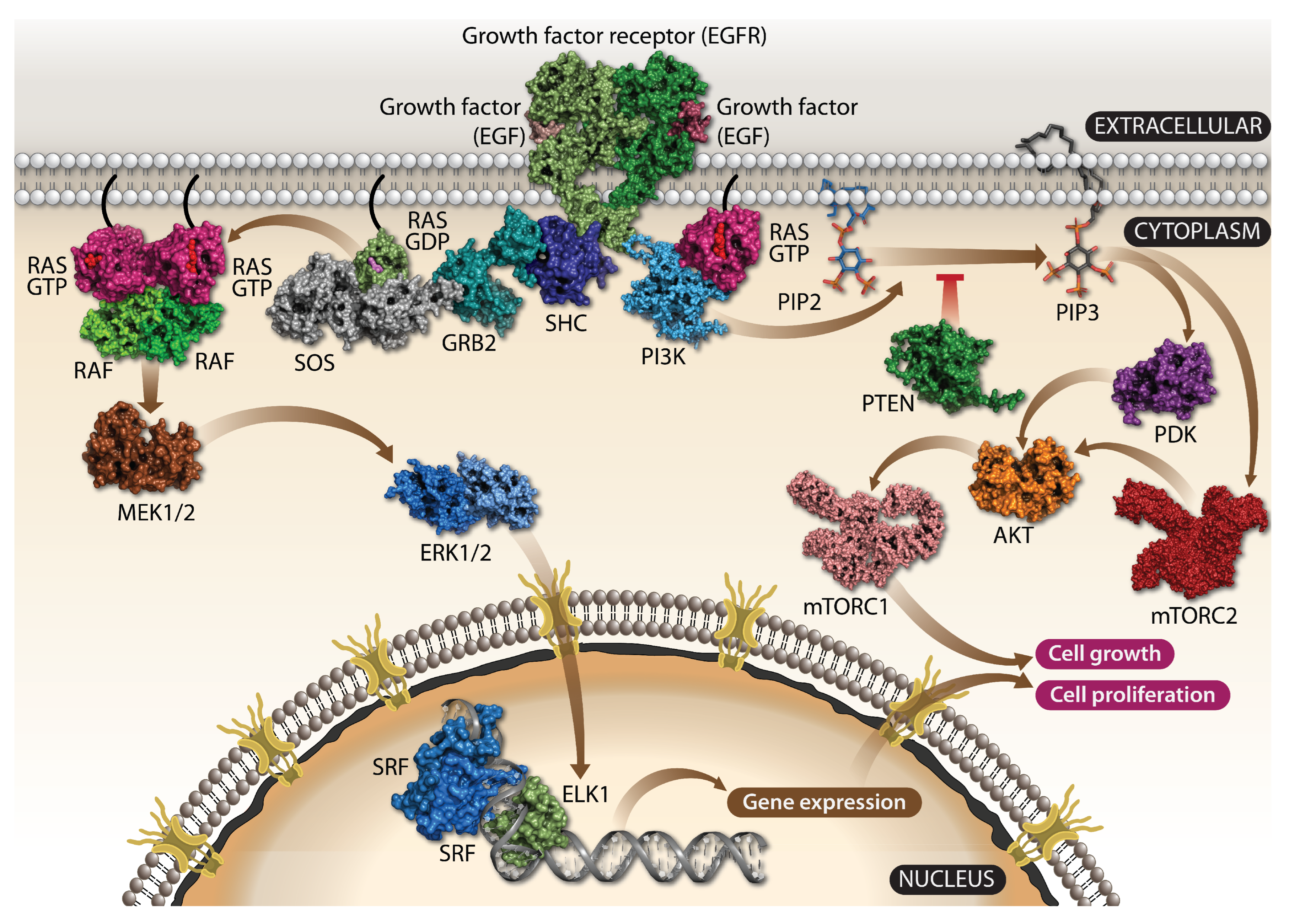
1.1. KRAS Is a Crucial Component of Growth Factor Signaling

The Kras gene was originally identified in the Kirsten rat sarcoma (Kras) virus DNA sequence, and its transforming oncogenic potential was described in the early 1980s [1,2]. RAS proteins are recruited from cytoplasm to the plasma membrane signaling nodes upon activation of a tyrosine kinase receptor where, together with various regulatory proteins, it activates signaling pathways coordinating cell growth and proliferation. A RAS protein translocation to the cell surface happens via one of two possible routes involving certain post-translational modifications, such as farnesylation of the cysteine residue of the RAS CAAX motif and α -carboxyl group methylation (Figure 1a) [3]. RAS proteins belong to small G protein groups and are transitioning between guanosine triphosphate (GTP)-bound active and guanosine diphosphate (GDP)-bound inactive forms [4,5]. Activation happens through exchange of GDP by GTP guanine nucleotides and is closely regulated by guanine exchange factors (GEFs) (Figure 1a) [6,7]. RAS activation (a GTP bound state) changes the protein conformation that allows its interactions with more than 20 different proteins from 10 effector families [4].
RAS triggers activation of the critical RAF/MEK/ERK (MAPK) kinase (Figure 2 left) and PI3K/AKT/mTOR signaling pathways (Figure 2 right) [5]. Signaling pathways are activated by a ligand binding with a specific receptor, such as the epidermal growth factor (EGF) with its receptor tyrosine kinase, EGFR, leading to its activation, mediated by the EGFR dimerisation and cross-phosphorylation of each other on tyrosine residues (also known as a ligand-dependent autophosphorylation). A specific tyrosine phosphorylated site on EGFR recruits growth factor receptor-bound protein 2 (GRB2) by its SH2 domain which serves as the adaptor protein by further recruiting the son of sevenless (SOS) and RAS proteins. An SOS protein is the guanosine nucleotide exchange factor (GEF) that serves as the RAS activator by catalyzing its GDP exchange to GTP and turning on the cascade of kinases starting from RAF to MEK and finally to ERK also known as MAPK (Figure 1c). The ERK kinases activate transcription factors promoting cell growth and proliferation [5]. The RAS family of proto-oncogenes comprises HRAS, KRAS, and NRAS, which are among the most mutated genes in human cancers (Figure 1a). These genes are ubiquitously expressed and share a significant sequence homology and mainly overlapping roles.

1.2. KRAS Is a Potent Oncogenic Driver

Recent statistical data indicate that approximately 25% of cases of cancer carry RAS mutations. Analysis of combined datasets from four leading cancer mutation databases detected KRAS in 19 out of 29 cancer types [8,9,10] and was marked as the “Everest” of oncogenes [11]. KRAS is the most frequently mutated isoform among the RAS family of genes and is associated with an overall percentage of 75–83% of all RAS-mutated cancers, whereas NRAS is detected in only 8–17% and HRAS in 3–7% of all cases. Human pancreatic ductal adenocarcinomas (PDAC) carry the highest frequency of KRAS mutations (about 95%) whereas in colon and lung cancers it is approximately 50% and 35%, respectively [10,12,13]. A high KRAS mutation load is detected in other types of cancers, such as multiple myeloma, ovarian, uterine, and stomach cancers (22%, 15%, 18%, and 16%, respectively), whereas NRAS mutations are frequently detected in melanoma (30%), multiple myeloma (18%), acute myelogenous leukemia, colorectal cancer (CRC) (10%), and thyroid cancer (8%), and HRAS is the least frequently mutated RAS gene detected in cancers of the bladder, head, and neck squamous cell carcinomas and the uterus (5% or less) [12]. These statistics largely vary due to the biological variation and populations used in different studies as well as the sampling bias within datasets. Among all mutations in KRAS and NRAS isoforms, the most dominant hotspots are located in codons G12, G13, and Q61 and comprise 96%–98% of all point mutations [14,15,16]. In the KRAS gene, about 81% mutations happen at position G12, and 14% at G13, but very few (2%) at codon Q61. On the contrary, 62% of NRAS tumor mutations are located in codon Q61, 23% in codon G12, and 11% G13. In HRAS mutant tumors, approximately 26% of mutations take place on the codon G12, 23% on G13 and 38% on Q61 [17]. Within 100 mutation sites known in all three RAS isoforms, the most common mutant alleles are located on G12 site in KRAS with 6 point mutations overall. The most common KRAS G12 mutations are identified as G12D (42%), G12V (28%), and G12C (14%), whereas G12A, G12R, and G12S are much less frequent [18,19]. A distribution of point mutations in KRAS also varies substantially in tumor tissues; for example in pancreas probability of the G12R mutation is 13% but it is only up to 2% in intestine. Moreover, different mutations alter RAS signaling with distinctive features as has been reported for the KRAS G12 and G13 mutants [19]. The KRAS mutations on Q61 site also promote change in morphology, growth transformation, signaling, and metabolism of cells [20]. It has been shown that different KRAS mutant alleles may have different clinical impacts on the prognosis of pancreatic and CRCs [21,22,23,24]. KRAS cancers are a typical example of oncogene “addiction” also known as KRAS dependency. Oncogene addiction is a phenomenon that arises when expression and activity of a single abnormally activated gene is necessary to be sustained despite the accumulation of other multiple oncogenic mutations [25]. A KRAS-dependent gene expression signature composed of 46 differentially regulated genes was able to correctly identify 15 out of 18 cell lines bearing KRAS mutations from non-KRAS mutations. This signature also allowed accurate prediction of KRAS dependency across well-differentiated KRAS human mutant cancer samples with a low misclassification error [26]. It has been concluded that a KRAS dependency is associated with epithelial differentiation status and its mechanism is epigenetically imprinted [26,27]. The other RAS Dependency Index (RDI) is computationally derived from a single sample gene set enrichment analysis (ssGSEA) by using an expression of sets of genes. A strong connection between RDI and patient survival rates and its potential clinical utility was described [28]. However, it needs further validation studies on larger patient cohorts.

1.3. Challenges of KRAS Targeting

The KRAS-driven cancers represent highly malignant oncologic disorders with a poor clinical outcome. A specific and potent targeting of this highly malignant oncogenic pathway is one of the most challenging and demanding tasks in oncology. Therapies already in use for treatment of KRAS-mutant cancers such as cetuximab, panitumumab, gefitinib and erlotinib present a big challenge in terms of the way cancers overcome the treatments and develop resistance [29]. Panitumumab and cetuximab are humanized anti-epidermal growth factor receptor (EGFR) monoclonal antibodies, which are more successful in patients with the wild-type rather than mutant KRAS tumors [30]. A retrospective overview showed that the cetuximab treatment is ineffective in cancers with KRAS mutations, and testing of KRAS genotype as a predictive biomarker is necessary for cetuximab or panitumumab therapy in CRC [31]. In past decades, there were no effective therapies available for KRAS cancers and they were “undruggable” because KRAS has no hydrophobic pockets for its inhibition [32,33]. However, recent years have brought considerable progress and several KRAS-mutant-specific inhibitors were developed such as ARS-853, AMG 510, MRTX849, and MRTX1133 (Figure 3) [34]. AMG 510 (generic name lumakras and sotorasib) was approved by the FDA in 2021 for treatment of a KRAS G12C mutation-positive non-small cell lung cancer (NSCLC). In silico drug discovery methods employing drug–gene network analysis, which screen the FDA-approved drug library, can lead to repurposed inhibitors of oncogenic KRAS. Decitabine is one such drug and was shown to be a potent growth inhibitor of KRAS-dependent pancreatic cancer cells and in patient-derived xenograft models. It uses pyrimidine biosynthesis as a metabolic vulnerability for KRAS-dependent PDAC [35]. Advances in drug development and formulation are promising in targeting highly malignant KRAS-mutant tumors. However, a dynamic development of cancer drug resistance remains a challenging problem in oncology.

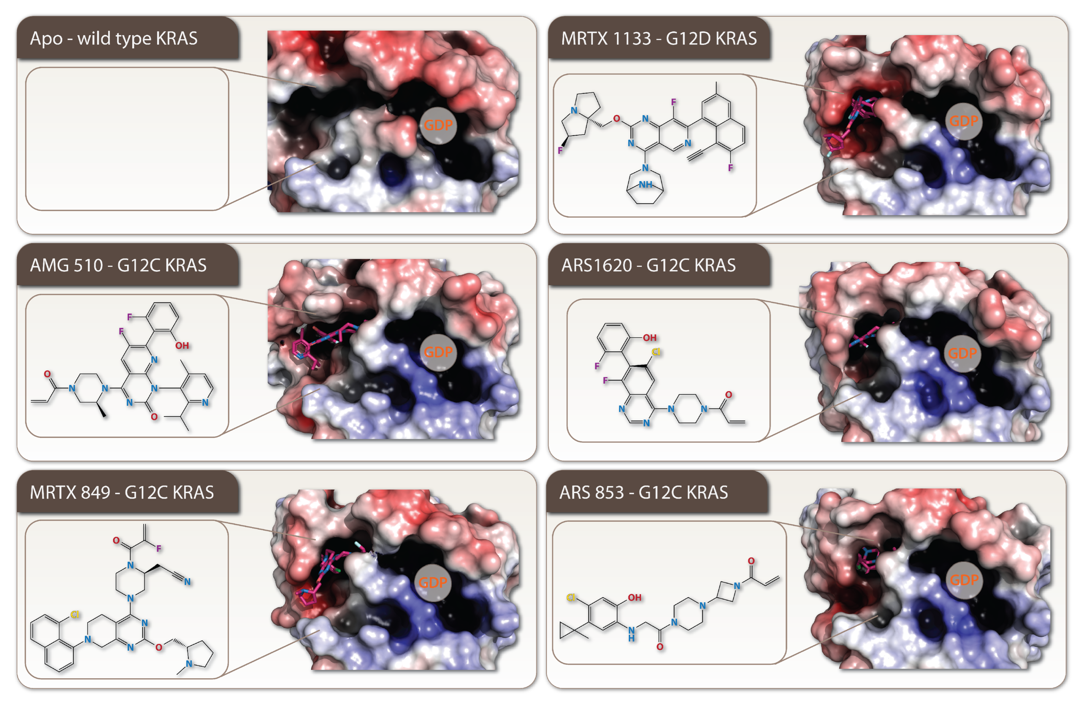
2. Targeting of the Specific KRAS-Mutants

Recent developments, however, attempted to overcome a shortcoming in KRAS targeting and the specific compounds with novel mechanisms of action were developed to a specific KRAS G12C mutant (Table 1). Novel inhibitors covalently target the G12C mutation in the switch II pocket of the protein, which blocks binding of the mutant KRAS with GTP (preventing effector interactions) and halts downstream signaling pathways, such as RAF/MEK/ERK [36].

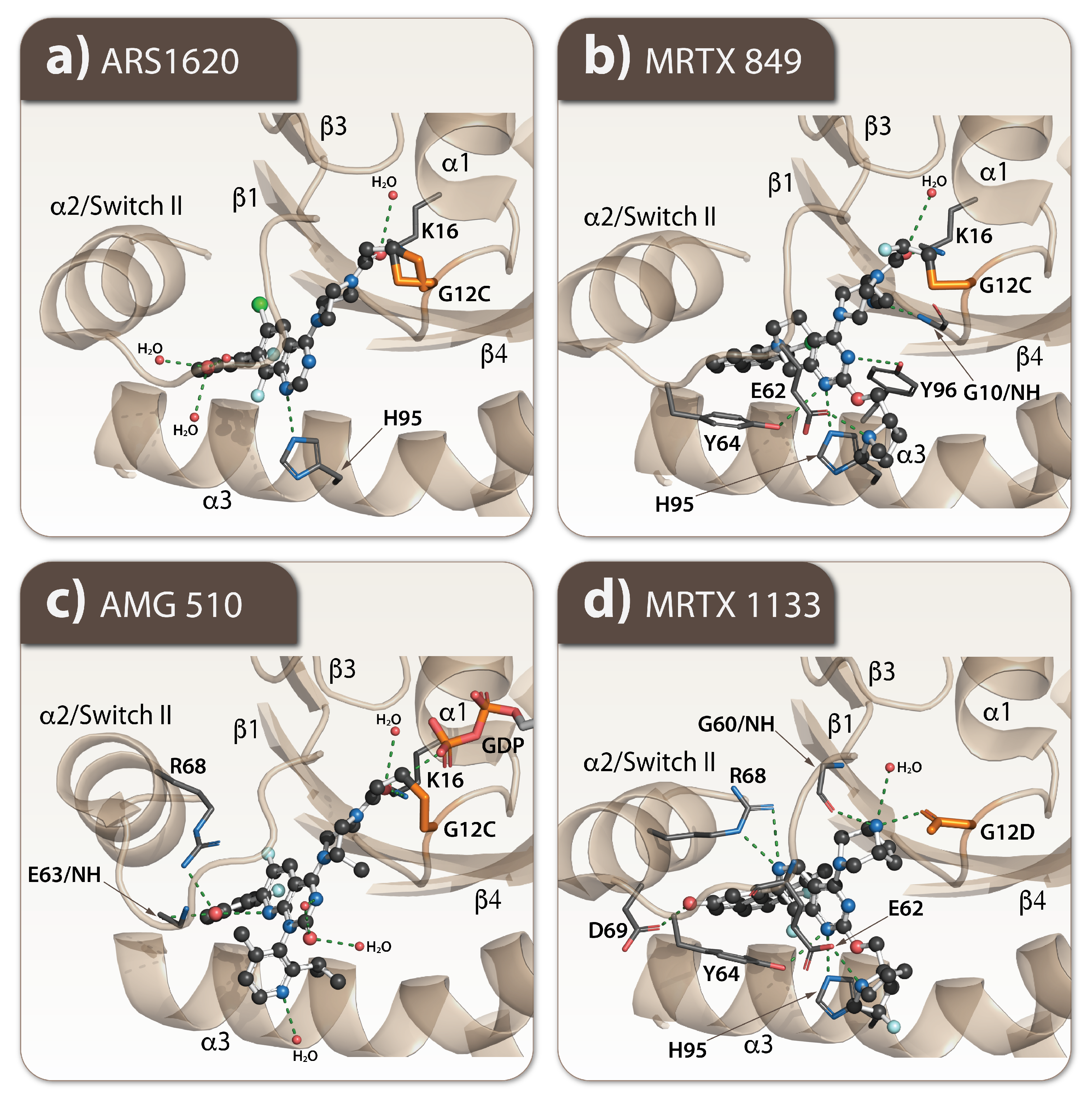
2.1. Every Common Specific KRAS-Mutant: A Case for the Drug Development

Structurally, KRAS consists of an effector binding lobe, comprising the first 86 residues, an allosteric lobe, and a carboxy-terminal region (Figure 1a). The effector lobe has the phosphate-binding loop within a stretch of amino acid residues 10–17 (P-loop), the switch I between amino acid residues 30–38, and the II loop spans between amino acid residues 60–76. The loops of the switch I and II, mediate the protein–protein interactions with downstream signaling effector protein kinase RAF1. The GTP hydrolysis activity of KRAS is performed by the Q61 site. The KRAS mutations at positions G12, G13, Q61, and A146 are located at the effector lobe and lead to a shift toward the active KRAS form through impairing nucleotide hydrolysis and/or activating nucleotide exchange [48]. Shokat and colleagues were the first to find a hidden pocket to target KRAS and effectively use it in therapy. This pocket is located in the switch II next to the G12C mutated site (Figure 1b right) [36]. A steric distance between the switch II and mutated C12 site made possible the development of covalent inhibitors of the switch II, thereby achieving allosteric inhibition of cysteine in G12C to prevent the nucleotide exchange catalyzed by GEFs and diminished the subsequent interaction between RAS and RAF [49]. The area of a covalent inhibition is pointed in the switch pocket.
The improved version of this drug, ARS-1620, demonstrated the tumor growth control in a KRAS G12C xenograft model [49] by its additional H-bond interaction with H95 in the switch pocket and a more favorable alignment of the warhead toward C12 (Figure 4c) [50].
There is also a different compound MRTX849 (Adagrasib) designed to target this KRAS G12C mutation. It acts by forming a hydrogen bond (H-bond) with D69, a salt bridge with E62 and a cation- π interaction with H95 in the switch pocket (Figure 4b) [51]. MRTX849 showed a 45% response rate and 8.2-month median duration of response in non-small lung cancer tumors [52]. A different mode of action is carried out by AMG510 (sorotasib); it does not form a H-bond with H95, but instead induces a conformational change of the histidine, resulting in the formation of a new cryptic sub-pocket formed by H95, Y96, and Q99 (Figure 4c). The compound demonstrated a 37.1% response rate, a progression-free survival of 6.8 months and a median overall survival of 12.5 months in a phase II clinical trial of 126 patients with advanced non-small-cell lung cancer [53]. MRTX1133 is a selective non-covalent inhibitor of KRAS G12D because the aspartate in KRAS G12D has a carboxyl group that is less nucleophilic than the sulfhydryl group of cysteine; therefore, it does not work for G12C (Figure 4d). MRTX1133 binds to the switch II pocket and inhibits the protein–protein interactions necessary for the activation of the downstream pathway of KRAS [44]. Other inhibitors, ARS-853 and AMG 510, target specifically KRAS G12C mutation and do not act on the KRAS G12V or G12D mutants occurring more frequently in human cancers [49,54].
The novel therapeutic development targeting KRAS-mutant cancers also works on discovery of pan-Kras inhibitors and proteolysis targeting chimeras (PROTAC) which avoid HRAS and NRAS [55,56]. Various inhibitors targeting upstream and downstream RAS signaling pathways were reviewed in [56,57]. Promising results of the pre-clinical studies using combinations of inhibitors simultaneously targeting several KRAS signaling regulators including SOS1, SHP2, and EGFR were also reported [58,59,60,61]. The phase I and IIb clinical trials of RMC-4630 (SHP2-inhibitor) plus LY3214996 (ERK-inhibitor) in patients with KRAS mutations in CRC, NSCLC, or PDAC are in progress (NCT04916236). Many other SHP2 inhibitors (TNO155, JAB-3068, JAB-3312, RLY-1971, BBP-398, ERAS-601, PF-07284892/ARRY-558, and SH3809) and SOS1 (BI 1701963, RMC-5845, BAY-293, SDGR5, GH52, ERAS-9, and SOS1i) are in the clinical and preclinical phases [56]. A potentially effective strategy is also to inhibit mutated KRAS gene expression at the mRNA level. A significant reduction in tumor size (pancreatic cancer xenografts) in mice was observed after CRISPR-Cas13a-mediated KRAS G12D mRNA knockdown. A specific KRAS mRNA silencing induced a potent apoptosis in in vitro models [62]. The RNA interference approach to silence KRAS oncogene expression was also tested [63,64]. Novel approaches such as siRNA encapsulated in exosomes and mRNA vaccines are in the clinical trials. Recent pre-clinical studies showed that fibroblast-derived exosomes loaded with G12D siRNA (iExosomes) efficiently attenuate PDAC tumor growth and extend tumor bearing mouse survival [65]. Two of KRAS mRNA vaccines are in phase I clinical trials: mRNA-5671/V941 co-developed by Moderna and Merck (NCT03948763) and ELI-002 by Elicio Therapeutics (for KRAS/NRAS-mutant solid tumor NCT04853017). Lipid nanoparticles encapsulated mRNA vaccines encoding mutant KRAS epitopes G12D, G12V, G13D, and G12C are taken up and translated in antigen-presenting cells, and then presented by MHC molecules on a cell surface [66].
Although some of the described novel therapies show promising results, it is evident that the drug resistance is a common feature of KRAS-mutant cancers that will be a most challenging task to overcome, making it a critical limiting factor in eradication of highly malignant cancers [67]. Therefore, a combination of the cancer targeting therapies with the focus to obtain a potent killing of KRAS-mutant cancer cells will be most effective, if a synergistic potentiation in the drug action outcomes can be realized.

2.2. How Resistance Gets Evolved to the Specific KRAS G12C Mutant Inhibitors

A potent targeting of specific KRAS G12C mutations with a high precision results in the further mutations that disrupt covalent or potentially non-covalent drug-binding and leads to drug resistance. The KRAS R68S and Y96C mutations located within the switch II pocket of the MRTX849 and AMG510-binding site interfere with the drug non-covalent binding interactions and decreases binding affinity substantially [68]. Another KRAS mutation within the same Y96D site alters the switch II pocket and reduces the H-bonding between the Y96 residue of KRAS and MRTX849 [69].
Alternatively, other KRAS mutations such as G13D, A59S, K117N, and A146P residing outside the drug-binding pocket are altering the functional activity of KRAS by enhancing its nucleotide exchange and maintaining its active GTP-bound state not accessible for the drug binding [68]. It has been also indicated that a conformation-specific KRAS G12C inhibitor leads to a rapid non-uniform adaptation where some cancer cells stabilize its active drug-insensitive state and maintain cancer cells in a drug-resistant state [70].
Various studies revealed that KRAS inhibition is possible to suppress by a feedback activation of upstream or downstream mediators and other negative regulators. Cells activated with EGF showed reactivation of KRAS in the cells, suggesting that EGFR mediates resistance to KRAS G12C inhibitors [71]. The changing activity of the downstream effectors of KRAS also leads to occurrence of drug resistance. For example, a sub-clonal evolution of MET amplification in KRAS G12C non-small cell lung cancer cells that have become resistant to AMG510 in vitro [72]. Another way of inducing resistance for AMG510 or MRTX849 might be the continued FAK activation, which leads to weak treatment outcomes by dysregulating FAK-YAP signaling. Focal adhesion kinase (FAK) is a non-receptor kinase which plays a role in cell growth regulation and transduction of signals [73]. Moreover, cells which acquired resistance to AMG510 seemed to undergo epithelial-to-mesenchymal transition (EMT). Such a kind of induction leads to activation of expression of several RTKs, such as ERBB3 and FGFR. Moreover, drug resistance induced by EMT causes the enhancement of PI3K/AKT and MAPK signaling [74]. Another mechanism that involves the development of so-called adaptive resistance, whereby KRAS-dependent tumors under the treatment pressure converse from one histological type to another, is driven by the primary cancer type. The transformation from adenocarcinoma to squamous cell carcinoma was observed during MRTX849 treatment without any other drug-resistance mechanisms [75]. Hallin et al. showed that a cell cycle dysregulation changed KRAS-mutant allele frequency, which is an additional factor that could suppress the therapeutic response to MRTX849 [38]. Hereby, we described only a few possible mechanisms of resistance toward novel KRAS-specific inhibitors with other existing and with the potential of KRAS-mutants to develop new complex ways to disable treatment efficacy. To overcome these, the different combinations of drugs are applied to block upstream, downstream, and other factors of regulation which are associated with KRAS signaling.

3. Deregulation of Metabolism in Cancer

The main source of energy and ATP production in primary cells is mitochondrial oxidative phosphorylation (OXPHOS), whereas in tumor cells the production of ATP by OXPHOS is low and most of the glucose is utilized by its fermentation to lactate even in the presence of oxygen, the process known as an aerobic glycolysis or Warburg effect (Figure 5) [76,77,78]. In normal cells, the pyruvate from glycolysis preferentially enters the mitochondrial matrix where it is oxidized to acetyl-coenzyme A (acetyl-CoA) by the pyruvate dehydrogenase (PDH) complex. Acetyl-CoA is then metabolized through tricarboxylic acid (TCA) cycle, followed by OXPHOS for a productive ATP generation (up to 38 ATP molecules per one molecule of glucose are generated) [79]. An abnormal glucose metabolism is inherent in cancer cells as one of the well-studied characteristics first described by Warburg who reported association between an elevated glucose uptake and decrease in oxygen consumption with an increased production of lactic acid in aerobiosis. In cancer cells, the majority of produced pyruvate is uncoupled from the mitochondrial TCA cycle and OXPHOS processes, and is converted by lactate dehydrogenase (LDH) to lactate. Accumulated lactate facilitates tumor progression and acidification of the tumor environment, which in turn promotes tumor proliferation, metastasis, and resistance to developed antitumor therapies [80].

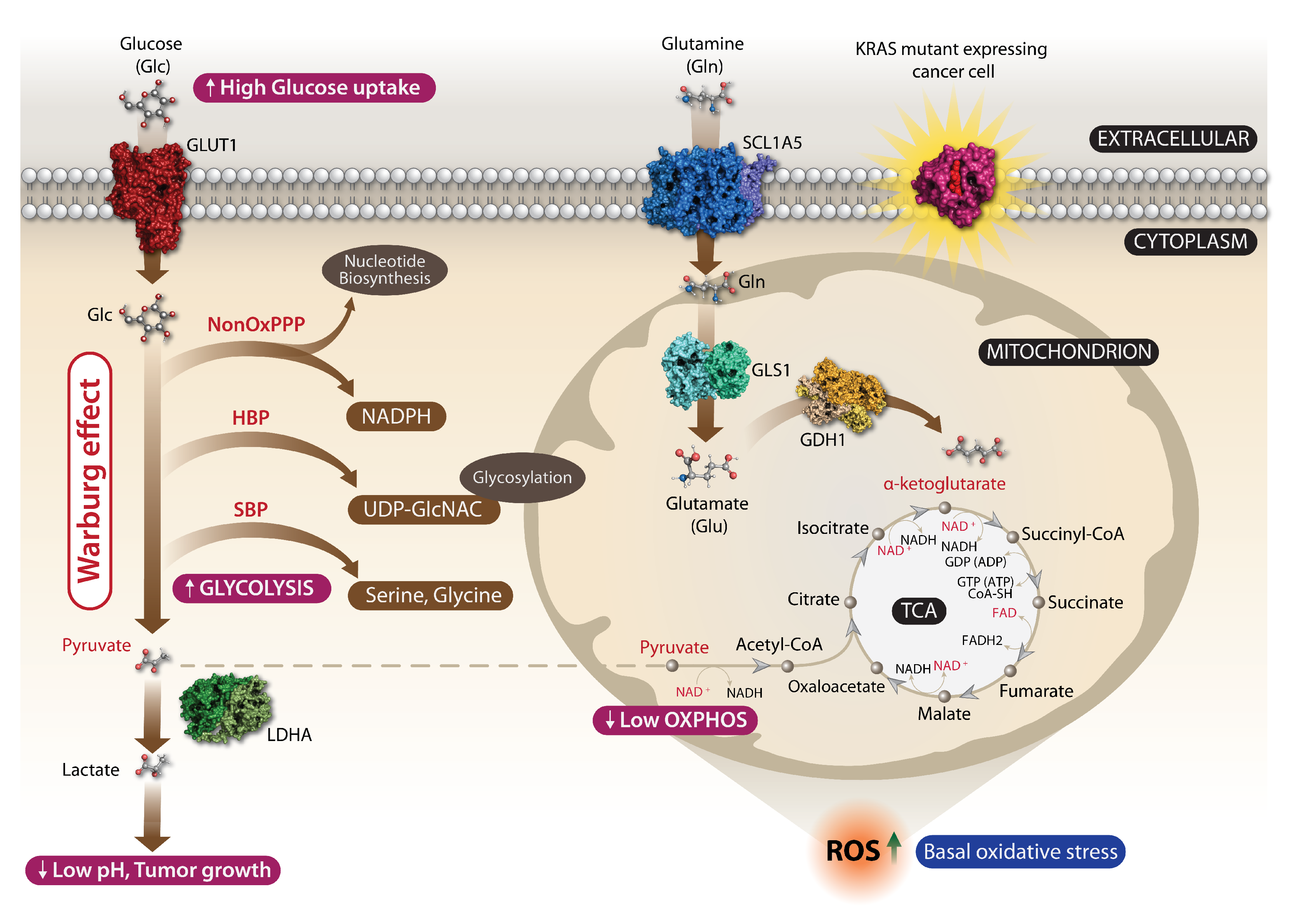
3.1. KRAS Cancer Mutations and Metabolic Reprogramming

An oncogenic KRAS sets the metabolic changes by hyperactivation of growth factor signaling that ultimately leads to increase of a cellular glycolytic flux facilitating tumor growth [81,82]. Intensive anabolic processes in cancer cells are maintained by an enhanced glucose consumption and its fermentation to lactate (aerobic glycolysis) [83]. It generates an anaerobic ATP in cancer cells supporting growth in hypoxic conditions of a tumor environment. The advantage of a high glycolytic flux is a continuous supply of glycolytic intermediates for the pentose phosphate pathway to gain an elevated synthesis of nucleotides and phospholipids for actively growing and proliferating cancer cells, which is highly relevant for KRAS-mutant cancers because of their glucose addiction.
The metabolic shift in KRAS-mutant cancer cells relies on a high absorption of glucose mediated by abundant glucose transporters (GLUT) where GLUT1 is considered as a key supplier of glucose in cancer cells promoting an aerobic glycolysis in tumorigenesis (Figure 5) [84]. Glucose metabolism intermediates are then channeled into non-canonical biosynthesis pathways such as the non-oxidative arm of a pentose phosphate pathway to produce ribose for biosynthesis of RNA/DNA and the hexosamine biosynthesis pathway (HBP) that produces glycosylation precursors [85,86,87]. A high reliance on glutamine of cancer cells for tumor growth was also reported, whereby the glutamine-fueled TCA cycle results in generation of ATP, reactive oxygen species (ROS), NADPH, amino acids, nucleotides, and lipids that are critical for KRAS oncogene-induced tumorigenicity [85,88,89,90,91].

3.2. A Basal Oxidative Stress Is a Liability of Malignant KRAS-Mutant Cancers

An altered cellular signaling increases the production of ROS as well as activating antioxidant programs, which are advantageous and drive tumorigenesis of KRAS-mutated cancers. An oncogenic KRAS can promote ROS generation by regulating HIFs, altering mitochondrial function by suppressing the respiratory chain complex I and III and inducing expression of the transferrin receptor. To balance the redox state, KRAS-mutant cancer cells display upregulation of major antioxidant enzymes (peroxiredoxin 3, thioredoxin peroxidase, catalase) and reduced glutathione (GSH) levels, as well as enhanced detoxification pathways and resistance to apoptotic death in response to oxidative compounds such as hydrogen peroxide (H2O2) [84]. It is still not clear how the RAS oncogene modulates the redox equilibrium or whether pro- or anti-oxidant elements contribute to a RAS-induced transformation and tumorigenicity. Expression of RAS itself can induce ROS production in cancer as a result of increased metabolic rate, genetic mutations, and hypoxia. A mitochondrial ROS generation is essential for KRAS-mediated cancerogenesis [92]. Apart from mitochondrial ROS, NADPH oxidase 1 (NOX1) increased expression and activity have been observed in KRAS-driven CRCs leading to additional ROS production. A NOX1-mediated ROS production is essential to support RAS transformation [93,94].
The translocalization of KRAS protein from the plasma membrane to the mitochondria was observed in KRAS G12V-expressing cells by a doxycycline inducible system. The expression of KRAS G12V protein causes a dysfunction of mitochondria by a significant decrease in mitochondrial respiratory activity (complex I), oxygen consumption, and mitochondrial membrane potential Δ Ψ m. In addition, an elevated ROS generation and adaptive upregulation of GSH synthesis in response to the sustained ROS production have been observed in this cellular model [95]. A KRAS hyperactivation alters a mitochondrial morphology caused by transformations in mitochondrial fusion and fission with subsequent changes in cellular metabolism [96]. However, oxidative stress is a vulnerable state of KRAS-mutant cells and can be used to induce cytotoxic response. A glucose withdrawal leads to increased oxidative stress driven by NOX1 and mitochondria and can activate a number of kinases such as ERK, JNK, and LYN in cells carrying KRAS mutations which are dependent on glucose for survival [97]. Glucose deprivation activates an amplification loop, with an increased phospho-tyrosine signaling, until ROS accumulates above a toxicity threshold. Once it exceeds the capacity of cell antioxidant systems, it triggers a cell death.

4. The Vitamin C Cancer Treatment Is Back: How a Famous Antioxidant Turns to a “Trojan Horse” Oxidant in KRAS-Mutant Cancer Cells

4.1. Vitamin C (Ascorbic Acid) in Cancer Treatment

Vitamin C (VC) or ascorbic acid is a water-soluble vitamin and antioxidant present in many vegetables and fruits. It plays an essential role in the majority of cellular processes. An increased risk of cancer mortality is associated with VC deficiency [98,99]. In high doses, VC is almost non-toxic to normal cells in in vitro experiments but highly toxic for a number of human tumor cell lines. The pioneering study by Linus Pauling and Ewan Cameron (double Nobel Prize winners) showed anticancer abilities of high doses of VC in the 1970s. The end-stage cancer patients were treated with 10 g per day of VC combined with chemotherapy, and their survival rate reached above one year [100]. The presence of KRAS mutations in cultured human CRC cells was associated with selective killing by high levels of oxidized VC, suggesting that KRAS status can at least partially explain response to VC [101]. Recent in vitro and in vivo preclinical studies of VC in various human cancers confirmed its efficacy and cytotoxic effects on cancer cells [102], but efficacy in clinical trials was limited [103,104].
Administration of VC to patients by an intravenous (IV) route is the most effective way to deliver a high dose of the drug. It appeared that an IV administration of VC achieved millimolar levels of the drug sufficient to provoke cytotoxicity in cancer cells [105]. In a phase I clinical study, the IV infusion of 100 g of VC reached 25–30 mM level of the drug in circulation that decreased only to a 10-mM range following 4 h. Such a high dose of VC displays antitumor activity sufficient to slow growth of cancer cells [106]. However, the oral administration of VC reached only 200 μ M with a further increase to 400 μ M by a special encapsulated formulation of VC [105,107]. Novel VC pharmaceutical formulations focus on increasing structural stability and cellular uptake [108,109].
Numerous studies demonstrated potential anticancer activity of VC alone or in combination of a high dose of VC with other conventional therapies. First, a human phase I clinical trial was conducted, with VC administration by IV with the radiotherapy enhanced radiosensitization of pancreatic cancer, while a protective effect from radiation was observed in a surrounding tissue [110]. It has been also reported that combining of a high dose of VC with the anti-cancer drugs eribulin mesylate, tamoxifen, fulvestrant, or trastuzumab was cytotoxic for breast cancer cells [111]. The efficacy of an immune checkpoint therapy (ICT), targeting PD-1/PD-L1 and CTLA-4 molecules in breast cancer or CRC cell lines was enhanced by the co-treatment with VC [112]. Other studies show that VC at 50 μ M can reduce viability of HCT116 cells when combined with the anti-neoplastic DNA-demethylating agents decitabine (DAC) and azacytidine (AZA) [113]. An intravenous administration of VC at 25–50 g per day every 1–2 weeks in patients with small cell lung cancer (SCLC) in combination with the alkali therapy results in median survival increased to 44.2 months compared to the control group with 17.7 month survival [114].
Calorie-restricting diets (low glucose or proteins) are referred to as fasting-mimicking diets (FMDs). The anti-tumor additive or synergistic effects were shown following the co-treatment of FMD with chemotherapy or number of targeted therapies in several preclinical breast, lung, and colorectal cancer models [115]. FMD contributes to sensitizing cancer cells to chemotherapy by increasing ROS production [116,117]. Importantly, it has been shown that FMD can increase anticancer activity of VC in KRAS-mutant CRCs. The synergistic effects of FMD and VC treatments were observed in cell culture and animal models in vivo. The combination of FMD and VC was the most effective and also most well-tolerated, with low toxicity [118]. FMD enhances a VC’s cytotoxicity in KRAS-mutant cancer cells by increasing reduced ferritin (a protein which binds iron), which further leads to an increase in reactive iron level, ROS production, and cell apoptosis. This effect can be further boosted by chemotherapy [118]. This promising combination should be further tested by well-designed randomized clinical trials in CRC and other KRAS-mutant tumors.

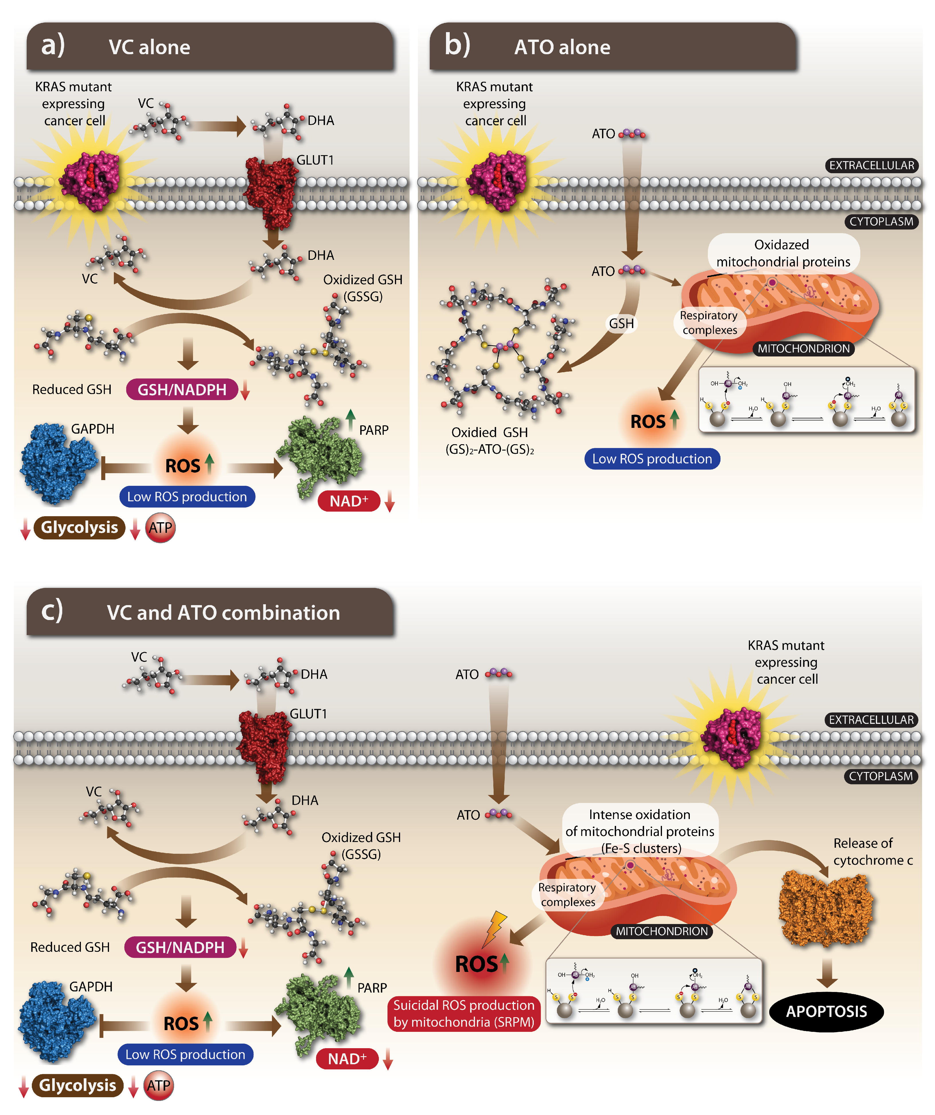
4.2. The Anti-Cancer Action of VC: How Does It Work

Metabolic reprogramming sensitizes KRAS-mutant cancer cells to a high dose of VC by meddling in an epigenetic regulation, hypoxia signaling, and intracellular iron metabolism (Figure 6a) [119]. The high dose of VC kills cancer cells by provoking an oxidative stress in highly malignant cancer cells including KRAS-mutant cancer cells, but how does a potent antioxidant VC become an oxidative factor in cancer cells? It is highly evident that a pro-oxidant action of VC depends on metal-ion redox chemistry. In particular, free iron was shown to be essential for VC-induced cytotoxicity [120]. The oxidative impact is carried out by the oxidized form of VC known as dehydroascorbate (DHA) and the distinct locations of the oxidation VC to the DHA reaction and by reducing DHA back to VC reaction to determine the oxidation of cancer cells. VC gets oxidized in a body to DHA. KRAS mutant cancer cells absorb intensively DHA by a glucose transporter GLUT1 because DHA structure resembles glucose. KRAS metabolic reprogramming associated with elevated expression of GLUT1 allows for an increased uptake of DHA to the cytoplasm, which leads to a disruption in the Warburg effect [120,121]. DHA inside a cancer cell is reduced back to VC at the cost of GSH oxidation. In a high scale, an active absorption of DHA, a decoyed form of glucose, results in a substantial exhaustion of a cellular GSH leading to oxidative stress coupled to ROS accumulation and inactivation of glyceraldehyde 3-phosphate dehydrogenase (GAPDH). A continuous inhibition of GAPDH activity induces oxidative damage via auto-oxidation, which is cytotoxic for highly glycolytic KRAS-mutant cancer cells [101,102].
Elevation of ROS to cytotoxic levels provokes irreversible damage to DNA and mitochondria and triggers apoptotic pathways in cancer cells [122,123,124,125]. In highly glycolytic KRAS-mutant cells, an accumulation of ROS provokes an apoptotic cell death via inhibition of GAPDH, a glycolytic enzyme that has an ability to reduce adenosine triphosphate (ATP) leading to energetic collapse and cell death which is not observed in KRAS wild type cells [101]. A highly glycolytic metabolism of KRAS-mutant cancer cells can be also disrupted by VC through downregulation of important metabolic checkpoints such as GLUT1 and PKM2 [121].
A redox damaging of VC also interferes with cell signaling and mitochondrial function. The effects of VC on EGFR/MAPK signaling pathway in KRAS-mutant CRC lines has been reported, indicating inhibition of MEK1 and ERK1/2 phosphorylation without changes in their protein expression [121]. VC alters a mitochondrial function in KRAS-mutant cancer cells. The VC-dependent modulation of pyruvate dehydrogenase (PDH) activity, as well as a TCA cycle, was reported. In this study, VC induced a significant ATP depletion, quick dissipation of mitochondrial membrane potential and decreased phosphorylation of PDH’s component E1- α on S293 site leading to an increase in PDH, citrate synthase activity, and also an increase in the conversion of pyruvate into acetyl-CoA. Moreover, the downregulation of PDK-1 has been linked to a VC-mediated hydroxylation of proline (P402) in hypoxia inducible factor-1 α (HIF-1 α ) [126]. It is further supported by finding that the VC treatment of pancreatic adenocarcinoma cells suppressed HIF-1 α , which is known to contribute to tumorigenesis. The effect was observed through activation of HIF hydroxylases, which in turn inhibits transcription of HIFs, resulting in delays in tumor growth [127]. It was also reported that high doses of VC are effective in suppressing Apc/Kras G12D mutant tumors [101]. Thus, VC is a potent suppressor of KRAS-mutant tumor growth by provoking an oxidative stress and meddling with cancer metabolism.

5. A Mild Oxidant ATO Is the Cancer Drug: The Mechanism of Action

5.1. The History of ATO: Harnessing the Poison for Cancer Treatment

An inorganic arsenic has long been used as the active ingredient in a traditional Chinese medicine for treatment of numerous conditions including tumors for over 2000 years [128]. In the early 17th century, arsenic trioxide (ATO) solution in potassium bicarbonate was developed by Thomas Fowler and used to treat diseases such as asthma, chorea, eczema, pemphigus, and psoriasis and was later used also to treat anemia, Hodgkin’s disease, and leukemia. Suppression of leukocytes by ATO was reported in 1878 [129,130,131] and later, a treatment of leukemias with ATO alone and in the early 20th century, combined with radiotherapy, was used [132]. For acute promyelocytic leukemia (APL), treatment with ATO was used for the first time in the early 1990s, when it showed high a clinical remission rate. An injectable formulation of ATO, known as trisenox, was approved in 2000 by the FDA for treating relapsed or refractory APLs. The majority of patients with APL achieve a complete remission [133,134]. The ATO treatment was not well-suited for solid tumors due to a dose-related toxicity and unfavorable pharmacokinetics. However, some promising results of ATO treatment have been observed in patients with refractory/relapsed CRC resistant to 5-flurouracil, which is the most widely used clinical treatment [135,136].

5.2. The Oxidizing Impact of ATO

Arsenic oxidants execute the substantial stress-related cellular changes by inducing apoptosis, halting proliferation, stimulating differentiation, and inhibiting angiogenesis through various mechanisms. The main mechanism of an antitumor ATO’s action is induction of an ROS-dependent apoptosis (Figure 6b). Exposure to ATO provokes an oxidant–antioxidant imbalance in cells, as it can promote generation of variety of ROS, such as H2O2, superoxide (O2¯•), nitric oxide (•NO), perhydroxyl (HOO•), dimethyl arsenic peroxyl ((CH3)2AsOO•), and dimethyl arsenic ((CH3)2As•) radicals causing oxidative stress. Accumulation of intracellular ROS alters mitochondrial membrane potential causing its dissipation, release of cytochrome c and activation of the caspases, ultimately inducing the mitochondrial apoptotic pathway [137]. The cytotoxic drugs including ATO often target mitochondria, which then initiate apoptotic processes. Arsenic is recognized as an uncoupler of mitochondrial oxidative phosphorylation. This effect is achieved by the ATO binding to reactive thiol groups on proteins (ligands) located on cysteine residues. Cysteine residues are critical to many unique mitochondrial functions, including ROS production. Mitochondrial proteins, including cysteine residues in these proteins, undergo redox changes through modifications to the thiol groups on cysteines. The localization of proteins and their interactions are greatly influenced by oxidative post-translational modifications (PTM) of cysteine residues, which in turn are ROS-dependent. These cysteine PTMs include oxidation to sulfenic/sulfinic/sulfonic acids, S-nitrosation, S-glutathionylation, and disulfide-bond formation [138]. An ATO binding to thiol groups of mitochondrial enzymes involved in electron transfer chain results in impaired tissue respiration. A direct binding of ATO to proteins results in thiol oxidation leading to changes of various protein conformation and to inhibition of their function.
Glutathione (GSH) is a non-protein thiol and a reducing substance essential for the detoxification of endogenous and exogenous oxidative compounds. It makes it an important element in the antioxidative redox system of cells, counterbalancing ROS produced by cellular metabolism or invading oxidants from outside. GSH directly neutralizes ATO by binding arsenic, and it also controls its metabolism by facilitating a cellular arsenic uptake, alteration of arsenic methylation and stimulation of excretion of methylated arsenic forms [139,140]. Arsenic can decrease GSH levels in three ways: first, by reducing arsenates to arsenites using electrons donated by GSH; secondly, by direct binding thiol groups of GSH; and thirdly, by inducing ROS which cause GSH oxidation [141]. Normal and cancer cells with reduced GSH levels are highly sensitive to ATO treatment [142]. In fibroblasts and APL cells, ATO dysregulates a redox balance by increasing intracellular ROS levels and simultaneous depletion of GSH [143]. It has been clearly demonstrated that cancer cells with lower levels of glutathione peroxidase and catalase are highly sensitive to arsenite. Moreover, the experimental conditions leading to lowering cellular GSH levels sensitize cells to arsenite with natural or acquired resistance to the oxidant, while cells with greater levels of reduced GSH are protected from an ATO-induced apoptosis [143,144].

5.3. The Action of ATO on a Cellular Metabolism and Its Nuclear Effects

ATO inhibits glycolysis by direct binding to the cysteine residues (C256 and C704) on hexokinase 2 (HK2) and halting its activity [145]. Pyruvate kinase (PKM2), another known target of ATO, can modulate oncogenic anaerobic glycolysis, known as the Warburg effect, inducing further tumorigenesis and proliferation of cancer cells because PKM2 deletion can enhance oxidative phosphorylation [146]. Moreover, the HK2 overexpression significantly rescues the cells from ATO-induced apoptosis [145,147,148]. Exposure to inorganic arsenic can significantly inhibit many other enzymes involved in glucose metabolism and further induce insulin resistance and diabetes [141]. ATO can also activate caspases, which can directly trigger many apoptotic molecular and structural changes [144,149,150,151] as well as can promote non-classical apoptosis pathways such as ETosis, extracellular DNA traps, and pyroptosis [152]. Moreover, an inorganic arsenic acts as an epigenetic modifier of genes involved in the critical cellular processes, such as cellular growth and immune response [153]. It can induce substantial epigenetic changes by the inhibition of DNA methyltransferases [154]. In CRC cells, ATO altered transcriptional activity of several unmethylated cell cycle regulatory genes including cyclin B1, E1, D1, and GADD45A, which causes cell cycle arrest [155]. The antitumor action of ATO can be also facilitated by up- or downregulation of miRNAs in cancer cells and consequently decreased growth, increased apoptosis, or suppression of cancer migration and invasion [156]. Moreover, downregulation of certain miRNAs can sensitize cancer cells to arsenic treatment [157,158]. Studies have shown that ATO inhibits the nuclear factor (NF)- κ B signaling pathway in leukemia cells and hepatocellular carcinoma stem cells by downregulating expression of p65, p50, p52, P65, c-rel, and RELB members of the NF- κ B pathway [159,160]. The diverse action of ATO highlights a necessity of further mechanistic studies to reveal its anticancer effects. The downside of ATO’s therapeutic effects is that it triggers severe side effects including cardio-toxicity, hepatotoxicity, neurotoxicity, and skin problems and affects kidney function, which limits its clinical applications. Combinations of ATO with other FDA-approved drugs have been tested, showing better results with less adverse effects.

6. ATO Potentiates an Oxidative Effect of VC and the Combination of Both Drugs Is Effectively Killing KRAS-Mutant Cancer Cells

The development of potent therapeutics targeting oncogenic KRAS signaling remains a most critical and challenging area in oncology. Although specific KRAS-mutant cancer inhibitors were developed recently with a hope of resolving the critical problem in cancer treatment, their applications gain only a partial impact because tumor resistance occurs at a high rate. It is likely that the targeting of specific KRAS-mutant cancers in combination with other established cancer drugs or treatments such as radiation or chemotherapy might lead to effective therapeutics. Considering that a direct targeting of oncogenic KRAS remains elusive and challenging, an unbalanced redox state of highly malignant KRAS-mutant cancers is an attractive area for therapeutics development.
A high dose of VC is a powerful leverage in an induction of a glucose-dependent oxidative stress in KRAS-mutant cancer cells. It was demonstrated that absorption of the oxidized form of VC, DHA-resembling glucose, is sufficient to exhaust a cellular GSH system because of an intracellular reduction of DHA back to VC mediated by oxidation of GSH. It is evident that a high dose of VC is a powerful approach in inducing the oxidative stress selectively in KRAS-mutant cancer cells due to their enhanced glucose absorption. In view of the glucose-dependent impact of VC, a potent cytotoxic impact of VC in KRAS-mutant cancer cells is observed mostly at a low glucose cell culture condition. This means that VC alone, even at a high dose, is effective in inducing a potent oxidative stress in KRAS-mutant cancer cells and reaching a potent cytotoxic impact in clinical settings that might not be feasible consistently because of DHA’s competition with glucose.
Considering the selectivity of VC in inducing an oxidizing impact in KRAS-mutant cancer cells, there were several cancer clinical trials initiated to test a high dose of VC or its combination with other common cancer drugs such as gemcitabine, paclitaxel, cisplatin, or docetaxel, and in some cases including the radiation treatment [102]. Although the clinical efficacy of the high-dose VC applications are actively examined, it is also sensible to enhance an oxidizing effect of VC with another oxidizing drug, ATO. The powerful synergistic impact of the VC and ATO combination was observed to effectively kill selected KRAS-mutant cancer cells and was recently reported [161]. The study clearly indicates that a potentiating impact of ATO in enhancing the oxidizing effect of VC is escalated to a potent cytotoxic impact selectively in KRAS-mutant cancer cells without affecting primary and other cancer cells. Importantly, the ATO and VC combination was effective at high-glucose cell culture conditions, indicating a potency of the drug combination without interference of glucose presence that has been evident for the action of VC alone. The advantage of the ATO and VC combination was also evident in in vivo xenograft study, when VC was effective at a much lower (1.5 g/kg) dose with ATO when compared to the VC dose injection alone (4 g/kg). Moreover, a synergistic action of ATO and VC in killing KRAS-mutant cancer cells (the human SW620 and LOVO colorectal adenocarcinoma cell lines) has also been described by another group [162]. If a glucose uptake determines the effect of VC, it is likely that the oxidizing effect of an ATO and VC combination will be more effective under fasting conditions.
A key finding of the ATO- and VC-induced cytotoxicity was a mechanism of killing KRAS-mutant cancer cells (Figure 6c) [161]. It has been shown that a potent cytotoxic impact was executed by a robust mitochondrial production of ROS that can be designated as a suicidal ROS production by mitochondria (SRPM). How an oxidizing impact of two drugs triggers a robust production of ROS by mitochondria or inducing SRPM is a central question to be actively pursued. A synergistic cytotoxic impact induced by the ATO and VC combination indicates that surpassing an oxidative threshold in KRAS-mutant cancer cells affects the redox-sensitive cellular systems including the most vulnerable mitochondrial oxidative phosphorylation system (OXPHOS) complexes. Taking it into account, a synergy of the ATO and VC combination in killing KRAS-mutant cancer cells might take place by the two-step actions. In the first step, a depletion of GSH is mediated by a high dose of VC that disarms a cellular anti-oxidative shield. In the second step, ATO acts in a full capacity by attacking thiol-reactive groups on cellular proteins with a preference toward the components of mitochondrial OXPHOS complexes [163]. It is likely that a direct oxidation of thiol-reactive proteins within OXPHOS complexes leads to uncontrolled ROS production or SRPM. While the exact mechanism is yet to be defined, the ATO-induced ROS production by mitochondria is coherent with the observations of the arsenic-induced mitochondrial cardiotoxicity [164] or damage in cellular model [165]. Within the mitochondrial OXPHOS complexes, the iron-sulfur (Fe-S) clusters are essential cofactors mediating electron transfer within the mitochondrial respiratory chain. The critical cysteines within OXPHOS complexes are engaged directly by their thiol-reactive groups in a proper coordination of Fe-S clusters and electron transfer [138,166], which are likely the most sensitive thiol-reactive groups for the ATO binding and functional interference.
If this is the case, ATO can directly attack most critical (Fe-S) clusters of OXPHOS, collide electron transport system and trigger ROS generation at a high scale, resulting in cytotoxic impact by SRPM. Thus, according to the proposed hypothesis, the oxidation of thiol-reactive groups within the mitochondrial OXPHOS complexes by ATO is a critical step resulting in aberrant mitochondrial function and provoking SRPM in KRAS-mutant cancer cells, whereas a high dose VC action leads to a GSH depletion step that eliminates an interference with the ATO’s oxidation action (Figure 6c).

7. A Remarkable Turn: The VC’s Enantiomer D-VC Works Better in the Tumor Xenograft Animal Model

A cytotoxic action of ascorbates is mediated by a glucose-dependent oxidizing effect that determines a selectivity in killing of highly malignant KRAS-mutant cancer cells. Among several analogs of VC, only its enantiomer D-VC in combination with ATO showed a similar potent cytotoxic effect in killing KRAS-mutant cancer cells in cell culture [161]. It is coherent with the original study carried out in 1993 describing a direct cell-killing action of VC or D-VC on fast growing malignant cancer cells by defining their mechanism of action indicating that “certain oxidation and degradation products of ascorbate were cytotoxic agents” for malignant cancer cells [167]. D-VC, also known as erythorbic acid, is a common food additive because it is a non-toxic antioxidant. A lower toxicity of D-VC versus VC is expected because more rapid clearance of D-VC from human bodies has been reported [168]. In addition, a lower toxicity of D-VC is also supported by the higher tolerance and survival of mice injected with high doses of D-VC versus the VC injection effects (Begimbetova and Sarbassov, unpublished data). More detailed comparative toxicological studies of VC and D-VC will be highly informative in defining a chirality-dependent toxicity of VC.
An unexpected finding was made when the animal xenograft studies of VC and D-VC were carried side by side, although no difference was detected in their cytotoxic effects in the cell culture setting. The KRAS-mutant tumor xenograft study in mice showed a clear distinction in the tumor eradicating potency between the VC and D-VC enantiomers. The injections of ATO, VC or D-VC alone had only weak and partial tumor suppressing effects, but the ATO and D-VC combination had much more potent KRAS-mutant tumor suppressing impact compared to the group of tumor-bearing mice injected with ATO and VC [161]. This finding shows that the ATO and D-VC drug combination is superior to the ATO and VC injections in suppressing KRAS-mutant tumors only in mouse models that has not been detected in cell culture condition. It indicates that D-VC, a non-natural enantiomer of VC, has distinctive pharmacokinetic properties increasing its tumor-suppressing capacity in the animal model. It is a rare case when a stereoisomeric analog shows a higher potency in animal models that is highly relevant for enhancing clinical applications of VC.
There were no reports of clinical applications of D-VC that explains a scarceness of information on a chirality-dependent pharmacokinetics of VC. One possible explanation of a D-VC superiority in vivo might be related to the report indicating the eight times-slower oxidation rate of D-VC to DHA compared to the oxidation rate of its natural enantiomer VC [169]. It might lead to advantage for the D-VC treatment by resulting in a higher accumulation of its oxidized form DHA in circulation and, subsequently, inducing a more potent oxidizing impact in KRAS-mutant cancer cells.

8. Conclusions

A combination of two known drugs, ATO and VC, works synergistically by enhancing a glucose-dependent oxidation effect and selective killing of KRAS-mutant cancer cells. How an oxidative stress provoked by the ATO and VC treatment triggered a SRPM is an attractive mechanism to explore. Understanding of SRPM will explain how a potent cytotoxic impact occurs in KRAS-mutant cancer cells. A promising ATO and VC or ATO and D-VC drug combination has to be studied in clinical trials to evaluate efficacy of the drug combination in suppressing KRAS-mutant cancers. In the previous cancer clinical trials, ATO was administered only with a low dose of VC. To alleviate the toxic effect of ATO at its maximum concentration (0.25 mg/kg), the antioxidant oral supplement of VC (1 g/day) has been provided [170,171,172]. At low dose, VC was not effective to potentiate the action of ATO. It will be critical to carry out the cancer clinical trials by administering ATO with a high dose of VC. A chirality-dependent action of VC also has to be addressed by performing the pharmacokinetics of VC and D-VC that will determine why D-VC is more effective in animal models.

Funding

The work was supported by the grant AP08857553 “Development of pancreatic cancer therapeutics by the oxidative drug combination targeting KRAS-mutant cancer cells” funded by the Ministry of Science and Higher Education of the Republic of Kazakhstan, to D.D.S. This research has been funded by Nazarbayev University under Faculty Development Competitive Research Grants No. 240919FD3904 to D.D.S, and 110119FD4520 to F.M.

Conflicts of Interest

The authors declare no conflict of interest.

References

  1. Tsuchida, N.; Ryder, T.; Ohtsubo, E. Nucleotide sequence of the oncogene encoding the p21 transforming protein of Kirsten murine sarcoma virus. Science 1982, 217, 937–939. [Google Scholar] [CrossRef] [PubMed]
  2. Tsuchida, N.; Uesugi, S. Structure and functions of the Kirsten murine sarcoma virus genome: Molecular cloning of biologically active Kirsten murine sarcoma virus DNA. J. Virol. 1981, 38, 720–727. [Google Scholar] [CrossRef] [Green Version]
  3. Hancock, J.; Cadwallader, K.; Paterson, H.; Marshall, C. A CAAX or a CAAL motif and a second signal are sufficient for plasma membrane targeting of ras proteins. Embo J. 1991, 10, 4033–4039. [Google Scholar] [CrossRef] [PubMed]
  4. Colicelli, J. Human RAS superfamily proteins and related GTPases. Sci. STKE 2004, 2004, RE13. [Google Scholar] [CrossRef] [Green Version]
  5. Zenonos, K.; Kyprianou, K. RAS signaling pathways, mutations and their role in colorectal cancer. World J. Gastrointest. Oncol. 2013, 5, 97. [Google Scholar] [CrossRef] [PubMed]
  6. Bos, J.L.; Rehmann, H.; Wittinghofer, A. GEFs and GAPs: Critical elements in the control of small G proteins. Cell 2007, 129, 865–877. [Google Scholar] [CrossRef] [Green Version]
  7. Simanshu, D.K.; Nissley, D.V.; McCormick, F. RAS proteins and their regulators in human disease. Cell 2017, 170, 17–33. [Google Scholar] [CrossRef] [Green Version]
  8. Smit, V.T.; Boot, A.J.; Smits, A.M.; Fleuren, G.J.; Cornelisse, C.J.; Bos, J.L. KRAS codon 12 mutations occur very frequently in pancreatic adenocarcinomas. Nucleic Acids Res. 1988, 16, 7773–7782. [Google Scholar] [CrossRef]
  9. Waddell, N.; Pajic, M.; Patch, A.-M.; Chang, D.K.; Kassahn, K.S.; Bailey, P.; Johns, A.L.; Miller, D.; Nones, K.; Quek, K. Whole genomes redefine the mutational landscape of pancreatic cancer. Nature 2015, 518, 495–501. [Google Scholar] [CrossRef] [Green Version]
  10. Aviel-Ronen, S.; Blackhall, F.H.; Shepherd, F.A.; Tsao, M.-S. K-ras mutations in non-small-cell lung carcinoma: A review. Clin. Lung Cancer 2006, 8, 30–38. [Google Scholar] [CrossRef]
  11. Russo, M.; Di Nicolantonio, F.; Bardelli, A. Climbing RAS, the everest of oncogenes. Cancer Discov. 2014, 4, 19–21. [Google Scholar] [CrossRef] [PubMed] [Green Version]
  12. Fang, B. RAS signaling and anti-RAS therapy: Lessons learned from genetically engineered mouse models, human cancer cells, and patient-related studies. Acta Biochim. Biophys. Sin. 2016, 48, 27–38. [Google Scholar] [CrossRef] [PubMed] [Green Version]
  13. Cekani, E.; Epistolio, S.; Dazio, G.; Cefalì, M.; Wannesson, L.; Frattini, M.; Froesch, P. Molecular Biology and Therapeutic Perspectives for K-Ras Mutant Non-Small Cell Lung Cancers. Cancers 2022, 14, 4103. [Google Scholar] [CrossRef] [PubMed]
  14. Chen, K.; Zhang, Y.; Qian, L.; Wang, P. Emerging strategies to target RAS signaling in human cancer therapy. J. Hematol. Oncol. 2021, 14, 1–23. [Google Scholar] [CrossRef] [PubMed]
  15. Bryant, K.L.; Mancias, J.D.; Kimmelman, A.C.; Der, C.J. KRAS: Feeding pancreatic cancer proliferation. Trends Biochem. Sci. 2014, 39, 91–100. [Google Scholar] [CrossRef] [Green Version]
  16. Vasan, N.; Boyer, J.L.; Herbst, R.S. A RAS renaissance: Emerging targeted therapies for KRAS-mutated non–small cell lung cancer. Clin. Cancer Res. 2014, 20, 3921–3930. [Google Scholar] [CrossRef] [Green Version]
  17. Prior, I.A.; Lewis, P.D.; Mattos, C. A comprehensive survey of Ras mutations in cancer. Cancer Res. 2012, 72, 2457–2467. [Google Scholar] [CrossRef] [Green Version]
  18. Mo, S.P.; Coulson, J.M.; Prior, I.A. RAS variant signalling. Biochem. Soc. Trans. 2018, 46, 1325. [Google Scholar] [CrossRef] [Green Version]
  19. Pantsar, T.; Rissanen, S.; Dauch, D.; Laitinen, T.; Vattulainen, I.; Poso, A. Assessment of mutation probabilities of KRAS G12 missense mutants and their long-timescale dynamics by atomistic molecular simulations and Markov state modeling. PLoS Comput. Biol. 2018, 14, e1006458. [Google Scholar] [CrossRef]
  20. Huynh, M.V.; Hobbs, G.A.; Schaefer, A.; Pierobon, M.; Carey, L.M.; Diehl, J.N.; DeLiberty, J.M.; Thurman, R.D.; Cooke, A.R.; Goodwin, C.M. Functional and biological heterogeneity of KRASQ61 mutations. Sci. Signal. 2022, 15, eabn2694. [Google Scholar] [CrossRef]
  21. Blons, H.; Emile, J.-F.; Le Malicot, K.; Julié, C.; Zaanan, A.; Tabernero, J.; Mini, E.; Folprecht, G.; Van Laethem, J.-L.; Thaler, J. Prognostic value of KRAS mutations in stage III colon cancer: Post hoc analysis of the PETACC8 phase III trial dataset. Ann. Oncol. 2014, 25, 2378–2385. [Google Scholar] [CrossRef] [PubMed]
  22. Lee, D.-W.; Kim, K.J.; Han, S.-W.; Lee, H.J.; Rhee, Y.Y.; Bae, J.M.; Cho, N.-Y.; Lee, K.-H.; Kim, T.-Y.; Oh, D.-Y. KRAS mutation is associated with worse prognosis in stage III or high-risk stage II colon cancer patients treated with adjuvant FOLFOX. Ann. Surg. Oncol. 2015, 22, 187–194. [Google Scholar] [CrossRef]
  23. Yoon, H.H.; Tougeron, D.; Shi, Q.; Alberts, S.R.; Mahoney, M.R.; Nelson, G.D.; Nair, S.G.; Thibodeau, S.N.; Goldberg, R.M.; Sargent, D.J. KRAS Codon 12 and 13 Mutations in Relation to Disease-Free Survival in BRAF–Wild-Type Stage III Colon Cancers from an Adjuvant Chemotherapy Trial (N0147 Alliance) Prognostic Impact of Specific KRAS Mutations in Colon Cancer. Clin. Cancer Res. 2014, 20, 3033–3043. [Google Scholar] [CrossRef] [PubMed] [Green Version]
  24. Park, H.E.; Yoo, S.-Y.; Cho, N.-Y.; Bae, J.M.; Han, S.-W.; Lee, H.S.; Park, K.J.; Kim, T.-Y.; Kang, G.H. Tumor microenvironment-adjusted prognostic implications of the KRAS mutation subtype in patients with stage III colorectal cancer treated with adjuvant FOLFOX. Sci. Rep. 2021, 11, 14609. [Google Scholar] [CrossRef] [PubMed]
  25. Weinstein, I.B. Addiction to oncogenes–the Achilles heal of cancer. Science 2002, 297, 63–64. [Google Scholar] [CrossRef]
  26. Singh, A.; Greninger, P.; Rhodes, D.; Koopman, L.; Violette, S.; Bardeesy, N.; Settleman, J. A gene expression signature associated with “K-Ras addiction” reveals regulators of EMT and tumor cell survival. Cancer Cell 2009, 15, 489–500. [Google Scholar] [CrossRef] [Green Version]
  27. Dumont, N.; Wilson, M.B.; Crawford, Y.G.; Reynolds, P.A.; Sigaroudinia, M.; Tlsty, T.D. Sustained induction of epithelial to mesenchymal transition activates DNA methylation of genes silenced in basal-like breast cancers. Proc. Natl. Acad. Sci. USA 2008, 105, 14867–14872. [Google Scholar] [CrossRef] [PubMed] [Green Version]
  28. Yi, M.; Nissley, D.V.; McCormick, F.; Stephens, R.M. ssGSEA score-based Ras dependency indexes derived from gene expression data reveal potential Ras addiction mechanisms with possible clinical implications. Sci. Rep. 2020, 10, 10258. [Google Scholar] [CrossRef]
  29. Cox, A.D.; Fesik, S.W.; Kimmelman, A.C.; Luo, J.; Der, C.J. Drugging the undruggable RAS: Mission possible? Nat. Rev. Drug Discov. 2014, 13, 828–851. [Google Scholar] [CrossRef] [Green Version]
  30. Sakai, D.; Taniguchi, H.; Sugimoto, N.; Tamura, T.; Nishina, T.; Hara, H.; Esaki, T.; Denda, T.; Sakamoto, T.; Okuda, H. Randomised phase II study of panitumumab plus irinotecan versus cetuximab plus irinotecan in patients with KRAS wild-type metastatic colorectal cancer refractory to fluoropyrimidine, irinotecan and oxaliplatin (WJOG 6510G). Eur. J. Cancer 2020, 135, 11–21. [Google Scholar] [CrossRef]
  31. Yokota, T. Are KRAS/BRAF mutations potent prognostic and/or predictive biomarkers in colorectal cancers? Anti-Cancer Agents Med. Chem. (Formerly Curr. Med.-Chem.-Anti-Cancer Agents) 2012, 12, 163–171. [Google Scholar] [CrossRef] [Green Version]
  32. Ostrem, J.M.; Shokat, K.M. Direct small-molecule inhibitors of KRAS: From structural insights to mechanism-based design. Nat. Rev. Drug Discov. 2016, 15, 771–785. [Google Scholar] [CrossRef] [PubMed]
  33. Wang, Y.; Kaiser, C.E.; Frett, B.; Li, H.-Y. Targeting mutant KRAS for anticancer therapeutics: A review of novel small molecule modulators. J. Med. Chem. 2013, 56, 5219–5230. [Google Scholar] [CrossRef] [PubMed] [Green Version]
  34. Maurer, T.; Garrenton, L.S.; Oh, A.; Pitts, K.; Anderson, D.J.; Skelton, N.J.; Fauber, B.P.; Pan, B.; Malek, S.; Stokoe, D. Small-molecule ligands bind to a distinct pocket in Ras and inhibit SOS-mediated nucleotide exchange activity. Proc. Natl. Acad. Sci. USA 2012, 109, 5299–5304. [Google Scholar] [CrossRef] [Green Version]
  35. Mottini, C.; Tomihara, H.; Carrella, D.; Lamolinara, A.; Iezzi, M.; Huang, J.K.; Amoreo, C.A.; Buglioni, S.; Manni, I.; Robinson, F.S. Predictive Signatures Inform the Effective Repurposing of Decitabine to Treat KRAS–Dependent Pancreatic Ductal AdenocarcinomaDecitabine Inhibits KRAS–Dependent Growth of Selected PDAC. Cancer Res. 2019, 79, 5612–5625. [Google Scholar] [CrossRef] [Green Version]
  36. Ostrem, J.M.; Peters, U.; Sos, M.L.; Wells, J.A.; Shokat, K.M. K-Ras (G12C) inhibitors allosterically control GTP affinity and effector interactions. Nature 2013, 503, 548–551. [Google Scholar] [CrossRef] [Green Version]
  37. Canon, J.; Rex, K.; Saiki, A.Y.; Mohr, C.; Cooke, K.; Bagal, D.; Gaida, K.; Holt, T.; Knutson, C.G.; Koppada, N. The clinical KRAS (G12C) inhibitor AMG 510 drives anti-tumour immunity. Nature 2019, 575, 217–223. [Google Scholar] [CrossRef]
  38. Hallin, J.; Engstrom, L.D.; Hargis, L.; Calinisan, A.; Aranda, R.; Briere, D.M.; Sudhakar, N.; Bowcut, V.; Baer, B.R.; Ballard, J.A. The KRASG12C Inhibitor MRTX849 Provides Insight toward Therapeutic Susceptibility of KRAS-Mutant Cancers in Mouse Models and PatientsTherapeutic Insight from the KRASG12C Inhibitor MRTX849. Cancer Discov. 2020, 10, 54–71. [Google Scholar] [CrossRef] [PubMed] [Green Version]
  39. Shi, Z.; Weng, J.; Fan, X.; Wang, E.; Zhu, Q.; Tao, L.; Han, Z.; Wang, Z.; Niu, H.; Jiang, Y. Discovery of D-1553, a novel and selective KRas-G12C inhibitor with potent anti-tumor activity in a broad spectrum of tumor cell lines and xenograft models. Cancer Res. 2021, 81, 932. [Google Scholar] [CrossRef]
  40. Shi, Z.; Weng, J.; Fan, X.; Zhu, Q.; Robb, E.; Moriarty, A.; Wick, M.; Jiang, Y.; Zhang, L.; Dai, X. Potent in vivo anti-tumor activity of D-1553 as a single agent and in combination with targeted therapeutics in a broad spectrum of patient-derived xenograft tumor models with KRas G12C mutation. Cancer Res. 2021, 81, 1056. [Google Scholar] [CrossRef]
  41. Weiss, A.; Lorthiois, E.; Barys, L.; Beyer, K.S.; Bomio-Confaglia, C.; Burks, H.; Chen, X.; Cui, X.; de Kanter, R.; Dharmarajan, L. Discovery, Preclinical Characterization, and Early Clinical Activity of JDQ443, a Structurally Novel, Potent, and Selective Covalent Oral Inhibitor of KRASG12C. Cancer Discov. 2022, 12, 1500–1517. [Google Scholar] [CrossRef] [PubMed]
  42. Weiss, A.; Voshol, H.; Porta, D.G.; Fedele, C.; Sterker, D.; De Kanter, R.; Stringer, R.; Widmer, T.; Loo, A.; Guthy, D.A. JDQ443, a covalent inhibitor of KRASG12C with a novel binding mode, shows broad antitumor activity in KRASG12C preclinical models as a single agent and in combination with inhibitors of SHP2, MEK or CDK4/6. Cancer Res. 2022, 82, 4026. [Google Scholar] [CrossRef]
  43. Wang, X.; Allen, S.; Blake, J.F.; Bowcut, V.; Briere, D.M.; Calinisan, A.; Dahlke, J.R.; Fell, J.B.; Fischer, J.P.; Gunn, R.J. Identification of MRTX1133, a noncovalent, potent, and selective KRASG12D inhibitor. J. Med. Chem. 2021, 65, 3123–3133. [Google Scholar] [CrossRef] [PubMed]
  44. Wang, J.; Martin-Romano, P.; Cassier, P.; Johnson, M.; Haura, E.; Lenox, L.; Guo, Y.; Bandyopadhyay, N.; Russell, M.; Shearin, E. Phase I Study of JNJ-74699157 in Patients with Advanced Solid Tumors Harboring the KRAS G12C Mutation. Oncologist 2022, 27, e536–e553. [Google Scholar] [CrossRef]
  45. Peng, S.-B.; Si, C.; Zhang, Y.; Van Horn, R.D.; Lin, X.; Gong, X.; Huber, L.; Donoho, G.; Curtis, C.; Strelow, J.M.; et al. Abstract 1259: Preclinical characterization of LY3537982, a novel, highly selective and potent KRAS-G12C inhibitor. Cancer Res. 2021, 81, 1259. [Google Scholar] [CrossRef]
  46. Kamerkar, S.; LeBleu, V.S.; Sugimoto, H.; Yang, S.; Ruivo, C.F.; Melo, S.A.; Lee, J.J.; Kalluri, R. Exosomes facilitate therapeutic targeting of oncogenic KRAS in pancreatic cancer. Nature 2017, 546, 498–503. [Google Scholar] [CrossRef] [PubMed] [Green Version]
  47. Wang, Q.J.; Yu, Z.; Griffith, K.; Hanada, K.-i.; Restifo, N.P.; Yang, J.C. Identification of T-cell receptors targeting KRAS-mutated human tumors. Cancer Immunol. Res. 2016, 4, 204–214. [Google Scholar] [CrossRef] [Green Version]
  48. Edkins, S.; O’Meara, S.; Parker, A.; Stevens, C.; Reis, M.; Jones, S.; Greenman, C.; Davies, H.; Dalgliesh, G.; Forbes, S. Recurrent KRAS codon 146 mutations in human colorectal cancer. Cancer Biol. Ther. 2006, 5, 928–932. [Google Scholar] [CrossRef] [Green Version]
  49. Janes, M.R.; Zhang, J.; Li, L.-S.; Hansen, R.; Peters, U.; Guo, X.; Chen, Y.; Babbar, A.; Firdaus, S.J.; Darjania, L. Targeting KRAS-mutant cancers with a covalent G12C-specific inhibitor. Cell 2018, 172, 578–589.e517. [Google Scholar] [CrossRef] [Green Version]
  50. Herdeis, L.; Gerlach, D.; McConnell, D.B.; Kessler, D. Stopping the beating heart of cancer: KRAS reviewed. Curr. Opin. Struct. Biol. 2021, 71, 136–147. [Google Scholar] [CrossRef]
  51. Fell, J.B.; Fischer, J.P.; Baer, B.R.; Ballard, J.; Blake, J.F.; Bouhana, K.; Brandhuber, B.J.; Briere, D.M.; Burgess, L.E.; Burkard, M.R. Discovery of tetrahydropyridopyrimidines as irreversible covalent inhibitors of KRAS-G12C with in vivo activity. Acs Med. Chem. Lett. 2018, 9, 1230–1234. [Google Scholar] [CrossRef] [PubMed]
  52. Jänne, P.; Rybkin, I.I.; Spira, A.; Riely, G.; Papadopoulos, K.; Sabari, J.; Johnson, M.; Heist, R.; Bazhenova, L.; Barve, M. KRYSTAL-1: Activity and safety of adagrasib (MRTX849) in advanced/metastatic non–small-cell lung cancer (NSCLC) harboring KRAS G12C mutation. Eur. J. Cancer 2020, 138, S1–S2. [Google Scholar] [CrossRef]
  53. Skoulidis, F.; Li, B.T.; Dy, G.K.; Price, T.J.; Falchook, G.S.; Wolf, J.; Italiano, A.; Schuler, M.; Borghaei, H.; Barlesi, F. Sotorasib for lung cancers with KRAS p. G12C mutation. N. Engl. J. Med. 2021, 384, 2371–2381. [Google Scholar] [CrossRef] [PubMed]
  54. Zeng, M.; Lu, J.; Li, L.; Feru, F.; Quan, C.; Gero, T.W.; Ficarro, S.B.; Xiong, Y.; Ambrogio, C.; Paranal, R.M. Potent and selective covalent quinazoline inhibitors of KRAS G12C. Cell Chem. Biol. 2017, 24, 1005–1016.e1003. [Google Scholar] [CrossRef] [Green Version]
  55. Qi, S.-M.; Dong, J.; Xu, Z.-Y.; Cheng, X.-D.; Zhang, W.-D.; Qin, J.-J. PROTAC: An effective targeted protein degradation strategy for cancer therapy. Front. Pharmacol. 2021, 12, 692574. [Google Scholar] [CrossRef]
  56. Hofmann, M.H.; Gerlach, D.; Misale, S.; Petronczki, M.; Kraut, N. Expanding the Reach of Precision Oncology by Drugging All KRAS-mutantsDrugging All KRAS-mutants. Cancer Discov. 2022, 12, 924–937. [Google Scholar] [CrossRef]
  57. Nagasaka, M.; Li, Y.; Sukari, A.; Ou, S.-H.I.; Al-Hallak, M.N.; Azmi, A.S. KRAS G12C Game of Thrones, which direct KRAS inhibitor will claim the iron throne? Cancer Treat. Rev. 2020, 84, 101974. [Google Scholar] [CrossRef]
  58. Fedele, C.; Li, S.; Teng, K.W.; Foster, C.J.; Peng, D.; Ran, H.; Mita, P.; Geer, M.J.; Hattori, T.; Koide, A. SHP2 inhibition diminishes KRASG12C cycling and promotes tumor microenvironment remodeling. J. Exp. Med. 2021, 218, e20201414. [Google Scholar] [CrossRef]
  59. Koga, T.; Suda, K.; Fujino, T.; Ohara, S.; Hamada, A.; Nishino, M.; Chiba, M.; Shimoji, M.; Takemoto, T.; Arita, T. KRAS secondary mutations that confer acquired resistance to KRAS G12C inhibitors, sotorasib and adagrasib, and overcoming strategies: Insights from in vitro experiments. J. Thorac. Oncol. 2021, 16, 1321–1332. [Google Scholar] [CrossRef]
  60. Ryan, M.B.; Fece de la Cruz, F.; Phat, S.; Myers, D.T.; Wong, E.; Shahzade, H.A.; Hong, C.B.; Corcoran, R.B. Vertical Pathway Inhibition Overcomes Adaptive Feedback Resistance to KRASG12C InhibitionCombined SHP2 and KRASG12C Inhibition. Clin. Cancer Res. 2020, 26, 1633–1643. [Google Scholar] [CrossRef]
  61. Amodio, V.; Yaeger, R.; Arcella, P.; Cancelliere, C.; Lamba, S.; Lorenzato, A.; Arena, S.; Montone, M.; Mussolin, B.; Bian, Y. EGFR Blockade Reverts Resistance to KRASG12C Inhibition in Colorectal CancerOvercoming KRASG12C Inhibitor Resistance in Colorectal Cancer. Cancer Discov. 2020, 10, 1129–1139. [Google Scholar] [CrossRef] [PubMed]
  62. Zhao, X.; Liu, L.; Lang, J.; Cheng, K.; Wang, Y.; Li, X.; Shi, J.; Wang, Y.; Nie, G. A CRISPR-Cas13a system for efficient and specific therapeutic targeting of mutant KRAS for pancreatic cancer treatment. Cancer Lett. 2018, 431, 171–181. [Google Scholar] [CrossRef] [PubMed]
  63. Pecot, C.V.; Wu, S.Y.; Bellister, S.; Filant, J.; Rupaimoole, R.; Hisamatsu, T.; Bhattacharya, R.; Maharaj, A.; Azam, S.; Rodriguez-Aguayo, C. Therapeutic Silencing of KRAS Using Systemically Delivered siRNAs in vivo KRAS Silencing with siRNA. Mol. Cancer Ther. 2014, 13, 2876–2885. [Google Scholar] [CrossRef] [PubMed] [Green Version]
  64. Yuan, T.L.; Fellmann, C.; Lee, C.-S.; Ritchie, C.D.; Thapar, V.; Lee, L.C.; Hsu, D.J.; Grace, D.; Carver, J.O.; Zuber, J. Development of siRNA Payloads to Target KRAS-Mutant CancerRNAi Therapy for KRAS-Mutant Cancer. Cancer Discov. 2014, 4, 1182–1197. [Google Scholar] [CrossRef] [PubMed] [Green Version]
  65. Mendt, M.; Kamerkar, S.; Sugimoto, H.; McAndrews, K.M.; Wu, C.-C.; Gagea, M.; Yang, S.; Blanko, E.V.R.; Peng, Q.; Ma, X. Generation and testing of clinical-grade exosomes for pancreatic cancer. JCI Insight 2018, 3, e99263. [Google Scholar] [CrossRef]
  66. Brea, E.J.; Oh, C.Y.; Manchado, E.; Budhu, S.; Gejman, R.S.; Mo, G.; Mondello, P.; Han, J.E.; Jarvis, C.A.; Ulmert, D. Kinase Regulation of Human MHC Class I Molecule Expression on Cancer CellsKinase Regulation of MHC-I in Tumors. Cancer Immunol. Res. 2016, 4, 936–947. [Google Scholar] [CrossRef] [Green Version]
  67. Zhong, L.; Li, Y.; Xiong, L.; Wang, W.; Wu, M.; Yuan, T.; Yang, W.; Tian, C.; Miao, Z.; Wang, T. Small molecules in targeted cancer therapy: Advances, challenges, and future perspectives. Signal Transduct. Target. Ther. 2021, 6, 1–48. [Google Scholar] [CrossRef]
  68. Awad, M.M.; Liu, S.; Rybkin, I.I.; Arbour, K.C.; Dilly, J.; Zhu, V.W.; Johnson, M.L.; Heist, R.S.; Patil, T.; Riely, G.J. Acquired resistance to KRASG12C inhibition in cancer. N. Engl. J. Med. 2021, 384, 2382–2393. [Google Scholar] [CrossRef]
  69. Tanaka, N.; Lin, J.J.; Li, C.; Ryan, M.B.; Zhang, J.; Kiedrowski, L.A.; Michel, A.G.; Syed, M.U.; Fella, K.A.; Sakhi, M. Clinical Acquired Resistance to KRASG12C Inhibition through a Novel KRAS Switch-II Pocket Mutation and Polyclonal Alterations Converging on RAS–MAPK ReactivationClinical Acquired Resistance to KRASG12C Inhibition. Cancer Discov. 2021, 11, 1913–1922. [Google Scholar] [CrossRef]
  70. Xue, J.Y.; Zhao, Y.; Aronowitz, J.; Mai, T.T.; Vides, A.; Qeriqi, B.; Kim, D.; Li, C.; de Stanchina, E.; Mazutis, L. Rapid non-uniform adaptation to conformation-specific KRAS (G12C) inhibition. Nature 2020, 577, 421–425. [Google Scholar] [CrossRef]
  71. Dunnett-Kane, V.; Nicola, P.; Blackhall, F.; Lindsay, C. Mechanisms of Resistance to KRASG12C Inhibitors. Cancers 2021, 13, 151. [Google Scholar] [CrossRef] [PubMed]
  72. Suzuki, S.; Yonesaka, K.; Teramura, T.; Takehara, T.; Kato, R.; Sakai, H.; Haratani, K.; Tanizaki, J.; Kawakami, H.; Hayashi, H. KRAS Inhibitor Resistance in MET-Amplified KRAS G12C Non–Small Cell Lung Cancer Induced By RAS-and Non–RAS-Mediated Cell Signaling Mechanisms. Clin. Cancer Res. 2021, 27, 5697–5707. [Google Scholar] [CrossRef]
  73. Zhang, B.; Zhang, Y.; Zhang, J.; Liu, P.; Jiao, B.; Wang, Z.; Ren, R. Focal Adhesion Kinase (FAK) Inhibition Synergizes with KRAS G12C Inhibitors in Treating Cancer through the Regulation of the FAK–YAP Signaling. Adv. Sci. 2021, 8, 2100250. [Google Scholar] [CrossRef] [PubMed]
  74. Akhave, N.S.; Biter, A.B.; Hong, D.S. Mechanisms of Resistance to KRASG12C-Targeted TherapyResistance to KRASG12C Inhibitors. Cancer Discov. 2021, 11, 1345–1352. [Google Scholar] [CrossRef]
  75. Chaft, J.E.; Litvak, A.; Arcila, M.E.; Patel, P.; D’Angelo, S.P.; Krug, L.M.; Rusch, V.; Mattson, A.; Coeshott, C.; Park, B. Phase II study of the GI-4000 KRAS vaccine after curative therapy in patients with stage I-III lung adenocarcinoma harboring a KRAS G12C, G12D, or G12V mutation. Clin. Lung Cancer 2014, 15, 405–410. [Google Scholar] [CrossRef]
  76. Liu, Y.; Xiang, F.; Huang, Y.; Shi, L.; Hu, C.; Yang, Y.; Wang, D.; He, N.; Tao, K.; Wu, K. Interleukin-22 promotes aerobic glycolysis associated with tumor progression via targeting hexokinase-2 in human colon cancer cells. Oncotarget 2017, 8, 25372. [Google Scholar] [CrossRef] [PubMed]
  77. Fang, S.; Fang, X. Advances in glucose metabolism research in colorectal cancer. Biomed. Rep. 2016, 5, 289–295. [Google Scholar] [CrossRef] [Green Version]
  78. Hanahan, D.; Weinberg, R.A. Hallmarks of cancer: The next generation. Cell 2011, 144, 646–674. [Google Scholar] [CrossRef] [Green Version]
  79. Gillies, R.J.; Gatenby, R.A. Adaptive landscapes and emergent phenotypes: Why do cancers have high glycolysis? J. Bioenerg. Biomembr. 2007, 39, 251–257. [Google Scholar] [CrossRef]
  80. Vaupel, P.; Schmidberger, H.; Mayer, A. The Warburg effect: Essential part of metabolic reprogramming and central contributor to cancer progression. Int. J. Radiat. Biol. 2019, 95, 912–919. [Google Scholar] [CrossRef]
  81. Mayers, J.R.; Torrence, M.E.; Danai, L.V.; Papagiannakopoulos, T.; Davidson, S.M.; Bauer, M.R.; Lau, A.N.; Ji, B.W.; Dixit, P.D.; Hosios, A.M. Tissue of origin dictates branched-chain amino acid metabolism in mutant Kras-driven cancers. Science 2016, 353, 1161–1165. [Google Scholar] [CrossRef] [PubMed] [Green Version]
  82. Yuneva, M.O.; Fan, T.W.; Allen, T.D.; Higashi, R.M.; Ferraris, D.V.; Tsukamoto, T.; Matés, J.M.; Alonso, F.J.; Wang, C.; Seo, Y. The metabolic profile of tumors depends on both the responsible genetic lesion and tissue type. Cell Metab. 2012, 15, 157–170. [Google Scholar] [CrossRef] [PubMed] [Green Version]
  83. Yun, J.; Rago, C.; Cheong, I.; Pagliarini, R.; Angenendt, P.; Rajagopalan, H.; Schmidt, K.; Willson, J.K.; Markowitz, S.; Zhou, S. Glucose deprivation contributes to the development of KRAS pathway mutations in tumor cells. Science 2009, 325, 1555–1559. [Google Scholar] [CrossRef] [PubMed] [Green Version]
  84. Chun, S.Y.; Johnson, C.; Washburn, J.G.; Cruz-Correa, M.R.; Dang, D.T.; Dang, L.H. Oncogenic KRAS modulates mitochondrial metabolism in human colon cancer cells by inducing HIF-1α and HIF-2α target genes. Mol. Cancer 2010, 9, 1–11. [Google Scholar] [CrossRef] [Green Version]
  85. Son, J.; Lyssiotis, C.A.; Ying, H.; Wang, X.; Hua, S.; Ligorio, M.; Perera, R.M.; Ferrone, C.R.; Mullarky, E.; Shyh-Chang, N. Glutamine supports pancreatic cancer growth through a KRAS-regulated metabolic pathway. Nature 2013, 496, 101–105. [Google Scholar] [CrossRef] [Green Version]
  86. Ying, H.; Kimmelman, A.C.; Lyssiotis, C.A.; Hua, S.; Chu, G.C.; Fletcher-Sananikone, E.; Locasale, J.W.; Son, J.; Zhang, H.; Coloff, J.L. Oncogenic Kras maintains pancreatic tumors through regulation of anabolic glucose metabolism. Cell 2012, 149, 656–670. [Google Scholar] [CrossRef] [Green Version]
  87. Nagarajan, A.; Malvi, P.; Wajapeyee, N. Oncogene-directed alterations in cancer cell metabolism. Trends Cancer 2016, 2, 365–377. [Google Scholar] [CrossRef] [Green Version]
  88. Eagle, H. Nutrition needs of mammalian cells in tissue culture. Science 1955, 122, 501–504. [Google Scholar] [CrossRef]
  89. Kovačević, Z.; Morris, H. The role of glutamine in the oxidative metabolism of malignant cells. Cancer Res. 1972, 32, 326–333. [Google Scholar]
  90. Hutton, J.E.; Wang, X.; Zimmerman, L.J.; Slebos, R.J.; Trenary, I.A.; Young, J.D.; Li, M.; Liebler, D.C. Oncogenic KRAS and BRAF drive metabolic reprogramming in colorectal cancer. Mol. Cell. Proteom. 2016, 15, 2924–2938. [Google Scholar] [CrossRef] [Green Version]
  91. Varshavi, D.; Varshavi, D.; McCarthy, N.; Veselkov, K.; Keun, H.C.; Everett, J.R. Metabolic characterization of colorectal cancer cells harbouring different KRAS mutations in codon 12, 13, 61 and 146 using human SW48 isogenic cell lines. Metabolomics 2020, 16, 51. [Google Scholar] [CrossRef] [PubMed]
  92. Weinberg, F.; Hamanaka, R.; Wheaton, W.W.; Weinberg, S.; Joseph, J.; Lopez, M.; Kalyanaraman, B.; Mutlu, G.M.; Budinger, G.S.; Chandel, N.S. Mitochondrial metabolism and ROS generation are essential for Kras-mediated tumorigenicity. Proc. Natl. Acad. Sci. USA 2010, 107, 8788–8793. [Google Scholar] [CrossRef] [PubMed] [Green Version]
  93. Mitsushita, J.; Lambeth, J.D.; Kamata, T. The superoxide-generating oxidase Nox1 is functionally required for Ras oncogene transformation. Cancer Res. 2004, 64, 3580–3585. [Google Scholar] [CrossRef] [PubMed] [Green Version]
  94. Laurent, E.; McCoy III, J.W.; Macina, R.A.; Liu, W.; Cheng, G.; Robine, S.; Papkoff, J.; Lambeth, J.D. Nox1 is over-expressed in human colon cancers and correlates with activating mutations in K-Ras. Int. J. Cancer 2008, 123, 100–107. [Google Scholar] [CrossRef] [Green Version]
  95. Hu, Y.; Lu, W.; Chen, G.; Wang, P.; Chen, Z.; Zhou, Y.; Ogasawara, M.; Trachootham, D.; Feng, L.; Pelicano, H. K-rasG12V transformation leads to mitochondrial dysfunction and a metabolic switch from oxidative phosphorylation to glycolysis. Cell Res. 2012, 22, 399–412. [Google Scholar] [CrossRef] [Green Version]
  96. Meng, N.; Glorieux, C.; Zhang, Y.; Liang, L.; Zeng, P.; Lu, W.; Huang, P. Oncogenic K-ras induces mitochondrial OPA3 expression to promote energy metabolism in pancreatic cancer cells. Cancers 2019, 12, 65. [Google Scholar] [CrossRef] [Green Version]
  97. Graham, N.A.; Tahmasian, M.; Kohli, B.; Komisopoulou, E.; Zhu, M.; Vivanco, I.; Teitell, M.A.; Wu, H.; Ribas, A.; Lo, R.S. Glucose deprivation activates a metabolic and signaling amplification loop leading to cell death. Mol. Syst. Biol. 2012, 8, 589. [Google Scholar] [CrossRef]
  98. Ullah, M.; H Bhat, S.; Hussain, E.; Abu-Duhier, F.; Ahmad, A.; M Hadi, S. Ascorbic acid in cancer chemoprevention: Translational perspectives and efficacy. Curr. Drug Targets 2012, 13, 1757–1771. [Google Scholar] [CrossRef]
  99. Lee, K.W.; Lee, H.J.; Surh, Y.-J.; Lee, C.Y. Vitamin C and cancer chemoprevention: Reappraisal. Am. J. Clin. Nutr. 2003, 78, 1074–1078. [Google Scholar] [CrossRef] [Green Version]
  100. Cameron, E.; Pauling, L. Supplemental ascorbate in the supportive treatment of cancer: Prolongation of survival times in terminal human cancer. Proc. Natl. Acad. Sci. USA 1976, 73, 3685–3689. [Google Scholar] [CrossRef] [Green Version]
  101. Yun, J.; Mullarky, E.; Lu, C.; Bosch, K.N.; Kavalier, A.; Rivera, K.; Roper, J.; Chio, I.I.C.; Giannopoulou, E.G.; Rago, C. Vitamin C selectively kills KRAS and BRAF mutant colorectal cancer cells by targeting GAPDH. Science 2015, 350, 1391–1396. [Google Scholar] [CrossRef]
  102. Ngo, B.; Van Riper, J.M.; Cantley, L.C.; Yun, J. Targeting cancer vulnerabilities with high-dose vitamin C. Nat. Rev. Cancer 2019, 19, 271–282. [Google Scholar] [CrossRef] [PubMed]
  103. Jacobs, C.; Hutton, B.; Ng, T.; Shorr, R.; Clemons, M. Is there a role for oral or intravenous ascorbate (vitamin C) in treating patients with cancer? A systematic review. Oncologist 2015, 20, 210–223. [Google Scholar] [CrossRef] [PubMed] [Green Version]
  104. Shenoy, N.; Creagan, E.; Witzig, T.; Levine, M. Ascorbic acid in cancer treatment: Let the phoenix fly. Cancer Cell 2018, 34, 700–706. [Google Scholar] [CrossRef] [PubMed] [Green Version]
  105. Padayatty, S.J.; Sun, H.; Wang, Y.; Riordan, H.D.; Hewitt, S.M.; Katz, A.; Wesley, R.A.; Levine, M. Vitamin C pharmacokinetics: Implications for oral and intravenous use. Ann. Intern. Med. 2004, 140, 533–537. [Google Scholar] [CrossRef]
  106. Hoffer, L.; Levine, M.; Assouline, S.; Melnychuk, D.; Padayatty, S.; Rosadiuk, K.; Rousseau, C.; Robitaille, L.; Miller Jr, W. Phase I clinical trial of iv ascorbic acid in advanced malignancy. Ann. Oncol. 2008, 19, 1969–1974. [Google Scholar] [CrossRef] [PubMed]
  107. Levine, M.; Padayatty, S.J.; Espey, M.G. Vitamin C: A concentration-function approach yields pharmacology and therapeutic discoveries. Adv. Nutr. 2011, 2, 78–88. [Google Scholar] [CrossRef] [Green Version]
  108. Hickey, S.; Roberts, H.J.; Miller, N.J. Pharmacokinetics of oral vitamin C. J. Nutr. Environ. Med. 2008, 17, 169–177. [Google Scholar] [CrossRef]
  109. Davis, J.L.; Paris, H.L.; Beals, J.W.; Binns, S.E.; Giordano, G.R.; Scalzo, R.L.; Schweder, M.M.; Blair, E.; Bell, C. Liposomal-encapsulated ascorbic acid: Influence on vitamin C bioavailability and capacity to protect against ischemia—Reperfusion injury. Nutr. Metab. Insights 2016, 9, NMI-S39764. [Google Scholar] [CrossRef] [Green Version]
  110. Alexander, M.S.; Wilkes, J.G.; Schroeder, S.R.; Buettner, G.R.; Wagner, B.A.; Du, J.; Gibson-Corley, K.; O’Leary, B.R.; Spitz, D.R.; Buatti, J.M. Pharmacologic ascorbate reduces radiation-induced normal tissue toxicity and enhances tumor radiosensitization in pancreatic cancer. Cancer Res. 2018, 78, 6838–6851. [Google Scholar] [CrossRef] [Green Version]
  111. Lee, S.J.; Jeong, J.-H.; Lee, I.H.; Lee, J.; Jung, J.H.; Park, H.Y.; Lee, D.H.; Chae, Y.S. Effect of high-dose vitamin C combined with anti-cancer treatment on breast cancer cells. Anticancer. Res. 2019, 39, 751–758. [Google Scholar] [CrossRef] [PubMed]
  112. Magrì, A.; Germano, G.; Lorenzato, A.; Lamba, S.; Chilà, R.; Montone, M.; Amodio, V.; Ceruti, T.; Sassi, F.; Arena, S. High-dose vitamin C enhances cancer immunotherapy. Sci. Transl. Med. 2020, 12, eaay8707. [Google Scholar] [CrossRef] [PubMed]
  113. Gerecke, C.; Schumacher, F.; Edlich, A.; Wetzel, A.; Yealland, G.; Neubert, L.K.; Scholtka, B.; Homann, T.; Kleuser, B. Vitamin C promotes decitabine or azacytidine induced DNA hydroxymethylation and subsequent reactivation of the epigenetically silenced tumour suppressor CDKN1A in colon cancer cells. Oncotarget 2018, 9, 32822. [Google Scholar] [CrossRef] [Green Version]
  114. Hamaguchi, R.; Narui, R.; Morikawa, H.; Wada, H. Improved Chemotherapy Outcomes of Patients With Small-cell Lung Cancer Treated With Combined Alkalization Therapy and Intravenous Vitamin C. Cancer Diagn. Progn. 2021, 1, 157. [Google Scholar] [CrossRef] [PubMed]
  115. Caffa, I.; Spagnolo, V.; Vernieri, C.; Valdemarin, F.; Becherini, P.; Wei, M.; Brandhorst, S.; Zucal, C.; Driehuis, E.; Ferrando, L. Fasting-mimicking diet and hormone therapy induce breast cancer regression. Nature 2020, 583, 620–624. [Google Scholar] [CrossRef] [PubMed]
  116. Lee, C.; Raffaghello, L.; Brandhorst, S.; Safdie, F.M.; Bianchi, G.; Martin-Montalvo, A.; Pistoia, V.; Wei, M.; Hwang, S.; Merlino, A. Fasting cycles retard growth of tumors and sensitize a range of cancer cell types to chemotherapy. Sci. Transl. Med. 2012, 4, 124ra127. [Google Scholar] [CrossRef] [Green Version]
  117. Bianchi, G.; Martella, R.; Ravera, S.; Marini, C.; Capitanio, S.; Orengo, A.; Emionite, L.; Lavarello, C.; Amaro, A.; Petretto, A. Fasting induces anti-Warburg effect that increases respiration but reduces ATP-synthesis to promote apoptosis in colon cancer models. Oncotarget 2015, 6, 11806. [Google Scholar] [CrossRef] [Green Version]
  118. Di Tano, M.; Raucci, F.; Vernieri, C.; Caffa, I.; Buono, R.; Fanti, M.; Brandhorst, S.; Curigliano, G.; Nencioni, A.; de Braud, F. Synergistic effect of fasting-mimicking diet and vitamin C against KRAS mutated cancers. Nat. Commun. 2020, 11, 1–11. [Google Scholar] [CrossRef]
  119. Kaźmierczak-Barańska, J.; Boguszewska, K.; Adamus-Grabicka, A.; Karwowski, B.T. Two faces of vitamin C—Antioxidative and pro-oxidative agent. Nutrients 2020, 12, 1501. [Google Scholar] [CrossRef]
  120. Chen, Q.; Espey, M.G.; Sun, A.Y.; Lee, J.-H.; Krishna, M.C.; Shacter, E.; Choyke, P.L.; Pooput, C.; Kirk, K.L.; Buettner, G.R. Ascorbate in pharmacologic concentrations selectively generates ascorbate radical and hydrogen peroxide in extracellular fluid in vivo. Proc. Natl. Acad. Sci. USA 2007, 104, 8749–8754. [Google Scholar] [CrossRef] [Green Version]
  121. Aguilera, O.; Muñoz-Sagastibelza, M.; Torrejón, B.; Borrero-Palacios, A.; del Puerto-Nevado, L.; Martínez-Useros, J.; Rodriguez-Remirez, M.; Zazo, S.; García, E.; Fraga, M. Vitamin C uncouples the Warburg metabolic switch in KRAS-mutant colon cancer. Oncotarget 2016, 7, 47954. [Google Scholar] [CrossRef] [PubMed]
  122. Gibson, A.R.; O’Leary, B.R.; Du, J.; Sarsour, E.H.; Kalen, A.L.; Wagner, B.A.; Stolwijk, J.M.; Falls-Hubert, K.C.; Alexander, M.S.; Carroll, R.S. Dual Oxidase-Induced Sustained Generation of Hydrogen Peroxide Contributes to Pharmacologic Ascorbate-Induced CytotoxicityPharmacologic Ascorbate Induces Dual Oxidases. Cancer Res. 2020, 80, 1401–1413. [Google Scholar] [CrossRef] [PubMed]
  123. Schoenfeld, J.D.; Sibenaller, Z.A.; Mapuskar, K.A.; Wagner, B.A.; Cramer-Morales, K.L.; Furqan, M.; Sandhu, S.; Carlisle, T.L.; Smith, M.C.; Hejleh, T.A. O2.- and H2O2-mediated disruption of Fe metabolism causes the differential susceptibility of NSCLC and GBM cancer cells to pharmacological ascorbate. Cancer Cell 2017, 31, 487–500.e8. [Google Scholar] [CrossRef] [Green Version]
  124. Chen, Q.; Espey, M.G.; Krishna, M.C.; Mitchell, J.B.; Corpe, C.P.; Buettner, G.R.; Shacter, E.; Levine, M. Pharmacologic ascorbic acid concentrations selectively kill cancer cells: Action as a pro-drug to deliver hydrogen peroxide to tissues. Proc. Natl. Acad. Sci. USA 2005, 102, 13604–13609. [Google Scholar] [CrossRef] [PubMed] [Green Version]
  125. Sabharwal, S.S.; Schumacker, P.T. Mitochondrial ROS in cancer: Initiators, amplifiers or an Achilles’ heel? Nat. Rev. Cancer 2014, 14, 709–721. [Google Scholar] [CrossRef] [Green Version]
  126. Cenigaonandia-Campillo, A.; Serna-Blasco, R.; Gómez-Ocabo, L.; Solanes-Casado, S.; Baños-Herraiz, N.; Del Puerto-Nevado, L.; Cañas, J.A.; Aceñero, M.J.; García-Foncillas, J.; Aguilera, Ó. Vitamin C activates pyruvate dehydrogenase (PDH) targeting the mitochondrial tricarboxylic acid (TCA) cycle in hypoxic KRAS-mutant colon cancer. Theranostics 2021, 11, 3595. [Google Scholar] [CrossRef]
  127. Wilkes, J.G.; O’Leary, B.R.; Du, J.; Klinger, A.R.; Sibenaller, Z.A.; Doskey, C.M.; Gibson-Corley, K.N.; Alexander, M.S.; Tsai, S.; Buettner, G.R. Pharmacologic ascorbate (P-AscH-) suppresses hypoxia-inducible Factor-1α (HIF-1α) in pancreatic adenocarcinoma. Clin. Exp. Metastasis 2018, 35, 37–51. [Google Scholar] [CrossRef] [Green Version]
  128. Liu, L.; Zhang, Y.; Yun, Z.; He, B.; Zhang, Q.; Hu, L.; Jiang, G. Speciation and bioaccessibility of arsenic in traditional Chinese medicines and assessment of its potential health risk. Sci. Total Environ. 2018, 619, 1088–1097. [Google Scholar] [CrossRef]
  129. Kwong, Y.; Todd, D. Delicious poison: Arsenic trioxide for the treatment of leukemia. Blood J. Am. Soc. Hematol. 1997, 89, 3487. [Google Scholar] [CrossRef]
  130. Aronson, S.M. Arsenic and old myths. RI Med. 1994, 77, 233–234. [Google Scholar]
  131. Forkner, C.E.; Scott, T.M. Arsenic as a therapeutic agent in chronic myelogenous leukemia: Preliminary report. J. Am. Med. Assoc. 1931, 97, 3–5. [Google Scholar] [CrossRef]
  132. Niu, C.; Yan, H.; Yu, T.; Sun, H.-P.; Liu, J.-X.; Li, X.-S.; Wu, W.; Zhang, F.-Q.; Chen, Y.; Zhou, L. Studies on treatment of acute promyelocytic leukemia with arsenic trioxide: Remission induction, follow-up, and molecular monitoring in 11 newly diagnosed and 47 relapsed acute promyelocytic leukemia patients. Blood J. Am. Soc. Hematol. 1999, 94, 3315–3324. [Google Scholar] [CrossRef]
  133. Soignet, S.; Frankel, S.; Tallman, M.; Douer, D.; Kantarjian, H.; Stone, R.; Sievers, E.; Kalayeio, M.; Coutre, S.; Ellison, R. US multicenter trial of arsenic trioxide (AT) in acute promyelocytic leukemia (APL). Blood 1999, 94, 698a. [Google Scholar]
  134. Soignet, S.L.; Maslak, P.; Wang, Z.-G.; Jhanwar, S.; Calleja, E.; Dardashti, L.J.; Corso, D.; DeBlasio, A.; Gabrilove, J.; Scheinberg, D.A. Complete remission after treatment of acute promyelocytic leukemia with arsenic trioxide. N. Engl. J. Med. 1998, 339, 1341–1348. [Google Scholar] [CrossRef] [PubMed]
  135. Podolsky, L.; Oh, M.; Subbarayan, P.R.; Francheschi, D.; Livingstone, A.; Ardalan, B. 5-Fluorouracil/Leucovorin and arsenic trioxide for patients with refractory/relapsed colorectal carcinoma: A clinical experience. Acta Oncol. 2011, 50, 602–605. [Google Scholar] [CrossRef]
  136. Ardalan, B.; Subbarayan, P.R.; Ramos, Y.; Gonzalez, M.; Fernandez, A.; Mezentsev, D.; Reis, I.; Duncan, R.; Podolsky, L.; Lee, K. A Phase I Study of 5-Fluorouracil/Leucovorin and Arsenic Trioxide for Patients with Refractory/Relapsed Colorectal CarcinomaArsenic Trioxide and 5-FU/Leucovorin for CRC. Clin. Cancer Res. 2010, 16, 3019–3027. [Google Scholar] [CrossRef] [Green Version]
  137. Palmeira, C.M.; Teodoro, J.S.; Amorim, J.A.; Steegborn, C.; Sinclair, D.A.; Rolo, A.P. Mitohormesis and metabolic health: The interplay between ROS, cAMP and sirtuins. Free. Radic. Biol. Med. 2019, 141, 483–491. [Google Scholar] [CrossRef]
  138. Bak, D.W.; Pizzagalli, M.D.; Weerapana, E. Identifying functional cysteine residues in the mitochondria. Acs Chem. Biol. 2017, 12, 947–957. [Google Scholar] [CrossRef] [Green Version]
  139. Hayakawa, T.; Kobayashi, Y.; Cui, X.; Hirano, S. A new metabolic pathway of arsenite: Arsenic–glutathione complexes are substrates for human arsenic methyltransferase Cyt19. Arch. Toxicol. 2005, 79, 183–191. [Google Scholar] [CrossRef]
  140. Scott, N.; Hatlelid, K.M.; MacKenzie, N.E.; Carter, D.E. Reactions of arsenic (III) and arsenic (V) species with glutathione. Chem. Res. Toxicol. 1993, 6, 102–106. [Google Scholar] [CrossRef]
  141. Kulshrestha, A.; Jarouliya, U.; Prasad, G.; Flora, S.; Bisen, P.S. Arsenic-induced abnormalities in glucose metabolism: Biochemical basis and potential therapeutic and nutritional interventions. World J. Transl. Med. 2014, 3, 96–111. [Google Scholar] [CrossRef]
  142. You, B.R.; Park, W.H. Arsenic trioxide induces human pulmonary fibroblast cell death via increasing ROS levels and GSH depletion. Oncol. Rep. 2012, 28, 749–757. [Google Scholar] [CrossRef] [PubMed] [Green Version]
  143. Jing, Y.; Dai, J.; Chalmers-Redman, R.M.; Tatton, W.G.; Waxman, S. Arsenic trioxide selectively induces acute promyelocytic leukemia cell apoptosis via a hydrogen peroxide-dependent pathway. Blood J. Am. Soc. Hematol. 1999, 94, 2102–2111. [Google Scholar]
  144. Akao, Y.; Nakagawa, Y.; Akiyama, K. Arsenic trioxide induces apoptosis in neuroblastoma cell lines through the activation of caspase 3 in vitro. FEBS Lett. 1999, 455, 59–62. [Google Scholar] [CrossRef]
  145. Zhang, H.-N.; Yang, L.; Ling, J.-Y.; Czajkowsky, D.M.; Wang, J.-F.; Zhang, X.-W.; Zhou, Y.-M.; Ge, F.; Yang, M.-K.; Xiong, Q. Systematic identification of arsenic-binding proteins reveals that hexokinase-2 is inhibited by arsenic. Proc. Natl. Acad. Sci. USA 2015, 112, 15084–15089. [Google Scholar] [CrossRef] [PubMed] [Green Version]
  146. Wong, N.; De Melo, J.; Tang, D. PKM2, a central point of regulation in cancer metabolism. Int. J. Cell Biol. 2013, 2013, 242513. [Google Scholar] [CrossRef] [PubMed] [Green Version]
  147. Zhang, T.; Lu, H.; Li, W.; Hu, R.; Chen, Z. Identification of arsenic direct-binding proteins in acute promyelocytic leukaemia cells. Int. J. Mol. Sci. 2015, 16, 26871–26879. [Google Scholar] [CrossRef]
  148. Zhang, X.; Yang, F.; Shim, J.-Y.; Kirk, K.L.; Anderson, D.E.; Chen, X. Identification of arsenic-binding proteins in human breast cancer cells. Cancer Lett. 2007, 255, 95–106. [Google Scholar] [CrossRef] [Green Version]
  149. Li, X.; Ding, X.; Adrian, T.E. Arsenic trioxide causes redistribution of cell cycle, caspase activation, and GADD expression in human colonic, breast, and pancreatic cancer cells. Cancer Investig. 2004, 22, 389–400. [Google Scholar] [CrossRef]
  150. Liu, Q.; Hilsenbeck, S.; Gazitt, Y. Arsenic Trioxide—Induced apoptosis in myeloma cells: P53-dependent G1 or G2/M cell cycle arrest, activation of caspase-8 or caspase-9, and synergy with APO2/TRAIL: Presented in preliminary form at the 43rd annual meeting of the American Society of Hematology, Orlando, FL, December 8, 2001.42. Blood J. Am. Soc. Hematol. 2003, 101, 4078–4087. [Google Scholar]
  151. Huang, X.-J.; Wiernik, P.H.; Klein, R.S.; Gallagher, R.E. Arsenic trioxide induces apoptosis of myeloid leukemia cells by activation of caspases. Med. Oncol. 1999, 16, 58–64. [Google Scholar] [CrossRef] [PubMed]
  152. An, H.; Heo, J.S.; Kim, P.; Lian, Z.; Lee, S.; Park, J.; Hong, E.; Pang, K.; Park, Y.; Ooshima, A. Tetraarsenic hexoxide enhances generation of mitochondrial ROS to promote pyroptosis by inducing the activation of caspase-3/GSDME in triple-negative breast cancer cells. Cell Death Dis. 2021, 12, 1–15. [Google Scholar] [CrossRef] [PubMed]
  153. Meakin, C.J.; Martin, E.M.; Fry, R.C. Epigenetic mechanisms underlying arsenic-induced toxicity. Curr. Opin. Toxicol. 2017, 6, 1–9. [Google Scholar] [CrossRef]
  154. Fu, H.-Y.; Shen, J.-Z.; Wu, Y.; Shen, S.-F.; Zhou, H.-R.; Fan, L.-P. Arsenic trioxide inhibits DNA methyltransferase and restores expression of methylation-silenced CDKN2B/CDKN2A genes in human hematologic malignant cells. Oncol. Rep. 2010, 24, 335–343. [Google Scholar]
  155. Eyvani, H.; Moghaddaskho, F.; Kabuli, M.; Zekri, A.; Momeny, M.; Tavakkoly-Bazzaz, J.; Alimoghaddam, K.; Ghavamzadeh, A.; Ghaffari, S.H. Arsenic trioxide induces cell cycle arrest and alters DNA methylation patterns of cell cycle regulatory genes in colorectal cancer cells. Life Sci. 2016, 167, 67–77. [Google Scholar] [CrossRef]
  156. Maimaitiyiming, Y.; Wang, Q.Q.; Hsu, C.-H.; Naranmandura, H. Arsenic induced epigenetic changes and relevance to treatment of acute promyelocytic leukemia and beyond. Toxicol. Appl. Pharmacol. 2020, 406, 115212. [Google Scholar] [CrossRef]
  157. Gu, J.; Zhu, X.; Li, Y.; Dong, D.; Yao, J.; Lin, C.; Huang, K.; Hu, H.; Fei, J. miRNA-21 regulates arsenic-induced anti-leukemia activity in myelogenous cell lines. Med. Oncol. 2011, 28, 211–218. [Google Scholar] [CrossRef]
  158. Liu, L.; Chen, R.; Huang, S.; Wu, Y.; Li, G.; Zhang, B.; Liu, Q.; Yin, D.; Liang, Y. miR-153 sensitized the K562 cells to As2O3-induced apoptosis. Med. Oncol. 2012, 29, 243–247. [Google Scholar] [CrossRef]
  159. Ghaffari, S.H.; Momeny, M.; Bashash, D.; Mirzaei, R.; Ghavamzadeh, A.; Alimoghaddam, K. Cytotoxic effect of arsenic trioxide on acute promyelocytic leukemia cells through suppression of NFkβ-dependent induction of hTERT due to down-regulation of Pin1 transcription. Hematology 2012, 17, 198–206. [Google Scholar] [CrossRef]
  160. Zhang, X.; Hu, B.; Sun, Y.F.; Huang, X.W.; Cheng, J.W.; Huang, A.; Zeng, H.Y.; Qiu, S.J.; Cao, Y.; Fan, J. Arsenic trioxide induces differentiation of cancer stem cells in hepatocellular carcinoma through inhibition of LIF/JAK1/STAT3 and NF-kB signaling pathways synergistically. Clin. Transl. Med. 2021, 11, e335. [Google Scholar] [CrossRef]
  161. Wu, X.; Park, M.; Sarbassova, D.A.; Ying, H.; Lee, M.G.; Bhattacharya, R.; Ellis, L.; Peterson, C.B.; Hung, M.C.; Lin, H.K. A chirality-dependent action of vitamin C in suppressing Kirsten rat sarcoma mutant tumor growth by the oxidative combination: Rationale for cancer therapeutics. Int. J. Cancer 2020, 146, 2822–2828. [Google Scholar] [CrossRef] [PubMed]
  162. Tian, W.; Wang, Z.; Tang, N.-n.; Li, J.-t.; Liu, Y.; Chu, W.-F.; Yang, B.-F. Ascorbic acid sensitizes colorectal carcinoma to the cytotoxicity of arsenic trioxide via promoting reactive oxygen species-dependent apoptosis and pyroptosis. Front. Pharmacol. 2020, 11, 123. [Google Scholar] [CrossRef] [PubMed]
  163. Finsterer, J.; Ohnsorge, P. Influence of mitochondrion-toxic agents on the cardiovascular system. Regul. Toxicol. Pharmacol. 2013, 67, 434–445. [Google Scholar] [CrossRef]
  164. Pace, C.; Dagda, R.; Angermann, J. Antioxidants protect against arsenic induced mitochondrial cardio-toxicity. Toxics 2017, 5, 38. [Google Scholar] [CrossRef] [PubMed] [Green Version]
  165. Yih, L.-H.; Huang, H.; Jan, K.; Lee, T.-C. Sodium arsenite induces ATP depletion and mitochondrial damage in HeLa cells. Cell Biol. Int. Rep. 1991, 15, 253–264. [Google Scholar] [CrossRef]
  166. Read, A.D.; Bentley, R.E.; Archer, S.L.; Dunham-Snary, K.J. Mitochondrial iron–sulfur clusters: Structure, function, and an emerging role in vascular biology. Redox Biol. 2021, 47, 102164. [Google Scholar] [CrossRef]
  167. Leung, P.Y.; Miyashita, K.; Young, M.; Tsao, C. Cytotoxic effect of ascorbate and its derivatives on cultured malignant and nonmalignant cell lines. Anticancer. Res. 1993, 13, 475–480. [Google Scholar] [PubMed]
  168. Sauberlich, H.E.; Tamura, T.; Craig, C.B.; Freeberg, L.E.; Liu, T. Effects of erythorbic acid on vitamin C metabolism in young women. Am. J. Clin. Nutr. 1996, 64, 336–346. [Google Scholar] [CrossRef] [Green Version]
  169. Baker, R.A.; Bruemmer, J. Oxidation of ascorbic acid by enzyme preparations from orange. Fla. State Hortic. Soc. Proc. 1968, 269–275. [Google Scholar]
  170. Bahlis, N.J.; McCafferty-Grad, J.; Jordan-McMurry, I.; Neil, J.; Reis, I.; Kharfan-Dabaja, M.; Eckman, J.; Goodman, M.; Fernandez, H.F.; Boise, L.H. Feasibility and correlates of arsenic trioxide combined with ascorbic acid-mediated depletion of intracellular glutathione for the treatment of relapsed/refractory multiple myeloma. Clin. Cancer Res. 2002, 8, 3658–3668. [Google Scholar]
  171. Campbell, R.A.; Chen, H.; Zhu, D.; Santos, J.C.; Bonavida, B.; Pang, S.; Said, J.; Berenson, J.R. Ascorbic Acid Overcomes Drug Resistance in Myeloma and Significantly Increases the Anti-Myeloma Effects of both Arsenic Trioxide and Melphalan in vitro and in vivo. Blood 2004, 104, 2470. [Google Scholar] [CrossRef]
  172. Subbarayan, P.R.; Lima, M.; Ardalan, B. Arsenic trioxide/ascorbic acid therapy in patients with refractory metastatic colorectal carcinoma: A clinical experience. Acta Oncol. 2007, 46, 557–561. [Google Scholar] [CrossRef] [PubMed]
Figure 1. The human RAS small GTPase isoforms: HRAS, NRAS, KRAS4A and KRAS4B. (a) The human RAS isoforms share high homology in their primary and secondary structures. The multiple sequence alignment was done by CLUSTALW. The RAS G-domain has an effector lobe (100% homology), allosteric lobe (90% homology), and a hypervariable region (15% homology). The residues in the effector and allosteric lobes that do not share homology are highlighted in red. Important regions are shown in the sequence alignment as colored squares and are responsible for Mg2+/nucleotide-, effector-binding or membrane interaction and dimerization. The majority of residues in the hypervariable region are responsible for interaction of RAS isoforms with the cytosolic membrane and some of the cysteine and serine residues are post-translationally modified by palmitoylation or phosphorylation. The very last carboxy-terminal tetrapeptide CXXX motif undergoes isoprenylation. The KRAS GTPase contains five α -helices (blue cylinders) and six β -strands (yellow arrows). The location of functionally important regions such as switch I (residues 30–38) and II (residues 60–76) are highlighted under the schematic secondary structures. (b) The three-dimensional structure of the wild-type KRAS GTPase based on crystal structure (PDBID 4OBE) shows the location of the functionally important regions such as switch I (red) and II (blue), the P-loop (orange) responsible for the binding and correctly positioning the α and β phosphates of the nucleotides and the base-binding loop (green). The surface representation shows the GTP/GDP-binding site, the inhibitor-binding pocket and the location of the critical G12 residue located in the P-loop. (c) The RAS GTPase cycle shown on the example of the available HRAS crystal structure complexes. HRAS is shown in light yellow. The GTP-bound HRAS is activated (PDBIS 6Q21) and initiates cellular signaling by recruiting PI3K, a lipid kinase, (PDBID 1HE8) and/or RAF, a serine/threonine-specific protein kinase (PDBID 4G0N). The GAP complex (PBDID 1WQ1) accelerates the catalytic hydrolysis of GTP to GDP, where the subsequent GDP-bound RAS reaches an inactive state (PDBID 4Q21). The release of a GDP from the inactive RAS and the subsequent binding of GTP is stimulated by the GEF complex, containing SOS (PDBID 1NVV). Abbreviations: PI3K, phosphoinositide 3-kinase; RAF, rapidly accelerated fibrosarcoma; GAP, GTPase activating protein; GEF, guanine nucleotide exchange factor; SOS, son of sevenless homologue.
Cells 11 03454 g001
Figure 2. The RAF-MEK-ERK and PI3K-AKT-mTOR signalings are key RAS effector pathways. The RAS signaling, which is involved among other things in cell proliferation, apoptosis, migration, fate specification, and differentiation, is the prototypical kinase signaling pathway that starts with the binding of the growth factor such as EGF to its parent receptor, which is a receptor tyrosine kinase located in the cellular membrane. EGFR dimerizes, activates, and recruits SOS, a GEF factor, to its phosphorylated C-terminus through GRB2 and SHC. GEF catalyzes the GDP/GTP exchange, which in turn activates RAS. The activated GTP-bound RAS dimer associates with RAF, promoting its dimerization and subsequent activation. The phosphorylated RAF activates MEK1/2 followed by ERK1/2 activation. One of the targets of ERK1/2 is the transcription factor ELK1, which is phosphorylated as well. Activated ELK1 recruits its cofactor, a dimer of SRF, leading to the upregulation of target genes that direct cellular proliferation. The second pathway that is activated involves PI3K, which phosphorylates PIP2 to PIP3. This reaction may be reversed by PTEN. PIP3 subsequently interacts with PDK1, which phosphorylates AKT, a serine-threonine kinase and then finally the mTORC1 complex is assembled, which regulates cell growth. Alternatively, PIP3 influences the assembly of the mTOR2, which phosphorylates AKT. Abbreviations: EGFR, epidermal growth factor receptor; SOS, son of sevenless; GEF, guanine nucleotide exchange factor; SHC, SH2-adaptor protein; GRB2, growth factor receptor-bound protein 2; MEK1/2, mitogen-activated protein kinase kinase 1 and 2; SRF, serum response factor; PI3K, phosphatidylinositol 3-kinase; PIP2, phosphatidylinositol-4,5-bisphosphate; PIP3, phosphatidylinositol-3,4,5-trisphosphate; PTEN, phosphatase and tensin homologue; PDK1, phosphoinositide-dependent kinase-1; mTOR, mammalian target of rapamycin; MAPK, mitogen-activated protein kinase.
Cells 11 03454 g002
Figure 3. The recent advances in RAS inhibitor design. Five representative examples of recently development RAS inhibitors AMG 510 (PBDID 6OIM), MRTX 849 (PBDID 6OIM), MRTX 1133 (PBDID 7RPZ), ARS 1620 (PBDID 5V9U), and ARS 853 (PBDID 5F2E) are shown. On the left, the molecular structure of the inhibitors as well as their binding mode in the inhibitor-binding pocket is depicted. For simplicity, the GDP molecule is not displayed, but its position is indicated on the electrostatics surface representation of RAS. As a reference the wild-type apo RAS is also shown (PDBID 4OBE).
Cells 11 03454 g003
Figure 4. Mechanism of interaction of RAS inhibitors. The molecular interaction of selected inhibitors is shown. All the three G12C mutants have covalently bound ligands compared to G12D, which instead forms an H-bond with its D12. Moreover, all ligand-bound G12C mutants use K16 as a contact point; its position is well-conserved in all three structures. In the G12D structure, this interaction is missing. (a) ARS 1620 (PBDID 5V9U) interacts with K16 and H95 and uses three water-mediated contacts. (b) MRTX 849 (PBDID 5F2E) besides K16 and H95 also interacts with E62, Y64, and Y96, the backbone of G10, and one water molecule. In addition, it interacts with the GDP molecule that may prevent its exchange to GTP. (c) AMG 510 interacts with K16, backbone of E63, R68 from Switch II, and creates three water-mediated contacts. (d) MRTX 1133 also interacts with R68 from Switch II, D69, Y64, H95, E62, and the backbone of G60 and forms one water-mediated contact.
Cells 11 03454 g004
Figure 5. The Warburg effect in RAS-expressing cancer cells. RAS-expressing cells are exposed in the tumor microenvironment to hypoxia, acidosis, and stromal cell formation that significantly affect the glycolysis as well as important signaling such as PI3K and HIF1 (not displayed). All these lead to an additional impairment in the efficiency of the TCA cycle, where the OXPHOS is decreased. The major alteration comes from the choice of cancer cells to preferentially metabolize anaerobically glucose, even under normoxia, which is referred to as the Warburg effect. RAS-expressing cells show high glucose uptake by GLUT1, a major increase of the glycolysis and the massive production of lactate catalyzed by LDHA, from which only a very small amount enters the mitochondria. The majority of lactate is transported outside of the cell, further promoting low pH and tumor growth. In parallel with the decreased overall flux of pyruvate through the TCA cycle, there is an increase of the utilization of glutamine, in particular its carbon backbone and amino moiety either by entering the TCA cycle as α -ketoglutarate or transamination to sustain biosynthetic metabolic pathways such as amino acid, nucleotide, and glutathione synthesis. Glutamine is transported to the cell through the SCL1A5 transporter, entering the mitochondria where GLS1 hydrolysis converts it to glutamate and ammonia. Glutamate is then converted by oxidative deamination to α -ketoglutarate and ammonia catalyzed by GDH1 and enters the TCA cycle. Abbreviations: PI3K, phosphatidylinositol 3-kinase; HIF1, hypoxia-inducible factor-1; TCA cycle, tricarboxylic acid/Krebbs cycle; OXPHOS, oxidative phosphorylation; GLUT1, glucose transporter-1; LDHA, lactate dehydrogenase A; NonOxPPP, non-oxidative penthose phosphate pathway; HBP, hexosamine biosynthetic pathway; SBP, serine biosynthetic pathway; SCL1A5, alanine-serine-cysteine transporter 2; GLS1, K-type mitochondrial glutaminase (phosphate-activated amidohydrolase); GDH1, mitochondrial glutamate dehydrogenase.
Cells 11 03454 g005
Figure 6. The cellular mechanism of VC, ATO and their co-treatment. (a) Increased uptake of DHA through GLUT1 causes oxidative stress as intracellular DHA is reduced to VC, depleting the pool of reduced GSH. This leads to oxidative stress with increased ROS production. The accumulation of ROS inactivates GAPDH, which in turn, in highly glycolytic KRAS-mutant cancer cells, leads to decreased glycolysis and ATP followed by an energetic crisis and subsequent cell deaths. In addition, the response to low GSH and NADPH and subsequent ROS production PARP is activated and NAD+ is decreased, leading to further inhibition of GADPH. (b) The main effect of ATO is to oxidize GSH so one molecule of ATO binds four molecules of oxidized GS (GS)2-ATO-(GS)2. A small portion of ATO enters the mitochondria, where it oxidizes the mitochondrial proteins, which leads to ROS production. (c) VC and ATO combination acts synergistically to enhance oxidative stress to cytotoxic effect. The VC depletes GSH by oxidizing it; thus ATO acts without much interference from GSH on the mitochondria by carrying out a direct intense oxidation of their Fe-S clusters, mostly located in OXPHOS complexes representing the respiratory electron transport chain. The heavy oxidation of the Fe-S clusters on OXPHOS impairs mitochondrial function triggering suicidal ROS production by mitochondria (SRPM) with subsequent release of cytochrome C from mitochondria and initiation of the apoptotic pathway and cell death. Abbreviations: GSH, glutathione (reduced); GS, glutathione (oxidized); ROS, reactive oxygen species; GAPDH, glyceraldehyde 3-phosphate dehydrogenase; PARP, poly ADP-ribose polymerase; SRPM, suicidal ROS production by mitochondria.
Cells 11 03454 g006
Table 1. Recent advances in targeting specific KRAS mutations.
NameMarkerReference
AMG510/Sotorasib (Amgen)KRAS G12C[37]
MRTX849/Adagrasib (Mirati)KRAS G12C[38]
D-1553 (Iventis Bio)KRAS G12C[39,40]
JDQ443 (Novartis)KRAS G12C[41,42]
MRTX1133 (Mirati)KRAS G12D[43]
JNJ-74699157 (Johnson & Johnson)KRAS G12C[44]
LY3499446 (Eli Lilly & Company)KRAS G12C[45] NCT04165031 discontinued
iExosomesKRAS G12D[46]
Anti-KRAS G12D mTCR PBLKRAS G12D[47]
Anti-KRAS G12V mTCR PBLKRAS G12V[47]
Publisher’s Note: MDPI stays neutral with regard to jurisdictional claims in published maps and institutional affiliations.

