
Genotoxicity Response of Fibroblast Cells and Human Epithelial Adenocarcinoma In Vitro Model Exposed to Bare and Ozone-Treated Silica Microparticles


 , , , , and
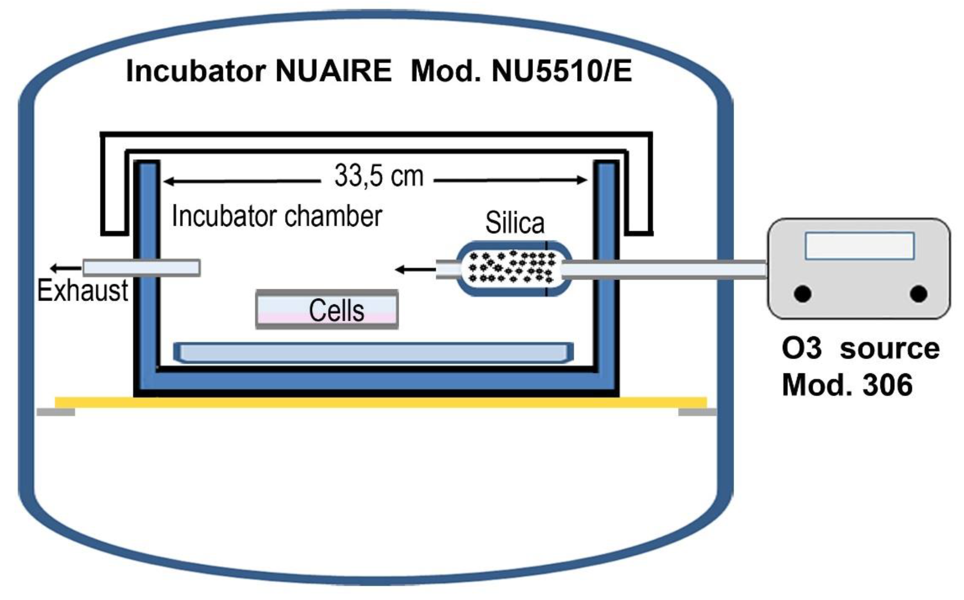
Abstract
:1. Introduction
2. Materials and Methods
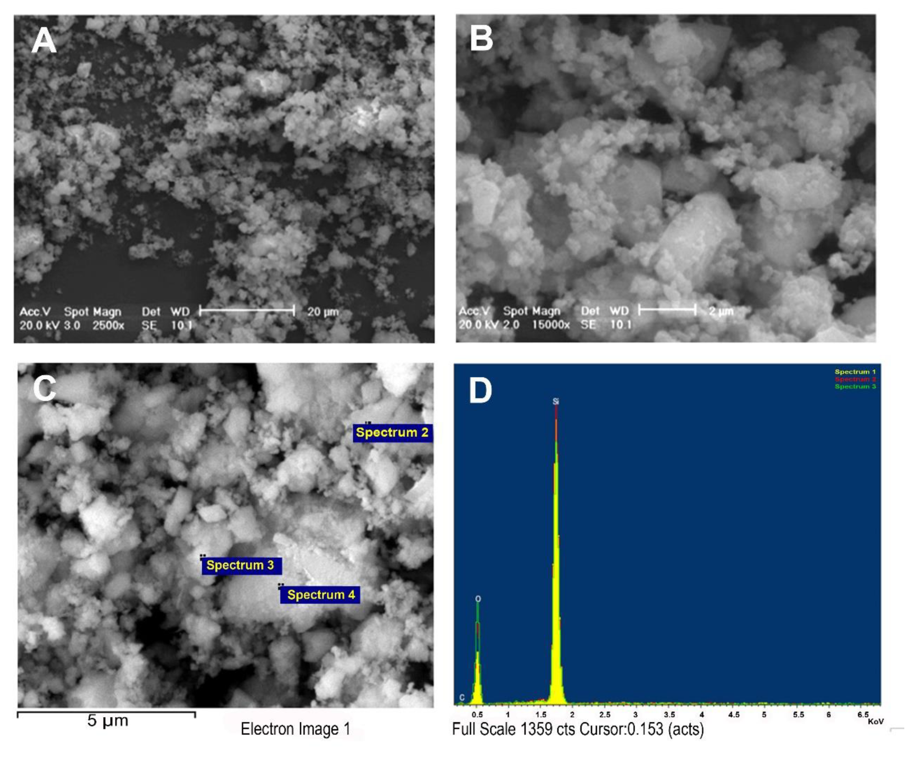
2.1. Analysis of the Silica Particles by SEM
2.2. Culture Chamber with Controlled Atmosphere
2.3. Cells Cultures
2.4. Cell Viability Assay
2.5. Cytokinesis-Block Micronucleus (CBMN) Assay
2.6. Alkaline Comet Assay
2.7. Statistical Analysis
3. Results
3.1. Silica Characterization
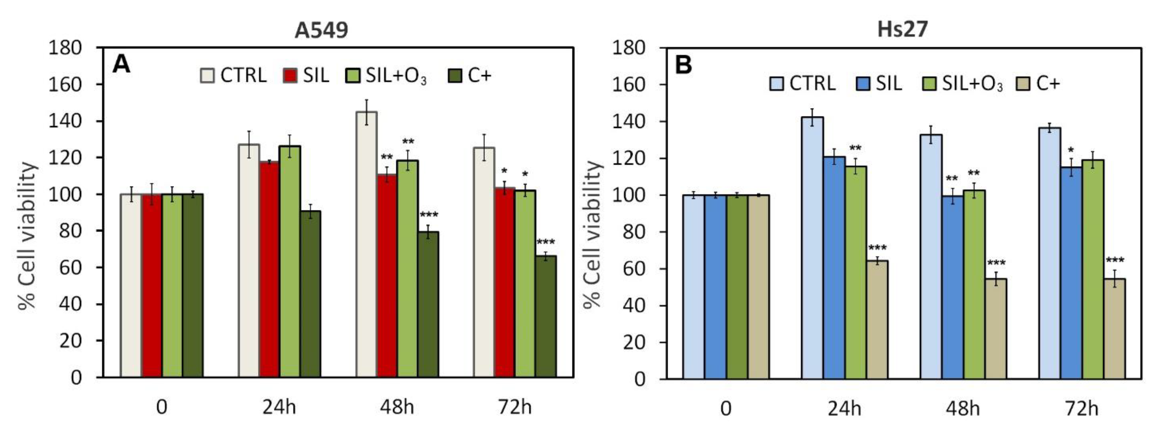
3.2. MTS Test
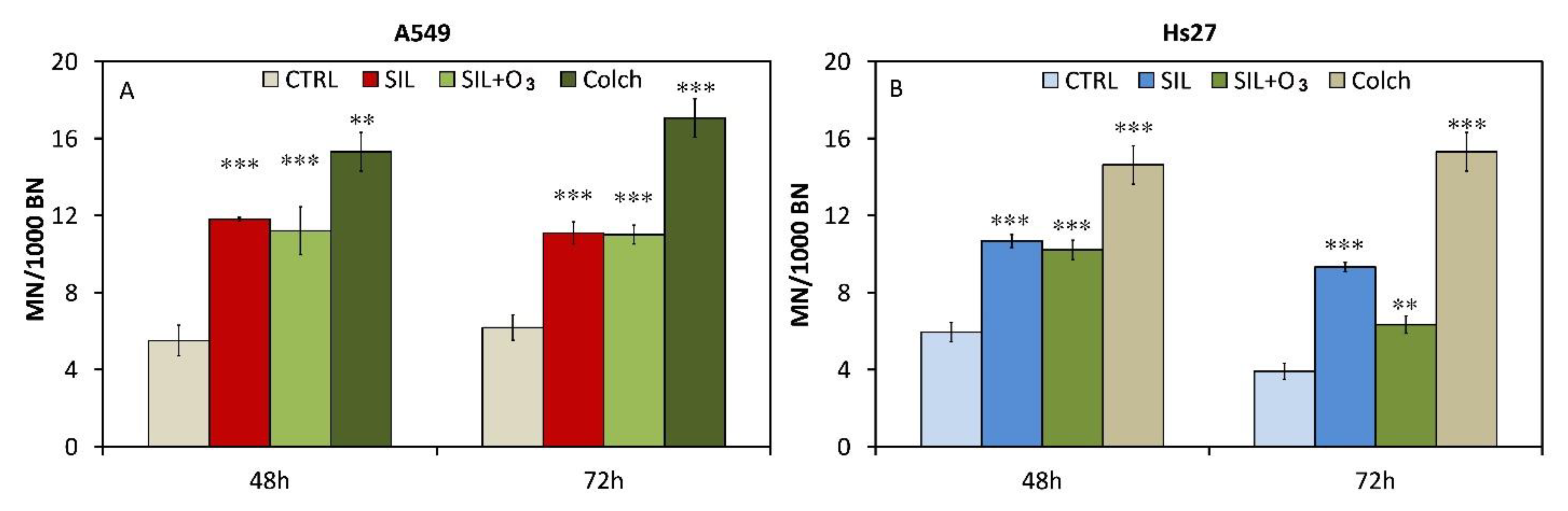
3.3. Micronuclei Test
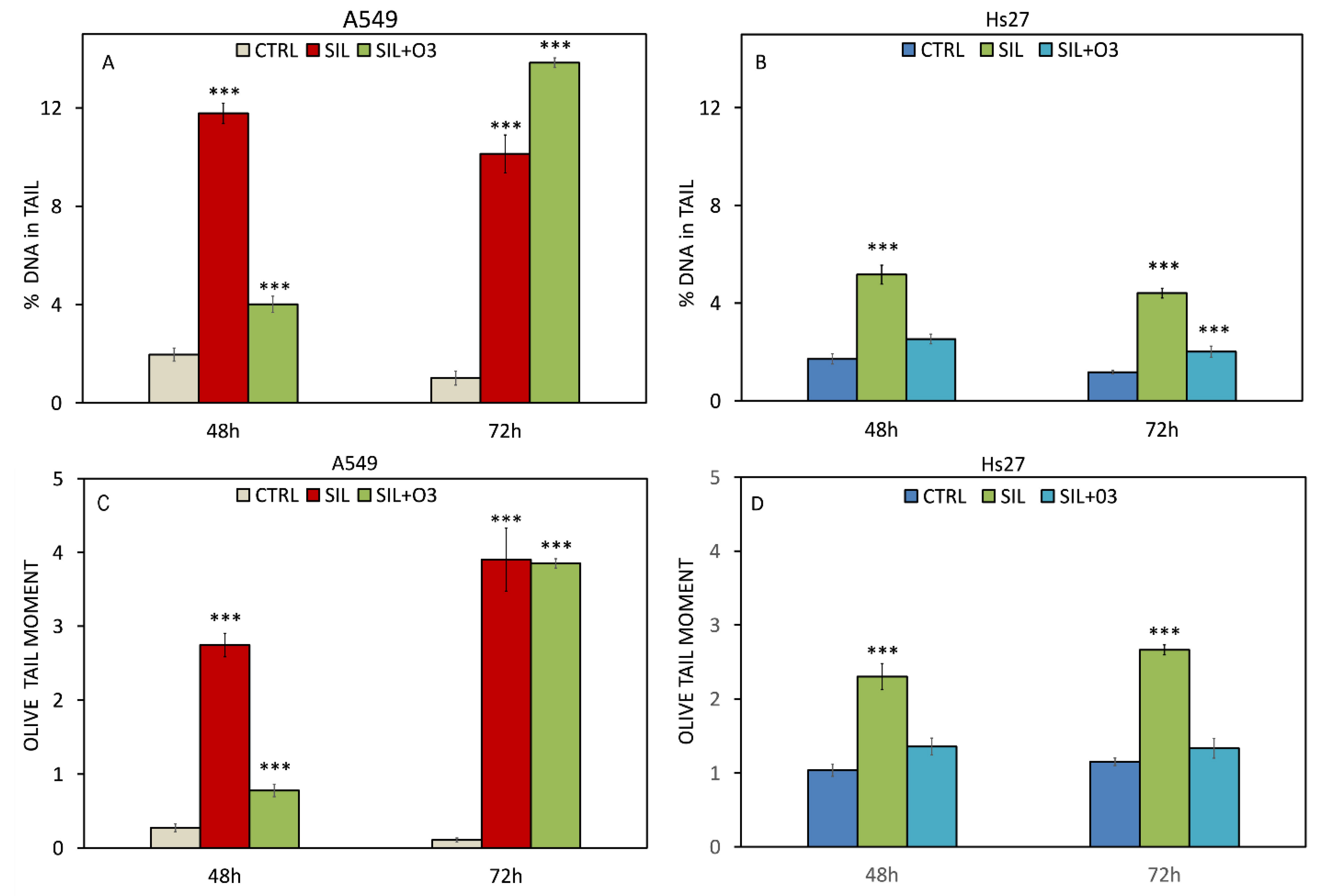
3.4. Alkaline Comet Assay
4. Discussion and Conclusions
Supplementary Materials
Author Contributions
Funding
Institutional Review Board Statement
Informed Consent Statement
Data Availability Statement
Conflicts of Interest
References
- Brulle, R.J.; Pellow, D.N. Environmental justice: Human health and environmental inequalities. Annu. Rev. Public Health 2006, 27, 103–124. [Google Scholar] [CrossRef] [Green Version]
- Godfrey, R.; Julien, M. Urbanisation and Health. Clin. Med. 2005, 5, 137–141. [Google Scholar]
- Schell, L.M.; Gallo, M.V.; Denham, M.; Ravenscroft, J. Effects of pollution on human growth and development: An introduction. J. Physiol. Anthropol. 2006, 25, 103–112. [Google Scholar] [CrossRef] [PubMed] [Green Version]
- Tran, V.V.; Park, D.; Lee, Y.C. Indoor Air Pollution, Related Human Diseases, and Recent Trends in the Control and Improvement of Indoor Air Quality. Int. J. Environ. Res. Public Health 2020, 17, 2927. [Google Scholar] [CrossRef] [PubMed] [Green Version]
- Jiang, X.Q.; Mei, X.D.; Feng, D. Air pollution and chronic airway diseases: What should people know and do? J. Thorac. Dis. 2016, 8, E31–E40. [Google Scholar]
- González-Martín, J.; Kraakman, N.J.R.; Pérez, C.; Lebrero, R.; Muñoz, R. A state-of-the-art review on indoor air pollution and strategies for indoor air pollution control. Chemosphere 2021, 262, 128376. [Google Scholar] [CrossRef]
- Shah, A.S.; Langrish, J.P.; Nair, H.; McAllister, D.A.; Hunter, A.L.; Donaldson, K.; Newby, D.E.; Mills, N.L. Global association of air pollution and heart failure: A systematic review and meta-analysis. Lancet 2013, 382, 1039–1048. [Google Scholar] [CrossRef] [Green Version]
- Ren, Z.; Liu, X.; Liu, T.; Chen, D.; Jiao, K.; Wang, X.; Suo, J.; Yang, H.; Liao, J.; Ma, L. Effect of ambient fine particulates (PM2.5) on hospital admissions for respiratory and cardiovascular diseases in Wuhan, China. Respir. Res. 2021, 22, 128. [Google Scholar] [CrossRef]
- Borgie, M.; Ledoux, F.; Verdin, A.; Cazier, F.; Greige, H.; Shirali, P.; Courcot, D.; Dagher, Z. Genotoxic and epigenotoxic effects of fine particulate matter from rural and urban sites in Lebanon on human bronchial epithelial cells. Environ. Res. 2015, 136, 352–362. [Google Scholar] [CrossRef]
- Zhang, H.H.; Li, Z.; Liu, Y.; Xinag, P.; Cui, X.Y.; Ye, H.; Hu, B.L.; Lou, L.P. Physical and chemical characteristics of PM2.5 and its toxicity to human bronchial cells BEAS-2B in the winter and summer. J. Zhejiang Univ. Sci. B 2018, 19, e317–e326. [Google Scholar] [CrossRef]
- Xie, Y.; Dai, H.; Dong, H.; Hanaoka, T.; Masui, T. Economic Impacts from PM2.5 Pollution-Related Health Effects in China: A Provincial-Level Analysis. Environ. Sci. Technol. 2016, 50, 4836–4843. [Google Scholar] [CrossRef]
- Zhang, Q.; Jiang, X.; Tong, D.; Davis, S.J.; Zhao, H.; Geng, G.; Feng, T.; Zheng, B.; Lu, Z.; Streets, D.G.; et al. Transboundary health impacts of transported global air pollution and international trade. Nature 2017, 543, 705–709. [Google Scholar] [CrossRef] [Green Version]
- Charlson, R.J.; Schwartz, S.E.; Hales, J.M.; Cess, R.D.; Coakley, J.A., Jr.; Hansen, J.E.; Hofmann, D.J. Climate forcing by anthropogenic aerosols. Science 1992, 255, 423–430. [Google Scholar] [CrossRef] [PubMed]
- Strak, M.; Janssen, N.A.; Gosens, I.; Cassee, F.R.; Lebret, E.; Godri, K.J.; Mudway, I.S.; Kelly, F.J.; Harrison, R.M.; Brunekreef, B.; et al. Airborne particulate matter and acute lung inflammation: Strak et al. Respond. Environ. Health Perspect. 2013, 121, A11–A12. [Google Scholar] [CrossRef] [PubMed] [Green Version]
- Peng, R.D.; Bell, M.L.; Geyh, A.S.; McDermott, A.; Zeger, S.L.; Samet, J.M.; Dominici, F. Emergency admissions for cardiovascular and respiratory diseases and the chemical composition of fine particle air pollution. Environ. Health Perspect. 2009, 117, 957–963. [Google Scholar] [CrossRef] [Green Version]
- Beeson, W.L.; Abbey, D.E.; Knutsen, S.F. Longterm concentrations of ambient air pollutants and incident lung cancer in California adults: Results from the air smog study. Environ. Health Perspect. 1998, 106, 813–822. [Google Scholar]
- Katsouyanni, K.; Pershagen, G. Ambient air pollution exposure and cancer. Cancer Causes Control 1997, 8, 284291. [Google Scholar] [CrossRef]
- Gold, D.R.; Litonjua, A.; Schwartz, J.; Lovett, E.; Larson, A.; Nearing, B.; Allen, G.; Verrier, M.; Cherry, R.; Verrier, R. Ambient pollution and heart rate variability. Circulation 2000, 101, 1267–1273. [Google Scholar] [CrossRef] [PubMed]
- Abbey, D.E.; Burchette, R.J.; Knutsen, S.F.; McDonnell, W.F.; Enright, P.L. Longterm particulate and other air pollutants and lung function in non-smokers. Am. J. Respir. Crit. Care Med. 1998, 158, 289–298. [Google Scholar] [CrossRef]
- Bartoli, C.R.; Wellenius, G.A.; Diaz, E.A.; Lawrence, J.; Coull, B.A.; Akiyama, I.; Lee, L.M.; Okabe, K.; Verrier, R.L.; Godleski, J.J. Mechanisms of inhaled fine particulate air pollution-induced arterial blood pressure changes. Environ. Health Perspect. 2009, 117, 361–366. [Google Scholar] [CrossRef]
- Anderson, P.J.; Wilson, D.J.; Irsch, A. Respiratory tract deposition ultrafine particles in subjects with obstructive or restrictive lung disease. Chest 1990, 97, 1115–1120. [Google Scholar] [CrossRef] [PubMed]
- Jin, R.H. Understanding Silica from the ViewpoInt. of Asymmetry. Chemistry 2019, 25, 6270–6283. [Google Scholar] [CrossRef] [PubMed]
- Cogliano, V.J.; Baan, R.; Straif, K.; Grosse, Y.; Lauby-Secretan, B.; El Ghissassi, F.; Bouvard, V.; Benbrahim-Tallaa, L.; Guha, N.; Freeman, C.; et al. Preventable exposures associated with human cancers. J. Natl. Cancer Inst. 2011, 103, 1827–1839. [Google Scholar] [CrossRef]
- IARC (International Agency for Research on Cancer). Silica, Some Silicates, Coal Dust and Para-Armid Fibrils. In IARC Monographs on the Evaluation of Carcinogenic Risk to Humans; International Agency for Research on Cancer: Lyon, France, 1997; Volume 68, pp. 41–242. [Google Scholar]
- Ding, M.; Chen, F.; Shi, X.; Yucesoy, B.; Mossman, B.; Vallyathan, V. Diseases caused by silica: Mechanisms of injury and disease development. Intl. Immunopharmacol. 2002, 2, 173–182. [Google Scholar] [CrossRef]
- Thomas, C.R.; Kelley, T.R. A brief review of silicosis in the United States. Environ. Health Insights 2010, 4, 21–26. [Google Scholar] [CrossRef]
- Ferrante, P. Asbestosis and silicosis hospitalizations in Italy (2001–2015): Results from the National Hospital Discharge Registry. Eur J. Public Health 2019, 29, 876–882. [Google Scholar] [CrossRef]
- Directive 2017/2398—Amendment of Directive 2004/37/EC on the Protection of Workers from the Risks Related to Exposure to Carcinogens or Mutagens at Work. Available online: https://www.eumonitor.eu/9353000/1/j9vvik7m1c3gyxp/vklpgng5lezp (accessed on 12 November 2021).
- Shi, X.; Castranova, V.; Halliwell, B.; Vallyathan, V. Reactive oxygen species and silica-induced carcinogenesis. J. Toxicol. Environ. Health B Crit. Rev. 1998, 1, 181–197. [Google Scholar] [CrossRef]
- Summer 2014 Ozone Assessment, EEA European Environment Agency Report. 2014. Available online: https://www.eea.europa.eu//publications/summer-2014-ozone-assessment (accessed on 13 December 2021).
- Myhre, G.; Shindell, F.M.; Bréon, W.; Collins, J.; Fuglestvedt, J.; Huang, D.; Koch, J.F.; Lamarque, D.; Lee, B.; Mendoza, T.; et al. Anthropogenic and Natural Radiative Forcing. In Climate Change 2013: The Physical Science Basis; Contribution of Working Group I to the Fifth Assessment Report of the Intergovernmental Panel on Climate Change; Cambridge University Press: Cambridge, UK; New York, NY, USA, 2013. [Google Scholar]
- Bell, M.L.; Goldberg, R.; Hogrefe, C.; Kinney, P.L.; Knowlton, K.; Lynn, B.; Rosenthal, J.; Rosenzweig, C.; Patz, J.A. Climate change, ambient ozone, and health in 50 US cities. Clim. Change 2007, 82, 61–76. [Google Scholar] [CrossRef]
- Nuvolone, D.; Petri, D.; Voller, F. The effects of ozone on human Health. Environ. Sci. Pollut. Res. Int. 2018, 25, 8074–8088. [Google Scholar] [CrossRef] [PubMed]
- Bell, M.L.; Zanobetti, A.; Dominici, F. Who is more affected by ozone pollution? A systematic review and meta-analysis. Am. J. Epidemiol. 2014, 180, 15–28. [Google Scholar] [CrossRef] [Green Version]
- Laskin, D.L.; Malaviya, R.; Laskin, J.D. Role of Macrophages in Acute Lung Injury and Chronic Fibrosis Induced by Pulmonary Toxicants. Toxicol. Sci. 2019, 168, 287–301. [Google Scholar] [CrossRef] [Green Version]
- Fenech, M. Cytokinesis-block micronucleus cytome assay. Nat. Protoc. 2007, 2, 1084–1104. [Google Scholar] [CrossRef] [PubMed] [Green Version]
- Fenech, M. The Lymphocyte Cytokinesis-Block Micronucleus Cytomeassay and Its Application in Radiation Biodosimetry. Health Physics. 2010, 26, 11–17. [Google Scholar]
- Bolognesi, C.; Fenech, M. Mussel micronucleus cytome assay. Nat. Protoc. 2012, 7, 1125–1137. [Google Scholar] [CrossRef]
- Tice, R.R.; Agurell, E.; Anderson, D.; Burlinson, B.; Hartmann, A.; Kobayashi, H.; Miyamae, Y.; Rojas, E.; Ryu, J.C.; Sasaki, Y.F. Single cell gel/comet assay: Guidelines for in vitro and in vivo genetic toxicology testing. Environ. Mol. Mutagen. 2000, 35, 206–221. [Google Scholar] [CrossRef]
- Grandi, C.; D’Ovidio, M.C.; Tomao, P. Impiego del comet test in medicina del lavoro e tossicologia industriale: Considerazioni e prospettive [Use of the comet test in occupational medicine and industrial toxicology: Considerations and prospects]. G. Ital. Med. Lav. Ergon. 2006, 28, 5–13. [Google Scholar] [PubMed]
- Kiskinis, E.; Suter, W.; Hartmann, A. High-throughput Comet assay using 96-well plates. Mutagen 2002, 17, 37–43. [Google Scholar] [CrossRef] [PubMed] [Green Version]
- Lichtveld, K.M.; Ebersviller, S.M.; Sexton, K.G.; Vizuete, W.; Jaspers, I.; Harvey Jeffries, E. In Vitro Exposures in Diesel Exhaust Atmospheres: Resuspension of PM from Filters versus Direct Deposition of PM from Air. Environ. Sci. Technol. 2012, 46, 9062–9070. [Google Scholar] [CrossRef] [PubMed]
- Mauderly, J.L.; Samet, J.M. Is the reevidence for synergy among air pollutants in causing health effects? Environ. Health Perspect. 2009, 117, 1–6. [Google Scholar] [CrossRef]
- Poma, A.; Colafarina, S.; Aruffo, E.; Zarivi, O.; Bonfigli, A.; Di Bucchianico, S.; Di Carlo, P. Effects of ozone exposure on human epithelial adenocarcinoma and normal fibroblasts cells. PLoS ONE 2017, 12, 0184519. [Google Scholar] [CrossRef] [Green Version]
- OECD (Organisation for Economic Cooperation and Development). Test No. 487: In Vitro Mammalian Cell Micronucleus Test. In OECD Guidelines for the Testing of Chemicals; OECD Publishing: Paris, France, 2016. [Google Scholar] [CrossRef]
- Fubini, B.; Hubbard, A. Reactive oxygen species (ROS) and reactive nitrogen species (RNS) generation by silica in inflammation and fibrosis. Free. Radic. Biol. Med. 2003, 34, 1507–1516. [Google Scholar] [CrossRef]
- Ouederni, A.; Limvorapituk, Q.; Bes, R.; Mora, J.C. Ozone decomposition on glass and silica. Ozone Sci. Eng. 1996, 18, 385–416. [Google Scholar] [CrossRef]
- Hsu, S.-Y.; Morris, R.; Cheng, F. Signaling Pathways Regulated by Silica Nanoparticles. Molecules 2021, 26, 1398. [Google Scholar] [CrossRef] [PubMed]


Publisher’s Note: MDPI stays neutral with regard to jurisdictional claims in published maps and institutional affiliations. |
© 2022 by the authors. Licensee MDPI, Basel, Switzerland. This article is an open access article distributed under the terms and conditions of the Creative Commons Attribution (CC BY) license (https://creativecommons.org/licenses/by/4.0/).
Share and Cite
Colafarina, S.; Di Carlo, P.; Zarivi, O.; Aloisi, M.; Di Serafino, A.; Aruffo, E.; Arrizza, L.; Limongi, T.; Poma, A. Genotoxicity Response of Fibroblast Cells and Human Epithelial Adenocarcinoma In Vitro Model Exposed to Bare and Ozone-Treated Silica Microparticles. Cells 2022, 11, 226. https://doi.org/10.3390/cells11020226
Colafarina S, Di Carlo P, Zarivi O, Aloisi M, Di Serafino A, Aruffo E, Arrizza L, Limongi T, Poma A. Genotoxicity Response of Fibroblast Cells and Human Epithelial Adenocarcinoma In Vitro Model Exposed to Bare and Ozone-Treated Silica Microparticles. Cells. 2022; 11(2):226. https://doi.org/10.3390/cells11020226
Chicago/Turabian StyleColafarina, Sabrina, Piero Di Carlo, Osvaldo Zarivi, Massimo Aloisi, Alessandra Di Serafino, Eleonora Aruffo, Lorenzo Arrizza, Tania Limongi, and Anna Poma. 2022. "Genotoxicity Response of Fibroblast Cells and Human Epithelial Adenocarcinoma In Vitro Model Exposed to Bare and Ozone-Treated Silica Microparticles" Cells 11, no. 2: 226. https://doi.org/10.3390/cells11020226