Review

Nicotinamide Adenine Dinucleotide (NAD) Metabolism as a Relevant Target in Cancer

1
Instituto de Biomedicina de Sevilla, IBIS, Hospital Universitario Virgen del Rocío, Universidad de Sevilla, Consejo Superior de Investigaciones Científicas, 41013 Sevilla, Spain
2
CIBERONC, Instituto de Salud Carlos III, 28029 Madrid, Spain
*
Author to whom correspondence should be addressed.
Cells 2022, 11(17), 2627; https://doi.org/10.3390/cells11172627
Submission received: 16 June 2022 / Revised: 25 July 2022 / Accepted: 16 August 2022 / Published: 24 August 2022
(This article belongs to the Special Issue Cancers: Genetics and Cellular Perspective)

Abstract

:
NAD+ is an important metabolite in cell homeostasis that acts as an essential cofactor in oxidation–reduction (redox) reactions in various energy production processes, such as the Krebs cycle, fatty acid oxidation, glycolysis and serine biosynthesis. Furthermore, high NAD+ levels are required since they also participate in many other nonredox molecular processes, such as DNA repair, posttranslational modifications, cell signalling, senescence, inflammatory responses and apoptosis. In these nonredox reactions, NAD+ is an ADP-ribose donor for enzymes such as sirtuins (SIRTs), poly-(ADP-ribose) polymerases (PARPs) and cyclic ADP-ribose (cADPRs). Therefore, to meet both redox and nonredox NAD+ demands, tumour cells must maintain high NAD+ levels, enhancing their synthesis mainly through the salvage pathway. NAMPT, the rate-limiting enzyme of this pathway, has been identified as an oncogene in some cancer types. Thus, NAMPT has been proposed as a suitable target for cancer therapy. NAMPT inhibition causes the depletion of NAD+ content in the cell, leading to the inhibition of ATP synthesis. This effect can cause a decrease in tumour cell proliferation and cell death, mainly by apoptosis. Therefore, in recent years, many specific inhibitors of NAMPT have been developed, and some of them are currently in clinical trials. Here we review the NAD metabolism as a cancer therapy target.

1. Nicotinamide Adenine Dinucleotide (NAD+)

NAD+ is a fundamental metabolite required for multiple oxidation–reduction (redox) reactions to maintain cellular homeostasis and energy production [1,2,3]. The NAD+ molecule is a dinucleotide composed of adenosine monophosphate (AMP) linked through a phosphate group to nicotinamide mononucleotide (NMN), which, in turn, is made up of the phosphate group, a ribose ring and nicotinamide [1,4]. The electrochemical properties of the nicotinamide moiety are essential for the classical redox functions of NAD+. Thus, the nicotinamide group can accept a hydride anion (H+, 2e−), reducing the molecule to NADH. In contrast, NADH is oxidised to NAD+ by donating that hydride anion [5,6]. Alternatively, the ribose ring of the AMP moiety of NAD+ can be phosphorylated by the enzyme NADK (NAD kinase), generating NADP+ (nicotinamide adenine dinucleotide phosphate), which can be reduced to NADPH [7]. All forms of NAD (NAD+, NADH, NADP+, NADPH) act as electron acceptors and donors in multiple redox reactions in the cell (Figure 1).
The NAD+ oxidised form is ubiquitous and the form most abundant in cells. However, its availability varies depending on intracellular factors, such as subcellular compartment, tissue and cell type, and intracellular glucose levels, but also on extracellular factors, such as caloric intake or expenditure, ageing or circadian rhythms. For example, it has been observed that free NAD+ concentrations in the nucleus and in the cytosol are similar since both compartments are connected, while the free NAD+ content in mitochondria is much higher (approximately 100 μM in nucleus/cytosol vs. 250–500 μM in mitochondria), ensuring a quick local regulation of NAD-dependent redox processes [8,9]. NAD+ concentration can also vary greatly during the day because of the string regulation by the circadian rhythm. The circadian clock complex CLOCK:BMAL1 induces the transcription of NAMPT, which is the limiting enzyme in the biosynthesis of NAD+, thus increasing the production of NAD+ every 24 h. SIRT1, which is an important NAD+-consuming factor, counteracts this effect by deacetylating BMAL1 and the promoter histones of other regulatory genes [10,11,12].

2. Redox Functions of NAD+

Initially, NAD+ was considered to have the role of a universal electron carrier in oxidation–reduction (redox) reactions of catabolic processes for energy generation in cells. One molecule of glucose during glycolysis will give rise to two NADH molecules, two ATP molecules, and two pyruvate molecules. Depending on the availability of oxygen, pyruvate can be reduced to lactate or oxidised to acetyl-CoA. In the absence of oxygen, the enzyme lactate dehydrogenase (LDH) reduces pyruvate to lactate using NADH as a cofactor, which is oxidised to regenerate NAD+. Conversely, in the presence of oxygen, pyruvate is oxidised to acetyl-CoA with the reduction of NAD+ to NADH [2,3,8]. Another process that also requires the reduction of NAD+ is the oxidation of fatty acids with the generation of acetyl-CoA [1,13]. In the mitochondria, after acetyl-CoA enters the Krebs cycle, more NAD+ is reduced to NADH by Krebs cycle enzymes. The mitochondrial NADH is oxidised by complex I (NADH:ubiquinone oxidoreductase) of the electron transport chain. Then, the electrons sequentially pass to ubiquinone, complex III, cytochrome C, and complex IV, generating a proton gradient across the inner mitochondrial membrane that ultimately leads to the production of more than 30 ATP molecules [1,2,3,8]. This process generates a large number of reactive oxygen species (ROS) that can inflict damage by oxidative reactions. Efficient mitochondrial metabolism requires an optimal NAD+/NADH ratio [14,15].
NADP+, NAD+ phosphorylated form and NADPH, reduced NADP+, are required for anabolic processes, including the synthesis of ribose-5-phosphate, used for nucleotide synthesis, via the PPP (pentose phosphate pathway) and lipid synthesis [16,17]. Furthermore, NADPH is essential to detoxify the high ROS levels produced in redox reactions [17]. The correct maintenance of redox homeostasis is an essential cellular process that requires a tight balance between the generation and elimination of ROS. This tight regulation is especially relevant in tumour cells, which have a higher production of ROS in general and need stricter control of this balance [18]. NADPH is also an important cofactor in the synthesis of fatty acids, which are necessary in the formation of the membrane during cell proliferation, using acetyl-CoA as a precursor [3] (Figure 1).

3. NAD+ as a Substrate in Nonredox Reactions

NAD+ and NADH can be reduced/oxidised in an almost unlimited manner [5]. However, the oxidised form, NAD+, is much more abundant than NADH, especially in the cytoplasm and nucleus (100-fold higher in those compartments, only 7 to 8-fold higher in the mitochondrial matrix) [19], highlighting the tight regulation of the process. High NAD+ levels are required since they participate in many other nonredox molecular processes, such as DNA repair, posttranslational modifications, cell signalling, senescence, inflammatory responses and apoptosis [1,2,4,6,17,20,21] In these nonredox reactions, NAD+ is a substrate for different types of enzyme families, mainly sirtuins (SIRTs), poly-(ADP-ribose) polymerases (PARPs) and cyclic ADP-ribose (cADPRSs). These enzymes use NAD+ as an ADP-ribose donor, releasing the catabolic product nicotinamide (NAM) [7,22] (Figure 2). The SIRT family consists of NAD+-dependent deacetylase enzymes that remove acetyl groups from lysine residues from proteins, such as histones, generating NAM and o-acetyl-ADP-ribose [22,23,24]. PARP family members transfer ADP-ribose long chain polymers from NAD+ to their target proteins involved in many different biological processes, such as metabolism, transcription or DNA repair [20]. The ectoenzymes CD38, CD157, CD39 and CD73 belong to the ADP-ribose synthases (cADPRS) family that consume NAD+ and NADP+, generating cyclic ADP-ribose (cADPR) or nicotinic acid adenine dinucleotide phosphate (NAADP), which act as secondary messengers of calcium signalling [19,23,25]. SIRTs, PARPs and cADPRSs are capable of coregulating each other, competing for the same subcellular NAD+ pool. SIRT1 can deacetylate PARP1 to inhibit its activity, thereby increasing the levels of NAD+, while PARP2 activity can inhibit the transcription of SIRTs [19,22,23]. Therefore, the activation of all these enzymes strongly depends on the availability of free NAD+. The importance of the role of NAD+ in these nonredox reactions has been well established, and recently, it has been suggested that NAD+ is the link connecting signal transduction and metabolism in physiological processes and many diseases [9].

4. Cancer and NAD+ Metabolism

Depending on internal genetic or epigenetic changes or microenvironmental conditions, cancer cells can reprogram their metabolism. This metabolic plasticity permits a high growth rate and survival with limited energy resources and adverse conditions. This metabolic plasticity rewiring allows an advantage during tumour progression and survival [26]. Higher ratios of the oxidised forms of NAD+ and NADP+ have been reported in tumour cells than in nontumor cells, suggesting an essential role for NAD+ in this plasticity [27]. Tumour cells are able to switch metabolism from OXPHOS to glycolysis, even under normal oxygen conditions (in a process known as the Warburg effect) [28,29]. Although OXPHOS metabolism generates energy in a more efficient way, producing 36 ATP molecules, tumour cells prefer glycolysis that generates less energy but in a faster way, producing two ATP molecules. It has been suggested that this fast energy production results in an important competitive advantage in sharing limited nutrients with the microenvironment and stromal cells [30,31]. However, this is not the only advantage of anaerobic glycolysis; it also allows tumour cells to synthesise macromolecules and counteract ROS production. In fact, part of the pyruvate that is formed is oxidised to acetyl-CoA to enter the Krebs cycle to produce the intermediate compounds of the cycle and use them as biosynthetic precursors. At the same time, these compounds are consumed and must be replenished primarily through the metabolism of glutamine, which is the main source of nitrogen in cells. Tumour cells depend on glutaminolysis for the synthesis of other molecules, such as nonessential amino acids, GSH, NAD+, NADPH and nucleic acids [29,31,32,33]. Some intermediates of glycolysis are also relevant for other processes promoted in cancer cells. For example, the PPP is essential for the synthesis of nucleic acids, DNA replication and the increase in tumour biomass [29,34]. In addition, the PPP is the main source of NADPH, which is required to reduce the ROS produced by accelerated metabolism, hypoxia or the accumulation of DNA damage in tumour cells. Radio- and chemotherapy also increase ROS levels, and in response, enzymes such as the PI3K/Akt/mTOR pathway, ATM, Ras or Src induce the expression of glucose-6-phosphate dehydrogenase (G6PDH), activating the PPP [16,34].
NAD+-dependent serine synthesis is another glycolysis-dependent pathway that is increased in cancer. The precursor of glycine and cysteine can be serine, which is involved in methionine and folic acid metabolism, redox balance, histone and DNA methylation and amino acid transport in the cells [35,36]. Metabolism in tumour cells depends highly on the PPP, serine biosynthesis and fatty acid synthesis. These metabolic pathways, which are dependent on NAD+ and NADP+, emerge from glycolysis, making NAD+ biosynthesis a main driver of cancer metabolism [1] (Figure 2).

5. NAD+ Biosynthesis

To meet the stringent NAD+ demands of redox and nonredox processes, NAD+ is generated from precursors such as tryptophan or vitamin B3 forms. These precursors are essential compounds that must be taken from the diet for the synthesis of NAD+. In fact, a chronic deficiency of these precursors can cause pellagra, an illness that can even lead to death. Interestingly, the essential role of NAD+ in cells was discovered when pellagra treatment was being investigated in the 1920s [3,19]. From these precursors, cells synthesise NAD+ by three pathways: the de novo pathway, the Preiss–Handler pathway and the nucleoside pathway. Additionally, cells can recycle the catabolic product released by NAD+-consuming enzymes (SIRTs, PARPs and cADPRs) and reconstitute the molecule through the salvage pathway, which is the most frequently used option for cells [8,9,10,11,37].

5.1. De Novo Pathway

NAD+ can be synthesised de novo from tryptophan, which is an essential amino acid that must be incorporated daily through the diet. In humans, this pathway, also called the kynurenine pathway, is present mainly in the liver and consists of several steps [9]. The limiting metabolite is quinolinic acid (QA). Its precursor, 2-amino-3-carboxymuconic acid semialdehyde (ACMS), can be oxidised to picolinic acid (PA) by the enzyme 2-amino-3-carboxymuconic acid semialdehyde decarboxylase (ACMSD) or enter a spontaneous cyclization, giving rise to QA. Compound QA is only formed when the ACMSD enzyme becomes saturated. Once formed, QA enters the Preiss–Handler pathway through the enzyme quinolinate phosphoribosyltransferase (QPRT) to continue NAD+ synthesis [3,7]. Trp is also required for the synthesis of other essential molecules, such as serotonin and melatonin, and approximately only 1.66% of dietary Trp is used for NAD+ biosynthesis. This low efficiency may explain why the de novo pathway is not the main source of NAD+ in humans.

5.2. The Preiss–Handler Pathway

NAD+ can also be synthesised from niacin and nicotinic acid (NA) present in the diet. Nicotinic acid phosphoribosyltransferase (NAPRT) is the rate-limiting enzyme in this pathway converting nicotinic acid to the intermediate nicotinic acid mononucleotide (NAMN). Consequently, NAMN is later transformed into NAAD by the enzyme NMNAT. This enzyme has three isoforms with different subcellular localizations: NMNAT-1 is found in the nucleus, NMNAT-2 is mainly present in the golgi apparatus and the cytosol and NMNAT3 is located in the cytosol and mitochondria [3]. Finally, the enzyme NAD synthetase (NADSYN) uses glutamine as a donor to amidate NAAD and form NAD+ [37].

5.3. The Salvage Pathway

Taking into account that the half-life of NAD+ is approximately 4–10 h and that the average daily intake of vitamin B3 is 15 mg, which does not cover physiological needs (for example, the liver needs approximately 8 g per day), cells have no choice but to recompose the NAD+ molecule through the salvage pathway [3]. Nicotinamide (NAM), the catabolic product released by NAD+-consuming enzymes, is recycled in two stages. The first step is the rate-limiting reaction of the pathway catalysed by NAMPT, transforming NAM to nicotinamide mononucleotide (NMN) in the presence of magnesium (Mg2+) and phosphoribosyl pyrophosphate (PRPP). In the second stage, the enzyme NMNAT reconstitutes NAD+ from NMN [38,39,40,41,42,43]. This pathway, the salvage pathway, is the main source of NAD+ generated in mammalian cells due to its high production efficiency, and less than 1% NAD+ is lost daily due to efficient recycling. This might explain why cells are completely dependent on this pathway [1,3,7,42,43].

5.4. The Nucleoside Pathway

Nicotinamide riboside (NR) and nicotinic acid riboside (NAR) are other NAD+ precursors that are acquired from the diet. Once they enter the body, the enzyme nicotinamide ribose kinase (NRK) is responsible for phosphorylating NAR, giving rise to NAMN (an intermediate in the Preiss–Handler pathway) and converting NR directly to NMN, bypassing the limiting reaction catalysed by NAMPT of the salvage route [7]. Until recently, it was thought that the cell membrane was impermeable to extracellular NMN generated by extracellular NAMPT and CD38. Thus, NMN must first be dephosphorylated to NR by the enzyme CD73, and then NR enters the cell through nucleoside transporters (ENTs) to equilibrate the pools [3]. However, Grozio et al. recently identified an NMN transporter in the cell membrane called Slc12a8 [44,45]. However, the expression of Slc12a8 is limited to the intestine and others have questioned its ability to transport NMN [46].

6. Nicotinamide Phosphoribosyltransferase (NAMPT)

Nicotinamide phosphoribosyltransferase (NAMPT), in addition to being the rate-limiting enzyme of the NAD+ salvage pathway, also acts as a cytokine-modulating inflammatory, metabolic, and immune responses [47,48]. In fact, NAMPT was discovered in lymphocytes in human peripheral blood and was initially called pre-B colony-stimulating factor (PBEF) since it is involved in the maturation of B lymphocytes in the presence of interleukin 7 (IL-7) and stem cell factor (SCF) [49,50]. This extracellular form of NAMPT is commonly called visfatin to differentiate it from the intracellular form. Visfatin is secreted mainly by adipocytes, macrophages, lymphocytes and inflamed endothelial cells and is involved in inflammatory processes and metabolic disorders such as obesity and cancer. For example, adipocytes release visfatin into the extracellular medium, where it exerts an insulin-mimetic function and regulates glucose and visceral fat levels related to obesity. In fact, high levels of visfatin have been detected in obese people [47,51]. Interestingly, the visfatin protein sequence lacks a secretion signal, so the protein is thought to be the same as intracellular NAMPT released after cell lysis [47,52]. The intracellular form of NAMPT is expressed in all tissues, mainly in those that require more energy, such as bone marrow, liver and muscle. Within the cell, NAMPT is located mainly in the cytoplasm and nucleus [43,50].
The NAMPT gene is located on chromosome 7 at the 7q22.3 locus and contains 11 exons and 10 introns [43,49,53]. Approximately 19 messenger RNAs and 14 variants have been predicted to be produced through alternative splicing (Figure 3). Of all these variants, only four likely produce proteins: variant 1 with a size of 491 amino acids (aa), variant 2 with 368 aa, variant 3 with 88 aa and variant 4 with 60 aa. The predominant variant is variant 1, NAMPT1, which is the only variant that possesses the enzymatic activity necessary for the conversion of NAM to NMN [53,54]. The protein contains two recognised domains: DUF5593, which comprises the N-terminal aa 10–116 and whose function is unknown, and NAPRTase, which comprises aa 188–466 and is responsible for the enzymatic activity of the protein [55,56]. The amino acids that make up the catalytic site are found at positions D219 (aspartic acid), G384 (glycine) and R392 (arginine). Additionally, amino acids R196 (arginine), H247 (histidine) and R311 (arginine) form the binding site of cofactor PRPP [57] (Figure 3A). To be functional, NAMPT1 must form a homodimer with two catalytic sites, each of which is made up of two aa from one chain and one aa from the other chain [58,59] (Figure 3B) In mammals, it appears that NAMPT can be activated by autophosphorylation. Thus, for NMN synthesis to occur, histidine 247 must first be phosphorylated by ATP hydrolysis and in the presence of Mg2+. Phosphorylated NAMPT has been shown to be more active than its unphosphorylated form. Once the enzyme is active, its substrates bind, first PRPP and then NAM, each to its corresponding binding site [53,60,61].
The other variants that have been predicted (NAMPT2, NAMPT3 and NAMPT4) do not contain these amino acids required for enzymatic activity assigned to NAMPT1, and it is currently unknown whether they have any function in the cell. In the present work, NAMPT will be mentioned, referring exclusively to the variant NAMPT1.

7. NAMPT in Cancer

To meet NAD+ demands, tumour cells must maintain high NAD+ levels, enhancing their synthesis mainly through the salvage pathway. The rate-limiting enzyme of this pathway, NAMPT, has been identified as an oncogene in some cancer types. In fact, increased NAMPT levels have been found in both haematologic malignancies [62,63] and various solid tumours, including colon [40], prostate [64], breast [65], thyroid [66], gastric [67] and glioblastoma tumours [41,42]. NAMPT is capable of increasing the tumorigenic properties of cells, such as cell proliferation, clone formation and resistance to apoptosis, thus allowing tumour initiation and progression [40,41]. The expression of NAMPT also correlates with the clinical data of cancer patients in some tumours. For example, in stomach, colon and glioblastoma cancer, high levels of NAMPT correlate with aggressive tumour stages, metastasis, treatment resistance and worse prognosis [40,41,64,67]. It also activates the secretion of interleukin 6 (IL-6), which induces the STAT3 signalling pathway, which favours the process of new blood vessel formation [62,68,69].
Visfatin (or extracellular NAMPT) is also implicated in cancer and its role is currently being studied. Although the exact molecular mechanism of visfatin remains unknown, its role is currently being studied. In fact, high levels of serum visfatin have been detected in patients with variant types of cancer, including postmenopausal breast cancer, endometrial cancer, gastric cancer and hepatocellular carcinoma. Visfatin has been proposed as a diagnostic marker in cancer and it is associated with poor patient survival [46,70,71,72]. Visfatin acts as a tumour cytokine promoting proliferation, neovascularization and metastasis in cancer. It has been described to regulate the function of adhesion molecules such as ICAM-1 and VCAM-1 through the activation of NF-κB in human endothelial cells [46]. It also upregulates matrix metalloproteinase, such as MMP-2 and MMP-9, and vascular endothelial growth factors, promoting angiogenesis via the STAT3/IL-6 signaling pathway [68,73]. Recently, it has been described that Visfatin can be secreted from cells via exosomes, which can promote the inflammatory and tumor microenvironment [74,75]. Similar to other adipocytokines (e.g., leptin, adiponectin and resistin), visfatin contributes to autocrine, paracrine and endocrine interactions between tumour cells and the microenvironment [46,76]. In breast cancer, it has been reported that visfatin induces THP-1 differentiation into M2 macrophages suppressing the immune response promoting tumor progression. Moreover, M2 macrophages secrete CXCL1 which elevated visfatin via a positive feedback loop. Visfatin/M2/CXCL1 contribute to tumor migration and invasion [77].
On the other hand, the exact molecular mechanism of visfatin remains unclear and its cellular receptor has been not found. However, visfatin presents insulin-mimetic effects, and it has been described that visfatin binds to and activates the insulin receptor of hepatocytes, myocytes and adipocytes [52]. There is controversy regarding if visfatin has the enzymatic activity as its intracellular form. Although the dimeric conformation of visfatin has been elucidated, it has been difficult to detect all of its substrates (PRPP, NAM and ATP) in serum [47,75]. Moreover, the effects of visfatin do not depend on the presence of NAM, nor are they reproducible by NMN addition or blocked by NAMPT inhibitors [67,78]. Lastly, the effects of visfatin are independent of the intracellular enzymatic activity of NAMPT.

8. Other NAD+-Dependent Enzymes in Cancer

NAMPT overexpression generates an increase in NAD+ content in tumour cells, which enhances the activity of NAD+-dependent and NAD+-consuming enzymes (SIRTs, PARPs and cADPRSs). The oncogenic role of NAMPT can be explained through the activation of these enzymes.

8.1. SIRTs in Cancer

The sirtuin family (SIRT1–7) regulates the activity of histones, DNA repair and transcription and other signalling proteins by deacetylating several substrates. Therefore, its role as a tumour suppressor or oncogene has been described to be dependent on the type of cancer [22,23,79]. The best characterised member is SIRT1, which is associated with longevity. Resveratrol, a powerful antioxidant and antiaging compound, is capable of increasing NAD+ levels, inducing the activation of SIRT1 [22]. This enzyme is also activated by caloric restriction, which reduces the risk of cancer [2,16]. SIRT1 activity is highly dependent on the NAD+/NADH ratio, rather than only the levels of NAD+, and can be inhibited by high levels of NAM [28,80] or by DBC1, an endogenous SIRT1 inhibitor. Many studies suggest that SIRT1 also acts as a tumour suppressor. SIRT1 has been shown to inhibit the alpha subunit of hypoxia-inducible factor 1 (HIF-1α), one of the factors that can activate glycolysis [28]. However, high levels of SIRT1 were found in some tumours, in which NAMPT activates SIRT1 expression by increasing NAD+ levels and decreasing NAM [69,81]. SIRT1 regulates other transcription factors, such as p53, FOXO3, E2F1, c-MYC and NF-κB. P53 regulates cell cycle arrest and apoptosis and can also inhibit G6PDH in the pentose phosphate pathway. SIRT1 can deacetylate and inhibit p53, thus allowing survival by ensuring the production of NADPH and ribose-5-P [17,82]. The proto-oncogene C-MYC can induce NAMPT expression and inhibit DBC1, thereby activating SIRT1. In turn, SIRT1 activates c-MYC by deacetylation, generating a positive feedback loop [29,81]. In addition, SIRT1 activates FOXO3 by deacetylation, contributing to resistance to oxidative stress [64,83]. Thus, the oncogenic role of SIRT1 depends on NAD+ levels. The other members of the sirtuin family, SIRT2–7, similar to SIRT1, can also act as oncogenes or tumour suppressors according to the type of tumour.

8.2. PARPs in Cancer

PARP1, known as the guardian of DNA integrity, is the most studied member of the PARP family [84]. The PARP1 enzyme is activated in response to DNA damage, requiring high levels of NAD+ in the cell, and is thus dependent on NAMPT activity. Excessive activation of PARP1, due to the action of genotoxic agents, for example, dramatically reduces ATP and NAD+ pools, regulating sirtuin activity and, therefore, cellular homeostasis. If the DNA damage is not repairable, PARP1 promotes apoptosis through the activation of p53 under normal conditions. However, PARP1 can also regulate oncogenic transcriptional factors. Depending on the context, PARP1 has been reported to act as an oncogene or tumour suppressor [17,84]. Most tumours overexpress PARP1 in response to increased DNA damage aggravated by radiation and chemotherapy treatment. In fact, high levels of PARP1 have been found in breast, ovarian and lung cancers and non-Hodgkin lymphoma [43,84]. In colon cancer, PARP1 has been found to have a dual oncogenic and suppressive function [81,85]. In addition to DNA damage, PARP1 is involved in other physiological cell processes, such as proliferation, cell cycle regulation, gene transcription, inflammation and cell fate. PARP1 regulates ERK, allowing the activation of proangiogenic and metastatic factors. PARP1 also regulates the transcription factor E2F1, which reduces apoptosis in tumour cells [86,87]. Inhibition of PARP1 by olaparib has been tested in clinical trials for tumours of different origins. Pharmacologically, tumour cells are in general more sensitive to PARP1 inhibition than healthy cells. This sensitivity may be because tumour cells lack alternative DNA repair mechanisms. However, resistance to PARP1 inhibitors has been observed [16].

8.3. cADPRSs in Cancer

CD38, belonging to the cADPRSs family of ectoenzymes, hydrolyzes NAD+ to produce cADPRs, which is involved in Ca2+ mobilization, regulation of the cell cycle and insulin signalling [17,88]. CD38 is the largest consumer of NAD+ in mammalian cells, since it requires 100 molecules of NAD+ to generate only one molecule of cADPR. For that reason, CD38 has been proposed as an important regulator of intracellular NAD+ levels. As we age, the levels of NAD+ decline sharply. It has been shown that this depletion is partly due to the increased expression of CD38 that occurs with age [85,89]. CD38 is a transmembrane protein whose C-terminal catalytic domain has two opposite orientations and, therefore, is capable of consuming both intracellular and extracellular NAD+ [86,90]. CD38 also generates NAM by metabolizing extracellular NMN, which is converted to NAD+ via visfatin or NAMPT and NMNAT after crossing the cell membrane [1,85,89]. CD38 has been described as a marker of negative prognosis in CLL and as a diagnostic cell marker in multiple myeloma [16,91]. It has also been observed that CD38 expression levels can modify the sensitivity and resistance of pancreatic cancer cells to the NAMPT inhibitor FK866. However, the functional relationships between NAMPT, NAD+ and CD38 are not yet well known [83,92]. CD73, another member of the cADPRS family, catalyses the conversion of extracellular NMN to NR, which is necessary to aid in the biosynthesis of intracellular NAD+ [89,93]. In addition, CD73 has a relevant role in the migration and invasion of tumour cells, acting as an adhesion molecule [90,94].

8.4. NAPRT in Cancer

High levels of NAPRT and NAMPT have been found in ovarian, prostate and pancreatic cancers [63,95,96], while in glioblastoma, neuroblastoma, chondrosarcoma, leukaemia and gastric and colon cancers, low levels of NAMPT and low levels of NAPRT have been found [63,97,98,99,100]. Comparing the pattern of expression of the NAPRT and NAMPT genes in human tumour cells with nontumor cells, there is high variability in the levels of NAPRT mRNA and protein in tumour cells [97,101]. This high expression variability depends on the tissue of origin. Thus, tumours from tissues with high NAPRT expression will have higher NAPRT amplification, and conversely, tumours from tissues with low NAPRT expression will be dependent on NAMPT activity [96,100,102].
NAPRT-negative tumours usually present with mutations in isocitrate dehydrogenase 1 and 2 (IDH1/2) and protein phosphatase 1D (PPM1D). IDH1/2 mutants use NADPH to convert α-ketoglutarate to D-2-hydroxyglutarate (D-2HG), which acts as an oncometabolite inhibiting NAPRT expression by the hypermethylation of its promoter [98,103,104]. PPM1D is an oncogene that is involved in the DNA damage response and checkpoints of the cell cycle. PPM1D mutants also lead to the inhibition of the NAPRT promoter by the methylation of CpG islands [101,105]. The inhibition of the Preiss–Handler pathway by NAPRT forces tumour cells to rely entirely on the salvage route. Therefore, NAMPT is usually overexpressed in these tumours since their cells rely on its activity to meet the demands for NAD+ [100,104]. In fact, cells with IDH1/2 and PPM1D mutations are more sensitive to treatment with NAMPT inhibitors due to the total depletion of NAD+ [103,104,105]. In tumours with high levels of NAPRT, tumour cells may have a better supply of NAD+, which allows for the activation of NAD+-dependent enzymes, including PARP1, which are required to protect against DNA damage and oxidative stress [106,107]. Overexpression of NAPRT may contribute to resistance to treatment with NAMPT inhibitors or alkylating agents that cause DNA damage. In vivo, xenografts have been shown to be less sensitive to the NAMPT inhibitor FK866 if NAPRT is overexpressed, whereas they are more sensitive when NAPRT is inhibited [100,106]. NAPRT can be inhibited by compounds such as 2-hydroxynicotinic acid (2-HNA). 2-HNA is an analogue of nicotinic acid that competitively blocks the enzyme [108,109]. It has been observed that this compound is capable of potentiating the cytotoxic effect of the NAMPT inhibitor FK866. Therefore, the ability to predict the response to NAMPT inhibitor treatments by low levels of NAPRT depends on the type of cancer [100,106].

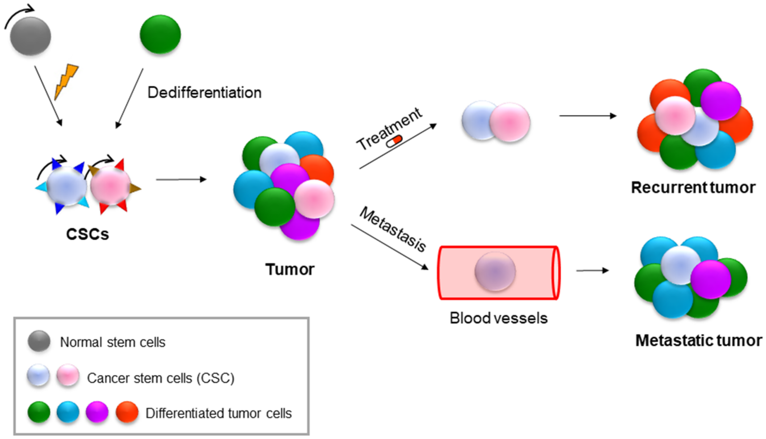
9. NAMPT in Cancer Stem Cells (CSCs)

Tumours are highly heterogeneous entities that are organised hierarchically into different subgroups of tumour cells. Cancer stem cells (CSCs) are at the top of this hierarchy. CSCs are a small subpopulation of cells with the ability to self-renew and differentiate into the different types of cells that form the bulk of the tumour. Similar to normal stem cells, these CSCs can self-renew and differentiate, but they do so in an uncontrolled manner. CSCs are responsible for tumour initiation and progression and for metastasis and resistance to therapies. CSCs can originate from normal stem cells or from differentiated tumour cells that are capable of acquiring stem cell capabilities through dedifferentiation mechanisms [110,111,112,113]. (Figure 4) The dedifferentiation process is similar to the process of reprogramming somatic cells into pluripotent cells (iPSCs), which is induced through the activation of some pluripotency transcription factors, such as OCT3/4, SOX2, KLF4 and c-MYC [114,115]. The interconversion capacity between both phenotypes, different cells and stem/pluripotent cells is known as cell plasticity. The cellular microenvironment and some signalling pathways activate and regulate this cellular plasticity. Some of the main pathways that regulate CSCs are Notch, Hippo, SHH, PI3K, WNT and NF-κB [110,113,116].
Both CsCs and iPSCs can show an active Warburg metabolic effect, which induces the hypoxic state necessary for the maintenance of the stem cell phenotype. Thus, the stem cell phenotype or pluripotency can be highly glycolytic when the cells are actively dividing [110,117,118]. However, much controversy exists regarding the typical metabolic profile of CSCs. In general, CSCs are in the G0 phase, or quiescence, a state in which they consume oxygen and depend mainly on OXPHOS metabolism, by which they obtain a greater antioxidant response to compensate for the high levels of ROS. This slow metabolism allows them to survive conventional therapies that mainly attack cells with high rates of proliferation. As sole survivors, CSCs change from their quiescent state to a more proliferative and glycolytic state, thus regenerating the tumour again [111,117,119]. In recent years, research has focused on searching for surface markers and transcriptional signatures specific to the CSC phenotype.
CSCs express specific antigens on their surface, similar to normal stem cells. These surface markers that characterise CSCs depend largely on the type of cancer, and the best known are CD44+CD24− and ALDH in breast cancer, CD44+ and CD133+ in colon and gastric cancer, CD34+CD38− in leukaemia, CD133+ in glioblastoma and sarcoma and CD13/CD45/CD90 in liver cancer [120,121]. However, the CSC population presents a high degree of heterogeneity, with different subgroups or categories even in the same tumour [110,113,116]. Finding new markers and genes that help us to identify CSC populations is vital to developing new and more specific therapeutic strategies. NAMPT enriches the CSC population of the tumour mainly through the salvage route, thus allowing an adequate supply of NAD+ [42,43]. NAMPT and NAD+ are involved in the processes of pluripotency and dedifferentiation to CSCs. It has been observed that media supplementation with NAM can prevent senescence and apoptosis and promote reprogramming and iPSC generation. Furthermore, reduced NAD+ levels in human stem cells can cause spontaneous differentiation and apoptosis [116,122]. The autofluorescence emitted by the NADH reduced form can be quantified by cytometry, and a subpopulation with high NADH has been related to the CD133+ subpopulation of CSCs in glioblastoma [123,124]. NAMPT not only increases tumorigenic properties but also allows the acquisition of the properties and phenotype of CSCs through the activation of OSKM factors, including SOX2, OCT4, KLF4 and NANOG [40,41]. In addition, NAMPT can regulate the canonical WNT pathway since the inhibition of NAMPT increases the levels of Axin, degrading β-catenin, which decreases tumorigenesis. The addition of NMN, the product of NAMPT, reverses the degradation of β-catenin and reduces the adhesion of mesenchymal cells, allowing them transform into cells with migratory and invasive capacities. NAMPT activates several transcription factors involved in EMT, such as SNAI1, TWIST and FOXC2, favouring tumour invasion and metastasis [40,41].
We found high levels of NAMPT expression in the tumorspheres generated from our head and neck squamous cell carcinoma (HNSCC) cell lines. NAMPT overexpression favoured the formation of clones and increased the expression levels of some genes involved in the regulation of stem cell pluripotency, such as SOX2 and NANOG, in HNSCC. However, in general, the reduction in NAMPT reduced the capacity for proliferation, clone formation and growth in conditions of competition with other cells and the in vivo growth of tumours generated in xenotransplantation models [1,2,40,41,42,43]. All these data indicate that NAMPT could be involved in the process of tumorigenesis in HNSCC.
NAMPT is a possible tumour and CSC marker in HNSCC. In several types of cancer, it has been described that NAMPT is capable of enriching the CSC population of the tumour through the processes of pluripotency and dedifferentiation, a mechanism by which cells acquire the properties and increase phenotype of tumour stem cells [40,41,43].
In addition, we found several genes that positively correlated with NAMPT overexpression from cells in the TCGA public database of HNSCC patients. These genes were CDH1, DESI1, MAP1B, TMEFF1, OCLN, TCF4, SMAD2, TFRC, AHNAK, PATP4A1, MSN, STEAP1, PGK1 and F11R [1,2].
CSCs are thought to be responsible for resistance to radiotherapy and chemotherapy and the recurrence rate of tumours in patients [110,111,112]. In the TCGA database, we found that high levels of NAMPT correlated with worse survival in patients with HNSCC. Therefore, NAMPT could also be an indicator of poor prognosis in HNSCC, as occurs in stomach cancer, colon cancer and glioblastoma [40,41,64,67].

10. NAMPT as a Therapeutic Strategy

NAMPT inhibition causes the depletion of NAD+ content in the cell, leading to the inhibition of ATP synthesis. This effect can cause a decrease in tumour cell proliferation and cell death, mainly by apoptosis. Therefore, in recent years, many specific inhibitors of NAMPT have been developed, and some of them are currently in clinical trials [1,2,3,4,5,6,7,8,9,10,11,12,13,14,15,16,17,18,19,20,21,22,23,24,25,26,27,28,29,30,31,32,33,34,35,36,37,38,39,40,41,42,43,62,83,125,126] (Figure 5, Table 1 and Table 2) The first inhibitor to be developed, being the most studied both in vitro and in vivo, was FK866/APO866, a partially noncompetitive inhibitor that binds to the interface between the two subunits of the NAMPT homodimer, partially covering the binding site of NAM [57]. NAMPT inhibition by FK866 can increase ROS levels and decrease glucose consumption, ATP production, fatty acid synthesis and, of course, NAD+ levels, negatively affecting the activity of all NAD+-dependent enzymes, such as SIRTs and PARPs [79]. FK866 treatment does not seem to affect mitochondrial NAD+ content; therefore, tumour cells could be more sensitive than nontumor cells [59]. In addition, FK866 cytotoxicity appears to be greater in some tumour types that express higher levels of NAMPT and are relatively more dependent on glycolysis [83,102]. As of 2007, FK866 is in phase 2 clinical trials (clinicaltrials.gov; NCT00435084, NCT00431912, NCT00432107). To palliate the adverse effects of FK866, it has been tried at suboptimal doses in combination with other drugs, such as sirtinol (SIRT1 inhibitor) and olaparib (PARP1 inhibitor) [40,41,97] Another very specific NAMPT inhibitor studied is GMX1778/CHS-828, which is administered orally. The GMX1777 prodrug is a cyanoguanidine compound that is rapidly converted to its active form GMX1778, which acts as both an inhibitor and a substrate. GMX1778 phosphorylated by NAMPT accumulates inside tumour cells, enhancing its antitumor effect [93,97]. GMX1778 was in phase 1 clinical trials for solid tumours (clinicaltrials.gov, Accessed on 1 April 2022; NCT00003979); however, it was withdrawn due to its toxic effects, mainly thrombocytopenia and gastrointestinal symptoms [127]. CB30865 and its analogue CB300919 were initially developed to inhibit thymidylate synthase (TS), but it was later found that this protein was not its target, although the compounds had a potent cytotoxic effect [128]. Years later, it was discovered that the target protein was NAMPT [129]; however, due to solubility problems, these inhibitors were not used in clinical trials [130]. The next generation of inhibitors showed better pharmacological properties, including oral administration, and higher specificity against their NAMPT target. Among these inhibitors are GNE617 and its analogues GNE618, GNE643 and GNE875, which are small molecules for oral administration that demonstrated high efficacy in vivo and were able to reduce the growth of NAPRT-deficient xenotransplants. However, they also showed some cardiac, retinal and gastrointestinal toxicity [131,132,133]. Other inhibitors are MPC-9528 and its analogue MPI0479883, which showed a potent inhibition of tumour progression in fibrosarcoma xenografts [134]. STF-118804 also showed efficacy in several cell lines and in acute lymphoblastic leukaemia xenografts [135]. TP201565, a potent analogue of GMX1778, showed cytotoxic effects in several cell lines in vitro [136].
However, despite the powerful antitumor effect shown by all these NAMPT inhibitors, they have not entered clinical trials because of toxicity and solubility problems due to their lipophilic chemical structures. For this reason, the strategy of incorporating polar and ionizable groups into their structures has been adopted to improve solubility. This approach, in addition to reducing toxicity, would reduce the permeability of these compounds through the cell membrane so that they could also act on the extracellular form of NAMPT (eNAMPT) [154]. GPP78 and MV78 are two analogues of FK866 that have served mainly as templates for the development of new, more soluble inhibitors [139,141,155]. MS0 is also an analogue of FK866 that was found to have less effective in vitro activity [145], but it served as a template for the development of better inhibitors (compounds MS1–31) [146]. LB-60-OF61, one of the latest inhibitors developed, has shown a high efficacy in cell lines that overexpress the MYC oncogene [138], which regulates and activates the expression of NAMPT and inhibits DBC1 [81], the endogenous inhibitor of SIRT1. Trans-3-(pyridin-3-yl)acrylamide-sulfonamide compounds are potent inhibitors that have shown an antiproliferative effect at subnanomolar doses in various cell lines in vitro [142]. A-1293201 and A-1307138 are two inhibitors that lack the aromatic nitrogen group mimetic to that of the natural substrate NAM. In the study, it was observed that these inhibitors maintained their antitumor effect despite not being phosphorylated by the target enzyme NAMPT, demonstrating that the formation of the phosphorylated adduct was not essential for the functionality of NAMPT inhibitors [143]. Depletion of NAD+ can be very toxic to nontumor cells, so using NAD+ precursors (e.g., nicotinic acid) as adjuvants has been considered [63,96]. Several studies have found that the coadministration of nicotinic acid (NA) with some NAMPT inhibitors (GMX1778, GNE617, LSN3154567, MPC-9528 and STF-31) helps prevent toxic effects, such as haematologic and retinal toxicity in rodents, while maintaining drug efficacy. However, the inhibitory effect was suppressed in NAPRT-positive tumours, that is, tumours that overexpressed this enzyme. Therefore, the coadministration of NA and NAMPT inhibitors has been proposed as a new therapeutic strategy, but only in tumours deficient in NAPRT [97,134,137,147]. Moreover, it has been observed that treatment with NAMPT inhibitors combined with other drugs, such as PARP inhibitors, may be useful to improve the efficacy of the inhibitory effect and reduce toxicity [153,154]. Therefore, the development of inhibitors with dual therapeutic targets could be a good therapeutic strategy. An example would be the compound STF-31, which is a double inhibitor of NAMPT and of the glucose transporter GLUT1. This additional inhibitory effect could be beneficial in triggering cell death in tumours with a high rate of glucose consumption. In fact, it has been observed that STF-31 is capable of selectively attacking renal carcinoma cells deficient in the von Hippel–Lindau (VHL) tumour suppressor gene [147,156]. Nampt-IN-3 is a double inhibitor of NAMPT and histone deacetylase (HDAC) enzymes, an enzyme family to which sirtuins belong [148]. This compound could be useful in tumours in which sirtuins act as oncogenes. Nampt-IN-5 is a NAMPT inhibitor that also acts on CYP3A4 activity, enhancing its cytotoxic effect both in vitro and in xenograft models [149]. In addition, new NAMPT inhibitors have been conjugated with antibodies, which are called ADCs (antibody–drug conjugates), with the aim of specifically targeting the drug to the tumour while avoiding damage to healthy tissues. To date, the antibodies with which inhibitors have been conjugated are antibodies directed against c-kit (receptor tyrosine kinase), which acts as a proto-oncogene in many types of cancer and is involved in processes related to CSCs. Some NAMPT-ADC1–4 conjugates have shown a high efficacy in vivo, so they could be good candidates to enter clinical trials [150,151]. Another targeted therapeutic strategy is called photoactivated chemotherapy (PACT), in which drugs are activated after irradiation with visible light in the tumour. Complexes of ruthenium with NAMPT inhibitors have been developed and have been shown to be effective in hypoxic parts of the tumour since the activation process of the prodrug is independent of oxygen concentration [152]. However, all these new NAMPT inhibitors require further preclinical study before entering clinical trials.
A few years ago, the inhibitor KPT-9274/ATG-019 entered phase 1 clinical trials for patients with advanced solid tumours or Hodgkin’s lymphoma (clinicaltrials.gov, Accessed on 1 April 2022; NCT02702492). This trial evaluated the clinical efficacy of the drug in monotherapy and in combination with niacin (vitamin B3) to palliate the toxic effects and in combination with nivolumab, a monoclonal antibody directed at PD1 (programmed death receptor of T lymphocytes). Research continued in a second phase 1 trial (clinicaltrials.gov; NCT04281420) that is still recruiting patients. In late 2021, KPT-9274 entered a new phase 1 trial to evaluate its efficacy as a monotherapy in patients with relapsed or refractory acute myeloid leukaemia (clinicaltrials.gov, Accessed on 1 April 2022; NCT04914845). Initially, KPT-9274 was discovered as an inhibitor of the PAK4 protein, which is involved in several oncogenic signalling pathways, such as Erk, Akt, Ras/MAPK and Wnt/β-catenin. Later, it was observed that KPT-9274 was also capable of inhibiting NAMPT, which is why it has been proposed as a candidate drug in tumours that express high levels of PAK4 and NAMPT [132,157,158,159,160]. OT-82 is another new NAMPT inhibitor that entered phase 1 clinical trials in 2019 to assess the safety and efficacy in patients with relapsed or refractory lymphoma (clinicaltrials.gov, Accessed on 1 April 2022; NCT03921879). In preclinical trials, OT-82 has shown better results in haematologic malignancies than in solid tumours. OT-82 enhances the efficacy of other drugs, such as cytarabine and dasatinib. Studies in mice and nonhuman primates indicated that OT-82 was well tolerated and showed no cardiac, neurological or retinal toxicity. Resistance to this drug was related to the high expression of CD38 in acute lymphocytic leukaemia cells [114,161].

Author Contributions

L.E.N. and A.C. conceived and designed this work. Write and edited the manuscript. All authors revised the manuscript. All authors have read and agreed to the published version of the manuscript.

Funding

This work was supported by grants from the Ministerio de Ciencia e Innovación (MCI), Plan Estatal de I+D+I 2021, a la Agencia Estatal de Investigación (AEI) y al Fondo Europeo de Desarrollo Regional (MCI/AEI/FEDER, UE): RTI2018-097455-B-I00; ID2021-122629OB-I00; from CIBER de Cáncer (CB16/12/00275), co-funded by FEDER from Regional Development European Funds (European Union); from Consejeria de Salud (PI-0397-2017) and Project P18-RT-2501 from 2018 competitive research projects call within the scope of PAIDI 2020—80% co-financed by the European Regional Development Fund (ERDF) from the Regional Ministry of Economic Transformation, Industry, Knowledge and Universities. Junta de Andalucía. Special thanks to the AECC (Spanish Association of Cancer Research) Founding Ref. GC16173720CARR for supporting this work. L.E.N. were funded by a FPU from Ministerio de Ciencia, Innovación y Universidades (MCIU) Plan Estatal de I+D+I 2016.

Institutional Review Board Statement

Not applicable.

Informed Consent Statement

Not applicable.

Data Availability Statement

No datasets were generated during the current study.

Acknowledgments

Especial thanks to the Fundación AECC and Fundación Eugenio Rodríguez Pascual.

Conflicts of Interest

The authors declare no conflict of interest.

Abbreviations

2-HNA: 2-hydroxynicotinic acid; aa: amino acid; ACMS: 2-amino-3-carboxymuconic acid semialdehyde; ACMSD: 2-amino-3-carboxymuconic acid semialdehyde decarboxylase; ADCs: antibody-drug conjugates; ADP-ribose: adenosine diphosphate ribose; AMP: adenosine monophosphate; cADPR: cyclic ADP-ribose; cADPRSs: CCND1: cyclin D1; HNSCC: head and neck squamous cancer; CRISPR-Cas9: clustered regularly interspaced short palindromic repeats—associated with the Cas9 protein; CSCs: cancer stem cells; CXCR4: CXC chemokine receptor type 4; D-2HG: D-2-hydroxyglutarate; DLT: dose limiting toxicity; EGFR: epidermal growth factor receptor; EMT: epithelial-mesenchymal transition; eNAMPT: extracellular NAMPT or visfatin; ENTs: equilibrative nucleoside transporters; G6PDH: glucose-6-phosphate dehydrogenase; GAPDH: glyceraldehyde-3-phosphate dehydrogenase; GSH: glutathione; GSSG: glutathione disulfide; HDAC: histone deacetylase; HIF-1α: hypoxia-inducible factor 1 alpha subunit; HNSCC: head and neck squamous cell carcinoma; IC50: half of the maximum inhibitory concentration; IDH1/2: isocitrate dehydrogenase 1 and 2; IGF-R1: insulin-like growth factor receptor 1; IL-6: interleukin 6; IL-7: interleukin 7; IP: intraperitoneal route; iPSCs: induced pluripotent cells; LDH: lactate dehydrogenase; Mg2+: magnesium; NA: nicotinic acid NAAD: nicotinic acid adenine dinucleotide; NAADP: nicotinic acid adenine dinucleotide phosphate; NAD+: nicotinamide adenine dinucleotide; NADH: reduced nicotinamide adenine dinucleotide; NADK: NAD kinase; NADP+: nicotinamide adenine dinucleotide phosphate; NADPH: reduced nicotinamide adenine dinucleotide phosphate; NADSYN: NAD synthetase; NAM: nicotinamide; NAMN: nicotinic acid mononucleotide; NAMPT: nicotinamide phosphoribosyltransferase; NAPRT: nicotinic acid phosphoribosyltransferase; NAR: nicotinic acid riboside; NF-κB: nuclear factor kappa light chain enhancer of activated B cells; NGS: next generation sequencing; NMN: nicotinamide mononucleotide; NMNAT: nicotinamide mononucleotide adenylyltransferase; NNMT: nicotinamide N-methyltransferase; NR: nicotinamide riboside; NRK: nicotinamide ribose kinase; OSKM: Yamanaka factors; OXPHOS: oxidative phosphorylation; PA: picolinic acid; PACT: photoactivated chemotherapy; PARPs: poly-(ADP-ribose); PBEF: pre-B colony-stimulating factor; PD1: programmed death receptor of T lymphocytes; PnC1: nicotinamidase; PPi: pyrophosphate; PPM1D: protein phosphatase 1D; PPP: pentose phosphate pathway; pRb: retinoblastoma protein; PRPP: phosphoribosyl pyrophosphate; QA: quinolinic acid; QPRT: quinolinate phosphoribosyltransferase;ROS: reactive oxygen species; SD: standard deviation; SDF-1: stromal cell-derived factor 1; SCF: stem cell factor; SIRTs: sirtuins; SNP: single nucleotide polymorphism; TCGA: The Cancer Genome Atlas Program; Trp: tryptophan; VHL: von Hippel-Lindau.

References

  1. Navas, L.E.; Carnero, A. NAD(+) metabolism, stemness, the immune response, and cancer. Signal Transduct. Target Ther. 2021, 6, 2. [Google Scholar] [CrossRef] [PubMed]
  2. Poljsak, B. NAD+ in Cancer Prevention and Treatment: Pros and Cons. J. Clin. Exp. Oncol. 2016, 4, 2. [Google Scholar] [CrossRef]
  3. Yang, Y.; Sauve, A.A. NAD(+) metabolism: Bioenergetics, signaling and manipulation for therapy. Biochim. Biophys. Acta 2016, 1864, 1787–1800. [Google Scholar] [CrossRef]
  4. Nakamura, M.; Bhatnagar, A.; Sadoshima, J. Overview of pyridine nucleotides review series. Circ. Res. 2012, 111, 604–610. [Google Scholar] [CrossRef] [PubMed]
  5. Guillot, B.; Lecomte, C.; Cousson, A.; Scherf, C.; Jelsch, C. High-resolution neutron structure of nicotinamide adenine dinucleotide. Acta Crystallogr. D. Biol. Crystallogr. 2001, 57, 981–989. [Google Scholar] [CrossRef] [PubMed]
  6. Htet, Y.; Tennyson, A.G. NAD(+) as a Hydride Donor and Reductant. J. Am. Chem. Soc. 2016, 138, 15833–15836. [Google Scholar] [CrossRef]
  7. Fletcher, R.S.; Lavery, G.G. The emergence of the nicotinamide riboside kinases in the regulation of NAD+ metabolism. J. Mol. Endocrinol. 2018, 61, R107–R121. [Google Scholar] [CrossRef]
  8. Kulkarni, C.A.; Brookes, P.S. Cellular Compartmentation and the Redox/Nonredox Functions of NAD(+). Antioxid. Redox Signal 2019, 31, 623–642. [Google Scholar] [CrossRef]
  9. Opitz, C.A.; Heiland, I. Dynamics of NAD-metabolism: Everything but constant. Biochem. Soc. Trans. 2015, 43, 1127–1132. [Google Scholar] [CrossRef]
  10. Nakahata, Y.; Sahar, S.; Astarita, G.; Kaluzova, M.; Sassone-Corsi, P. Circadian control of the NAD+ salvage pathway by CLOCK-SIRT1. Science 2009, 324, 654–657. [Google Scholar] [CrossRef]
  11. Masri, S. Sirtuin-dependent clock control: New advances in metabolism, aging and cancer. Curr. Opin. Clin. Nutr. Metab. Care 2015, 18, 521–527. [Google Scholar] [CrossRef] [PubMed]
  12. Ramsey, K.M.; Yoshino, J.; Brace, C.S.; Abrassart, D.; Kobayashi, Y.; Marcheva, B.; Hong, H.K.; Chong, J.L.; Buhr, E.D.; Lee, C.; et al. Circadian clock feedback cycle through NAMPT-mediated NAD+ biosynthesis. Science 2009, 324, 651–654. [Google Scholar] [CrossRef] [PubMed]
  13. Carracedo, A.; Cantley, L.C.; Pandolfi, P.P. Cancer metabolism: Fatty acid oxidation in the limelight. Nat. Rev. Cancer 2013, 13, 227–232. [Google Scholar] [CrossRef] [PubMed]
  14. Dan Dunn, J.; Alvarez, L.A.; Zhang, X.; Soldati, T. Reactive oxygen species and mitochondria: A nexus of cellular homeostasis. Redox Biol. 2015, 6, 472–485. [Google Scholar] [CrossRef]
  15. Lee, B.W.L.; Ghode, P.; Ong, D.S.T. Redox regulation of cell state and fate. Redox Biol. 2019, 25, 101056. [Google Scholar] [CrossRef]
  16. Chiarugi, A.; Dolle, C.; Felici, R.; Ziegler, M. The NAD metabolome—A key determinant of cancer cell biology. Nat. Rev. Cancer 2012, 12, 741–752. [Google Scholar] [CrossRef]
  17. Braidy, N.; Berg, J.; Clement, J.; Khorshidi, F.; Poljak, A.; Jayasena, T.; Grant, R.; Sachdev, P. Role of Nicotinamide Adenine Dinucleotide and Related Precursors as Therapeutic Targets for Age-Related Degenerative Diseases: Rationale, Biochemistry, Pharmacokinetics, and Outcomes. Antioxid. Redox Signal 2019, 30, 251–294. [Google Scholar] [CrossRef]
  18. Perillo, B.; Di Donato, M.; Pezone, A.; Di Zazzo, E.; Giovannelli, P.; Galasso, G.; Castoria, G.; Migliaccio, A. ROS in cancer therapy: The bright side of the moon. Exp. Mol. Med. 2020, 52, 192–203. [Google Scholar] [CrossRef]
  19. Rajman, L.; Chwalek, K.; Sinclair, D.A. Therapeutic Potential of NAD-Boosting Molecules: The In Vivo Evidence. Cell Metab. 2018, 27, 529–547. [Google Scholar] [CrossRef]
  20. Kim, M.Y.; Zhang, T.; Kraus, W.L. Poly(ADP-ribosyl)ation by PARP-1: ‘PAR-laying’ NAD+ into a nuclear signal. Genes Dev. 2005, 19, 1951–1967. [Google Scholar] [CrossRef] [Green Version]
  21. Langelier, M.F.; Zandarashvili, L.; Aguiar, P.M.; Black, B.E.; Pascal, J.M. NAD(+) analog reveals PARP-1 substrate-blocking mechanism and allosteric communication from catalytic center to DNA-binding domains. Nat. Commun. 2018, 9, 844. [Google Scholar] [CrossRef] [PubMed]
  22. Houtkooper, R.H.; Canto, C.; Wanders, R.J.; Auwerx, J. The secret life of NAD+: An old metabolite controlling new metabolic signaling pathways. Endocr. Rev. 2010, 31, 194–223. [Google Scholar] [CrossRef] [PubMed]
  23. Canto, C.; Menzies, K.J.; Auwerx, J. NAD(+) Metabolism and the Control of Energy Homeostasis: A Balancing Act between Mitochondria and the Nucleus. Cell Metab. 2015, 22, 31–53. [Google Scholar] [CrossRef] [PubMed]
  24. Gomes, A.P.; Price, N.L.; Ling, A.J.; Moslehi, J.J.; Montgomery, M.K.; Rajman, L.; White, J.P.; Teodoro, J.S.; Wrann, C.D.; Hubbard, B.P.; et al. Declining NAD(+) induces a pseudohypoxic state disrupting nuclear-mitochondrial communication during aging. Cell 2013, 155, 1624–1638. [Google Scholar] [CrossRef]
  25. Moreschi, I.; Bruzzone, S.; Melone, L.; De Flora, A.; Zocchi, E. NAADP+ synthesis from cADPRP and nicotinic acid by ADP-ribosyl cyclases. Biochem. Biophys. Res. Commun. 2006, 345, 573–580. [Google Scholar] [CrossRef]
  26. Hanahan, D.; Weinberg, R.A. Hallmarks of cancer: The next generation. Cell 2011, 144, 646–674. [Google Scholar] [CrossRef]
  27. Moreira, J.D.; Hamraz, M.; Abolhassani, M.; Bigan, E.; Peres, S.; Pauleve, L.; Nogueira, M.L.; Steyaert, J.M.; Schwartz, L. The Redox Status of Cancer Cells Supports Mechanisms behind the Warburg Effect. Metabolites 2016, 6, 33. [Google Scholar] [CrossRef]
  28. Liberti, M.V.; Locasale, J.W. The Warburg Effect: How Does it Benefit Cancer Cells? Trends Biochem. Sci. 2016, 41, 211–218. [Google Scholar] [CrossRef]
  29. Cantor, J.R.; Sabatini, D.M. Cancer cell metabolism: One hallmark, many faces. Cancer Discov. 2012, 2, 881–898. [Google Scholar] [CrossRef]
  30. Yaku, K.; Okabe, K.; Hikosaka, K.; Nakagawa, T. NAD Metabolism in Cancer Therapeutics. Front. Oncol. 2018, 8, 622. [Google Scholar] [CrossRef] [Green Version]
  31. DeBerardinis, R.J.; Lum, J.J.; Hatzivassiliou, G.; Thompson, C.B. The biology of cancer: Metabolic reprogramming fuels cell growth and proliferation. Cell Metab. 2008, 7, 11–20. [Google Scholar] [CrossRef] [PubMed]
  32. Mates, J.M.; Di Paola, F.J.; Campos-Sandoval, J.A.; Mazurek, S.; Marquez, J. Therapeutic targeting of glutaminolysis as an essential strategy to combat cancer. Semin. Cell Dev. Biol. 2020, 98, 34–43. [Google Scholar] [CrossRef]
  33. Bott, A.J.; Maimouni, S.; Zong, W.X. The Pleiotropic Effects of Glutamine Metabolism in Cancer. Cancers 2019, 11, 770. [Google Scholar] [CrossRef] [PubMed]
  34. Patra, K.C.; Hay, N. The pentose phosphate pathway and cancer. Trends Biochem. Sci. 2014, 39, 347–354. [Google Scholar] [CrossRef] [PubMed]
  35. Amelio, I.; Cutruzzola, F.; Antonov, A.; Agostini, M.; Melino, G. Serine and glycine metabolism in cancer. Trends Biochem. Sci. 2014, 39, 191–198. [Google Scholar] [CrossRef]
  36. Mattaini, K.R.; Sullivan, M.R.; Vander Heiden, M.G. The importance of serine metabolism in cancer. J. Cell Biol. 2016, 214, 249–257. [Google Scholar] [CrossRef]
  37. Cambronne, X.A.; Stewart, M.L.; Kim, D.; Jones-Brunette, A.M.; Morgan, R.K.; Farrens, D.L.; Cohen, M.S.; Goodman, R.H. Biosensor reveals multiple sources for mitochondrial NAD(+). Science 2016, 352, 1474–1477. [Google Scholar] [CrossRef]
  38. Hara, N.; Yamada, K.; Terashima, M.; Osago, H.; Shimoyama, M.; Tsuchiya, M. Molecular identification of human glutamine- and ammonia-dependent NAD synthetases. Carbon-nitrogen hydrolase domain confers glutamine dependency. J. Biol. Chem. 2003, 278, 10914–10921. [Google Scholar] [CrossRef]
  39. Lucena-Cacace, A.; Carnero, A. Nicotinamide phosphoribosyltransferase: Biology, role in cancer, and novel drug target. Cancer Transl. Med. 2018, 4, 109–116. [Google Scholar] [CrossRef]
  40. Lucena-Cacace, A.; Otero-Albiol, D.; Jimenez-Garcia, M.P.; Munoz-Galvan, S.; Carnero, A. NAMPT Is a Potent Oncogene in Colon Cancer Progression that Modulates Cancer Stem Cell Properties and Resistance to Therapy through Sirt1 and PARP. Clin. Cancer Res. 2018, 24, 1202–1215. [Google Scholar] [CrossRef] [Green Version]
  41. Lucena-Cacace, A.; Otero-Albiol, D.; Jimenez-Garcia, M.P.; Peinado-Serrano, J.; Carnero, A. NAMPT overexpression induces cancer stemness and defines a novel tumor signature for glioma prognosis. Oncotarget 2017, 8, 99514–99530. [Google Scholar] [CrossRef] [PubMed]
  42. Jiménez-García, M.P.; Verdugo-Sivianes, E.M.; Lucena-Cacace, A.J.C.T.M. Nicotinamide adenine dinucleotide+ metabolism biomarkers in malignant gliomas. Cancers 2016, 2, 189–196. [Google Scholar]
  43. Lucena-Cacace, A.; Umeda, M.; Navas, L.E.; Carnero, A. NAMPT as a Dedifferentiation-Inducer Gene: NAD(+) as Core Axis for Glioma Cancer Stem-Like Cells Maintenance. Front. Oncol. 2019, 9, 292. [Google Scholar] [CrossRef] [PubMed]
  44. Grozio, A.; Mills, K.F.; Yoshino, J.; Bruzzone, S.; Sociali, G.; Tokizane, K.; Lei, H.C.; Cunningham, R.; Sasaki, Y.; Migaud, M.E.; et al. Slc12a8 is a nicotinamide mononucleotide transporter. Nat. Metab. 2019, 1, 47–57. [Google Scholar] [CrossRef]
  45. Wu, L.E.; Sinclair, D.A. The elusive NMN transporter is found. Nat. Metab. 2019, 1, 8–9. [Google Scholar] [CrossRef]
  46. Wang, Y.Y.; Hung, A.C.; Lo, S.; Yuan, S.F. Adipocytokines visfatin and resistin in breast cancer: Clinical relevance, biological mechanisms, and therapeutic potential. Cancer Lett. 2021, 498, 229–239. [Google Scholar] [CrossRef]
  47. Grolla, A.A.; Travelli, C.; Genazzani, A.A.; Sethi, J.K. Extracellular nicotinamide phosphoribosyltransferase, a new cancer metabokine. Br. J. Pharmacol. 2016, 173, 2182–2194. [Google Scholar] [CrossRef]
  48. Sun, Z.; Lei, H.; Zhang, Z. Pre-B cell colony enhancing factor (PBEF), a cytokine with multiple physiological functions. Cytokine Growth Factor Rev. 2013, 24, 433–442. [Google Scholar] [CrossRef]
  49. Ognjanovic, S.; Bao, S.; Yamamoto, S.Y.; Garibay-Tupas, J.; Samal, B.; Bryant-Greenwood, G.D. Genomic organization of the gene coding for human pre-B-cell colony enhancing factor and expression in human fetal membranes. J. Mol. Endocrinol. 2001, 26, 107–117. [Google Scholar] [CrossRef]
  50. Samal, B.; Sun, Y.; Stearns, G.; Xie, C.; Suggs, S.; McNiece, I. Cloning and characterization of the cDNA encoding a novel human pre-B-cell colony-enhancing factor. Mol. Cell Biol. 1994, 14, 1431–1437. [Google Scholar] [CrossRef]
  51. Fukuhara, A.; Matsuda, M.; Nishizawa, M.; Segawa, K.; Tanaka, M.; Kishimoto, K.; Matsuki, Y.; Murakami, M.; Ichisaka, T.; Murakami, H.; et al. Visfatin: A protein secreted by visceral fat that mimics the effects of insulin. Science 2005, 307, 426–430. [Google Scholar] [CrossRef] [PubMed]
  52. Dakroub, A.; Nasser, S.A.; Younis, N.; Bhagani, H.; Al-Dhaheri, Y.; Pintus, G.; Eid, A.A.; El-Yazbi, A.F.; Eid, A.H. Visfatin: A Possible Role in Cardiovasculo-Metabolic Disorders. Cells 2020, 9, 2444. [Google Scholar] [CrossRef] [PubMed]
  53. Zhang, L.Q.; Heruth, D.P.; Ye, S.Q. Nicotinamide Phosphoribosyltransferase in Human Diseases. J. Bioanal Biomed. 2011, 3, 13–25. [Google Scholar] [CrossRef] [PubMed]
  54. Chen, H.; Xia, T.; Zhou, L.; Chen, X.; Gan, L.; Yao, W.; Peng, Y.; Yang, Z. Gene organization, alternate splicing and expression pattern of porcine visfatin gene. Domest. Anim. Endocrinol. 2007, 32, 235–245. [Google Scholar] [CrossRef] [PubMed]
  55. Takahashi, R.; Nakamura, S.; Nakazawa, T.; Minoura, K.; Yoshida, T.; Nishi, Y.; Kobayashi, Y.; Ohkubo, T. Structure and reaction mechanism of human nicotinamide phosphoribosyltransferase. J. Biochem. 2010, 147, 95–107. [Google Scholar] [CrossRef] [PubMed]
  56. Dulyaninova, N.G.; Podlepa, E.M.; Toulokhonova, L.V.; Bykhovsky, V.Y. Salvage pathway for NAD biosynthesis in Brevibacterium ammoniagenes: Regulatory properties of triphosphate-dependent nicotinate phosphoribosyltransferase. Biochim. Biophys. Acta 2000, 1478, 211–220. [Google Scholar] [CrossRef]
  57. Kim, M.K.; Lee, J.H.; Kim, H.; Park, S.J.; Kim, S.H.; Kang, G.B.; Lee, Y.S.; Kim, J.B.; Kim, K.K.; Suh, S.W.; et al. Crystal structure of visfatin/pre-B cell colony-enhancing factor 1/nicotinamide phosphoribosyltransferase, free and in complex with the anti-cancer agent FK-866. J. Mol. Biol 2006, 362, 66–77. [Google Scholar] [CrossRef]
  58. Wang, T.; Zhang, X.; Bheda, P.; Revollo, J.R.; Imai, S.; Wolberger, C. Structure of Nampt/PBEF/visfatin, a mammalian NAD+ biosynthetic enzyme. Nat. Struct. Mol. Biol. 2006, 13, 661–662. [Google Scholar] [CrossRef]
  59. Khan, J.A.; Tao, X.; Tong, L. Molecular basis for the inhibition of human NMPRTase, a novel target for anticancer agents. Nat. Struct. Mol. Biol. 2006, 13, 582–588. [Google Scholar] [CrossRef]
  60. Burgos, E.S.; Ho, M.C.; Almo, S.C.; Schramm, V.L. A phosphoenzyme mimic, overlapping catalytic sites and reaction coordinate motion for human NAMPT. Proc. Natl. Acad. Sci. USA 2009, 106, 13748–13753. [Google Scholar] [CrossRef]
  61. Burgos, E.S.; Schramm, V.L. Weak coupling of ATP hydrolysis to the chemical equilibrium of human nicotinamide phosphoribosyltransferase. Biochemistry 2008, 47, 11086–11096. [Google Scholar] [CrossRef] [PubMed] [Green Version]
  62. Bi, T.Q.; Che, X.M. Nampt/PBEF/visfatin and cancer. Cancer Biol. Ther. 2010, 10, 119–125. [Google Scholar] [CrossRef] [PubMed]
  63. Olesen, U.H.; Hastrup, N.; Sehested, M. Expression patterns of nicotinamide phosphoribosyltransferase and nicotinic acid phosphoribosyltransferase in human malignant lymphomas. APMIS 2011, 119, 296–303. [Google Scholar] [CrossRef] [PubMed]
  64. Wang, B.; Hasan, M.K.; Alvarado, E.; Yuan, H.; Wu, H.; Chen, W.Y. NAMPT overexpression in prostate cancer and its contribution to tumor cell survival and stress response. Oncogene 2011, 30, 907–921. [Google Scholar] [CrossRef] [PubMed]
  65. Zhang, H.; Zhang, N.; Liu, Y.; Su, P.; Liang, Y.; Li, Y.; Wang, X.; Chen, T.; Song, X.; Sang, Y.; et al. Epigenetic Regulation of NAMPT by NAMPT-AS Drives Metastatic Progression in Triple-Negative Breast Cancer. Cancer Res. 2019, 79, 3347–3359. [Google Scholar] [CrossRef] [PubMed]
  66. Sawicka-Gutaj, N.; Waligorska-Stachura, J.; Andrusiewicz, M.; Biczysko, M.; Sowinski, J.; Skrobisz, J.; Ruchala, M. Nicotinamide phosphorybosiltransferase overexpression in thyroid malignancies and its correlation with tumor stage and with survivin/survivin DEx3 expression. Tumour Biol. 2015, 36, 7859–7863. [Google Scholar] [CrossRef]
  67. Bi, T.Q.; Che, X.M.; Liao, X.H.; Zhang, D.J.; Long, H.L.; Li, H.J.; Zhao, W. Overexpression of Nampt in gastric cancer and chemopotentiating effects of the Nampt inhibitor FK866 in combination with fluorouracil. Oncol. Rep. 2011, 26, 1251–1257. [Google Scholar] [CrossRef]
  68. Kim, J.Y.; Bae, Y.H.; Bae, M.K.; Kim, S.R.; Park, H.J.; Wee, H.J.; Bae, S.K. Visfatin through STAT3 activation enhances IL-6 expression that promotes endothelial angiogenesis. Biochim. Biophys. Acta 2009, 1793, 1759–1767. [Google Scholar] [CrossRef]
  69. Kim, S.R.; Bae, Y.H.; Bae, S.K.; Choi, K.S.; Yoon, K.H.; Koo, T.H.; Jang, H.O.; Yun, I.; Kim, K.W.; Kwon, Y.G.; et al. Visfatin enhances ICAM-1 and VCAM-1 expression through ROS-dependent NF-kappaB activation in endothelial cells. Biochim. Biophys. Acta 2008, 1783, 886–895. [Google Scholar] [CrossRef]
  70. Cymbaluk-Ploska, A.; Chudecka-Glaz, A.; Pius-Sadowska, E.; Sompolska-Rzechula, A.; Machalinski, B.; Menkiszak, J. Circulating Serum Level of Visfatin in Patients with Endometrial Cancer. Biomed. Res. Int. 2018, 2018, 8576179. [Google Scholar] [CrossRef]
  71. Pazgan-Simon, M.; Kukla, M.; Zuwala-Jagiello, J.; Derra, A.; Bator, M.; Menzyk, T.; Lekstan, A.; Grzebyk, E.; Simon, K. Serum visfatin and vaspin levels in hepatocellular carcinoma (HCC). PLoS ONE 2020, 15, e0227459. [Google Scholar] [CrossRef] [Green Version]
  72. Lu, G.W.; Wang, Q.J.; Xia, M.M.; Qian, J. Elevated plasma visfatin levels correlate with poor prognosis of gastric cancer patients. Peptides 2014, 58, 60–64. [Google Scholar] [CrossRef] [PubMed]
  73. Lin, T.C. The role of visfatin in cancer proliferation, angiogenesis, metastasis, drug resistance and clinical prognosis. Cancer Manag. Res. 2019, 11, 3481–3491. [Google Scholar] [CrossRef] [PubMed]
  74. Lu, Y.B.; Chen, C.X.; Huang, J.; Tian, Y.X.; Xie, X.; Yang, P.; Wu, M.; Tang, C.; Zhang, W.P. Nicotinamide phosphoribosyltransferase secreted from microglia via exosome during ischemic injury. J. Neurochem. 2019, 150, 723–737. [Google Scholar] [CrossRef] [PubMed]
  75. Audrito, V.; Messana, V.G.; Brandimarte, L.; Deaglio, S. The Extracellular NADome Modulates Immune Responses. Front. Immunol. 2021, 12, 704779. [Google Scholar] [CrossRef]
  76. Vona-Davis, L.; Rose, D.P. Adipokines as endocrine, paracrine, and autocrine factors in breast cancer risk and progression. Endocr. Relat. Cancer 2007, 14, 189–206. [Google Scholar] [CrossRef]
  77. Wang, Y.Y.; Chen, H.D.; Lo, S.; Chen, Y.K.; Huang, Y.C.; Hu, S.C.; Hsieh, Y.C.; Hung, A.C.; Hou, M.F.; Yuan, S.F. Visfatin Enhances Breast Cancer Progression through CXCL1 Induction in Tumor-Associated Macrophages. Cancers 2020, 12, 3526. [Google Scholar] [CrossRef]
  78. Li, Y.; Zhang, Y.; Dorweiler, B.; Cui, D.; Wang, T.; Woo, C.W.; Brunkan, C.S.; Wolberger, C.; Imai, S.; Tabas, I. Extracellular Nampt promotes macrophage survival via a nonenzymatic interleukin-6/STAT3 signaling mechanism. J. Biol. Chem. 2008, 283, 34833–34843. [Google Scholar] [CrossRef]
  79. Aman, Y.; Qiu, Y.; Tao, J.; Fang, E.F. Therapeutic potential of boosting NAD+ in aging and age-related diseases. Transl. Med. Aging 2018, 2, 30–37. [Google Scholar] [CrossRef]
  80. Hwang, E.S.; Song, S.B. Nicotinamide is an inhibitor of SIRT1 in vitro, but can be a stimulator in cells. Cell Mol. Life Sci. 2017, 74, 3347–3362. [Google Scholar] [CrossRef]
  81. Menssen, A.; Hydbring, P.; Kapelle, K.; Vervoorts, J.; Diebold, J.; Luscher, B.; Larsson, L.G.; Hermeking, H. The c-MYC oncoprotein, the NAMPT enzyme, the SIRT1-inhibitor DBC1, and the SIRT1 deacetylase form a positive feedback loop. Proc. Natl. Acad. Sci. USA 2012, 109, E187–E196. [Google Scholar] [CrossRef] [PubMed] [Green Version]
  82. Behrouzfar, K.; Alaee, M.; Nourbakhsh, M.; Gholinejad, Z.; Golestani, A. Extracellular NAMPT/visfatin causes p53 deacetylation via NAD production and SIRT1 activation in breast cancer cells. Cell Biochem. Funct. 2017, 35, 327–333. [Google Scholar] [CrossRef] [PubMed]
  83. Kennedy, B.E.; Sharif, T.; Martell, E.; Dai, C.; Kim, Y.; Lee, P.W.; Gujar, S.A. NAD(+) salvage pathway in cancer metabolism and therapy. Pharmacol. Res. 2016, 114, 274–283. [Google Scholar] [CrossRef] [PubMed]
  84. Wang, L.; Liang, C.; Li, F.; Guan, D.; Wu, X.; Fu, X.; Lu, A.; Zhang, G. PARP1 in Carcinomas and PARP1 Inhibitors as Antineoplastic Drugs. Int. J. Mol. Sci. 2017, 18, 2111. [Google Scholar] [CrossRef]
  85. Dorsam, B.; Seiwert, N.; Foersch, S.; Stroh, S.; Nagel, G.; Begaliew, D.; Diehl, E.; Kraus, A.; McKeague, M.; Minneker, V.; et al. PARP-1 protects against colorectal tumor induction, but promotes inflammation-driven colorectal tumor progression. Proc. Natl. Acad. Sci. USA 2018, 115, E4061–E4070. [Google Scholar] [CrossRef]
  86. Schiewer, M.J.; Goodwin, J.F.; Han, S.; Brenner, J.C.; Augello, M.A.; Dean, J.L.; Liu, F.; Planck, J.L.; Ravindranathan, P.; Chinnaiyan, A.M.; et al. Dual roles of PARP-1 promote cancer growth and progression. Cancer Discov. 2012, 2, 1134–1149. [Google Scholar] [CrossRef]
  87. Schiewer, M.J.; Mandigo, A.C.; Gordon, N.; Huang, F.; Gaur, S.; de Leeuw, R.; Zhao, S.G.; Evans, J.; Han, S.; Parsons, T.; et al. PARP-1 regulates DNA repair factor availability. EMBO Mol. Med. 2018, 10, e8816. [Google Scholar] [CrossRef]
  88. Chini, E.N. CD38 as a regulator of cellular NAD: A novel potential pharmacological target for metabolic conditions. Curr. Pharm. Des. 2009, 15, 57–63. [Google Scholar] [CrossRef]
  89. Camacho-Pereira, J.; Tarrago, M.G.; Chini, C.C.S.; Nin, V.; Escande, C.; Warner, G.M.; Puranik, A.S.; Schoon, R.A.; Reid, J.M.; Galina, A.; et al. CD38 Dictates Age-Related NAD Decline and Mitochondrial Dysfunction through an SIRT3-Dependent Mechanism. Cell Metab. 2016, 23, 1127–1139. [Google Scholar] [CrossRef]
  90. Zhao, Y.J.; Lam, C.M.; Lee, H.C. The membrane-bound enzyme CD38 exists in two opposing orientations. Sci. Signal. 2012, 5, ra67. [Google Scholar] [CrossRef]
  91. Malavasi, F.; Deaglio, S.; Damle, R.; Cutrona, G.; Ferrarini, M.; Chiorazzi, N. CD38 and chronic lymphocytic leukemia: A decade later. Blood 2011, 118, 3470–3478. [Google Scholar] [CrossRef] [PubMed] [Green Version]
  92. Chini, C.C.; Guerrico, A.M.; Nin, V.; Camacho-Pereira, J.; Escande, C.; Barbosa, M.T.; Chini, E.N. Targeting of NAD metabolism in pancreatic cancer cells: Potential novel therapy for pancreatic tumors. Clin. Cancer Res. 2014, 20, 120–130. [Google Scholar] [CrossRef] [PubMed]
  93. Grozio, A.; Sociali, G.; Sturla, L.; Caffa, I.; Soncini, D.; Salis, A.; Raffaelli, N.; De Flora, A.; Nencioni, A.; Bruzzone, S. CD73 protein as a source of extracellular precursors for sustained NAD+ biosynthesis in FK866-treated tumor cells. J. Biol. Chem. 2013, 288, 25938–25949. [Google Scholar] [CrossRef] [PubMed]
  94. Sadej, R.; Skladanowski, A.C. Dual, enzymatic and non-enzymatic, function of ecto-5′-nucleotidase (eN, CD73) in migration and invasion of A375 melanoma cells. Acta Biochim. Pol. 2012, 59, 647–652. [Google Scholar]
  95. Cerna, D.; Li, H.; Flaherty, S.; Takebe, N.; Coleman, C.N.; Yoo, S.S. Inhibition of nicotinamide phosphoribosyltransferase (NAMPT) activity by small molecule GMX1778 regulates reactive oxygen species (ROS)-mediated cytotoxicity in a p53- and nicotinic acid phosphoribosyltransferase1 (NAPRT1)-dependent manner. J. Biol. Chem. 2012, 287, 22408–22417. [Google Scholar] [CrossRef]
  96. Audrito, V.; Messana, V.G.; Deaglio, S. NAMPT and NAPRT: Two Metabolic Enzymes with Key Roles in Inflammation. Front. Oncol. 2020, 10, 358. [Google Scholar] [CrossRef]
  97. Watson, M.; Roulston, A.; Belec, L.; Billot, X.; Marcellus, R.; Bedard, D.; Bernier, C.; Branchaud, S.; Chan, H.; Dairi, K.; et al. The small molecule GMX1778 is a potent inhibitor of NAD+ biosynthesis: Strategy for enhanced therapy in nicotinic acid phosphoribosyltransferase 1-deficient tumors. Mol. Cell Biol. 2009, 29, 5872–5888. [Google Scholar] [CrossRef]
  98. Peterse, E.F.P.; van den Akker, B.; Niessen, B.; Oosting, J.; Suijker, J.; de Jong, Y.; Danen, E.H.J.; Cleton-Jansen, A.M.; Bovee, J. NAD Synthesis Pathway Interference Is a Viable Therapeutic Strategy for Chondrosarcoma. Mol. Cancer Res. 2017, 15, 1714–1721. [Google Scholar] [CrossRef]
  99. Lee, J.; Kim, H.; Lee, J.E.; Shin, S.J.; Oh, S.; Kwon, G.; Kim, H.; Choi, Y.Y.; White, M.A.; Paik, S.; et al. Selective Cytotoxicity of the NAMPT Inhibitor FK866 toward Gastric Cancer Cells with Markers of the Epithelial-Mesenchymal Transition, Due to Loss of NAPRT. Gastroenterology 2018, 155, 799–814.e13. [Google Scholar] [CrossRef]
  100. Li, X.Q.; Lei, J.; Mao, L.H.; Wang, Q.L.; Xu, F.; Ran, T.; Zhou, Z.H.; He, S. NAMPT and NAPRT, Key Enzymes in NAD Salvage Synthesis Pathway, Are of Negative Prognostic Value in Colorectal Cancer. Front. Oncol. 2019, 9, 736. [Google Scholar] [CrossRef]
  101. Duarte-Pereira, S.; Pereira-Castro, I.; Silva, S.S.; Correia, M.G.; Neto, C.; da Costa, L.T.; Amorim, A.; Silva, R.M. Extensive regulation of nicotinate phosphoribosyltransferase (NAPRT) expression in human tissues and tumors. Oncotarget 2016, 7, 1973–1983. [Google Scholar] [CrossRef] [PubMed]
  102. Chowdhry, S.; Zanca, C.; Rajkumar, U.; Koga, T.; Diao, Y.; Raviram, R.; Liu, F.; Turner, K.; Yang, H.; Brunk, E.; et al. NAD metabolic dependency in cancer is shaped by gene amplification and enhancer remodelling. Nature 2019, 569, 570–575. [Google Scholar] [CrossRef] [PubMed]
  103. Tateishi, K.; Wakimoto, H.; Iafrate, A.J.; Tanaka, S.; Loebel, F.; Lelic, N.; Wiederschain, D.; Bedel, O.; Deng, G.; Zhang, B.; et al. Extreme Vulnerability of IDH1 Mutant Cancers to NAD+ Depletion. Cancer Cell 2015, 28, 773–784. [Google Scholar] [CrossRef] [PubMed]
  104. Pramono, A.A.; Rather, G.M.; Herman, H.; Lestari, K.; Bertino, J.R. NAD- and NADPH-Contributing Enzymes as Therapeutic Targets in Cancer: An Overview. Biomolecules 2020, 10, 358. [Google Scholar] [CrossRef]
  105. Fons, N.R.; Sundaram, R.K.; Breuer, G.A.; Peng, S.; McLean, R.L.; Kalathil, A.N.; Schmidt, M.S.; Carvalho, D.M.; Mackay, A.; Jones, C.; et al. PPM1D mutations silence NAPRT gene expression and confer NAMPT inhibitor sensitivity in glioma. Nat. Commun. 2019, 10, 3790. [Google Scholar] [CrossRef]
  106. Piacente, F.; Caffa, I.; Ravera, S.; Sociali, G.; Passalacqua, M.; Vellone, V.G.; Becherini, P.; Reverberi, D.; Monacelli, F.; Ballestrero, A.; et al. Nicotinic Acid Phosphoribosyltransferase Regulates Cancer Cell Metabolism, Susceptibility to NAMPT Inhibitors, and DNA Repair. Cancer Res. 2017, 77, 3857–3869. [Google Scholar] [CrossRef]
  107. Hara, N.; Yamada, K.; Shibata, T.; Osago, H.; Hashimoto, T.; Tsuchiya, M. Elevation of cellular NAD levels by nicotinic acid and involvement of nicotinic acid phosphoribosyltransferase in human cells. J. Biol. Chem. 2007, 282, 24574–24582. [Google Scholar] [CrossRef]
  108. Ghanem, M.S.; Monacelli, F.; Nencioni, A. Advances in NAD-Lowering Agents for Cancer Treatment. Nutrients 2021, 13, 1665. [Google Scholar] [CrossRef]
  109. Gaut, Z.N.; Solomon, H.M. Uptake and metabolism of nicotinic acid by human blood platelets. Effects of structure analogs and metabolic inhibitors. Biochim. Biophys. Acta 1970, 201, 316–322. [Google Scholar] [CrossRef]
  110. Carnero, A.; Lleonart, M. The hypoxic microenvironment: A determinant of cancer stem cell evolution. Bioessays 2016, 38 (Suppl. S1), S65–S74. [Google Scholar] [CrossRef]
  111. Peiris-Pages, M.; Martinez-Outschoorn, U.E.; Pestell, R.G.; Sotgia, F.; Lisanti, M.P. Cancer stem cell metabolism. Breast Cancer Res. 2016, 18, 55. [Google Scholar] [CrossRef] [PubMed]
  112. Bao, B.; Ahmad, A.; Azmi, A.S.; Ali, S.; Sarkar, F.H. Overview of cancer stem cells (CSCs) and mechanisms of their regulation: Implications for cancer therapy. Curr. Protoc. Pharmacol. 2013, 14, 25. [Google Scholar] [CrossRef] [PubMed] [Green Version]
  113. Espinosa-Sanchez, A.; Suarez-Martinez, E.; Sanchez-Diaz, L.; Carnero, A. Therapeutic Targeting of Signaling Pathways Related to Cancer Stemness. Front. Oncol. 2020, 10, 1533. [Google Scholar] [CrossRef]
  114. Korotchkina, L.; Kazyulkin, D.; Komarov, P.G.; Polinsky, A.; Andrianova, E.L.; Joshi, S.; Gupta, M.; Vujcic, S.; Kononov, E.; Toshkov, I.; et al. OT-82, a novel anticancer drug candidate that targets the strong dependence of hematological malignancies on NAD biosynthesis. Leukemia 2020, 34, 1828–1839. [Google Scholar] [CrossRef] [PubMed]
  115. Takahashi, K.; Yamanaka, S. Induction of pluripotent stem cells from mouse embryonic and adult fibroblast cultures by defined factors. Cell 2006, 126, 663–676. [Google Scholar] [CrossRef] [PubMed]
  116. Fessler, E.; Dijkgraaf, F.E.; De Sousa, E.M.F.; Medema, J.P. Cancer stem cell dynamics in tumor progression and metastasis: Is the microenvironment to blame? Cancer Lett. 2013, 341, 97–104. [Google Scholar] [CrossRef] [PubMed]
  117. Snyder, V.; Reed-Newman, T.C.; Arnold, L.; Thomas, S.M.; Anant, S. Cancer Stem Cell Metabolism and Potential Therapeutic Targets. Front. Oncol. 2018, 8, 203. [Google Scholar] [CrossRef]
  118. Teslaa, T.; Teitell, M.A. Pluripotent stem cell energy metabolism: An update. EMBO J. 2015, 34, 138–153. [Google Scholar] [CrossRef]
  119. Sancho, P.; Barneda, D.; Heeschen, C. Hallmarks of cancer stem cell metabolism. Br. J. Cancer 2016, 114, 1305–1312. [Google Scholar] [CrossRef]
  120. Deshmukh, A.; Deshpande, K.; Arfuso, F.; Newsholme, P.; Dharmarajan, A. Cancer stem cell metabolism: A potential target for cancer therapy. Mol. Cancer 2016, 15, 69. [Google Scholar] [CrossRef]
  121. Karsten, U.; Goletz, S. What makes cancer stem cell markers different? Springerplus 2013, 2, 301. [Google Scholar] [CrossRef] [PubMed]
  122. Son, M.J.; Son, M.Y.; Seol, B.; Kim, M.J.; Yoo, C.H.; Han, M.K.; Cho, Y.S. Nicotinamide overcomes pluripotency deficits and reprogramming barriers. Stem Cells 2013, 31, 1121–1135. [Google Scholar] [CrossRef] [PubMed]
  123. Yuan, Y.; Yan, Z.; Miao, J.; Cai, R.; Zhang, M.; Wang, Y.; Wang, L.; Dang, W.; Wang, D.; Xiang, D.; et al. Autofluorescence of NADH is a new biomarker for sorting and characterizing cancer stem cells in human glioma. Stem Cell Res. Ther. 2019, 10, 330. [Google Scholar] [CrossRef] [PubMed]
  124. Bonuccelli, G.; De Francesco, E.M.; de Boer, R.; Tanowitz, H.B.; Lisanti, M.P. NADH autofluorescence, a new metabolic biomarker for cancer stem cells: Identification of Vitamin C and CAPE as natural products targeting “stemness”. Oncotarget 2017, 8, 20667–20678. [Google Scholar] [CrossRef]
  125. Cui, Q.; Wang, J.Q.; Assaraf, Y.G.; Ren, L.; Gupta, P.; Wei, L.; Ashby, C.R., Jr.; Yang, D.H.; Chen, Z.S. Modulating ROS to overcome multidrug resistance in cancer. Drug Resist. Updat. 2018, 41, 1–25. [Google Scholar] [CrossRef]
  126. Panieri, E.; Santoro, M.M. ROS homeostasis and metabolism: A dangerous liason in cancer cells. Cell Death Dis. 2016, 7, e2253. [Google Scholar] [CrossRef]
  127. Von Heideman, A.; Berglund, A.; Larsson, R.; Nygren, P. Safety and efficacy of NAD depleting cancer drugs: Results of a phase I clinical trial of CHS 828 and overview of published data. Cancer Chemother. Pharmacol. 2010, 65, 1165–1172. [Google Scholar] [CrossRef]
  128. Skelton, L.A.; Ormerod, M.G.; Titley, J.C.; Jackman, A.L. Cell cycle effects of CB30865, a lipophilic quinazoline-based analogue of the antifolate thymidylate synthase inhibitor ICI 198583 with an undefined mechanism of action. Cytometry 1998, 33, 56–66. [Google Scholar] [CrossRef]
  129. Fleischer, T.C.; Murphy, B.R.; Flick, J.S.; Terry-Lorenzo, R.T.; Gao, Z.H.; Davis, T.; McKinnon, R.; Ostanin, K.; Willardsen, J.A.; Boniface, J.J. Chemical proteomics identifies Nampt as the target of CB30865, an orphan cytotoxic compound. Chem. Biol. 2010, 17, 659–664. [Google Scholar] [CrossRef]
  130. Bavetsias, V.; Skelton, L.A.; Yafai, F.; Mitchell, F.; Wilson, S.C.; Allan, B.; Jackman, A.L. The design and synthesis of water-soluble analogues of CB30865, a quinazolin-4-one-based antitumor agent. J. Med. Chem. 2002, 45, 3692–3702. [Google Scholar] [CrossRef]
  131. O’Brien, T.; Oeh, J.; Xiao, Y.; Liang, X.; Vanderbilt, A.; Qin, A.; Yang, L.; Lee, L.B.; Ly, J.; Cosino, E.; et al. Supplementation of nicotinic acid with NAMPT inhibitors results in loss of in vivo efficacy in NAPRT1-deficient tumor models. Neoplasia 2013, 15, 1314–1329. [Google Scholar] [CrossRef] [PubMed]
  132. Xiao, Y.; Elkins, K.; Durieux, J.K.; Lee, L.; Oeh, J.; Yang, L.X.; Liang, X.; DelNagro, C.; Tremayne, J.; Kwong, M.; et al. Dependence of tumor cell lines and patient-derived tumors on the NAD salvage pathway renders them sensitive to NAMPT inhibition with GNE-618. Neoplasia 2013, 15, 1151–1160. [Google Scholar] [CrossRef] [PubMed]
  133. Zabka, T.S.; Singh, J.; Dhawan, P.; Liederer, B.M.; Oeh, J.; Kauss, M.A.; Xiao, Y.; Zak, M.; Lin, T.; McCray, B.; et al. Retinal toxicity, in vivo and in vitro, associated with inhibition of nicotinamide phosphoribosyltransferase. Toxicol. Sci. 2015, 144, 163–172. [Google Scholar] [CrossRef]
  134. Baichwal, V.R.; Willardsen, J.A.; Lockman, J.W.; Murphy, B.R.; Gordillo, R.; Fleischer, T.C.; Bradford, C.; Papac, D.I.; Mather, G.G.; Carlson, R.O. Activity of the cancer metabolism inhibitor MPC-9528 in xenograft models: Comparison of different dosing schedules. J. Clin. Oncol. 2011, 29, 10529. [Google Scholar] [CrossRef]
  135. Matheny, C.J.; Wei, M.C.; Bassik, M.C.; Donnelly, A.J.; Kampmann, M.; Iwasaki, M.; Piloto, O.; Solow-Cordero, D.E.; Bouley, D.M.; Rau, R.; et al. Next-generation NAMPT inhibitors identified by sequential high-throughput phenotypic chemical and functional genomic screens. Chem. Biol. 2013, 20, 1352–1363. [Google Scholar] [CrossRef]
  136. Olesen, U.H.; Petersen, J.G.; Garten, A.; Kiess, W.; Yoshino, J.; Imai, S.; Christensen, M.K.; Fristrup, P.; Thougaard, A.V.; Bjorkling, F.; et al. Target enzyme mutations are the molecular basis for resistance towards pharmacological inhibition of nicotinamide phosphoribosyltransferase. BMC Cancer 2010, 10, 677. [Google Scholar] [CrossRef]
  137. Zhao, G.; Green, C.F.; Hui, Y.H.; Prieto, L.; Shepard, R.; Dong, S.; Wang, T.; Tan, B.; Gong, X.; Kays, L.; et al. Discovery of a Highly Selective NAMPT Inhibitor That Demonstrates Robust Efficacy and Improved Retinal Toxicity with Nicotinic Acid Coadministration. Mol. Cancer Ther. 2017, 16, 2677–2688. [Google Scholar] [CrossRef]
  138. Estoppey, D.; Hewett, J.W.; Guy, C.T.; Harrington, E.; Thomas, J.R.; Schirle, M.; Cuttat, R.; Waldt, A.; Gerrits, B.; Yang, Z.; et al. Identification of a novel NAMPT inhibitor by CRISPR/Cas9 chemogenomic profiling in mammalian cells. Sci. Rep. 2017, 7, 42728. [Google Scholar] [CrossRef]
  139. Esposito, E.; Impellizzeri, D.; Mazzon, E.; Fakhfouri, G.; Rahimian, R.; Travelli, C.; Tron, G.C.; Genazzani, A.A.; Cuzzocrea, S. The NAMPT inhibitor FK866 reverts the damage in spinal cord injury. J. Neuroinflammation 2012, 9, 66. [Google Scholar] [CrossRef]
  140. Bai, J.; Liao, C.; Liu, Y.; Qin, X.; Chen, J.; Qiu, Y.; Qin, D.; Li, Z.; Tu, Z.C.; Jiang, S. Structure-Based Design of Potent Nicotinamide Phosphoribosyltransferase Inhibitors with Promising In Vitro and In Vivo Antitumor Activities. J. Med. Chem. 2016, 59, 5766–5779. [Google Scholar] [CrossRef]
  141. Travelli, C.; Aprile, S.; Mattoteia, D.; Colombo, G.; Clemente, N.; Scanziani, E.; Terrazzino, S.; Alisi, M.A.; Polenzani, L.; Grosa, G.; et al. Identification of potent triazolylpyridine nicotinamide phosphoribosyltransferase (NAMPT) inhibitors bearing a 1,2,3-triazole tail group. Eur. J. Med. Chem. 2019, 181, 111576. [Google Scholar] [CrossRef]
  142. Zhang, K.; Ni, Y.; Chen, J.; Tu, Z.; Wu, X.; Chen, D.; Yao, H.; Jiang, S. Discovery of trans-3-(pyridin-3-yl)acrylamide-derived sulfamides as potent nicotinamide phosphoribosyltransferase (NAMPT) inhibitors for the potential treatment of cancer. Bioorg. Med. Chem. Lett. 2019, 29, 1502–1506. [Google Scholar] [CrossRef] [PubMed]
  143. Wilsbacher, J.L.; Cheng, M.; Cheng, D.; Trammell, S.A.J.; Shi, Y.; Guo, J.; Koeniger, S.L.; Kovar, P.J.; He, Y.; Selvaraju, S.; et al. Discovery and Characterization of Novel Nonsubstrate and Substrate NAMPT Inhibitors. Mol. Cancer Ther. 2017, 16, 1236–1245. [Google Scholar] [CrossRef] [PubMed]
  144. Mudd, S.R.; Voorbach, M.J.; Cheng, D.; Cheng, M.; Guo, J.; Gao, W.; Buchanan, F.G.; Tse, C.; Wilsbacher, J. Utilization of (18)F-Fluorodeoxyglucose-Positron Emission Tomography to Understand the Mechanism of Nicotinamide Phosphoribosyltransferase Inhibitors In Vivo. J. Pharmacol. Exp. Ther. 2019, 371, 583–589. [Google Scholar] [CrossRef]
  145. Zhang, S.L.; Xu, T.Y.; Yang, Z.L.; Han, S.; Zhao, Q.; Miao, C.Y. Crystal structure-based comparison of two NAMPT inhibitors. Acta Pharmacol. Sin. 2018, 39, 294–301. [Google Scholar] [CrossRef] [PubMed]
  146. Xu, T.Y.; Zhang, S.L.; Dong, G.Q.; Liu, X.Z.; Wang, X.; Lv, X.Q.; Qian, Q.J.; Zhang, R.Y.; Sheng, C.Q.; Miao, C.Y. Discovery and characterization of novel small-molecule inhibitors targeting nicotinamide phosphoribosyltransferase. Sci. Rep. 2015, 5, 10043. [Google Scholar] [CrossRef] [PubMed]
  147. Kraus, D.; Reckenbeil, J.; Veit, N.; Kuerpig, S.; Meisenheimer, M.; Beier, I.; Stark, H.; Winter, J.; Probstmeier, R. Targeting glucose transport and the NAD pathway in tumor cells with STF-31: A re-evaluation. Cell Oncol. 2018, 41, 485–494. [Google Scholar] [CrossRef]
  148. Dong, G.; Chen, W.; Wang, X.; Yang, X.; Xu, T.; Wang, P.; Zhang, W.; Rao, Y.; Miao, C.; Sheng, C. Small Molecule Inhibitors Simultaneously Targeting Cancer Metabolism and Epigenetics: Discovery of Novel Nicotinamide Phosphoribosyltransferase (NAMPT) and Histone Deacetylase (HDAC) Dual Inhibitors. J. Med. Chem. 2017, 60, 7965–7983. [Google Scholar] [CrossRef]
  149. Palacios, D.S.; Meredith, E.L.; Kawanami, T.; Adams, C.M.; Chen, X.; Darsigny, V.; Palermo, M.; Baird, D.; George, E.L.; Guy, C.; et al. Scaffold Morphing Identifies 3-Pyridyl Azetidine Ureas as Inhibitors of Nicotinamide Phosphoribosyltransferase (NAMPT). ACS Med. Chem. Lett. 2019, 10, 1524–1529. [Google Scholar] [CrossRef]
  150. Karpov, A.S.; Abrams, T.; Clark, S.; Raikar, A.; D’Alessio, J.A.; Dillon, M.P.; Gesner, T.G.; Jones, D.; Lacaud, M.; Mallet, W.; et al. Nicotinamide Phosphoribosyltransferase Inhibitor as a Novel Payload for Antibody-Drug Conjugates. ACS Med. Chem. Lett. 2018, 9, 838–842. [Google Scholar] [CrossRef]
  151. Neumann, C.S.; Olivas, K.C.; Anderson, M.E.; Cochran, J.H.; Jin, S.; Li, F.; Loftus, L.V.; Meyer, D.W.; Neale, J.; Nix, J.C.; et al. Targeted Delivery of Cytotoxic NAMPT Inhibitors Using Antibody-Drug Conjugates. Mol. Cancer Ther. 2018, 17, 2633–2642. [Google Scholar] [CrossRef] [PubMed]
  152. Lameijer, L.N.; Ernst, D.; Hopkins, S.L.; Meijer, M.S.; Askes, S.H.C.; Le Dévédec, S.E.; Bonnet, S. A Red-Light-Activated Ruthenium-Caged NAMPT Inhibitor Remains Phototoxic in Hypoxic Cancer Cells. Angew. Chem. 2017, 56, 11549–11553. [Google Scholar] [CrossRef] [PubMed] [Green Version]
  153. Heske, C.M.; Davis, M.I.; Baumgart, J.T.; Wilson, K.; Gormally, M.V.; Chen, L.; Zhang, X.; Ceribelli, M.; Duveau, D.Y.; Guha, R.; et al. Matrix Screen Identifies Synergistic Combination of PARP Inhibitors and Nicotinamide Phosphoribosyltransferase (NAMPT) Inhibitors in Ewing Sarcoma. Clin. Cancer Res. 2017, 23, 7301–7311. [Google Scholar] [CrossRef]
  154. Galli, U.; Colombo, G.; Travelli, C.; Tron, G.C.; Genazzani, A.A.; Grolla, A.A. Recent Advances in NAMPT Inhibitors: A Novel Immunotherapic Strategy. Front. Pharmacol. 2020, 11, 656. [Google Scholar] [CrossRef] [PubMed]
  155. Aprile, S.; Galli, U.; Tron, G.C.; Del Grosso, E.; Travelli, C.; Grosa, G. Data on metabolic stability, aqueous solubility and CYP inhibition of novel triazole-based nicotinamide phosphoribosyltransferase (NAMPT) inhibitors. Data Brief. 2020, 28, 105034. [Google Scholar] [CrossRef]
  156. Adams, D.J.; Ito, D.; Rees, M.G.; Seashore-Ludlow, B.; Puyang, X.; Ramos, A.H.; Cheah, J.H.; Clemons, P.A.; Warmuth, M.; Zhu, P.; et al. NAMPT is the cellular target of STF-31-like small-molecule probes. ACS Chem. Biol. 2014, 9, 2247–2254. [Google Scholar] [CrossRef]
  157. Abu Aboud, O.; Chen, C.H.; Senapedis, W.; Baloglu, E.; Argueta, C.; Weiss, R.H. Dual and Specific Inhibition of NAMPT and PAK4 By KPT-9274 Decreases Kidney Cancer Growth. Mol. Cancer Ther. 2016, 15, 2119–2129. [Google Scholar] [CrossRef]
  158. Aboukameel, A.; Muqbil, I.; Senapedis, W.; Baloglu, E.; Landesman, Y.; Shacham, S.; Kauffman, M.; Philip, P.A.; Mohammad, R.M.; Azmi, A.S. Novel p21-Activated Kinase 4 (PAK4) Allosteric Modulators Overcome Drug Resistance and Stemness in Pancreatic Ductal Adenocarcinoma. Mol. Cancer Ther. 2017, 16, 76–87. [Google Scholar] [CrossRef]
  159. Rane, C.; Senapedis, W.; Baloglu, E.; Landesman, Y.; Crochiere, M.; Das-Gupta, S.; Minden, A. A novel orally bioavailable compound KPT-9274 inhibits PAK4, and blocks triple negative breast cancer tumor growth. Sci. Rep. 2017, 7, 42555. [Google Scholar] [CrossRef]
  160. Radu, M.; Semenova, G.; Kosoff, R.; Chernoff, J. PAK signalling during the development and progression of cancer. Nat. Rev. Cancer 2014, 14, 13–25. [Google Scholar] [CrossRef]
  161. Somers, K.; Evans, K.; Cheung, L.; Karsa, M.; Pritchard, T.; Kosciolek, A.; Bongers, A.; El-Ayoubi, A.; Forgham, H.; Middlemiss, S.; et al. Effective targeting of NAMPT in patient-derived xenograft models of high-risk pediatric acute lymphoblastic leukemia. Leukemia 2020, 34, 1524–1539. [Google Scholar] [CrossRef] [PubMed]
Figure 1. NAD+ metabolism. Tumour cells depend more on glycolysis (in blue) than on OXPHOS from the mitochondria (in dashed black). The pentose phosphate pathway (in purple), serine biosynthesis (in green), and fatty acid synthesis (in red) are highly activated in cancer and depend on glycolysis, which is the axis of cancer metabolism. Glutaminolysis (in brown) is also increased as the main source of nitrogen in the cell. The NAD+/NADH and NADP+/NADPH cofactors participate in all these pathways, allowing rapid energy production, the elimination of excess ROS, and the synthesis of macromolecules to support tumour proliferation and development.
Cells 11 02627 g001
Figure 2. Different pathways in NAD+ metabolism. De novo pathway, the Preiss–Handler pathway, the salvage pathway and the nucleoside pathway are responsible for maintaining cellular NAD pools to be used in redox and non-redox reactions.
Cells 11 02627 g002
Figure 3. Structure of the NAMPT gene and protein. (A) The NAMPT gene contains 11 exons and 10 introns that are translated into 4 possible variants by alternative splicing or splicing. NAMPT1 is the predominant variant and the only one that has enzymatic activity. The protein contains 2 domains, DUF5593 and NAPRTase, in which the amino acids that form the catalytic site of NAM (D219, G384 and R392) and PRPP (R196, H247 and R311) are located. H247 aa must first be phosphorylated by ATP hydrolysis for NMN synthesis to take place. (B) Crystal structure of the NAMPT functional homodimer (3DHF; https://www.rcsb.org/structure/3DHF, Accessed on 1 April 2022). Catalytic sites are marked with dashed circles.
Cells 11 02627 g003
Figure 4. Brief scheme of CSC contribution to recurrence and metastasis.
Cells 11 02627 g004
Figure 5. Scheme of the proliferation of NAMPT inhibitors in preclinical or clinical stages.
Cells 11 02627 g005
Table 1. NAMPT inhibitors in clinical trials.
PhDrugTypeConditionClinicalTrials.gov IdentifierTreatmentName of the Study
IIFK866Non-
competitive
MelanomaNCT00432107AloneStudy to Assess APO866 for the Treatment of Advanced Melanoma (2006, completed)
Cutaneous T-cell LymphomaNCT00431912AloneStudy of APO866 for the Treatment of Cutaneous T-cell Lymphoma (2007, completed)
B-cell Chronic Lymphocytic LeukemiaNCT00435084AloneStudy to Assess the Safety and Tolerability of APO866 for the Treatment of Refractory B-CLL (2007, completed)
IGMX1778/CHS-828Oral competitiveSolid tumorsNCT00003979AloneCHS 828 in Treating Patients with Solid Tumors (1999, Withdrawn)
IOT-82OralRelapsed or refractory lymphomaNCT03921879Dose escalation and expansionSafety and Efficacy of OT-82 in Participants with Relapsed or Refractory Lymphoma (2019, recruiting)
IKPT-9274/ATG-019Non-
competitive oral dual inhibitor of PAK4 and NAMPT
Solid tumors, non-Hodgkin’s lymphomaNCT02702492Alone or co-administered with Niacin or NivolumabPAK4 and NAMPT in Patients with Solid Malignancies or Non-Hodgkin’s Lymphoma (2016, terminated)
Solid tumors, non-Hodgkin’s lymphomaNCT04281420Alone or co-administered with NiacinStudy of Evaluating Dual Inhibitor of PAK4 and NAMPT ATG-019 in Advanced Solid Tumors or Non-Hodgkin’s Lymphoma (2020, recruiting)
Acute Myeloid LeukemiaNCT04914845AloneKPT-9274 in Patients with Relapsed and Refractory Acute Myeloid Leukemia (2021, recruiting)
Ph: clinical phase.
Table 2. NAMPT inhibitors in preclinical studies.
Preclinical DrugsTypeIC50In Vivo Treatments
GNE617
GNE618
GNE643
GNE875
Oral competitive5 nM20–30 mg/kg orally in mice [131,133]
STF-118804Competitive<10 nM50 mg/kg by subcutaneous injections in mice [135]
Nampt-IN-1/LSN3154567Competitive3.1 nM2 mg/kg in mice, 1–2.5 mg/kg or 5 mg/kg (with NA) in dogs [137]
CB30865
CB300919
Competitive1–10 nM0.25 mg/kg by intraperitoneal injection in mice [130]
LB-60-OF61Competitive30 nMIn MYC-overexpressing cell lines [138]
GPP78/CAY10618Competitive3 nM10 mg/kg by intraperitoneal injection in mice [139]
Compound 30Competitive0.13–25.3 nM15 mg/kg by intravenous injection [140]
TP201565Competitive In several human cell lines [136]
MV78Competitive3.1 nM[141]
trans-3-(pyridin-3-yl) acrylamide- sulfamidesCompetitive0.2–5 nMIn several human cell lines [142]
MPC-9528 MPI0479883Competitive0.06 nM75 mg/kg in mice [134]
A-1293201
A-1307138
Oral Competitive11–900 nM7.5, 15 or 30 mg/kg orally in mice [143,144]
MS0-MS31Competitive0.9–96 nMIn some human cell lines [145,146]
STF-31Dual inhibitor of GLUT1 and NAMPT1 µMIn several human cell lines [147]
Nampt-IN-3Dual inhibitor of NAMPT and HDAC31–55 nM25 mg/kg by intraperitoneal injection in mice [148]
Nampt-IN-5Dual inhibitor of NAMPT and CYP3A40.7–3.9 nM5–30 mg/kg oral gavage in mice [149]
NAMPT inhibitor-ADC1–4Drug-linker conjugates for ADC (anti-c-Kit)0.1 pM–
10 nM
3–10 mg/kg by intraperitoneal injection in mice [150,151]
Water-soluble ruthenium complexesPro-drug photoactivated
chemotherapy (PACT)
In skin and lung human tumour cell lines [152]
Niraparib/Olaparib +NAMPT inhibitorsCombination with synergistic effect 50 mg/kg orally in mice (PARPi) [153]
Publisher’s Note: MDPI stays neutral with regard to jurisdictional claims in published maps and institutional affiliations.

