Article

Differential Expression of miRNAs and Their Predicted Target Pathways in Cochlear Nucleus Following Chronic Noise Exposure in Rats

Department of Otorhinolaryngology-Head & Neck Surgery, College of Medicine, CHA University, Seongnam 13496, Korea
*
Author to whom correspondence should be addressed.
Cells 2022, 11(15), 2266; https://doi.org/10.3390/cells11152266
Submission received: 26 May 2022 / Revised: 20 July 2022 / Accepted: 20 July 2022 / Published: 22 July 2022
(This article belongs to the Special Issue Frontiers in Neuroinflammation)

Abstract

:
Several recent preclinical studies have reported that dynamic changes in miRNA expression contribute to hearing function. This study aims to investigate miRNA expression changes in the cochlear nuclei (CN) of rats following chronic noise exposure. Eight-week-old rats (n = 14) were exposed to noise for 4 weeks. The control rats (n = 14) were raised under identical conditions without noise. Two months after noise exposure, the auditory brainstem response (ABR) was examined, and the cochlea and CN were harvested. In the CN, the expression levels of arc, neurocan, and brevican were measured (n = 6 per group). Furthermore, the expression levels of miRNAs and their predicted target genes were measured in the CN (n = 8 per group). ABR thresholds were elevated after 4 weeks of noise exposure, which were maintained for 3 months. In CN, the protein expression of arc and brevican was higher in the noise-exposed group than in the control group (0.95 [standard deviation (SD) = 0.53] vs. 3.19 [SD = 1.00], p < 0.001 for arc and 1.02 [SD = 0.10] vs. 1.66 [SD = 0.24], p < 0.001 for brevican). The noise-exposed rats exhibited lower expression levels of miR-758-5p, miR-15b-5p, miR-212-3p, miR-199a-5p, and miR-134-3p than the control rats (all p < 0.001). The AMPK signaling pathway was predicted to be regulated by these miRNAs. The predicted target genes AKT3, SIRT1, and PRKAA1 were highly expressed in noise-exposed rats. In CN of noise-exposed rats, the miRNAs of miR-758-5p, miR-15b-5p, miR-212-3p, miR-199a-5p, and miR-134-3p were reduced and related to AMPK signaling including AKT3 and SIRT1 expression. These modulation of signaling pathways could mediate the increased expression of brevican in the CN of noise-exposed rats.

1. Introduction

The cochlear nucleus (CN) is an important auditory brainstem that controls auditory perception and synaptic plasticity following external stimuli such as noise exposure [1]. The evolutionary auditory hindbrain, including the CN, represents therian-specific features of the auditory nervous system, which may contribute to the high-frequency hearing ability (>15–20 kHz) [2]. The restriction of synaptic plasticity and maintenance of high-frequency synaptic relays have been reported to be linked with high density of perineuronal nets (PNs) in the CN [3]. The chondroitin sulfate proteoglycans (CSPGs), such as brevicans and neurocans, are composed of PNs [4]. Changes in the PNs following noise exposure have been reported in the primary auditory cortex [5,6]. However, the changes in PNs or CSPGs have not been explored in the auditory brainstem of noise-induced hearing loss model.
As short non-coding transcripts, miRNAs orchestrate the transcription or translation of genes during development and pathological conditions, including auditory deprivation [7,8]. As miRNAs are abundant in the central nervous system and harbor multiple target genes, they are thought to regulate the neural pathways of neurogenesis and synaptogenesis [9,10]. For instance, miRNAs may suppress the translation of synaptic mRNAs before exposure to external stimuli such as chronic stress, which can induce synaptic plasticity [11].
Several recent preclinical studies have reported that dynamic changes in miRNA expression contribute to hearing function [7,8,12,13]. miRNAs are presumed to regulate the development of hearing function [7]. Among the 12 abundant miRNAs in the cochlea, 9 show dynamic changes during developmental periods and 11 are differentially expressed in the CN and superior olivary complex of the auditory hind brain [7]. Noise exposure has been suggested to alter miRNA expression in the CN [8]. Even as short as 2 h of noise exposure could induce changes in miRNA expression in the CN, although hearing thresholds were recovered [8]. However, changes in miRNA expression following chronic noise exposure have not been investigated.
Previous studies demonstrated the changes of CSPGs including brevican in auditory nervous system following noise-induced hearing loss [5,6]. The mediating role of miRNAs were suggested for the changes of CSPGs in visual cortex [14]. Therefore, we hypothesized that chronic noise exposure might induce changes in the expression of miRNAs that mediate synaptic plasticity, such as change of CSPGs, in the CN. To test this hypothesis, adult rats were exposed to chronic noise, and miRNA levels were examined. The target genes and signaling pathways for the dysregulated miRNAs were predicted, and the expression levels of CSPGs, neurocan, and brevican were estimated in the CN of noise-exposed rats.

2. Results

The noise was periodically exposed for 4 weeks in the noise-exposed rats (Figure 1). The hearing levels were measured at 1 week, 2 weeks, 2 months, and 3 months after the noise exposure.
The threshold shifts were maintained in the noise-exposure group at 1 week, 2 weeks, 2 months, and 3 months after the noise exposure (all p < 0.05) (Figure 2).
The protein expression levels of arc were higher in the CN of noise-exposed rats than in those of the control rats (0.95 [SD = 0.53] vs. 3.19 [SD = 1.00], p < 0.001) (Figure 3). Brevican was also expressed at higher levels in the CN of noise-exposed rats than in those of the control rats (1.02 [SD = 0.10] vs. 1.66 [SD = 0.24], p < 0.001) (Figure 3).
The expression levels of miR-758-5p, miR-15b-5p, miR-212-3p, miR-199a-5p, and miR-134-3p were lower in the noise-exposed rats than in the control rats (1.27 [SD = 0.37] vs. 0.04 [SD = 0.05], p < 0.001 for miR-758-5p; 1.03 [SD = 0.54] vs. 0.24 [SD = 0.07], p < 0.001 for miR-15b-5p; 1.15 [SD = 0.45] vs. 0.10 [ 0.04], p < 0.001 for miR-212-3p; 0.92 [SD = 0.39] vs. 0.10 [SD = 0.05], p < 0.001 for miR-212-3p; 0.92 [SD = 0.39] vs. 0.10 [SD = 0.05], p < 0.001 for miR-199a-5p; 1.13 [SD = 0.54] vs. 0.07 [SD = 0.04], p < 0.001 for miR-134-3p) (Figure 4).
Pathways involving miR-758-5p, miR-15b-5p, miR-212-3p, miR-199a-5p, and miR-134-3p were analyzed (Figure 5). FoxO signaling, dorsoventral axis formation, AMPK signaling, lysine degradation, focal adhesion, and PI3K-Akt signaling were found to be associated with these downregulated miRNAs.
Among the predicted pathways, the AMPK signaling pathway was explored for related genes based on the previous studies which reported the modulation of AMPK signaling following noise-induced hearing loss [15,16]. Each miRNA was investigated for its associated target genes of the AMPK signaling pathway (Table 1). The predicted target genes of miR-758-5p–related AMPK signaling were G6PC, PRKAA1, and SIRT1. The predicted target genes of miR-15b-5p, i.e., FASN, IGF1R, PPP2R5C, CCND1, PPP2R1A, PIK3R1, INSR, and IGF1, were related to AMPK signaling. The predicted target genes of miR-212-3p, i.e., PPP2R5C, AKT3, PIK3CA, FOXO3, and CPT1B, were related to AMPK signaling. The predicted target genes of miR-199a-5p, i.e., PRKAG2, RAB10, PRKAA2, SIRT1, CREB2, and PPP2R5C, were involved in AMPK signaling. The predicted target gene of miR-134-3p was PPP2R5D.
Among the predicted genes related to AMPK signaling, PPP2R5C, AKT3, RAB10, SIRT1, PRKAA1, and PPP2R5D were examined for their mRNA expression levels using qRT-PCR (Figure 6). The mRNA expression levels of AKT3, SIRT1, and PRKAA1 were higher in the noise-exposed rats than in the control rats (1.05 [SD = 0.38] vs. 1.76 [SD = 0.78], p = 0.039 for AKT3; 1.00 [SD = 0.16] vs. 1.78 [SD = 0.76], p = 0.013 for SIRT1; 1.01 [SD = 0.11] vs. 1.42 [SD = 0.17], p < 0.001 for PRKAA1).
The protein expression level of SIRT1 was higher in the noise-exposed rats than control rats (1.00 [SD = 0.17] vs. 1.50 [SD = 0.15], p < 0.001, Figure 7). The protein expression levels related with apoptosis were also higher in the noise-exposed rats than control rats (1.00 [SD = 0.16] vs. 1.55 [SD = 0.37], p = 0.013 for caspase 3; 1.00 [SD = 0.33] vs. 1.53 [SD = 0.31], p = 0.016 for cleaved caspase 3).

3. Discussion

In the present study, the CN of noise-exposed rats demonstrated an increased expression of brevican. Five miRNAs, miR-758-5p, miR-15b-5p, miR-212-3p, miR-199a-5p, and miR-134-3p, were downregulated in the CN of noise-exposed rats. These downregulated miRNAs were related to the AMPK signaling pathway. Among the predicted target genes related to the AMPK signaling pathway, the mRNA expression of AKT3 and SIRT1 was increased in the CN of noise-exposed rats. These changes particularly in AMPK signaling pathway can be related with the increased expression of brevican.
In this study, several miRNAs were found to be differentially expressed in CN following chronic noise exposure. A number of prior studies described the plausible mechanisms of these miRNAs with neural plastic changes [17,18,19,20,21,22]. miR-15b was reported to control epigenetics by inhibiting the methylation of cyclin D1 through the downregulation of a key enzyme (methylcytosine dioxygenase) in the ten-eleven translocation family, thereby dysregulating the cell cycle and differentiation of neural progenitors and accelerating neurogenesis [17]. In patients with Alzheimer’s disease, blood levels of miR-15b were downregulated compared to those in the control participants [18]. miR-212 has been characterized as a regulator of synaptic plasticity following external stimuli [19]. The dysregulation of miR-212, both overexpression and conditional knockout, induces anxiety-like behaviors following acute and chronic stress exposure [19]. In the animal models with dysregulated miR-212 expression, SIRT1 and AKT were suggested to be involved as target genes of miR-212 [19]. In addition, the expression levels of miR-212 were reduced in neutrally derived plasma exosomes from patients with Alzheimer’s disease [20]. miR-199a was suggested to inhibit neuritin in the hippocampus and cortex in the early stage of an Alzheimer’s disease model (APPSWE, PSEN1dE9 transgenic mice) [21]. miR-134 is mainly localized in the synapto-dendritic regions and is presumed to repress synaptic plasticity [10]. Following exposure to unpredictable chronic mild stress, miR-134 inhibits the phosphorylation of synapse-associated proteins of LIM-domain kinase 1 and cofilin, which reduces synaptic plasticity and induces depression-like behavior [22]. Thus, reduced miR-134 expression in the noise-exposed rats in the current study implied neural plastic changes induced by the noise stimulus.
The AMPK pathway involving SIRT1 and AKT was suggested as a target pathway for the downregulated miRNAs of CN in noise-exposed rats in the current study. AMPK and SIRT1 are evolutionarily similar proteins that share common target molecules to regulate metabolism and inflammation [23]. AMPK expression in the cochlear spiral ligament was transiently enhanced following noise exposure. AMPK is activated by pathological stresses such as hypoxia, oxidative stress, and noise exposure [16]. Upon the activation of AMPK, the expression of SIRT1 increases and induces signaling cascades involving AKT [24].
In the present study, SIRT1 expression was increased in the CN of noise-exposed rats. SIRT1 is a homolog of sirtuins (SIRT1–SIRT7), which are predominantly expressed in the central nervous system [25]. As an oxidized nicotinamide adenine dinucleotide-dependent histone and nonhistone deacetylase, SIRT1 responds to various external stimuli via deacetylation of several transcription factors associated with inflammation and apoptosis [26,27]. SIRT1 has been suggested to be crucial to determine stress-induced anhedonia, as it enhances anxiety and depression [28]. Although the pathophysiological mechanism is still elusive, activation of SIRT1 is supposed to reinforce signaling cascades, such as ERK1/2 cascades, and inhibit neurogenesis [28,29]. SIRT1 activates AKT expression and promotes axonogenesis [30]. In animal models exposed to external stimuli, such as cerebral ischemia and exercise, SIRT1 was upregulated with the activation of AKT signaling cascades and pro-apoptotic caspase 3 expression [24,31].
In this study, expression of AKT3 was increased in the CN of noise-exposed rats. AKT was upregulated in selective nerve fibers of the osseous spiral lamina of the cochleae in noise-exposed guinea pigs [32]. AKT is activated by distress, including ischemia and noise exposure, and activates a number of survival pathways, including counteracting apoptosis [33,34]. As most cell types of the organ of Corti, stria vascularis, and supporting cells exhibited reduced expression of AKT following noise exposure, it was presumed that noise distress induced pro-apoptotic responses, and selective nerve fibers that showed increased AKT expression might be more responsive to noise stimuli, which led to early spatial activation of the antiapoptotic pathway [32]. Dysregulation of AKT signaling has been demonstrated in the cochleae of animals with noise-induced, aminoglycoside-induced, and aging-related hearing loss [35,36,37,38].
The expression of brevican is increased in the CN of noise-exposed rats of the present study. Although the molecular mechanisms underlying the changes in brevican expression in the CN are elusive, the elevated AKT levels could be associated with the increased expression of brevican in the CN of noise-exposed rats. Although the molecular mechanisms linking AMPK signaling involving SIRT1 and AKT with CSPGs is still elusive, a prior study suggested the AMPK signaling induced regulation of CSPGs in brain tumor [39]. In addition, It has been reported to induce the expression of neurocan and brevican in astrocytes [40]. High levels of CSPGs, such as Neurocan and Brevican, inhibit axonal growth by inducing astrogliosis and glial scarring [41,42]. Thus, the high levels of AKT might be related to the high expression of brevican in this study. In addition, increased expression of brevican may induce glial scarring and inhibition of axonal regeneration in the CN following chronic noise exposure. Although the auditory threshold shifts were examined, the quantitative analyses on the cochleae cannot be conducted in this study due to the small number of study group. Because the noise exposure condition of the current study was enough to induce permanent threshold shifts, we can presume the cochlear dysfunction and injuries through all turns of cochleae. Further studies are needed to delineate the specific miRNAs inducing molecular cascades involving AKT and brevican in the CN following chronic noise exposure. Moreover, the clinical relevance of these molecular pathways in chronic noise-induced hearing loss condition without total deafness should be unraveled in follow-up research.

4. Materials and Methods

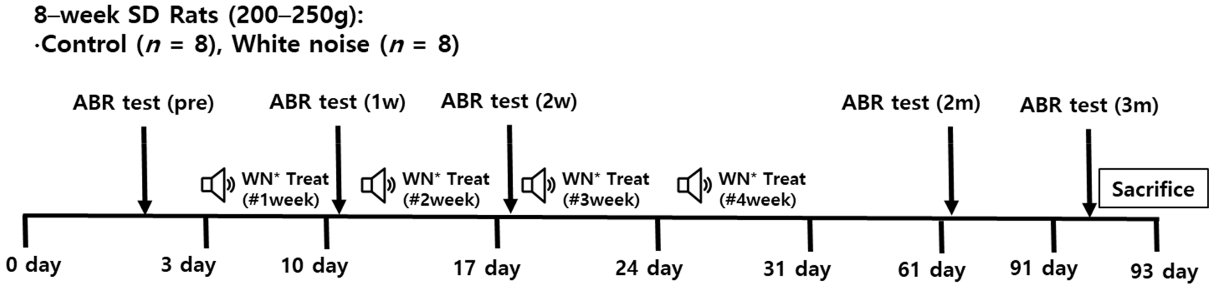
4.1. Noise and Comparison Groups

The Institutional Animal Care and Use Committee of CHA University Medical School (IACUC190046) approved this study. All experimental procedures followed the guidelines of the Institutional Animal Care and Use Committee of CHA University Medical School. The rats (postnatal eight-week Sprague-Dawley) were divided into control and noise-exposed groups (Figure 1). The rats in the noise-exposed group were exposed to wideband noise (2–20 kHz, 115 dB SPL, 4 h/day for 3 days/week for 4 weeks) in free-field condition using a speaker placed at the ceiling of the noise box (Tucker-Davis Technologies; Alachua, FL, USA). The 115 dB SPL of noise was applied to result in a permanent threshold shift. We chose the profound hearing loss animals to exclude the neural plasticity due to the recovery of hearing loss, which can be mixed up with the neural plasticity from the auditory deprivation. Rats were not anesthetized during noise exposure and the noise box was soundproofed. Rats in the control group were raised with ambient noise of approximately 40–60 dB SPL. Hearing levels were estimated using auditory brainstem response (ABR) thresholds. Two months after noise exposure, all rats were euthanized and the cochleae and CN were dissected [43].

4.2. Hearing Level Measurement

Hearing levels were measured using SmartEP Intelligent Hearing System (Miami, FL, USA) [44]. ABR measurements were conducted under anesthesia (40 mg/kg zoletil and 10 mg/kg xylazine). The reference, ground, and measuring electrodes were placed in the vertex, contralateral thigh, and ipsilateral retroauricular space, respectively. A plastic earphone was placed into the ipsilateral ear canal and connected to an EC1 electrostatic speaker. Pure tone sound stimuli were delivered to earphones at frequencies of 4, 8, 16, and 32 kHz (duration: 1562 µs; envelope: Blackman; stimulation rate: 21.1/s; amplitude: 90–20 dB SPL, 1024 sweeps). The hearing threshold was set as the lowest sound amplitude-evoked wave II [45].

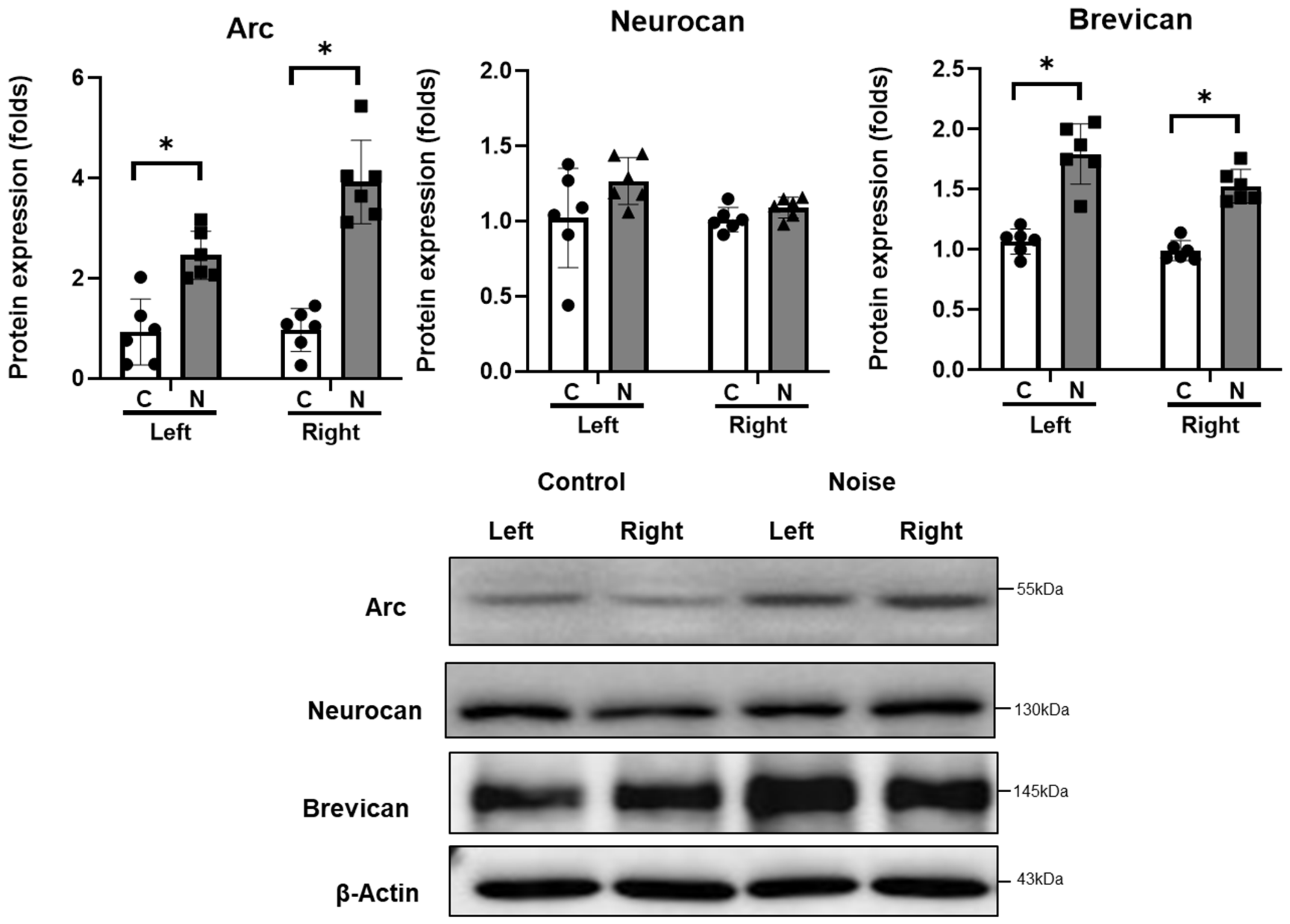
4.3. Protein Expression in CN

The expression levels of arc, neurocan, brevican, SIRT1, caspase 3, and cleaved caspase 3 were evaluated in the CN. The protein was purified from CN tissue using a lysis buffer (Pre-prep, Intron). Equal amounts of protein samples were subjected to 8–10% sodium dodecyl sulfate-polyacrylamide gel electrophoresis. After transferring the resolved proteins onto polyvinylidene difluoride membranes (Merck Millipore, Burlington, MA, USA), they were incubated with 1:1000 anti-arc (Santa Cruz, Dallas, TX, USA, sc166461), anti-neurocan (Mybiosource, San Diego, CA, USA, MBS822310), anti-brevican (Santa Cruz, Dallas, TX, USA, sc135849), anti-SIRT1 (Santa Cruz, Dallas, TX, USA, sc74465), caspase 3 (Cell Signaling Technology, Danvers, MA, USA, 9662s), cleaved caspase 3 (Cell Signaling Technology, 9661s), and anti-rabbit monoclonal β-actin antibodies (Cell Signaling Technology, D6A8). Horseradish peroxidase (HRP)-conjugated secondary antibodies were treated with 1:2000 anti-rabbit IgG, HRP-linked (Cell Signaling Technology, 7074s), and goat anti-mouse IgG H&L (HRP) (Abcam, Cambridge, England, ab97023). The membranes were visualized using an enhanced chemiluminescence kit (Bio-Rad, Hercules, CA, USA). Protein bands were quantified using the ImageJ software (National Institutes of Health, Bethesda, MD, USA).

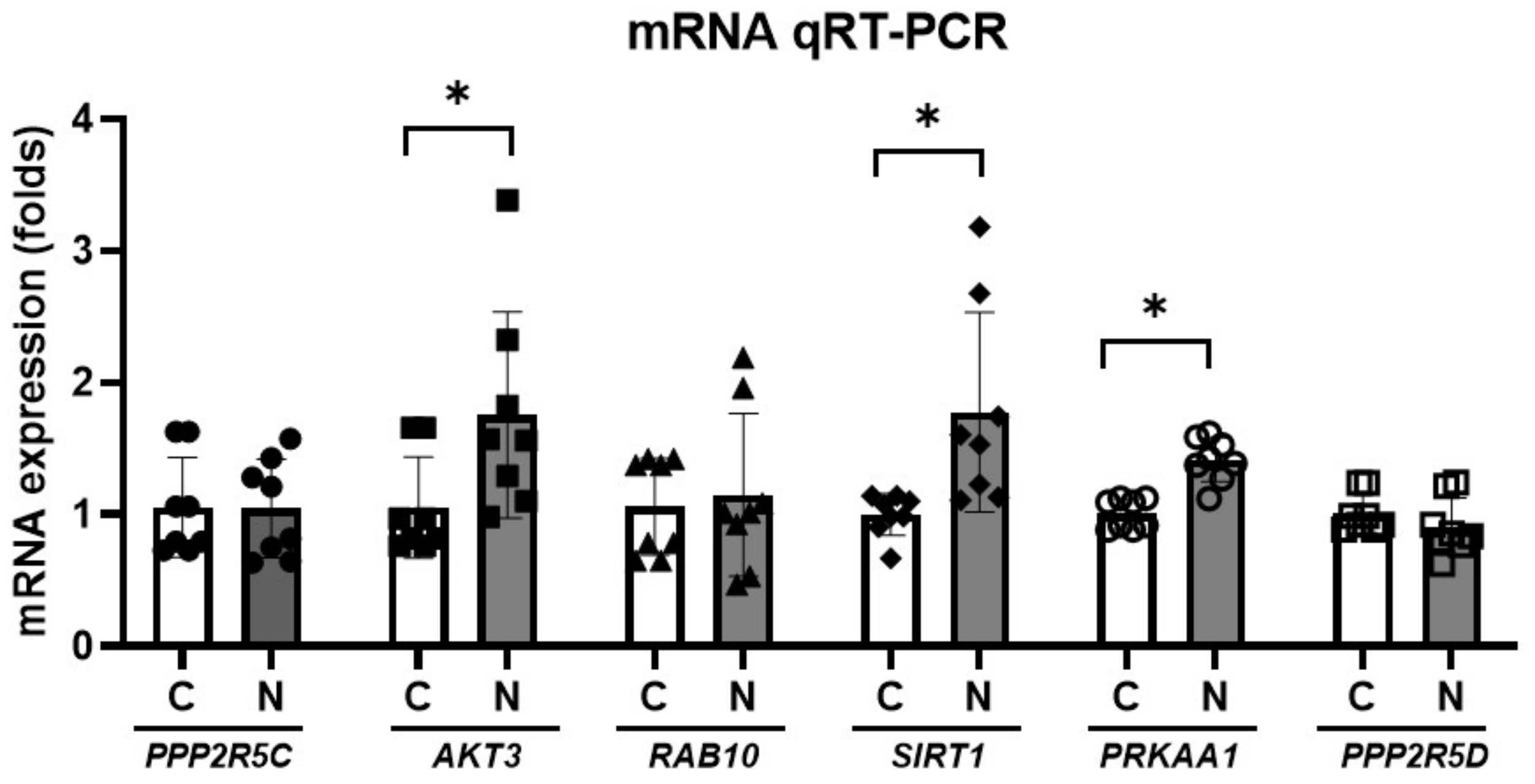
4.4. miRNA Expression in the CN

The miRNAs that showed expression changes following noise exposure or hearing loss in the central nervous system were examined in the CN of rats exposed to chronic noise. CN from eight rats per group were analyzed. MDNA was synthesized from purified miRNAs using the miScript® II RT kit (Qiagen, Hilden, Germany). PCR was conducted using the miScript SYBR® Green PCR kit (Qiagen) with the miScript Primer Assay reagents (Qiagen). U6 small nuclear RNA was used as the reference. ABI 7500 real-time PCR system (Applied Biosystems, Foster City, CA, USA) was used. The expression levels of miRNAs were quantified using cycle threshold (Ct) values and the 2−∆∆Ct method.

4.5. The Predicted Target Genes for miRNAs

The predicted target pathways and related genes for the dysregulated miRNAs were accessed using DIANA-mirPath v.3 (Available online: http://snf-515788.vm.okeanos.grnet.gr/ (accessed on 7 June 2021)). KEGG pathways and the related target genes were searched with a threshold of 0.8 and a false discovery rate correction [46].
The mRNA expression of the predicted target genes was analyzed using quantitative reverse-transcription-PCR (qRT-PCR) [47,48]. Following reverse transcription of cDNA from purified RNA, the reagent was prepared using TOPreal™ qPCR 2× PreMIX (SYBR Green with low ROX; Enzynomics; Daejeon, Korea). PCR was conducted using the ViiA7 real-time PCR system (Applied Biosystems, Carlsbad, CA, USA). The primer sets used in this study are listed in Table 2. Glyceraldehyde 3-phosphate dehydrogenase (GAPDH) was used as the reference gene. mRNA expression levels were quantified using the 2ΔΔCt method.

4.6. Statistical Analysis

The hearing threshold shifts after the noise exposure were compared between nose-exposed and control rats using a unpaired t-test. The mRNA and miRNA expression levels in noise-exposed and control rats were compared using the unpaired t-test. The results are presented as mean ± standard deviation (SD). All analyses were conducted using the SPSS software (ver. 21.0; IBM Corp.; Armonk, NY, USA). Statistical significance was set at p ≤ 0.05.

5. Conclusions

The CN of noise-exposed rats exhibited elevated expression of arc and brevican. The levels of several miRNAs, including miR-758-5p, miR-15b-5p, miR-212-3p, miR-199a-5p, and miR-134-3p, were low in the CN of rats exposed to chronic noise. The AMPK signaling pathway involving AKT3 and SIRT1 was predicted to be activated in noise-exposed rats.

Author Contributions

Conceptualization, S.Y.K.; methodology, S.Y.K.; formal analysis, S.Y.K., J.J. and S.M.L.; writing—original draft preparation, S.Y.K.; writing—review and editing, S.Y.K., C.H.L. and S.M.L.; funding acquisition, S.Y.K. and C.H.L. All authors have read and agreed to the published version of the manuscript.

Funding

This research was supported by funding from the National Research Foundation (NRF) of Korea (2020R1A2C4002594 (Approval date: 1 March 2020)) and a grant of the Research Driven Hospital R&D project, funded by the CHA Bundang Medical Center (grant number: BDCHA R&D 2021-003) The APC was funded by BDCHA R&D 2021-003.

Institutional Review Board Statement

The Institutional Animal Care and Use Committee of CHA University (IACUC190046) approved the performed animal experiments. The conditions of animal rearing, drug administration, and sacrifice complied with the regulations of the Institutional Animal Care and Use Committee of CHA University.

Informed Consent Statement

Not applicable.

Data Availability Statement

The data presented in this study are available upon request from the corresponding author.

Conflicts of Interest

The authors declare no conflict of interest. The funders had no role in the design of the study; in the collection, analyses, or interpretation of data; in the writing of the manuscript; or in the decision to publish the results.

References

  1. Manohar, S.; Ramchander, P.; Salvi, R.; Seigel, G. Synaptic Reorganization Response in the Cochlear Nucleus Following Intense Noise Exposure. Neuroscience 2018, 399, 184–198. [Google Scholar] [CrossRef] [PubMed]
  2. Manley, G.A. The mammalian Cretaceous cochlear revolution. Hear. Res. 2017, 352, 23–29. [Google Scholar] [CrossRef] [PubMed]
  3. Weinrich, L.; Sonntag, M.; Arendt, T.; Morawski, M. Neuroanatomical characterization of perineuronal net components in the human cochlear nucleus and superior olivary complex. Hear. Res. 2018, 367, 32–47. [Google Scholar] [CrossRef]
  4. Favuzzi, E.; Marques-Smith, A.; Deogracias, R.; Winterflood, C.M.; Sánchez-Aguilera, A.; Mantoan, L.; Maeso, P.; Fernandes, C.; Ewers, H.; Rico, B. Activity-Dependent Gating of Parvalbumin Interneuron Function by the Perineuronal Net Protein Brevican. Neuron 2017, 95, 639–655.e10. [Google Scholar] [CrossRef]
  5. Nguyen, A.; Khaleel, H.M.; Razak, K.A. Effects of noise-induced hearing loss on parvalbumin and perineuronal net expression in the mouse primary auditory cortex. Hear. Res. 2017, 350, 82–90. [Google Scholar] [CrossRef]
  6. Park, S.S.; Lee, D.H.; Lee, S.M.; Lee, C.H.; Kim, S.Y. Noise exposure alters MMP9 and brevican expression in the rat primary auditory cortex. BMC Neurosci. 2020, 21, 16. [Google Scholar] [CrossRef] [PubMed]
  7. Krohs, C.; Bordeynik-Cohen, M.; Messika-Gold, N.; Elkon, R.; Avraham, K.B.; Nothwang, H.G. Expression pattern of cochlear microRNAs in the mammalian auditory hindbrain. Cell Tissue Res. 2021, 383, 655–666. [Google Scholar] [CrossRef] [PubMed]
  8. Park, S.; Han, S.H.; Kim, B.G.; Suh, M.W.; Lee, J.H.; Oh, S.H.; Park, M.K. Changes in microRNA Expression in the Cochlear Nucleus and Inferior Colliculus after Acute Noise-Induced Hearing Loss. Int. J. Mol. Sci. 2020, 21, 8792. [Google Scholar] [CrossRef]
  9. Giraldez, A.J.; Cinalli, R.M.; Glasner, M.E.; Enright, A.; Thomson, J.M.; Baskerville, S.; Hammond, S.M.; Bartel, D.P.; Schier, A.F. MicroRNAs Regulate Brain Morphogenesis in Zebrafish. Science 2005, 308, 833–838. [Google Scholar] [CrossRef] [PubMed] [Green Version]
  10. Schratt, G.M.; Tuebing, F.; Nigh, E.A.; Kane, C.G.; Sabatini, M.E.; Kiebler, M.; Greenberg, M.E. A brain-specific microRNA regulates dendritic spine development. Nature 2006, 439, 283–289. [Google Scholar] [CrossRef]
  11. Higuchi, F.; Uchida, S.; Yamagata, H.; Abe-Higuchi, N.; Hobara, T.; Hara, K.; Kobayashi, A.; Shintaku, T.; Itoh, Y.; Suzuki, T.; et al. Hippocampal MicroRNA-124 Enhances Chronic Stress Resilience in Mice. J. Neurosci. 2016, 36, 7253–7267. [Google Scholar] [CrossRef] [PubMed]
  12. Schlüter, T.; Berger, C.; Rosengauer, E.; Fieth, P.; Krohs, C.; Ushakov, K.; Steel, K.P.; Avraham, K.B.; Hartmann, A.K.; Felmy, F.; et al. miR-96 is required for normal development of the auditory hindbrain. Hum. Mol. Genet. 2018, 27, 860–874. [Google Scholar] [CrossRef] [PubMed]
  13. Friedman, L.M.; Dror, A.A.; Mor, E.; Tenne, T.; Toren, G.; Satoh, T.; Biesemeier, D.J.; Shomron, N.; Fekete, D.M.; Hornstein, E.; et al. MicroRNAs are essential for development and function of inner ear hair cells in vertebrates. Proc. Natl. Acad. Sci. USA 2009, 106, 7915–7920. [Google Scholar] [CrossRef] [PubMed] [Green Version]
  14. Napoli, D.; Lupori, L.; Mazziotti, R.; Sagona, G.; Bagnoli, S.; Samad, M.; Sacramento, E.K.; Kirkpartick, J.; Putignano, E.; Chen, S.; et al. MiR-29 coordinates age-dependent plasticity brakes in the adult visual cortex. EMBO Rep. 2020, 21, e50431. [Google Scholar] [CrossRef] [PubMed]
  15. Hill, K.; Yuan, H.; Wang, X.; Sha, S.H. Noise-Induced Loss of Hair Cells and Cochlear Synaptopathy Are Mediated by the Activation of AMPK. J. Neurosci. 2016, 36, 7497–7510. [Google Scholar] [CrossRef] [PubMed] [Green Version]
  16. Nagashima, R.; Yamaguchi, T.; Kuramoto, N.; Ogita, K. Acoustic overstimulation activates 5′-AMP-activated protein kinase through a temporary decrease in ATP level in the cochlear spiral ligament prior to permanent hearing loss in mice. Neurochem. Int. 2011, 59, 812–820. [Google Scholar] [CrossRef]
  17. Noack, F.; Calegari, F. Micro- RNA s meet epigenetics to make for better brains. EMBO Rep. 2014, 15, 1224–1225. [Google Scholar] [CrossRef] [Green Version]
  18. Wu, H.Z.Y.; Thalamuthu, A.; Cheng, L.; Fowler, C.; Masters, C.L.; Sachdev, P.; Mather, K.A. Differential blood miRNA expression in brain amyloid imaging-defined Alzheimer’s disease and controls. Alzheimers Res. Ther. 2020, 12, 59. [Google Scholar] [CrossRef]
  19. Aten, S.; Page, C.E.; Kalidindi, A.; Wheaton, K.; Niraula, A.; Godbout, J.P.; Hoyt, K.; Obrietan, K. miR-132/212 is induced by stress and its dysregulation triggers anxiety-related behavior. Neuropharmacology 2018, 144, 256–270. [Google Scholar] [CrossRef]
  20. Cha, D.J.; Mengel, D.; Mustapic, M.; Liu, W.; Selkoe, D.J.; Kapogiannis, D.; Galasko, D.; Rissman, R.A.; Bennett, D.A.; Walsh, D.M. miR-212 and miR-132 Are Downregulated in Neurally Derived Plasma Exosomes of Alzheimer’s Patients. Front. Neurosci. 2019, 13, 1208. [Google Scholar] [CrossRef] [Green Version]
  21. Song, D.; Li, G.; Hong, Y.; Zhang, P.; Zhu, J.; Yang, L.; Huang, J. miR-199a decreases Neuritin expression involved in the development of Alzheimer’s disease in APP/PS1 mice. Int. J. Mol. Med. 2020, 46, 384–396. [Google Scholar] [CrossRef] [PubMed]
  22. Fan, C.; Zhu, X.; Song, Q.; Wang, P.; Liu, Z.; Yu, S.Y. MiR-134 modulates chronic stress-induced structural plasticity and depression-like behaviors via downregulation of Limk1/cofilin signaling in rats. Neuropharmacology 2018, 131, 364–376. [Google Scholar] [CrossRef] [PubMed]
  23. Ruderman, N.B.; Xu, X.J.; Nelson, L.; Cacicedo, J.M.; Saha, A.K.; Lan, F.; Ido, Y. AMPK and SIRT1: A long-standing partnership? Am. J. Physiol. Endocrinol. Metab. 2010, 298, E751–E760. [Google Scholar] [CrossRef]
  24. Lin, J.Y.; Kuo, W.W.; Baskaran, R.; Kuo, C.H.; Chen, Y.A.; Chen, W.S.; Ho, T.J.; Day, C.H.; Mahalakshmi, B.; Huang, C.Y. Swimming exercise stimulates IGF1/PI3K/Akt and AMPK/SIRT1/PGC1α survival signaling to suppress apoptosis and inflammation in aging hippocampus. Aging 2020, 12, 6852–6864. [Google Scholar] [CrossRef]
  25. Ramadori, G.; Lee, C.E.; Bookout, A.L.; Lee, S.; Williams, K.; Anderson, J.; Elmquist, J.K.; Coppari, R. Brain SIRT1: Anatomical Distribution and Regulation by Energy Availability. J. Neurosci. 2008, 28, 9989–9996. [Google Scholar] [CrossRef]
  26. Vaziri, H.; Dessain, S.K.; Eaton, E.N.; Imai, S.-I.; Frye, R.A.; Pandita, T.K.; Guarente, L.; Weinberg, R.A. hSIR2SIRT1 Functions as an NAD-Dependent p53 Deacetylase. Cell 2001, 107, 149–159. [Google Scholar] [CrossRef] [Green Version]
  27. Jeong, H.; Cohen, D.E.; Cui, L.; Supinski, A.; Savas, J.N.; Mazzulli, J.R.; Yates, J.R.; Bordone, L.; Guarente, L.; Krainc, D. Sirt1 mediates neuroprotection from mutant huntingtin by activation of the TORC1 and CREB transcriptional pathway. Nat. Med. 2011, 18, 159–165. [Google Scholar] [CrossRef] [PubMed]
  28. Ferland, C.L.; Hawley, W.R.; Puckett, R.E.; Wineberg, K.; Lubin, F.D.; Dohanich, G.P.; Schrader, L.A. Sirtuin Activity in Dentate Gyrus Contributes to Chronic Stress-Induced Behavior and Extracellular Signal-Regulated Protein Kinases 1 and 2 Cascade Changes in the Hippocampus. Biol. Psychiatry 2013, 74, 927–935. [Google Scholar] [CrossRef] [Green Version]
  29. Prozorovski, T.; Schulze-Topphoff, U.; Glumm, R.; Baumgart, J.; Schröter, F.; Ninnemann, O.; Siegert, E.; Bendix, I.; Brüstle, O.; Nitsch, R.; et al. Sirt1 contributes critically to the redox-dependent fate of neural progenitors. Nat. Cell Biol. 2008, 10, 385–394. [Google Scholar] [CrossRef]
  30. Li, X.H.; Chen, C.; Tu, Y.; Sun, H.T.; Zhao, M.L.; Cheng, S.X.; Qu, Y.; Zhang, S. Sirt1 Promotes Axonogenesis by Deacetylation of Akt and Inactivation of GSK3. Mol. Neurobiol. 2013, 48, 490–499. [Google Scholar] [CrossRef]
  31. Teertam, S.K.; Babu, P.P. Differential role of SIRT1/MAPK pathway during cerebral ischemia in rats and humans. Sci. Rep. 2021, 11, 6339. [Google Scholar] [CrossRef] [PubMed]
  32. Selivanova, O.; Brieger, J.; Heinrich, U.-R.; Mann, W. Akt and c-Jun N-terminal kinase are regulated in response to moderate noise exposure in the cochlea of guinea pigs. ORL J. Otorhinolaryngol. Relat. Spec. 2007, 69, 277–282. [Google Scholar] [CrossRef] [PubMed]
  33. Plas, D.R.; Thompson, C.B. Akt-dependent transformation: There is more to growth than just surviving. Oncogene 2005, 24, 7435–7442. [Google Scholar] [CrossRef] [Green Version]
  34. Duan, J.; Cui, J.; Zheng, H.; Xi, M.; Guo, C.; Weng, Y.; Yin, Y.; Wei, G.; Cao, J.; Wang, Y.; et al. Aralia taibaiensis Protects against I/R-Induced Brain Cell Injury through the Akt/SIRT1/FOXO3a Pathway. Oxidative Med. Cell. Longev. 2019, 2019, 7609765. [Google Scholar] [CrossRef] [PubMed] [Green Version]
  35. Chen, J.; Yuan, H.; Talaska, A.E.; Hill, K.; Sha, S.-H. Increased Sensitivity to Noise-Induced Hearing Loss by Blockade of Endogenous PI3K/Akt Signaling. J. Assoc. Res. Otolaryngol. 2015, 16, 347–356. [Google Scholar] [CrossRef] [PubMed] [Green Version]
  36. Sha, S.-H.; Chen, F.-Q.; Schacht, J. PTEN attenuates PIP3/Akt signaling in the cochlea of the aging CBA/J mouse. Hear. Res. 2010, 264, 86–92. [Google Scholar] [CrossRef] [PubMed] [Green Version]
  37. Chung, W.-H.; Pak, K.; Lin, B.; Webster, N.; Ryan, A.F. A PI3K Pathway Mediates Hair Cell Survival and Opposes Gentamicin Toxicity in Neonatal Rat Organ of Corti. J. Assoc. Res. Otolaryngol. 2006, 7, 373–382. [Google Scholar] [CrossRef] [Green Version]
  38. Lai, R.; Li, W.; Hu, P.; Xie, D.; Wen, J. Role of Hsp90/Akt pathway in the pathogenesis of gentamicin-induced hearing loss. Int. J. Clin. Exp. Pathol. 2018, 11, 4431–4438. [Google Scholar]
  39. Kim, Y.; Kang, H.; Powathil, G.; Kim, H.; Trucu, D.; Lee, W.; Lawler, S.; Chaplain, M. Role of extracellular matrix and microenvironment in regulation of tumor growth and LAR-mediated invasion in glioblastoma. PLoS ONE 2018, 13, e0204865. [Google Scholar] [CrossRef]
  40. Jahan, N.; Hannila, S.S. Transforming growth factor β-induced expression of chondroitin sulfate proteoglycans is mediated through non-Smad signaling pathways. Exp. Neurol. 2015, 263, 372–384. [Google Scholar] [CrossRef]
  41. Silver, J.; Miller, J.H. Regeneration beyond the glial scar. Nat. Rev. Neurosci. 2004, 5, 146–156. [Google Scholar] [CrossRef] [PubMed]
  42. Bradbury, E.J.; Moon, L.; Popat, R.J.; King, V.R.; Bennett, G.S.; Patel, P.N.; Fawcett, J.; McMahon, S. Chondroitinase ABC promotes functional recovery after spinal cord injury. Nature 2002, 416, 636–640. [Google Scholar] [CrossRef] [PubMed]
  43. Han, K.H.; Cho, H.; Han, K.R.; Mun, S.K.; Kim, Y.K.; Park, I.; Chang, M. Role of microRNA-375-3p-mediated regulation in tinnitus development. Int. J. Mol. Med. 2021, 48, 136. [Google Scholar] [CrossRef] [PubMed]
  44. Kim, S.Y.; Heo, H.; Kim, D.H.; Kim, H.J.; Oh, S.H. Neural Plastic Changes in the Subcortical Auditory Neural Pathway after Single-Sided Deafness in Adult Mice: A MEMRI Study. BioMed Res. Int. 2018, 2018, 8624745. [Google Scholar] [CrossRef] [PubMed] [Green Version]
  45. Alvarado, J.C.; Fuentes-Santamaría, V.; Jareño-Flores, T.; Blanco, J.L.; Juiz, J.M. Normal variations in the morphology of auditory brainstem response (ABR) waveforms: A study in wistar rats. Neurosci. Res. 2012, 73, 302–311. [Google Scholar] [CrossRef]
  46. Vlachos, I.S.; Zagganas, K.; Paraskevopoulou, M.D.; Georgakilas, G.; Karagkouni, D.; Vergoulis, T.; Dalamagas, T.; Hatzigeorgiou, A.G. DIANA-miRPath v3.0: Deciphering microRNA function with experimental support. Nucleic Acids Res. 2015, 43, W460–W466. [Google Scholar] [CrossRef] [PubMed]
  47. Kim, S.Y.; Kim, J.K.; Park, S.H.; Kim, B.G.; Jang, A.S.; Oh, S.H.; Lee, J.H.; Suh, M.W.; Park, M.K. Effects of inhaled particulate matter on the central nervous system in mice. NeuroToxicology 2018, 67, 169–177. [Google Scholar] [CrossRef]
  48. Bustin, S.A.; Benes, V.; Garson, J.A.; Hellemans, J.; Huggett, J.; Kubista, M.; Mueller, R.; Nolan, T.; Pfaffl, M.W.; Shipley, G.L.; et al. The MIQE Guidelines: Minimum Information for Publication of Quantitative Real-Time PCR Experiments. Clin. Chem. 2009, 55, 611–622. [Google Scholar] [CrossRef] [Green Version]
Figure 1. Design of the present study (n = 8 per group). Noise was experienced by the rats for 4 weeks, and hearing levels were measured via auditory brainstem response (ABR) tests. The qRT-PCR of miRNA (n = 8), the predicted target genes (n = 8 per group), and Western blotting (n = 6) were conducted using cochlear nuclei of the noise-exposed and control rats. * White noise (115 dB SPL) 3 h/day 3 times a week.
Cells 11 02266 g001
Figure 2. Hearing threshold shifts after noise exposure (n = 8 per group). The noise exposed rats demonstrated hearing threshold shifts at 1 week, 2 weeks, 2 months, and 3 months post noise exposure at 4, 8, 16, and 32 kHz. (* p < 0.05; unpaired t-test between the control and noise-exposed groups; C: control rats; N: noise rats).
Cells 11 02266 g002
Figure 3. Arc, neurocan, and brevican expression in the cochlear nuclei after noise exposure (n = 6 per group). Arc and brevican expression was increased in the noise-exposed group (* p < 0.05; unpaired t-test between control and noise-exposed rats; C: control rats; N: noise rats).
Cells 11 02266 g003
Figure 4. miR-758-5p, miR-15b-5p, miR-212-3p, miR-199a-5p, and miR-134-3p expression was reduced in the noise-exposed rats (n = 8 per group; * p < 0.05; unpaired t-test between control and noise-exposed rats).
Cells 11 02266 g004
Figure 5. Pathways related to the dysregulated miRNAs in the cochlear nuclei of noise-exposed rats.
Cells 11 02266 g005
Figure 6. mRNA expression levels of PPP2R5C, AKT3, RAB10, SIRT1, PRKAA1, and PPP2R5D were higher in the noise-exposed rats than in the control rats (n = 8 per group; * p < 0.05; unpaired t-test between control and noise-exposed rats).
Cells 11 02266 g006
Figure 7. SIRT1, caspase 3, and cleaved caspase 3 expression in the cochlear nuclei after noise exposure (n = 6 per group). SIRT1, caspase 3, and cleaved caspase 3 expressions were higher in the noise-exposed group (* p < 0.05; unpaired t-test between control and noise-exposed rats; C: control rats; N: noise rats).
Cells 11 02266 g007
Table 1. The predicted target genes related to the AMPK signaling pathway of the downregulated miRNAs in the cochlear nuclei of noise-exposed rats.
Related miRNAGene NameScore
hsa-miR-15b-5pFASNfatty acid synthase0.935
IGF1Rinsulin like growth factor 1 receptor0.866
PPP2R5Cprotein phosphatase 2 regulatory subunit B’gamma0.845
CCND1cyclin D10.873
PPP2R1Aprotein phosphatase 2 scaffold subunit Aalpha0.847
PIK3R1phosphoinositide-3-kinase regulatory subunit 10.989
INSRinsulin receptor0.947
IGF1insulin like growth factor 10.859
AKT3AKT serine/threonine kinase 31
PPARGC1APPARG coactivator 1 alpha0.806
FOXO1forkhead box O10.913
CAB39calcium binding protein 390.8
PPP2R1Bprotein phosphatase 2 scaffold subunit Abeta0.885
RAB10RAB10, member RAS oncogene family0.941
PFKFB46-phosphofructo-2-kinase/fructose-2,6-biphosphatase 40.872
STRADASTE20 related adaptor alpha0.81
hsa-miR-758-5pCD36CD36 molecule0.901
G6PCglucose-6-phosphatase catalytic subunit 10.865
PRKAA1protein kinase AMP-activated catalytic subunit alpha 10.84
hsa-miR-199a-5pSIRT1sirtuin 10.9
PRKAG2protein kinase AMP-activated non-catalytic subunit gamma 20.831
RAB10RAB10, member RAS oncogene family0.954
hsa-miR-212-3p|PRKAA2protein kinase AMP-activated catalytic subunit alpha 20.844
SIRT1sirtuin 11
CREB5cAMP responsive element binding protein 50.982
PPP2R5Cprotein phosphatase 2 regulatory subunit B’gamma0.889
AKT3AKT serine/threonine kinase 30.92
PIK3CAphosphatidylinositol-4,5-bisphosphate 3-kinase catalytic subunit alpha0.864
FOXO3forkhead box O30.997
CPT1Bcarnitine palmitoyltransferase 1B0.892
hsa-miR-134-3pPPP2R5Dprotein phosphatase 2 regulatory subunit B’delta0.965
Table 2. Oligonucleotide primer sequences for quantitative reverse transcriptase polymerase chain reaction.
GenePrimer Sequence (Forward)Primer Sequence (Reverse)Annealing Temperature (°C)Product Size (bp)
PPP2R5C5′-CTAGCCAAAGCGAATCCCCA-3′5′-GAGTCTCGTCGCTCACTGTC-3′6093
AKT35′-CCACCTGAAAAGTATGACGACG-3′5′-TAAGAGCGAGGACTGGTGGA-3′60133
RAB105′-TTGTTTGCCCCCACTACTCC-3′5′-TAAATGAGGGGCTGACACCG-3′60185
SIRT15′-GCAGGTTGCGGGAATCCAA-3′5′-GGCAAGATGCTGTTGCAAA-3′60155
PRKAA15′-GGGTGAAGATCGGCCACTAC-3′5′-CTCTCTGCGGATTTTCCCGA-3′6062
PPP2R5D5′-CGAGTCGGGTCGCTAAGAAG-3′5′-ACACTCAGAGTCAAAGGGCG-3′60164
GAPDH5′-AGTGCCAGCCTCGTCTCATA-3′5′-AAGAGAAGGCAGCCCTGGTA-3′6093
Publisher’s Note: MDPI stays neutral with regard to jurisdictional claims in published maps and institutional affiliations.

