Article

The Inhibitory Mechanism of Eugenol on Lasiodiplodia theobromae and Its Induced Disease Resistance of Passion Fruit

1
Food and Pharmaceutical Engineering Institute, Guiyang University, Guiyang 550005, China
2
School of Chemistry and Food Science, Nanchang Normal University, Nanchang 330023, China
*
Author to whom correspondence should be addressed.
Agronomy 2023, 13(5), 1408; https://doi.org/10.3390/agronomy13051408
Submission received: 27 March 2023 / Revised: 8 May 2023 / Accepted: 18 May 2023 / Published: 19 May 2023

Abstract

:
The inhibitory effects and mechanisms of eugenol were studied in pathogenic fungi that were isolated from passion fruit affected with naturally occurring fruit rot; additionally, the effect of eugenol treatment on fruit rot resistance in passion fruit was investigated. The corresponding results demonstrated that the pathogenic fungus causing passion fruit rot was Lasiodiplodia theobromae and that the minimal inhibitory concentration of eugenol against L. theobromae was 0.30 mg/mL. Eugenol significantly inhibited mycelial growth and spore germination of this fruit rot fungus. Further, nucleic acid release, electrical conductivity, and protein and soluble sugar content of the fruit rot fungus gradually increased with increasing eugenol concentrations. Propidium iodide staining revealed that the cell membrane integrity of L. theobromae hyphae was disrupted when treated with eugenol. In addition, eugenol treatment inhibited the spread of disease spot diameter after inoculation with L. theobromae, effectively increased APX, SOD, CAT, POD, 4CL, C4H, and PAL activities, promoted the accumulation of disease-stage-related proteins CHI and GLU, and increased the total phenol and flavonoid content during storage in passion fruit. Overall, these results suggest that eugenol has good application prospects for the effective control of fruit rot in passion fruit.

1. Introduction

Passion fruit (Passiflora edulis Sims.), also known as granadilla, is the edible fruit of an herbaceous climbing plant in the family Passifloraceae [1]. It is rich in nutrients and is composed of polysaccharides, flavonoids, vitamins, and various minerals and fibres, resulting in a high commercial value of the fruit, which is highly favoured by consumers [2,3,4,5]. Passion fruit possesses antioxidant, anti-inflammatory, and anti-cancer properties, and can reduce the risk of cardiovascular diseases, cancers, and metabolic disorders [6,7]. Additionally, it is in great demand in the fresh fruit, processed food, and juice market [8]; however, the harvest period of passion fruit is concentrated in the high-temperature and rainy seasons, such as summer and autumn. It undergoes vigorous respiration and transpiration metabolism after harvest, and the fruit peel is susceptible to mechanical damage and microbial infection, which accelerate fruit decay and strongly impact the storage quality of passion fruit, thereby drastically reducing the economic benefits of the passion fruit industry [9].
Currently, the reported diseases of passion fruit primarily include anthracnose, brown spots, and fruit rot disease. Fruit rot readily occurs during passion fruit storage; specifically, this disease occurs during the ripening period of this fruit, with oil spots being the main symptom appearing on the fruit surface. With the development of brown rot, these spots enlarge and become oval-shaped, with a light blue-brown colour and water stains at the edges of these spots. As the disease progresses to a later stage, the colour of the diseased fruit deepens and the spots become larger, ultimately leading to a deterioration in passion fruit quality and making it an unfavourable fruit for fresh storage [10,11,12]. Few studies have been conducted on the control of postharvest infection in passion fruit during storage; therefore, there is an urgent need to develop effective preservatives to improve the postharvest quality of passion fruit and to efficiently address the invasion of disease-causing fungi during storage.
Eugenol, a natural preservative, has been increasingly recognized for its efficient antibacterial properties and its corresponding ability to maintain freshness during the post-harvest storage of agricultural products. Through scanning electron microscopy, Zhang [13] observed that 31.25 μM eugenol treatment resulted in cell membrane contraction and disruption in Porphyromonas gingivalis. Jafri [14] determined that 100 μg/mL eugenol could significantly impair the cell membrane structure of Bacillus subtilis and Bacillus licheniformis. Further, Devi [15] demonstrated that 0.0125% eugenol could efficiently eliminate Salmonella typhimurium within 60 min of treatment by damaging the cytoplasmic membrane. Cui [16] established that eugenol affects the number of metabolites in the tricarboxylic acid (TCA) cycle pathways of Listeria, resulting in bacterial death. However, the mechanism by which eugenol inhibits the growth of the pathogenic fungi that causes passion fruit rot remains unclear; ultimately, this greatly limits the practical application of eugenol.
Therefore, this study investigated the effects of different concentrations of eugenol on Lasiodiplodia theobromae. Mycelial growth, cell membrane, spore germination, and fruit rot of passion fruit; additionally, the potential mechanism by which eugenol inhibits the fruit rot pathogen and its effect on fruit rot resistance were established. Ultimately, this provided a theoretical basis to consistently control post-harvest fruit rot and develop new technology to preserve and maintain the freshness of passion fruit.

2. Materials and Methods

2.1. Isolation and Identification of Pathogenic Fungi

2.1.1. Isolation and Identification of Pathogenic Fungi and Detection of Pathogenicity

Naturally infected passion fruits were disinfected with a 75% ethanol solution for 30 s and removed from the solution to dry. These fruits were then placed in clean water for 30 s to wash away excess ethanol solution and dried on a laminar flow bench. Tissue (approximately 5 mm2) was collected from the margin between naturally infected and healthy passion fruit; The tissue was used for pathogen isolation and transferred to potato dextrose agar (PDA) plates. 50 PDA plates were used for isolation and purification of different pathogenic fungi. These plates were then placed in a 25 °C incubator and monitored regularly for colony growth. Colonies were separated on PDA plates every three days until different purified strains. We used criteria such as pathogenicity assay, morphological observation, and ITS sequence analysis of pathogenic fungi to select specific strains and named the selected strains as strain A.
The pathogenicity of the isolated pathogen was tested using Koch’s postulates. The pathogenic culture with a diameter of 5 mm was inoculated on the PDA plate and cultured until the mycelium grew effectively. Ninety fresh and healthy passion fruits were used in the experiment. The surface was wiped with 75% alcohol for disinfection, washed with distilled water for alcohol, dried, and the centre of the passion fruit surface was punctured with a double punch. Pathogenic fungal cake was used inoculated to the centre of the passion fruit surface damage area, allowing the mycelium side to contact the wound area of the passion fruit; Additionally, inoculate non pathogenic cake onto healthy passion fruit as a control. Subsequently, the inoculated fruits were maintained at a constant temperature of 28 °C and a relative humidity of 95% for 7 days, followed by continuous observation and the recording of symptom development. Once symptoms manifested, the infected area of the fruit was collected and reisolated to complete Koch’s postulate test and confirm whether the re-isolated fungus was the pathogen.

2.1.2. Morphological Identification of Pathogenic Fungi

Observations and recordings of the colony culture characteristics, including colony colour and shape, were performed under a microscope (Murzider optical microscope, Guangdong Murzider Technology Co., Ltd., Dongguan, China). Preliminary identification of the pathogens was performed according to the colony characteristics of the various pathogen strains on PDA; the morphological structures of the conidia were observed under an optical microscope.

2.1.3. ITS Sequence Analysis of Pathogenic Fungi

Edwards’ cetyltrimethylammonium bromide (CTAB) method was used to extract DNA from this strain [17]. The universal primers ITS1 (5′-TCCGTAGGTGAACCTGCGG-3′) and ITS4 (5′-TCCTCCGCTTATTGATATGC-3′) of the fungal rDNA-ITS were used as PCR amplification primers. The amplification system was 25 µL 2 × Taq PCR Master Mix, 1 µL DNA, 2 µL 10 mmoL/L upstream primer and 2 µL 10 mmoL/L downstream primer, and 20 µL ultrapure water. PCR cycling conditions were 95 °C predenaturation for 5 min, 95 °C for 30 s, 58 °C for 30 s, 72 °C for 30 s for a total of 35 cycles, 72 °C repair extension for 5 min, and then stored at 4 °C. Following the PCR product analysis, agarose gel electrophoresis was performed by Sangon Biotech Co., Ltd. (Guangzhou, China). The sequencing results were subjected to Basic Local Alignment Search Tool (BLAST, U.S. National Library of Medicine) comparative analysis using the NCBI database; phylogenetic tree construction was performed with species that exhibited similar homology, using Molecular Evolutionary Genetics Analysis (MEGA).

2.2. Effect of Eugenol on the Mycelial Growth of the Passion Fruit Rot Pathogen

Mahdi’s method was used to determine the minimal inhibitory concentration (MIC) [18]. A solution of 10.00 mg/mL of eugenol (Shanghai Yuanyuan Biotechnology Co., Ltd., Shanghai, China) was prepared by weighing 0.1 g of eugenol dissolved in 10 mL of anhydrous ethanol; a total of 10.00 mg/mL of eugenol solution was aspirated in PDA medium to obtain PDA medium with concentrations of 0 (control), 0.05, 0.15, 0.30, 0.45, and 0.60 mg/mL, respectively. Fresh colonies of the plant pathogen Lasiodiplodia theobromae with a diameter of 5 mm were collected and placed centrally on the respective PDA plates. Each Petri dish contained a single colony, and each treatment was performed in triplicate. The results were observed after 24 h of inversion culture. The MIC was established as the lowest dilution, at which fungal growth was completely inhibited.
The effect of eugenol on the inhibition of pathogenic fungi in passion fruit rot was measured using a fungal growth inhibition rate assay [19]. Eugenol stock solutions were mixed with the PDA culture medium at specific ratios to prepare culture media with final concentrations of 0, 1/2 MIC, MIC, and 3/2 MIC of eugenol. The purified test fungi were inoculated onto the drug-containing culture medium and inverted. Each treatment was repeated three times. The PDA plate with the inoculated fungi was placed in a 28 °C incubator; the fungal colony growth state was observed and recorded each day. The diameter of the fungal colony was measured using the cross-method (The cross method is to use a vernier calliper to cross measure the colony diameter and take the average value for counting.), and the inhibition rate was calculated according to Equation (1).
Inhibition   rate   % = control   spot   diameter treatment   spot   diameter control   spot   diameter × 100

2.3. Effect of Different Concentrations of Eugenol on Spore Germination of the Passion Fruit Rot Pathogen

The method conducted by Ge was slightly modified to determine the effect of eugenol treatment on spore germination of the fruit rot pathogen [19]. The spore suspension was prepared as follows: The L. theobromae colony PDA plate that was cultured in a 28 °C incubator for 7 d was washed with 5–10 mL of sterile distilled water and filtered through 4 layers of sterile gauze; finally, the concentration was adjusted to 1.0 × 10⁶ CFU/mL. Then, 50 μL of the pathogenic spore suspension was spotted onto the eugenol plates with concentrations at 0 (control group), 1/2 MIC (treatment group 1), MIC (treatment group 2), or 3/2 MIC (treatment group 3); each treatment was repeated three times. The plates were incubated in 28 °C incubators for 12 h and the germination of the pathogenic spores was observed under an optical microscope every 4 h.

2.4. Effect of Eugenol on Cell Membrane Integrity of the Passion Fruit Rot Pathogen

The fungal filamentous samples were collected as follows: after 5 d of 28 °C incubation on a PDA plate, 5 mL sterile water and 5 μL 0.01% Tween-80 solution was poured into the culture dish; then, fungal filaments were gently scraped off using sterilised swabs. Four layers of gauze are used to filter the solution to remove hyphae and obtain the filtrate. The filtrate was centrifuged at 10,000 r/min for 10 min to remove sediment. Resuspended the spores of the pathogenic fungi causing passion fruit rot in a phosphate-buffered salt (PBS) solution. The blood cell counting board was used to regulate the spore concentration to 106 CFU/mL and prepare spore suspensions for future use. Twenty millilitres of spore suspension was injected into sterile PDB medium, incubated at 25 °C, and centrifuged at 160 r/min for 48 h; samples were then centrifuged at 10,000 r/min for 15 min to remove the supernatant to obtain the fungal mycelium

2.4.1. Determination of Relative Conductivity, Nucleic Acid Release, Intracellular Protein Release, and Soluble Sugar Release by Fruit Rot Pathogens

To measure cell membrane permeability, relative conductivity was measured according to Liu’s method [20], intracellular nucleic acid leakage was determined using Tao’s method [21], intracellular protein release and soluble sugar release were determined using Chen’s method [22], and Malondialdehyde (MDA) content was determined using Wei’s method [23].
Fungal filaments were aseptically collected in 25 mL of sterile water; then a specific proportion of eugenol stock solution was added to a final concentration of 0, 1/2 MIC, MIC, or 3/2 MIC. These samples were placed in a shaker incubator at 25 °C with 160 r/min shaking for 0, 2, 4, 6, and 8 h; at these time points, the samples were centrifuged, and the corresponding supernatants were reserved for later use.
To determine the relative conductivity, the extracellular conductivity L/(μS/cm) of shaken cultures treated for 2, 4, 6 and 8 h was first measured using a DDS-11A conductivity meter (Shanghai Raycom Xinjing Instruments Co., Ltd., Shanghai, China), the extracellular conductivity is considered to be L0 at 0 h, and the conductivity measured after boiling in boiling water for 10 min and cooling to 25 °C was considered L′. The experiment was repeated three times. Relative conductivity was used to indicate the permeability of the cell membrane and was calculated using Equation (2):
Relative   electrical   conductivity   % = L L 0   L L 0 × 100
The amount of intracellular nucleic acid released was determined using a UV spectrophotometer (Agilent Cary 3500 UV-Vis spectrophotometer, Guangdong Shengze Technology Co., Ltd., Dongguan, China) at a wavelength of 260 nm; the absorbance was positively correlated with the amount of intracellular nucleic acid released from the fruit rot pathogen after eugenol treatment. The higher the absorbance observed, the more intracellular nucleic acids released. This experiment was repeated three times, and sterile water was used as the control.
One millilitre of the supernatant was added to a tube, and 5.0 mL of Coomassie Brilliant Blue G-250 (Shanghai Yuanye Biology Co., Ltd., Shanghai, China) was added and mixed thoroughly. The solution was allowed to stand for two minutes, and the absorbance was measured at 595 nm. The extracellular protein concentration was calculated using a standard curve of bovine serum protein; this was expressed in μg/mL.
An aliquot of 0.4 mL of the supernatant was transferred to a test tube; then, 3.6 mL of sterile distilled water, 0.4 mL of naphthol-acetic acetate solution, and 1.2 mL of concentrated sulfuric acid were sequentially added to form the supernatant solution. The contents of the tube were mixed thoroughly and allowed to cool to 25 °C before measuring absorbance at 625 nm. Soluble sugar release in the samples was calculated against the standard curve of sucrose, which was expressed as μg/mL.
MDA content was measured using 2 mL of the supernatant solution mixed with 2 mL of thiobarbituric acid (6.7 g/L). After boiling for 20 min, the absorbance of the reaction mixture was measured at 450 nm, 532 nm, and 600 nm. Equation (3) was used to determine MDA content.
MDA   content   μ mol / L = 6.45 × OD 532 OD 600 0.56 × OD 450

2.4.2. Propidium Iodide (PI) Staining to Observe the Effect of Eugenol on the DNA of Passion-Fruit-Rot-Causing Fungi

In this study, Qin’s method of PI staining was utilized with some modifications [24]. Collect, dry, and store the cultivated fungal hyphae for future use. The filaments were added to PI, with a final PI concentration of 10 μg/mL; samples were incubated in the dark at 4 °C for 30 min, then washed twice with a phosphate-buffered solution. The samples were then observed and imaged using a fluorescence microscope. Three fields were randomly observed for each sample and the test was repeated three times.

2.5. Inoculation Treatment for Induction of Eugenol-Dependent Passion Fruit Rot Resistance

Effect of Eugenol on the Incidence of Fruit Rot and Spot Diameter in Passion Fruit

Passion fruit were delivered back to the laboratory on the day of harvesting and maintained overnight at 25 °C; on the second day, fruits were selected that were of equal size, the same maturity, and free of visible injuries or diseases. Two treatments were conducted:
Treatment I: Surface sterilisation with 75% ethanol, followed by immersion in different concentrations of eugenol solution (0, 1/2 MIC, MIC, or 3/2 MIC) for 15 min, removal, and air-drying. Symmetrical puncture wounds at the equator of the fruit were made with a sterile inoculating needle, and 10 μL of the spore suspension was inoculated at each wound and allowed to stand for 2 h. After natural air-drying of the fruits, they were packaged in polyethylene bags and placed in an incubator (25 ± 1 °C temperature, 90 ± 5% humidity) for 8 days. Images and lesion diameters were recorded daily, and the corresponding degrees of induction were calculated.
Treatment II: Fresh and healthy passion fruit was used in the experiment and divided into two groups. The control group and the treatment group are immersed in distilled water without eugenol and MIC concentration of eugenol for 15 min, then air dry and set aside. After air drying, the two sets of fruits are treated using the same method as treatment I. Passion fruit surfaces were disinfected with 75% ethanol, and sterile inoculation needles were used to create wounds in the equatorial region of the passion fruit. After natural drying, 10 μL (106 CFU/mL) of the Lasiodiplodia theobromae spore suspension was inoculated into each wound. After natural drying, the fruits were stored in an incubator at 25 ± 1 °C and 90 ± 5% relative humidity for 12 days; the diameter of the lesion was measured every 2 days based on the cross-shape formula. A lesion closer to 1 cm was used to determine defence enzyme activity and the concentration of antifungal substances. The induction rate was calculated as follows:
Induction   rate   % = fruit   spot   diameter   of   control   group fruit   spot   diameter   of   treatment   spot fruit   spot   diameter   of   treatment   group × 100

2.6. Effect of Eugenol Treatment on the Shrivelling of Passion Fruit

One of the most important indices for evaluating the quality of stored fruit is the degree of fruit shrivelling. Over time the surface of the fruit loses water and wrinkles, which can strongly affect its quality of appearance, and thus, its commercial value. Twelve passion fruit replicates were randomly divided into four groups and soaked in different concentrations of eugenol solution (0, 0.5 MIC, MIC, and 1.5 MIC) for 15 min; these were then removed and dried. After natural drying, the fruit was stored in polyethylene bags and placed in an incubator (25 °C temperature, 95% relative humidity) for 8 days. The area of shrivelled fruit was observed daily, and the shrinkage index was calculated using Equation (5). Shrinkage was graded as follows: 0, no shrinkage; 1, 0–25% shrinkage; 2, 25–50% shrinkage; 3, 50–75% shrinkage; and 4, 75–100% shrinkage.
Shrinkage   index = shrinkage × number   of   passion   fruit   at   this   level total   number   of   passion   fruit

2.7. Effect of Eugenol Treatment on Defence Enzyme Activity in Passion Fruit

To further determine the effect of eugenol on the resistance of passion fruit to fruit rot, we evaluated the relationship between the activities of defence enzymes in passion fruit that was treated with eugenol and the corresponding resistance induction. The prepared crude enzyme extract is used to determine the levels of defence enzyme catalase (CAT), superoxide dismutase (SOD) and peroxidase (POD) [25,26]. Approximately 3 g of tissue sample was weighed into a pre-cooled mortar; next, 5 mL of 0.1 mol/L EDTA, 5% (w/v) polyvinylpyrrolidone, and 1% (v/v) Triton X-100 phosphate buffers (pH 7.5) were added. The sample was then homogenised and transferred to a centrifuge tube and was centrifuged at 4 °C and 12,000 r/min for 20 min; the resultant supernatant was the crude enzyme solution.
The CAT reaction system consisted of 2.9 mL of 20 mmol/L H2O2 solution and 0.1 mL of enzyme extract; absorbance was measured at 240 nm, and the reaction system recorded absorbance at 30 s intervals. Therefore, 6 consecutive data points were measured and repeated 3 times; 1 unit of CAT enzyme activity was defined as when the absorbance change value decreased by 0.01 per minute per gram of sample.
SOD activity was determined according to the method conducted by Xia [26]; the reaction system contained 0.1 mL crude enzyme extract, 50 mmol/L sodium phosphate buffer (pH 7.8), 130 mmol/L methionine, 750 μmol/L nitroblue tetrazolium (NBT), 100 μmol/L EDTA-Na2, and 20 μmol/L riboflavin. SOD activity was set to zero in the absence of light and absorbance was measured at 560 nm; one unit of SOD activity was defined as the volume of enzyme corresponding to 50% NBT reduction inhibition.
POD was determined according to the method conducted by Zhang [27]; the reaction system contained 3.0 mL of 25 mmol/L guaiacol solution and 0.5 mL of enzyme extract, followed by the addition of 200 μL of 0.5 mol/L H2O2. One unit of POD activity was defined as an increase in the absorbance of 0.01 units per minute at 470 nm under the specified measurement conditions.
Ascorbate Peroxidase (APX) activity was determined according to the method conducted by Zheng [28]; the reaction system consisted of 4.6 mL of 0.1 mol/L sodium phosphate buffer, 0.1 mL of 9 mmol/L of ascorbic acid, 0.2 mL of crude enzyme solution, and 0.1 mL of 3% H2O2. This experiment was repeated three times; the decrease in absorbance of the reaction system by 0.01 per minute at 290 nm was considered as one enzyme activity unit (U).
Phenylalanine Ammonia Lyase (PAL) was determined according to the method proposed by Zhang [29]; the reaction contained 0.5 mL of enzyme solution, 0.5 mL of 20 mmol/L phenylalanine solution, and 3.0 mL of 50 mmol/L borate buffer (pH 8.8, containing 5 mmol/L β-mercaptoethanol), 2 mmol/L EDTA, and 40 g/L polyvinylpyrrolidone. The reaction tube was placed in a water bath and was heated to 37 °C for 60 min, then 0.1 mL of 6 mol/L HCl was added to terminate the reaction; finally, the absorbance of the reaction solution was measured at 290 nm. The decrease in absorbance of the reaction system by 0.01 per minute at 290 nm was used as one enzyme activity unit (U).
Cinnamic Acid 4-hydroxylase (C4H) activity was determined according to the method proposed by Li [30]; the reaction system contained 50 μL of enzyme solution, 100 μL of 0.5 mmol/L D-glucose-6-phosphate disodium salt, 1 mL of 2 mmol/L trans-cinnamic acid, 100 μL of 0.5 mmol/L disodium oxide coenzyme II, and 2 mL of 50 mmol/L extraction buffer (pH 7.5, containing 2 mmol/L β-mercaptoethanol). After heating in a water bath at 37 °C for 1 h, the reaction was terminated by adding 200 μL of 6 mol/L HCl; the absorbance of the reaction solution was measured at 340 nm. The increase in absorbance of the reaction system at 340 nm by 0.01 per minute was considered as one enzyme activity unit (U).
For 4-coumarate Coenzyme, a Ligase (4CL), the reaction system was 0.1 mL of enzyme solution, 0.3 mL of 50 μmol/L adenosine triphosphate (ATP), 0.3 mL of 5 μmol/L coenzyme A (CoA), and 0.15 mL of 0.3 μmol/L p-coumaric acid. This reaction solution was heated in a water bath at 40 °C for 10 min, and the absorbance of the reaction solution was measured at 333 nm. The increase in absorbance of the reaction system at 333 nm by 0.01 per minute was considered as one enzyme activity unit (U) [31].
The β-1,3-glucanase (GLU) activity was determined by a colorimetric method using 3,5-dinitrosalicylic acid. The reaction system included 100 μL of 0.1% kombucha polysaccharide (prepared with 0.05 mol/L pH 5.5 sodium acetate-acetic acid buffer), 100 μL of enzyme solution; this was mixed well and maintained at 37 °C for 30 min. This experiment was repeated three times. One unit of β-1,3-glucanase activity was defined as 1 μmol of reducing sugar per g of tissue per h [32].
Chitinase (CHI) activity was determined by referring to the method used in various studies [33,34]. The reaction system included 0.4 mL of supernatant and 40 μL of 1% snailase solution; the reaction was conducted at 37 °C for 30 min, and the amount of N-acetylglucosamine produced was measured to obtain total CHI activity. The experiment was repeated three times, and the amount of enzyme required to produce 1 μmol N-acetylglucosamine per minute was used as one enzyme activity unit (U).

2.8. Effect of Eugenol Treatment on the Total Phenolic and Flavonoid Content of Passion Fruit

The total phenolic content was determined via the forinthenol method [35] using gallic acid as the standard curve; total phenolic content of the samples was converted to gallic acid per 100 g of fresh mass of the sample. The reaction system contained 1 mL of enzyme extract and 3 mL of 0.25 mol/L forinthenol, which was added to a 25 mL volumetric flask and shaken well. After standing for 30 s, 6 mL of 12% sodium carbonate solution was added and distilled water was added to bring the solution to the scale line. This mixture was then left for 1 h. Absorbance was measured at 765 nm, and total phenol content was expressed as milligrams contained in each gram of fruit tissue.
The flavonoid content was determined using the method described by Wang [36]. Three grams of fruit pulp was ground with pre-chilled 1% hydrochloric acid methanol solution, transferred to a 20 mL graduated tube, and add distilled water to the mark of volumetric flask The filtrate was centrifuged, and the crude enzyme solution was collected. The absorbance was measured at 325 nm, and flavonoid content was expressed as milligrams contained in each gram of fruit tissue.

2.9. Statistical Analysis of Data

All experiments were performed in triplicate, and data were expressed as mean ± standard deviation. Duncan’s multiple range test was used for statistical analysis, and significance was set at p < 0.05. Data were plotted using the Origin 2021 (OriginLab Ltd., Northampton, MA, USA) software.

3. Results

3.1. Isolation, Identification, and Pathogenicity Detection of Pathogenic Fungi

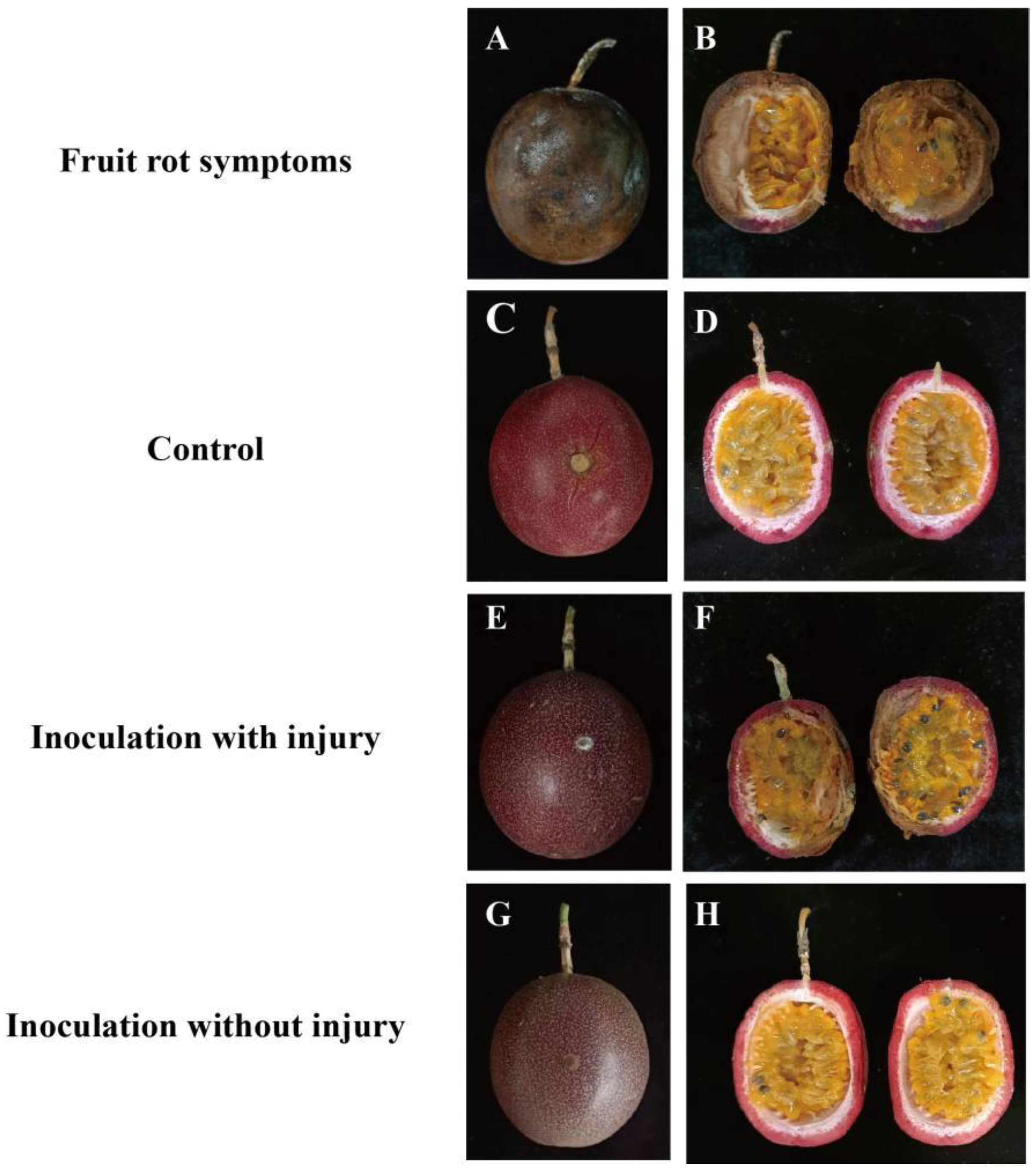
3.1.1. Symptoms of Naturally Occurring Passion Fruit Rot Disease

In the early stages of the disease, irregular brown spots appeared on the surface of the fruit, accompanied by softening of the fruit skin (Figure 1A,B). Greyish-white or white velvet-form mycelia were observed around these brown spots. As the number of storage days increased, the spots gradually darkened and spread to form depressions; rotting occurred inside the diseased fruits, and grey-black mould appeared at the edge of the fruit skin. In severe cases, the diseased fruit exhibited dehydration and shrinkage (Figure 1B).

3.1.2. Pathogenicity of the Passion Fruit Rot Pathogen

Inoculate blank PDA containing no pathogenic fungal filter cake onto passion fruit as a control, and inoculate PDA containing purified pathogenic fungal filter cake onto healthy fruit as a treatment group; the corresponding disease appeared after five days of incubation at a constant temperature of 28 °C and 95% relative humidity. (Figure 1C–H). Healthy passion fruit grafted with the passion fruit rot pathogen displayed symptoms on day five, including white velvet-like fungal filaments on the grafted surface with a round-shaped colony, dry texture that can be easily picked up, and abundant aerial filaments (Figure 1E). After seven days of culture, the symptoms of the grafted fruit tissue, including lesions and internal mould, were consistent with fruit rot disease (Figure 1F), whereas fruit in the control group remained healthy (Figure 1C,D). Tissue samples collected from infected areas were identified, purified, and re-isolated. The colony characteristics and spores of the isolate were identical to those of the original inoculation. According to Koch’s postulates, the original inoculation was the passion fruit rot pathogen. In addition, no disease symptoms were observed in the grafted area of the uninjured passion fruits (Figure 1G,H). Therefore, it was inferred that the causal agents of passion fruit rot are primarily propagated via wounds.

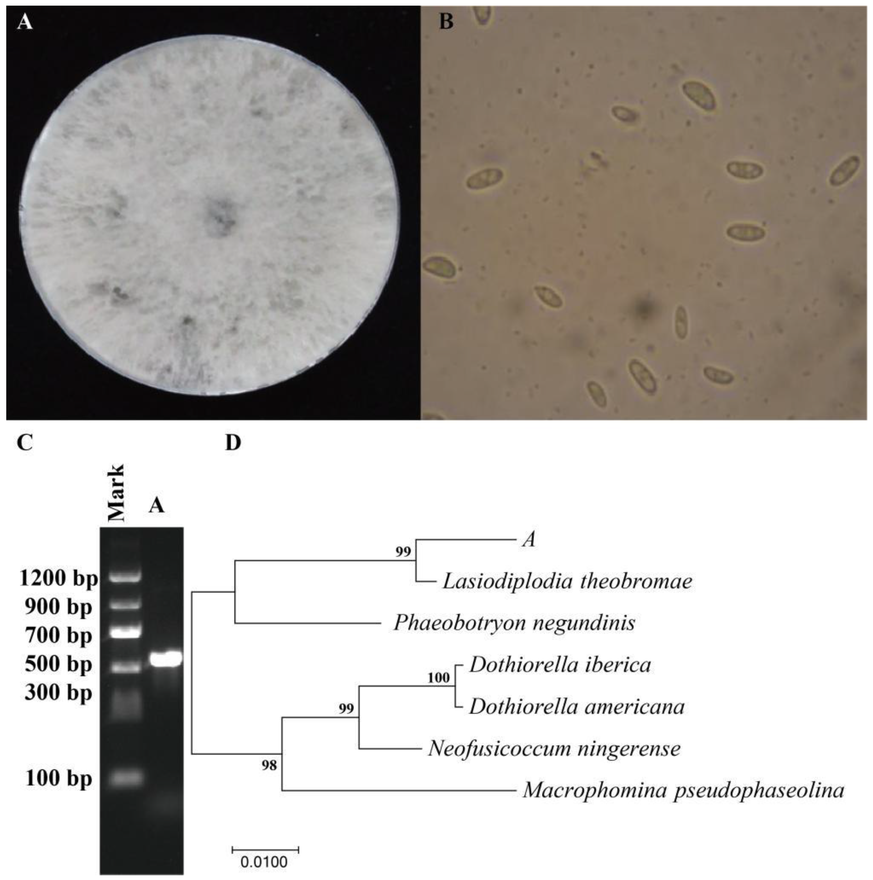
3.1.3. Identification of Pathogenic Fungi of Passion Fruit Rot Disease

The colonies of strain A are shown in Figure 2A. This fungus grows rapidly on potato dextrose agar (PDA) plates, and can completely occupy a culture dish within 4 days of culturing at 25 °C. Initially, the colonies were white, but eventually turned light black. The colony texture was dry and easy to lift, the aerial mycelia were fluffy and dense, and a light black colour was observed at the base that deepened over time. Using an optical microscope, the small conidia were determined to be ellipsoidal (Figure 2B).
The corresponding electrophoresis pattern of the pathogen strain indicates that the rDNA ITS length of the strain is approximately 500–700 bp (Figure 2C). The neighbour-joining phylogenetic tree of strain A was constructed using MEGA-X; this indicated a 99% homology between strain A and Lasiodiplodia theobromae (Figure 2D). The combined morphological characteristics and molecular identification results suggest that strain A is the same pathogen that causes guava fruit rot, L. theobromae.

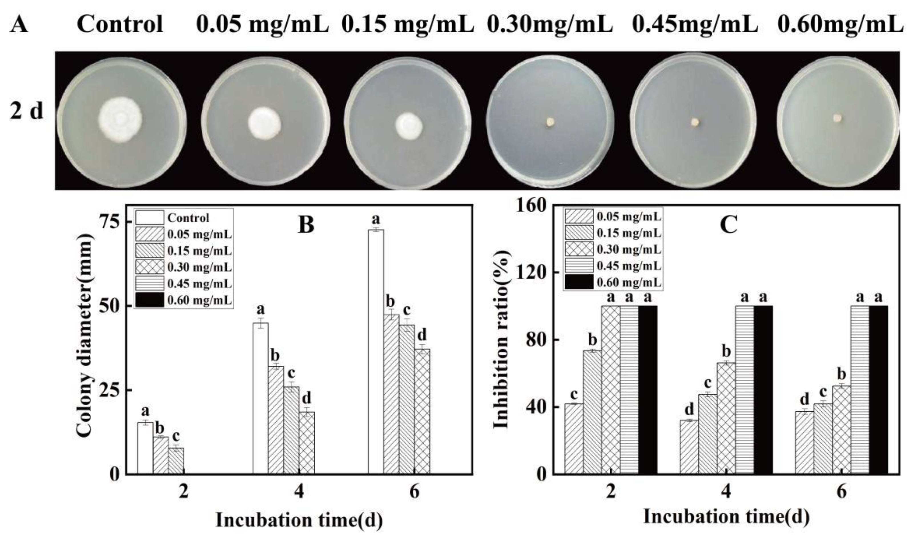
3.2. Inhibitory Effect of Eugenol on the Passion Fruit Rot Pathogen

As shown in Figure 3A, the higher the concentration of eugenol, the greater its inhibitory effect on the pathogenic fruit rot fungus. After incubation for 2 d, the diameter of the L. theobromae filamentous fungus under 0.30 mg/mL eugenol treatment was 0 mm; therefore, the minimal inhibitory concentration (MIC) value for L. theobromae was determined to be 0.30 mg/mL. As shown in Figure 3, with an increase in eugenol concentration, the inhibitory effect of eugenol on the pathogenic fruit rot fungi increased.
After 4 days of treatment with different concentrations of eugenol, the pathogenic fungal colony diameter of the control group was 44.87 mm, which was 1.73 times larger than that of the pathogenic fungal colony diameter of 0.30 mg/mL eugenol treatment. Compared with the control group, 0.30 mg/mL eugenol treatment had a significant inhibitory effect on the pathogenic fungal colony diameter (p < 0.05); after 6 days of treatment, the pathogenic fungal colony diameter of the control group was 72.64 mm, while that of the 0.30 mg/mL eugenol treatment was 37.13 mm. The colony diameter of pathogenic fungi in the control group was 1.95 times that of 0.30 mg/mL eugenol treatment. The 0.30 mg/mL eugenol treatment significantly inhibited the pathogenic fungal colony diameter compared to the control group (p < 0.05) (Figure 3B).
Furthermore, when treated with eugenol for 4 days, the inhibitory effect of 0.30 mg/mL eugenol on the growth of pathogenic fungi was 66.23%, while the inhibitory effects of 0.05 and 0.15 mg/mL eugenol on the growth of pathogenic fungi were 32.05%, 47.48%, and the inhibitory effects of 0.30 mg/mL eugenol were significantly higher than those of 0.05 and 0.15 mg/mL eugenol (p < 0.05) (Figure 3C). Throughout the entire cultivation process, the inhibition rate of filamentous fungi with 0.30 mg/mL, 0.45 mg/mL, and 0.60 mg/mL eugenol was 100%.

3.3. Effect of Eugenol on the Cell Membrane of the Passion Fruit Rot Pathogen

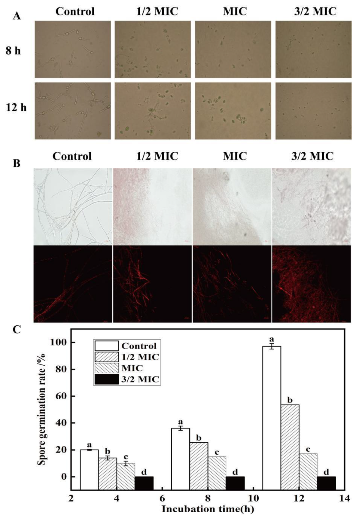
3.3.1. Effect of Eugenol on Spore Germination and Cell Membrane of the Passion Fruit Rot Pathogen

As shown in Figure 4A, compared with the control group, the spore germination rate of fruit rot pathogens in the control group after eugenol treatment was higher, and the bud tube elongation was evident. Spore germination and bud tube elongation were inhibited in the eugenol-treated group. In addition, with an increase in eugenol concentration, the spore germination rate of fruit rot pathogenic fungi decreased. After 4 h of treatment with 1/2 MIC and MIC concentrations of eugenol, the spore germination rates of pathogenic fungi were 13.97% and 9.87%, respectively, compared to 20.05% in the control group; After 8 h of treatment, the spore germination rate of the control group was 36%, which was 1.4 times and 2.4 times higher than the spore germination rate of pathogenic fungi treated with 1/2 MIC and MIC mass concentrations of eugenol (Figure 4C).
As shown in Figure 4B, the cell membrane structure of the fruit rot pathogen in the control group was complete, ordered, and relatively full; the cell membrane in this control group also contained high water content and high morphological integrity, displaying little propidium iodide (PI) staining (red) and a low degree of cell membrane damage. With an increase in eugenol concentration, the cell membrane structure began to deform, fold, and curl, the cell gap gradually increased, and the cell wall was clearly perforated, indicating that the integrity of the cell membrane of the fruit rot pathogen was damaged (Figure 4B).

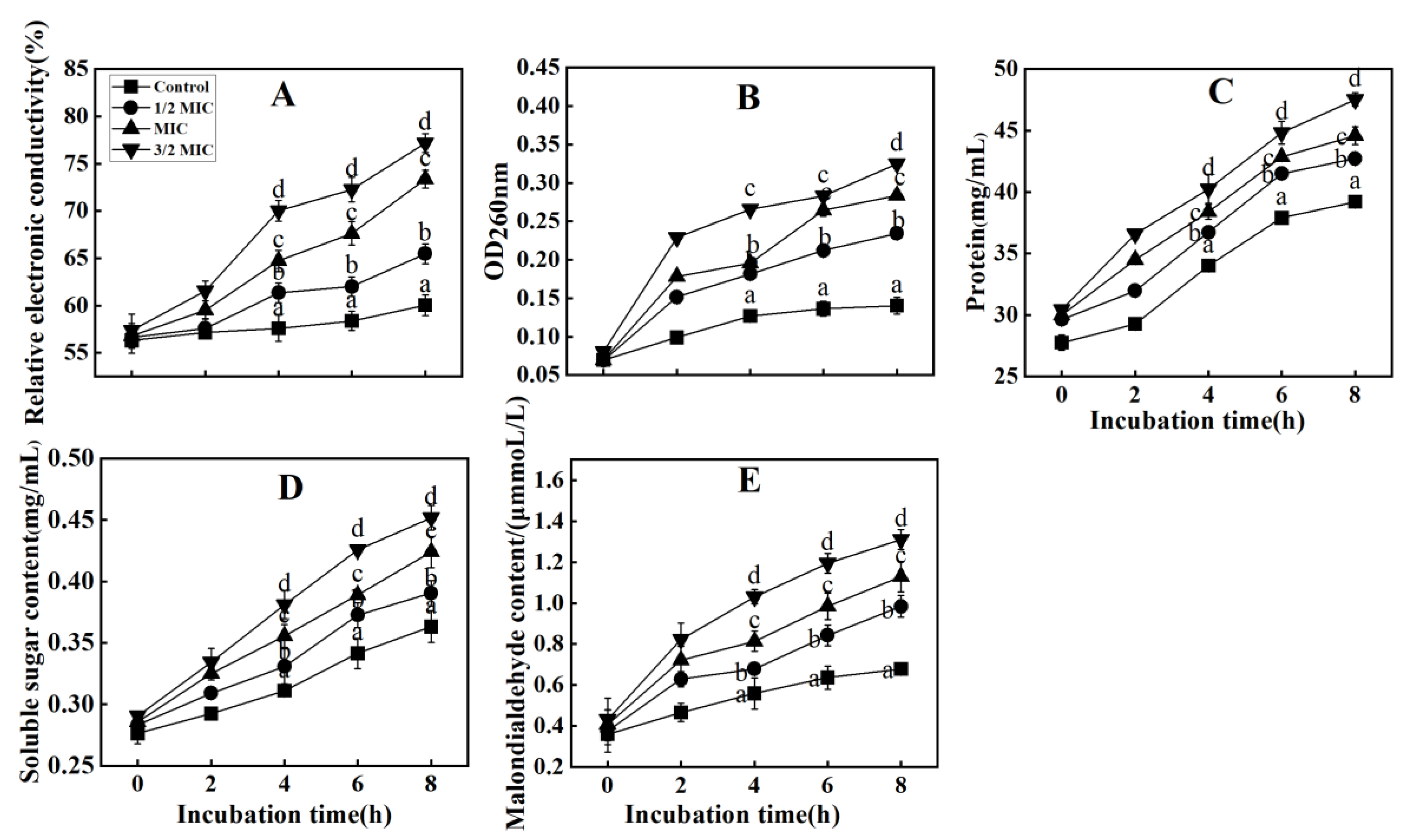
3.3.2. Effect of Eugenol on Mycelial Nucleic Acid Release and Electrical Conductivity of the Mycorrhizal Fungi That Causes Fruit Rot in Passion Fruit

Changes in the cellular nucleic acid material released by L. theobromae may indicate the effects of eugenol treatment on fungal membrane permeability. As shown in Figure 5A, the relative permeability of the fungal membranes treated with 1/2 MIC, MIC, and 3/2 MIC of eugenol was significantly higher compared with the control group. After 4 h of treatment, the relative permeability of the control group ceased to increase, whereas that of the treatment groups continued to increase. After 8 h, the relative permeability of L. theobromae treated with 1/2 MIC, MIC and 3/2 MIC of eugenol reached 71.64%, 76.67% and 79.59%, respectively, while the relative permeability of the control group reached 61.93%, i.e., 1.15 times, 1.23 times, and 1.28 times higher than that of the control group; Duncan’s multi interval test results showed that the eugenol treatment had a significant impact on the conductivity of pathogenic fungi (p < 0.05) (Figure 5A).
Figure 5B presents that the nucleic acid release level of the control group became stable after 4 h of treatment; alternatively, the nucleic acid release level of the treated groups increased gradually. As the treatment time increased, the difference between the nucleic acid release levels of the control and treated groups increased, with the control group exhibiting a gradual decrease in nucleic acid release levels compared to the treated group. When treated for 6 and 8 h, the nucleic acid release of pathogenic fungi treated with 0.30 mg/mL eugenol was significantly higher than that of the control group (p < 0.05). Furthermore, the nucleic acid release level increased as concentration of eugenol increased. These results indicate that eugenol affects the cell membranes of filamentous fungi, increasing membrane permeability and affecting the normal metabolic activities of filamentous fungi, thereby increasing the release of nucleic acids.

3.3.3. Effects of Eugenol on Intracellular Protein Release, Soluble Sugar Release, and Malondialdehyde (MDA) Content in the Pathogenic Passion Fruit Rot Fungi

Figure 5C shows that the amount of protein released in both the control and treatment groups gradually increased; additionally, the difference between the control and treatment groups increased with prolonged treatment time, with that of the control gradually becoming lower than that in the treatment group. Treatment with eugenol significantly increased the amount of protein released into the fungal cells (p < 0.05). After 8 h treatment with 1/2 MIC, MIC, or 3/2 MIC eugenol, the released protein in the fungal strain L. theobromae reached 42.73 mg/mL, 44.56 mg/mL, and 47.53 mg/mL, respectively, whereas that in the control group was 39.20 mg/mL. They are 1.09 times, 1.13 times, and 1.16 times higher than the control group, respectively. Among them, compared with the control group, MIC or 3/2 MIC eugenol treatment had a significant impact on protein release (p < 0.05).
The release of soluble sugar from the control and treated strains gradually increased with an prolonged treatment time; additionally, the difference between soluble sugar release from the control and treated strains increased, with the control strain gradually exhibiting a lower soluble sugar release than that of the treatment strain (Figure 5D). After treatment with thymol at 3/2 MIC for 6 h, the release of soluble sugar from the L. theobromae pathogen rapidly increased to 0.43 mg/mL, while that of the control strain was maintained at 0.34 mg/mL. Compared with the control group, the 3/2 MIC eugenol treatment significantly increased the release of soluble sugars (p < 0.05). During the whole incubation period, the release of soluble sugar from the control strain was consistently lower than that of the treatment strain.
As shown in Figure 5E, MDA levels increased steadily throughout the cultivation and incubation periods in both the control and thymol treatment groups. The MDA content in L. theobromae pathogen in the 6 h, 3/2 MIC thymol treatment group increased rapidly to 1.19 mmol/L, while that in the control group was 0.64 mmol/L, which was three times higher than the control group. The 3/2 MIC thymol treatment significantly increased the MDA content of pathogenic fungi (p < 0.05). Overall, the MDA content in the thymol-treated strain at 2–8 h was significantly higher than that in the untreated group; moreover, the higher the thymol concentration, the faster the increase in MDA content, indicating a concentration-dependent phenomenon. Therefore, this suggests that thymol acts on the mycelial cell membrane, increases membrane permeability, and affects the normal metabolic activity of mycelia, thereby increasing the release of intracellular proteins and soluble sugars.

3.4. Mechanism of Eugenol-Dependent Resistance to Passion Fruit Rot after Harvest

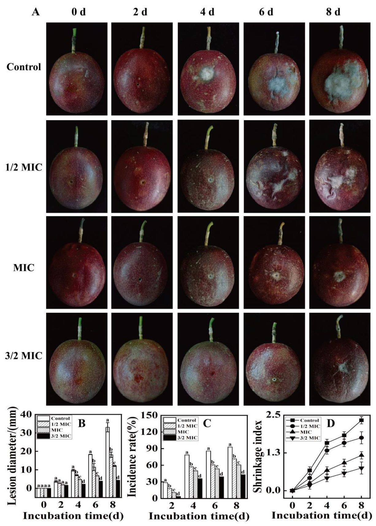
3.4.1. Effect of Different Eugenol Concentrations on the Resistance of Passion Fruit to Fruit Rot

The effect of different concentrations of eugenol with respect to inducing resistance to fruit rot in passion fruit is shown in Figure 6. The control fruit group was infected four days after inoculation with the pathogen; the inoculated fruit surfaces exhibited brownish fungal spots, and the lesions gradually expanded as the inoculation time was extended (Figure 6A). On the 6th day after inoculation in the control group, the surface of the fruits exhibited obvious white woolly fungal mycelia, and by day 8, the incidence of the lesion reached 92.85% (Figure 6A), with observations of the lesions indenting inward, rotten skin, and acidic odours. Compared with the control, different concentrations of eugenol suppressed fruit rot to varying degrees, with the resistance efficacy being exhibited in a dose-dependent manner. The low-concentration eugenol treatment groups (1/2 MIC and MIC) also exhibited infection 4 days after inoculation, although the infection rate and symptoms were evidently lower than those of the controls (p < 0.05). In the 3/2 MIC eugenol treatment group, infection occurred on the sixth day; however, on day 6, the incidence of lesions was still 46.43% lower than that in the control group. Compared to the control group, the 3/2 MIC eugenol treatment group significantly inhibited the occurrence of diseases (p < 0.05).
The incidence of lesions and the corresponding average lesion diameters in the eugenol-treated samples were significantly lower than those in the control group. Compared to the control group, eugenol effectively delayed the onset of crown rot in passion fruit and reduced the lesion diameter after inoculation with the pathogen (Figure 6A,B). In addition, eugenol treatment reduced the formation of fungal filaments by this pathogen. After the pathogen was inoculated on the fruit, all filaments of the control group continued to grow for four days, whereas the filaments of the treated group were effectively inhibited (Figure 6A). After the passion fruit was soaked in eugenol, the incidence of fruit rot decreased significantly; after eight days of treatment, the MIC and 3/2 MIC treatment groups possessed a fruit rot incidence of 60.71% and 42.86%, respectively, while the corresponding incidence in the control group reached up to 92.85% (Figure 6C). Compared with the control group, MIC and 3/2 MIC eugenol treatments significantly reduced the occurrence of fruit rot (p < 0.05). In addition, after the passion fruit was soaked in eugenol and inoculated with the pathogen, the lesion diameter decreased; on the 8th day, the lesion diameter of the 3/2 MIC treatment was 4.37 mm, while the lesion diameter of the control group was 33.03 mm (Figure 6B); the lesion diameter of the control group was 7.5 times higher than that of the 3/2 MIC eugenol treated group, and the 3/2 MIC eugenol treatment significantly inhibited fruit lesion (p < 0.05).
A change in the shrinkage index is related to the nutritional quality of the fruit and is one of the indicators used to measure the postharvest appearance quality of fruits. The shrinkage index of postharvest passion fruit consistently increased with the extension of storage time; nonetheless, eugenol treatment delayed the growth rate of the shrinkage index (Figure 6D). The shrinkage index of the control group showed a rapid upward trend during the postharvest storage period; specifically, its shrinkage index was 2.33 on the 8th day of storage, which was 3.11 times higher than that of the 3/2 MIC treatment group. Compared with the control group, 3/2 eugenol treatment can significantly reduce the shrinkage rate of fruits (p < 0.05). Throughout the storage period, the shrinkage index of the 3/2 MIC treatment group was consistently lower than that of the other treatment groups, indicating that compared to the 1/2 MIC and MIC treatments, the 3/2 MIC treatment could better maintain the appearance of the fruit and reduce its dehydration rate, thus improving its commodity value.

3.4.2. Effect of Eugenol on Disease-Resistant Substances and Defence-Related Enzyme Activities in Passion Fruit

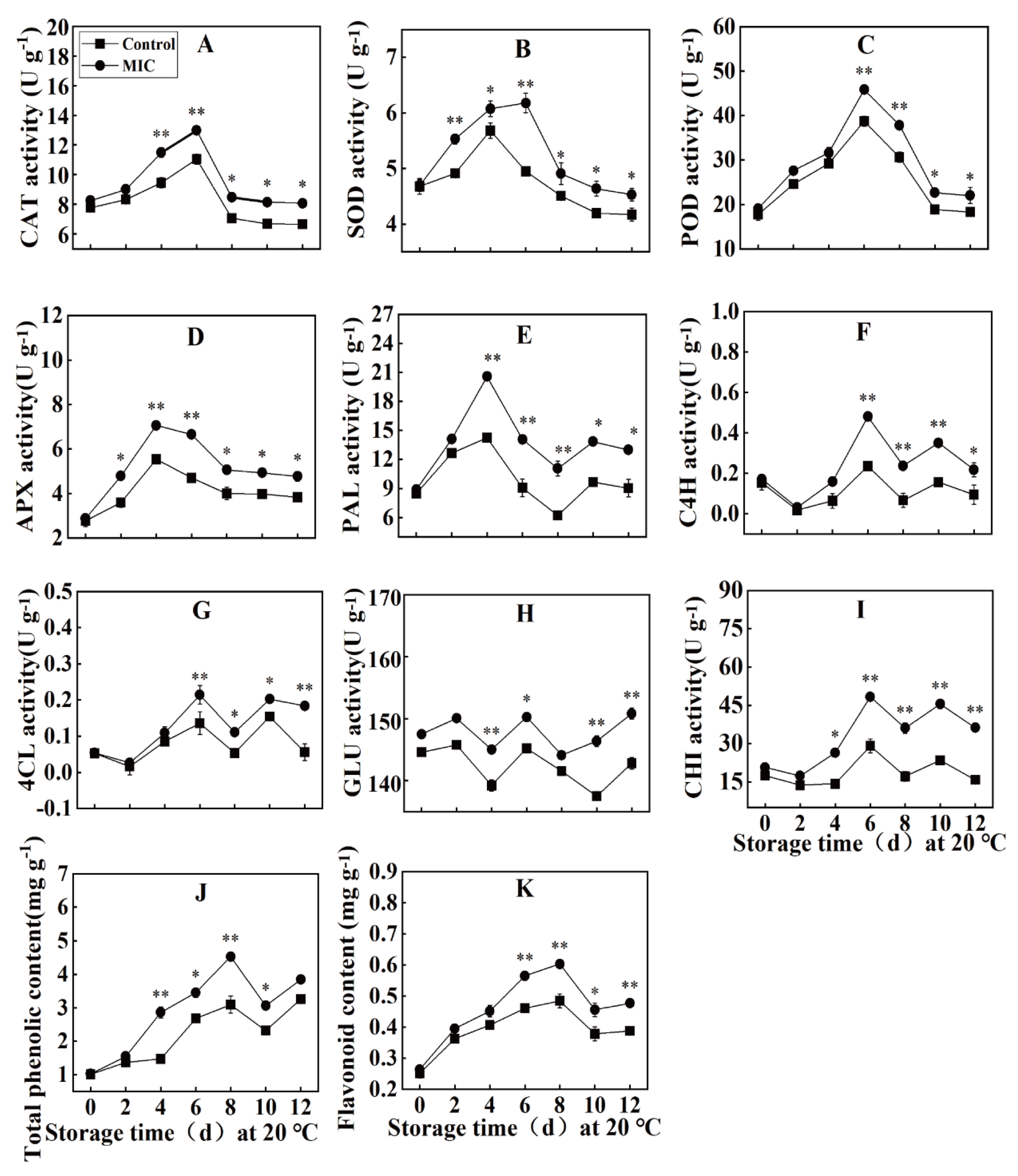
Effect of Eugenol on CAT, SOD, POD, and APX Activities in Passion Fruit

As shown in Figure 7A, during the storage period, the CAT activity of the control (CK) and eugenol groups of passion fruit exhibited a trend of an initial increase and then decline. On the 4th and 6th days of storage, the CAT activity of the eugenol group passion fruit was 11.49 U/g and 12.98 U/g, respectively, which were 1.21 times and 1.17 times higher, respectively, than those in the CK group. With the extension of storage time, the SOD activity in passion fruit tissues demonstrated a trend of an initial increase and then decrease (Figure 7B). Throughout the storage period, the SOD activity of eugenol-treated passion fruit was higher than that of the CK group. On the 2nd, 4th, and 6th days of storage, the SOD activity of the eugenol-treated group was 5.53, 6.07, and 6.18 U/g, respectively, which was 1.13, 1.05, and 1.24 times higher, respectively, than that of the CK group. Eugenol treatment significantly improved the SOD activity in post-harvest passion fruit. During the storage period, the POD activity of passion fruit exhibited a trend of an initial increase, followed by a decrease (Figure 7C). On the 6th day of the storage, the POD activity of the eugenol group passion fruit reached its peak, with an enzymatic activity of 45.81 U/g, which was 1.18 times higher than that of the CK group. On days 10 and 12 of storage, the enzyme activity of the passion fruit in the eugenol group decreased to 22.66 U/g and 22.0 U/g, respectively, which were 1.21 and 1.20 times higher than that of the CK group. As shown in Figure 7D, the changes in APX activity during the storage of passion fruit were similar to those of POD. At the beginning of storage (0–4 d), the APX enzyme activity gradually increased and peaked on the 4th day of storage. The APX enzyme activity of the passion fruit in the eugenol group was 7.06 U/g, which was significantly higher than that in the CK group (5.03 U/g). Subsequently, the APX enzyme activity gradually decreased. On days 10 and 12 of storage, the APX enzyme activities of the eugenol group were 4.94 and 4.77 U/g, respectively, which were 1.24 and 1.25 times higher, respectively, than those in the CK group. Overall, eugenol treatment maintained high APX activity in passion fruit tissues.

Effect of Eugenol on the Activity of PAL, C4H, and 4CL in Passion Fruit

Figure 7E shows that the PAL activity in passion fruit initially increased and then decreased during storage. The PAL activity in the eugenol−treated group was consistently higher than that in the CK group. From 0 to 4 days of storage, PAL activity increased rapidly to its maximum value, with the 4−day treatment increasing by 2.31 and 1.67 times compared to day 0 in the eugenol−treated and CK groups, respectively. During the 4−8 day storage period, overall PAL activity decreased; however, the PAL activity of the eugenol−treated group was higher than that of the CK group during this period. On the 12th day of storage, the PAL activity in the eugenol−treated group was 30.43% higher than that in the CK group.
The C4H activity of passion fruit during storage exhibited an upward and then downward trend, reaching a peak on day 6, with activities of 0.48 U/g and 0.23 U/g for the eugenol−treated and CK groups, respectively; these levels were increased by 65% and 53%, respectively, compared to those at day 0 of storage, (Figure 7F). In addition, significant differences were observed between the CK and eugenol groups during storage from day 6 to day 10. Overall, eugenol treatment maintained a relatively high C4H activity in passion fruit tissues.
As shown in Figure 7G, the 4CL activity of passion fruit during storage exhibited an initial increase, then decrease, followed by a further slow increase before a final decrease; the 4CL activity reached a peak on day 6, with the corresponding activities of the eugenol−treated and CK treatments being observed as 0.21 U/g and 0.14 U/g, respectively. After day 6, the 4CL activity of both the eugenol and CK treatments decreased with increasing storage time; however, the 4CL activity of the eugenol treatment was consistently higher than that of CK. On the 12th day of storage, the 4CL activity of the eugenol−treated group was 0.20 U/g, which was 1.33-fold higher than that of the CK group. In conclusion, eugenol treatment increased 4CL activity and improved disease resistance in passion fruit.

Effect of Eugenol on the GLU and CHI Activity in Passion Fruit

Throughout the storage period, the GLU activity of passion fruit treated with eugenol was higher than that of the CK group (Figure 7H). On days 4, 6, and 8 of storage, the GLU activity was 145.02, 150.23, and 144.12 U/g, respectively, which was 1.04, 1.03, and 1.02 times higher than that of the CK group, respectively. Therefore, the eugenol treatment significantly increased GLU activity in post-harvest passion fruit tissues.
As shown in Figure 7I, during storage, the CHI activity of passion fruit in the eugenol−treated group was higher than that of the CK group. On the 6th, 8th, and 10th days of storage, the CHI activity of eugenol−treated passion fruit was 48.29, 36.08, and 45.53 U/g, respectively, which was 1.66, 2.01, and 1.94 times higher, respectively, than that of the CK group. Thus, it was determined that eugenol treatment could effectively maintain high CHI activity in passion fruit.

Effect of Eugenol on the Total Phenolic and Flavonoid Content of Passion Fruit

Figure 7J indicates that the total phenolic content in passion fruit initially increased and then decreased during storage. At the early stage of storage (0−8 d), the total phenolic content increased continuously in both the eugenol−treated and CK groups, which reached a peak of 4.53 mg/g and 3.08 mg/g, respectively, on day 8. During later-stage storage (10−12 d), the total phenolic content began to decrease; however, the eugenol−treated passion fruit maintained a higher level than that in the CK group. For example, the total phenolic content in the eugenol−treated group was 1.32 and 1.17 times than that of the CK group on storage days 10 and 12, respectively. These results suggest that eugenol treatment can disrupt the decrease in the total phenolic content of passion fruit.
It is evident from Figure 7K that the trend of changes in flavonoid content in passion fruit during storage was similar to that of total phenolics, with an initial rise followed by a decline. During the early stage of storage (0−8 days), the flavonoid content of passion fruit in the eugenol−treated and CK groups both increased continuously, reaching a peak content of 0.60 mg/g and 0.48 mg/g, respectively, on day 8. As storage progressed to the late stage (10−12 days), the flavonoid content began to decrease; however, the flavonoid content of passion fruits in the eugenol-treated group remained higher than that in the CK group, with corresponding ratios of 1.47 and 1.23 at days 10 and 12, respectively. This indicates that eugenol may suppress the decline in flavonoid content in passion fruit.

4. Discussion

Eugenol, the main constituent of clove oil, possesses antimicrobial activity and exhibits good application potential in the preservation of a variety of fruits and vegetables, such as tomatoes [37] and blueberries [38]. Ju [39] determined that eugenol and citral can eliminate Aspergillus niger via the TCA cycle. Similarly, Hu [40] established that eugenol effectively suppressed Aspergillus and Penicillium fungi. Ju [41] also determined that eugenol could inhibit the growth of A. niger, thus demonstrating its potential use as a preservative in extending the shelf life of bread. Scanning electron microscopy and fluorescence microscopy revealed significant changes in the morphological characteristics of A. niger hyphae, such as cell membrane rupture, accompanied by an increase in extracellular conductance and protein and nucleic acid porosity. The present study determined that various concentrations of eugenol were effective in inhibiting the growth of L. theobromae hyphae and spore germination of L. theobromae hyphae in a dose-dependent manner. When eugenol concentration reached 0.30 mg/mL, L. theobromae hyphal growth and spore germination were completely inhibited. Numerous studies have been conducted to explore the antifungal activity of eugenol and plants; however, few studies have reported the antifungal mode of action of eugenol and its effects on fruit rot pathogens.
Cells possess selective semi-permeable membranes; however, certain antipathogenic substances can damage the cell membrane permeability, thereby disrupting normal substance transportation [42]. This study determined that upon treatment with eugenol, the pathogen causing fruit rot exhibited a notably increased content leakage in a time and concentration-dependent manner. This indicates that eugenol damages the cell membrane of fruit rot pathogens, increasing their permeability and allowing for the escape of proteins and nucleic acids. However, PI cannot pass through intact membranes; therefore, when membrane integrity is lost, PI enters the cell and binds to DNA, thereby exhibiting red fluorescence. Membrane PI staining can be studied based on this characteristic [43]. Shu [44] determined that upon treatment with ellagic acid, PI staining revealed a greater number of filaments emitting fluorescence and more intense filaments. This is consistent with the findings of the current study, in which an increase in the amount and intensity of PI fluorescence was observed as eugenol dosage increased.
Eugenol treatment can improve fruit quality and enhance the activity of disease-associated enzymes during storage, thereby increasing disease resistance and prolonging the corresponding storage period. Under eugenol treatment, the resistance of tomato fruit [45] to yellow leaf curl virus could be significantly enhanced, and the expression of phenylalanine ammonia-lyase and CHI-related genes in pomegranate [46] could be significantly upregulated. The activity of disease resistance–related enzymes in passion fruit gradually decreases during the ripening process, and then the fruit gradually decays with aging. Treatment with eugenol in the current study could better maintain the activity of disease-resistant enzymes and increase the disease resistance of the fruit, which is similar to the results obtained by Yang [47] in citrus fruits. Overall, eugenol reduces the decay caused by pathogen infection by inducing the expression of resistance genes. The results of the current study demonstrated that eugenol treatment increased the activity of CAT, SOD, APX, and POD in passion fruit; additionally, the corresponding enzyme activity was significantly higher in the eugenol-treated group than in the control group during the inoculation period, resulting in stronger defence ability. This showed that eugenol induced disease resistance and activation of antioxidant enzymes in passion fruit; however, further studies are needed to clarify these mechanisms.

5. Conclusions

In conclusion, L. theobromae was identified as the pathogen of passion fruit rot. Eugenol can significantly inhibit the growth and spore germination of L. theobromae by affecting the integrity of the cell membrane of this pathogen. The MIC of eugenol for this pathogenic fungus was determined to be 0.30 mg/mL by using the doubling dilution method. In addition, eugenol treatment was determined to reduce the incidence of passion fruit rot and lesion diameter, improve the activities of disease-resistance enzymes APX, SOD, CAT, POD, 4CL, C4H, PAL, CHI, and GLU, and promote the accumulation of total phenolics and flavonoids in passion fruit. These results suggest that exogenous eugenol induces disease resistance in passion fruit by increasing the activity of defence enzymes, disease-stage-related proteins, and antifungal materials. Therefore, eugenol has good prospects for commercial use in the control of passion fruit rot.

Author Contributions

Conceptualization, Y.S. and L.S.; methodology, Y.S.; software, Y.S.and D.L.; validation, L.B.and L.S.; formal analysis, Y.S.; writing—original draft preparation, Y.S.; supervision, L.B. All authors have read and agreed to the published version of the manuscript.

Funding

This research was funded by The Science and Technology Foundation of Guizhou Province (Grant No.[2021]119), the Master’s Research Fund Project of Guiyang University (GYU-YJS [2021]-39), and the Talent Program for “Academic Leaders in Disciplines” of Guiyang University (GYURC-07).

Data Availability Statement

Data are available from the authors upon request.

Conflicts of Interest

The authors declared that there are no conflict of interest.

References

  1. Correia, A.D.O.; Alexandre, R.S.; Pfenning, L.H.; Cabanez, P.A.; Ferreira, A.; Ferreira, M.; de Lima, P.A.M.; de Mello, M.T.; Otoni, W.C.; Lopes, J.C. Passiflora mucronata, a passion fruit wild species resistant to fusariosis and a potential rootstock for commercial varieties. Sci. Hortic. 2022, 302, 111174. [Google Scholar] [CrossRef]
  2. De Oliveira, A.B.; de Almeida, L.; Mônica Maria, D.A.L.; Carlos, F.H.M.; Luciana, D.S.O.; Kellina, O.D.S.; Enéas, G.F.; Laurent, U.; Maria, R.A.D.M. Effects of organic and conventional farming systems on quality and antioxidant metabolism of passion fruit during maturation. Sci. Hortic. 2017, 222, 84–89. [Google Scholar] [CrossRef]
  3. Jordán, M.J.; Goodner, K.L.; Shaw, P.E. Characterization of the aromatic profile in aqueous essence and fruit juice of yellow passion fruit (Passiflora edulis sims F. Flavicarpa degner) by GC-MS and GC/O. J. Agric. Food Chem. 2002, 50, 1523–1528. [Google Scholar] [CrossRef] [PubMed]
  4. Porto-Figueira, P.; Freitas, A.; Cruz, C.J.; Figueira, J.; Câmara, J.S. Profiling of passion fruit volatiles: An effective tool to discriminate between species and varieties. Food Res. Int. 2015, 77, 408–418. [Google Scholar]
  5. Alemwati, P.; Sagar, V.; Ram, K.P.; Ram, A.; Ram, R.S.; Sanjay, K.S. Physiological and quality changes during postharvest ripeningof purple passion fruit (Passiflora edulis Sims). Fruits 2014, 69, 19–30. [Google Scholar]
  6. Mcevoy, C.T.; Wallace, I.R.; Hamill, L.L.; Hunter, S.J.; Neville, C.E.; Patterson, C.; Woodside, J.V.; Young, I.S.; McKinley, M.C. Increasing fruit and vegetable intake has no dose-response effect on conventional cardiovascular risk factors in overweight adults at high risk of developing cardiovascular disease. J. Nutr. 2015, 145, 1464–1471. [Google Scholar] [CrossRef]
  7. Sun, Y.B.; Jiang, C.Q.; Kar, K.C.; Wei, S.Z.; Gabriel, M.; Tai, H.L.; Schooling, C.M. Fruit and vegetable consumption and cardiovascular risk factors in older Chinese: The guangzhou biobank cohort study. PLoS ONE 2015, 10, e0135380. [Google Scholar] [CrossRef]
  8. Araujo, L.S.; Rodrigues, C.E.M.; Soares, T.L.; Idália, S.D.; Jesus, O.N.D. Effect of time and storage conditions on the physical and physico-chemical characteristics of the pulp of yellow and purple passion fruit. Food Sci. Technol. 2017, 37, 500–506. [Google Scholar] [CrossRef]
  9. Shiomi, S.; Kubo, Y.; Wamocho, L.S.; Koaze, H.; Nakamura, R.; Inaba, A. Postharvest ripening and ethylene biosynthesis in purple passion fruit. Postharvest Biol. Technol. 1996, 8, 188–207. [Google Scholar] [CrossRef]
  10. Du, Y.X.; Shi, N.N.; Chen, W.L.; Ruan, H.C.; Yang, X.J.; Gan, L. Identification of colletotrichum brevisporum causing anthracnose on passion fruit. Can. J. Plant Pathol. 2017, 39, 1367725. [Google Scholar] [CrossRef]
  11. Dai, Y.L.; Wang, C.C.; Lin, H.L. First report of Septoria Blotch of passion fruit caused by Septoria passifloricola in Taiwan. Plant Dis. 2021, 105, 700. [Google Scholar] [CrossRef] [PubMed]
  12. Zhang, W.; Niu, X.L.; Yang, J.Y. First report of postharvest fruit rot on passion fruit (Passiflora edulis) caused by Lasiodiplodia theobromae in China’s mainland. Plant Dis. 2020, 105, 1198. [Google Scholar] [CrossRef]
  13. Zhang, Y.; Wang, Y.; Zhu, X.J.; Cao, P.; Shaomin, W.; Lu, Y.H. Antibacterial and antibiofilm activities of eugenol from essential oil of Syzygium aromaticum (L.) Merr. & L.M. Perry (clove) leaf against periodontal pathogen Porphyromonas gingivalis. Microb. Pathog. 2017, 113, 396–402. [Google Scholar] [PubMed]
  14. Jafri, H.; Mohd, S.A.K.; Iqbal, A. In vitro efficacy of eugenol in inhibiting single and mixed-biofilms of drug-resistant strains of Candida albicans and Streptococcus mutans. Phytomedicine 2018, 54, 206–213. [Google Scholar] [CrossRef] [PubMed]
  15. Devi, K.P.; Nisha, S.A.; Sakthivel, R.; Pandian, S.K. Eugenol (an essential oil of clove) acts as an antibacterial agent against Salmonella typhi by disrupting the cellular membrane. J. Ethnopharmacol. 2010, 1301, 107–115. [Google Scholar] [CrossRef] [PubMed]
  16. Cui, H.Y.; Zhang, C.H.; Li, C.Z.; Lin, L. Antimicrobial mechanism of clove oil on Listeria monocytogenes. Food Control 2018, 94, 140–146. [Google Scholar] [CrossRef]
  17. Edwards, K.; Johnstone, C.; Thompson, C. A simple and rapid method for the preparation of plant genomic DNA for PCR analysis. Nucleic Acids Res. 1991, 19, 1349. [Google Scholar] [CrossRef]
  18. Yahyazadeh, M.; Omidbaigi, R.; Zare, R.; Taheri, H. Effect of some essential oils on mycelial growth of Penicillium digitatum Sacc. World J. Microbiol. Biotechnol. 2008, 24, 1445–1450. [Google Scholar] [CrossRef]
  19. Ge, Y.H.; Chen, Y.R.; Li, C.Y.; Wei, M.L.; Lv, J.Y. Inhibitory effects of sodium silicate on the fungal growth and secretion of cell wall-degrading enzymes by Trichothecium roseum. J. Phytopathol. 2017, 165, 620–625. [Google Scholar] [CrossRef]
  20. Liu, K.; Zhou, X.; Fu, M. Inhibiting effects of ε-poly-lysine (ε-PL) on Pencillium digitatum and its involved mechanism. Postharvest Biol. Technol. 2017, 123, 94–101. [Google Scholar] [CrossRef]
  21. Tao, N.G.; Yang, Q.L.; Jia, L. Citral inhibits mycelial growth of Penicillium italicum by a membrane damage mechanism. Food Control 2014, 41, 116–121. [Google Scholar] [CrossRef]
  22. Chen, C.Y.; Qi, W.W.; Peng, X.; Chen, J.Y.; Wan, C.P. Inhibitory Effect of 7-Demethoxytylophorine on Penicillium italicum and its Possible Mechanism. Microorganisms 2019, 7, 36. [Google Scholar] [CrossRef] [PubMed]
  23. Wei, M.; Ge, Y.H.; Li, C.Y.; Chen, Y.R.; Wang, W.H.; Duan, B.; Li, X. Antifungal activity of ε-poly-L-lysine on Trichothecium roseum in vitro and its mechanisms. Physiol. Mol. Plant Pathol. 2018, 103, 23–27. [Google Scholar] [CrossRef]
  24. Qin, G.Z.; Zong, Y.Y.; Chen, Q.L.; Hua, D.L.; Tian, S.P. Inhibitory effect of boron against Botrytis cinerea on table grapes and its possible mechanisms of action. Int. J. Food Microbiol. 2010, 138, 45–150. [Google Scholar] [CrossRef]
  25. Chen, H.Z.; Cheng, Z.; Wisniewski, M.; Liu, Y.S.; Liu, J. Ecofriendly hot water treatment reduces postharvest decay and elicits defense response in kiwifruit. Environ. Sci. Pollut. Res. 2015, 22, 15037–15045. [Google Scholar] [CrossRef]
  26. Xia, Y.X.; Chen, T.; Qin, G.Z.; Li, B.Q.; Tian, S.P. Synergistic action of antioxidative systems contributes to the alleviation of senescence in kiwifruit. Postharvest Biol. Technol. 2016, 111, 15–24. [Google Scholar] [CrossRef]
  27. Zhang, M.; Xu, L.Y.; Zhang, L.Y.; Zhang, L.; Guo, Y.H.; Qi, X.; He, L. Effects of quercetin on postharvest blue mold control in kiwifruit. Sci. Hortic. 2018, 228, 18–25. [Google Scholar] [CrossRef]
  28. Zheng, F.L.; Zheng, W.W.; Li, L.M.; Pan, S.M.; Liu, M.C.; Zhang, W.W.; Liu, H.S.; Zhu, C.Y. Chitosan Controls Postharvest Decay and Elicits Defense Response in Kiwifruit. Food Bioprocess Technol. 2017, 10, 1937–1945. [Google Scholar] [CrossRef]
  29. Zhang, W.L.; Jiang, H.T.; Cao, J.K.; Jiang, W.B. UV-C treatment controls brown rot in postharvest nectarine by regulating ROS metabolism and anthocyanin synthesis. Postharvest Biol. Technol. 2021, 180, 111613. [Google Scholar] [CrossRef]
  30. Li, S.G.; Xu, Y.H.; Bi, Y.; Zhang, B.; Shen, S.L.; Jiang, T.J.; Zheng, X.L. Melatonin treatment inhibits gray mold and induces disease resistance in cherry tomato fruit during postharvest. Postharvest Biol. Technol. 2019, 157, 110962. [Google Scholar] [CrossRef]
  31. Li, G.J.; Zhu, S.H.; Wu, W.X.; Zhang, C.; Peng, Y.; Wang, Q.G.; Shi, J.Y. Exogenous nitric oxide induces disease resistance against Monilinia fructicola through activating the phenylpropanoid pathway in peach fruit. J. Sci. Food Agric. 2017, 97, 17–21. [Google Scholar] [CrossRef] [PubMed]
  32. Bautista-Rosales, P.U.; Calderon-Santoyo, M.; Servín-Villegas, R.; Ochoa-Alvarez, N.A.; Vazquez-Juarez, R.; Ragazzo-Sanchez, J.A. Biocontrol action mechanisms of Cryptococcus laurentii on Colletotrichum gloeosporioides of mango. J. Crop Protection 2014, 65, 194–201. [Google Scholar] [CrossRef]
  33. Boller, T.; Gehri, A.; Mauch, F.; Vögeli, U. Chitinase in bean leaves: Induction by ethylene, purification, properties, and possible function. Planta 1983, 157, 22–31. [Google Scholar] [CrossRef] [PubMed]
  34. Berger, L.R.; Reynolds, D.M. The chitinase system of a strain of Streptomyces griseus. Biochim. Biophys. Acta (BBA)—Lipids Lipid Metab. 1958, 29, 522–534. [Google Scholar]
  35. Ramful, D.; Tarnus, E.; Aruoma, O.I.; Bourdon, E.; Bahorun, T. Polyphenol composition, vitamin C content and antioxidant capacity of Mauritian citrus fruit pulps. Food Res. Int. 2011, 44, 2088–2099. [Google Scholar] [CrossRef]
  36. Wang, H.; Chen, Y.H.; Lin, H.T.; Lin, M.S.; Chen, Y.H.; Lin, Y.F. 1-Methylcyclopropene containing-papers suppress the disassembly of cell wall polysaccharides in anxi persimmon fruit during storage. Int. J. Biol. Macromol. 2020, 151, 723–729. [Google Scholar] [CrossRef]
  37. Mirdehghan, S.H.; Valero, D. Bioactive compounds in tomato fruit and its antioxidant activity as affected by incorporation of Aloe, eugenol, and thymol in fruit package during storage. Int. J. Food Prop. 2017, 20, 1798–1806. [Google Scholar]
  38. Yang, R.P.; Miao, J.Y.; Shen, Y.T.; Cai, N.; Wan, C.; Zou, L.Q.; Chen, C.Y.; Chen, J.Y. Antifungal effect of cinnamaldehyde, eugenol and carvacrol nanoemulsion against Penicillium digitatum and application in postharvest preservation of citrus fruit. LWT 2021, 141, 110924. [Google Scholar] [CrossRef]
  39. Ju, J.; Lei, Y.; Guo, Y.H.; Yu, H.; Cheng, Y.L.; Yao, W.R. Eugenol and citral kills Aspergillus niger through the tricarboxylic acid cycle and its application in food preservation. LWT 2023, 173, 114226. [Google Scholar] [CrossRef]
  40. Hu, Q.; Zhou, M.F.; Wei, S.Y. Progress on the antimicrobial activity research of clove oil and eugenol in the food antisepsis field. J. Food Sci. 2018, 83, 1476–1483. [Google Scholar] [CrossRef]
  41. Ju, J.; Xie, Y.F.; Yu, H.; Guo, Y.H.; Cheng, Y.L.; Zhang, R.R.; Yao, W.R. Synergistic inhibition effect of citral and eugenol against Aspergillus niger and their application in bread preservation. Food Chem. 2020, 310, 125974. [Google Scholar] [CrossRef]
  42. Li, W.S.; Yuan, S.Z.; Sun, J.; Li, Q.; Jiang, W.; Cao, J. Ethyl p-coumarate exerts antifungal activity in vitroand in vivo against fruit Alternaria alternata via membrane-targeted mechanism. Int. J. Food Microbiol. 2018, 278, 26–35. [Google Scholar] [CrossRef] [PubMed]
  43. Yang, R.X.; Fan, X.J.; Cai, X.Q.; Hu, F.P. The inhibitory mechanisms by mixtures of two endophytic bacterial strains isolated from Ginkgo biloba against pepper phytophthora blight. Biol. Control 2015, 85, 59–67. [Google Scholar] [CrossRef]
  44. Shu, C.; Zhao, H.D.; Jiao, W.X.; Liu, B.D.; Cao, J.K.; Jiang, W.B. Antifungal efficacy of ursolic acid in control of Alternaria alternata causing black spot rot on apple fruit and possible mechanisms involved. Sci. Hortic. 2019, 256, 108636. [Google Scholar] [CrossRef]
  45. Sun, W.J.; Lv, W.J.; Li, L.N.; Yin, G.; Hang, X.F.; Xue, Y.F.; Chen, J.; Shi, Z.Q. Eugenol confers resistance to Tomato yellow leaf curl virus (TYLCV) by regulating the expression of SlPer1 in tomato plants. New Biotechenology 2016, 33, 345–354. [Google Scholar] [CrossRef]
  46. Kumar, P.; Dashyal, M.S.; Doddaraju, P.; Meti, B.S.; Girigowda, M. Differential gene responses in different varieties of pomegranate during the pathogenesis of Xanthomonas axonopodis pv. punicae. 3 Biotech 2021, 11, 180. [Google Scholar] [CrossRef]
  47. Yang, R.P.; Miao, J.Y.; Chen, X.; Chen, C.Y.; Simal-Gandara, J.; Chen, J.Y.; Wan, C.P. Essential oils nano-emulsion confers resistance against Penicillium digitatum in ‘Newhall’ navel orange by promoting phenylpropanoid metabolism. Ind. Crops Prod. 2022, 187, 115297. [Google Scholar] [CrossRef]
Figure 1. Images showing (A,B) symptoms of naturally occurring passion fruit rot disease, (C,D) a blank control, (E,F) symptoms of invasive inoculation of pathogenic fungi, and (G,H) symptoms of non-invasive inoculation of pathogenic fungi in passion fruit.
Agronomy 13 01408 g001
Figure 2. (A) Pathogen colony morphology, (B) spore morphology, (C) electrophoresis diagram, and (D) phylogenetic tree of the passion fruit rot pathogen.
Agronomy 13 01408 g002
Figure 3. Effect of eugenol on (A) mycelial growth, (B) colony diameter, and (C) mycelial inhibition rate of pathogenic fungi. Different lowercase letters in the same sampling are significantly different to each other (p < 0.05).
Agronomy 13 01408 g003
Figure 4. (A) The effect of eugenol on spore germination and the hyphal cell membrane of the passion fruit rot pathogen. (B) The effect of spore germination and PI dye on the hyphal cell membrane of the passion fruit rot pathogen. (C) Effect of different mass concentrations of eugenol treatment on the germination rate of spores. Different lowercase letters (a, b, c, d) in the same sampling are significantly different to each other (p < 0.05).
Agronomy 13 01408 g004
Figure 5. Effects of eugenol on the (A) conductivity, (B) nucleic acid release, (C) intracellular protein release, (D) soluble sugar release, and (E) MDA content of pathogenic fungi. Different lowercase letters in the same sampling are significantly different to each other (p < 0.05).
Agronomy 13 01408 g005
Figure 6. (A) Images showing the effects of eugenol on passion fruit rot and corresponding graphs showing the effect of eugenol specifically on the (B) spot diameter, (C) incidence rate, and (D) shrinkage of passion fruit rot. Different lowercase letters in the same sampling are significantly different from each other (p < 0.05).
Agronomy 13 01408 g006
Figure 7. Effects of eugenol treatment on the content of (A) CAT, (B) SOD, (C) POD, (D) APX, (E) PAL, (F) C4H, (G) 4CL, (H) GLU, (I) CHI, (J) total phenol, and (K) flavone in passion fruit. An asterisk indicates a significant difference between groups (p < 0.05), and two asterisks indicate a highly significant difference between groups (p < 0.01).
Agronomy 13 01408 g007
Disclaimer/Publisher’s Note: The statements, opinions and data contained in all publications are solely those of the individual author(s) and contributor(s) and not of MDPI and/or the editor(s). MDPI and/or the editor(s) disclaim responsibility for any injury to people or property resulting from any ideas, methods, instructions or products referred to in the content.

