Article

Morphological Features and Biological Activity of Different Extracts of Echinops spinosissimus Grown in Saudi Arabia

by
Luluah M. Al Masoudi
1 and
Ahmed M. Hashim
2,*
1
Biology Department, College of Science, Taif University, P.O. Box 11099, Taif 21944, Saudi Arabia
2
Botany Department, Faculty of Science, Ain Shams University, Cairo 11865, Egypt
*
Author to whom correspondence should be addressed.
Agronomy 2023, 13(2), 573; https://doi.org/10.3390/agronomy13020573
Submission received: 12 January 2023 / Revised: 10 February 2023 / Accepted: 13 February 2023 / Published: 17 February 2023
(This article belongs to the Special Issue Research Progress and Application Prospect of Medicinal Plants)

Abstract

:
Based on a shortage of available data on Echinops spinosissimus in Saudi Arabia, the current study’s aim was to present some new information on the topic. Plant samples were collected from different locations in the northeast of Mecca. Out of fifteen species from this genus found in Saudi Arabia, one species was targeted in the current study. It was noted as a perennial subshrub that is 30–80 cm in length. Its stem is gray, striate, and slightly covered with glandular hairs. The epidermis is converted into cork cells in older stem parts. The vascular system showed a continuous siphonostelic structure and dissected vascular bundles. The lamina is abaxially rounded and straight. The pollen grains are monads, radially symmetric, medium-sized, and a prolate spheroidal shape with an aculeate–foveolate exine structure. Based on its historical pharmaceutical properties, the phytochemical properties were studied, and it was noted that ethyl acetate was the best solvent for producing high amounts of bioactive compounds such as phenols, flavonoids, and alkaloids. The obtained extracts appeared to exhibit high activity against Gram-positive pathogenic bacteria. These extracts were identified by using HPLC and GC-MS. Many bioactive compounds were detected, such as protocatechuic acid, gallic acid, rutin, vanillic acid, quercetin, and kaempferol. Additionally, four main compounds, including hexadecanoic, stearic, oleic, and linoleic acids, were detected via GC-MS. The total antioxidants of E. spinosissimus extracts showed that the ethyl acetate extract exhibited a high total antioxidant capacity and free radical-scavenging properties.

1. Introduction

The Compositae family (Asteraceae) contains 20,000 species of flowering plants. These plants are distributed in Africa, the Mediterranean, and Asia, with most being annuals, perennials, vines, ground covers, shrubs, and a few small trees [1,2]. This family belongs to the subclass Asteridae in the order Asterales. Thus, it is characterized by its diversity and worldwide distribution. Echinops L. is one of the family’s genera, with 15 species found in Saudi Arabia. About 130 species of the genus Echinops L., which is found in tropical and North Africa, are members of the Cardueae tribe of the Asteraceae family. A majority of the taxa (76) are located in the Iranian plateau, with many found in the Mediterranean Basin and the Middle East, spreading eastward to China and Japan [3,4,5]. A majority of taxonomic groups recognized today are defined by their floral morphology. Khafagi [6] studied the morphological and anatomical characteristics of 37 species of Aster. Bremer [7] studied the floral characteristics, including flower color, shape, size, anther shape, and appendages, as well as stigma shape, which are considered to be major distinguishing characteristics between species. Although the macro- and micromorphological aspects of Echinops spinosissimus have already been investigated, the anatomical features of the stem and leaves, pollen and achene morphology, and phytochemical screening have not been investigated in detail, which would aid in defining the taxonomical characteristics of this species.
Based on phytochemical studies of E. spinosissimus, it is known that this species contains biologically active compounds such as thiophenes, terpenoids, sterols, fatty acids, and alkanes [8]. Due to its effectiveness in treating infections, intestinal worm infestation, hemorrhoids, migraine, diarrhea, and heart pain, E. spinosissimus is typically referred to as a complete pharmacy [9]. Utilizing its antimicrobial activity toward multidrug-resistant microbes has grown to be the main goal facing researchers in this area of science. Therefore, studies have searched for a novel natural compound from plants [10,11,12] and microbial sources [13,14], or have developed chemical drugs to avoid the microbial-resistant system [15,16,17]. In this regard, the genus Echinops is used as a traditional treatment for different infectious diseases, including sepsis, trachoma, gonorrhea, typhoid, and ulcerative lymphangitis [18,19]. Additionally, it is used to treat different diseases that could be caused by bacteria and/or fungi, including respiratory diseases, fever, leucorrhea, earache, and toothache. Thus, Echinops spp. have been examined for their antimicrobial properties. The previous studies on this genus showed that both Gram-negative and Gram-positive bacteria were sensitive to its extracts and isolated compounds [20]. Free radicals can cause oxidative damage, and antioxidants have been demonstrated to prevent this damage by interacting with free radicals, chelating with catalytic metals, and scavenging oxygen. Oxidation stress is one of the primary health issues faced today. E. spinosissimus has many phenolic and flavonoid substances, which are considered to be antioxidant agents [21,22]. The present study aimed to provide primary documentation on the morphological, anatomical, pollen, and achene characteristics of E. spinosissimus in Saudi Arabia by using both light microscopy and scanning electron microscopy (SEM), as well as phytochemical screening, to fill the gap in our knowledge regarding E. spinosissimus.

2. Materials and Methods

2.1. Morphological and Anatomical Characteristics of Echinops spinosissimus

Fresh, whole plant materials (10 individuals) were collected from various localities in the northeast of Mecca (e.g., Wady Galil, Arnah and Wady AlKherar) during the period of 2021–2022 (Figure 1). Specimens were identified, and the synonyms were derived from the International Plant Names Index (IPNI; http://www.ipni.org/ipni/plantnamesearchpage.do, accessed on 5 January 2022) and Plants of the World Online (Plants of the World Online/Kew Science). Morphological description was made directly from fresh materials. For anatomical studies, 3–5 specimens of internodes and mature blades were taken from basal leaflets. The materials were prepared for microtome sectioning with a 10–15 µm thickness using the conventional Johansen procedure [23]. Sections were mounted in Canada balsam and dyed with safranin-light green standard double stain [24].
Using a Canon PowerShot A470 camera with 7.1 megapixels, the pollen grains (five to ten pollen grains per individual) were inspected, measured, and photographed. The pollen size, shape, and aperture type were also evaluated, along with the equatorial diameter (E) and polar axis (P). Non-acetolyzed pollen grains were applied to a metallic stub using double-sided sellotape for SEM study, then coated with gold in a sputtering chamber before being scanned and photographed for exine and aperture ornamentations using a JEOL JSM-IT100 scanning electron microscope. The terminology used to describe the morphology of pollen grains was in accordance with that provided by Punt et al. [25] and Grant-Downton [26].
Five to ten mature achene and seeds from each individual were inspected under a stereomicroscope and captured on camera using a Canon PowerShot A470 with 7.1 megapixels. The calibration and seed measurements were performed using the stage micrometer and ImageJ program. The stereomicroscope was used to examine the hilum location, seed length, and width. The mature achene was mounted onto SM stubs for SEM study, coated with gold, analyzed, and captured using the JEOL JSM-IT100 scanning electron microscope. Terminology was used as specified [27,28].

2.2. Preparation of E. spinosissimus Extract

The flower heads, leaves, and stems were cleaned, air dried at room temperature in the shade, and then pulverized into a powder via mechanical mills. Ethyl alcohol, ethyl acetate, or hexane solvents were used to prepare different extracts by mixing each (200 mL) with the dried powder (50 g) at a temperature of 28 °C for 72 h of incubation. The extracts were concentrated at 30 °C and at a lower pressure on a rotating evaporator. Firstly, active charcoal was used to remove the color from the crude concentrated extracts for each solvent; after that, the final volume of methanol was added, and the samples were then subjected to a phytochemical examination [29]. To find the best solvent for the most accurate quantitative detection of nutraceutical secondary metabolites, a qualitative phytochemical investigation was conducted.

2.3. Initial Phytochemical Research

A preliminary, qualitative phytochemical screening will aid in future study, as it will provide knowledge on the chemical components present in the different extracts of the studied species. As described in the literature [29,30], phytochemical screening was carried out. Flavonoids were investigated using the aluminum chloride colorimetric method, total phenolics and tannins were investigated using the Folin–Ciocalteu method, and saponins were investigated using the vanillin–HCl reagent. Alkaloids were evaluated using 10% acetic acid and ammonium hydroxide reagents. Total lipids were analyzed using methanol/chloroform. The detection of total soluble sugars (TSSs) was performed in extracts of the E. spinosissimus plant using the anthrone method [31].

2.4. Determination of Antioxidant Properties

The samples for assessing antioxidant properties were created by mixing 0.2 mg of the lyophilized E. spinosissimus extract with 1 milliliter of distilled water. The samples’ hydroxyl radical-scavenging activity (%) and 2, 2-azinobis (3-ethylbenzothiazoline-6-sulfonate) diammonium salt (ABTS) levels were assessed.

2.4.1. Determination of DPPH Radical Scavenging

Using the DPPH method outlined by Eweys et al. [12], the antioxidant activity of the E. spinosissimus extract was assessed. Briefly, 3.9 mL of freshly made DPPH solution (22 mg of DPPH in 50 mL methanol) was added to 0.1 mL of the sample. The mixture was vortexed for 30 s and then left at room temperature for 30 min in a dark place. Using a UV-Vis spectrophotometer, the decolorization of the DPPH reaction mixture was found to occur at 515 nm (JASCO serial NO. A114761798, Japan). The percentage of absorbance decline was used to calculate the DPPH free radical-scavenging activity (%).

2.4.2. ABTS Radical-Scavenging Assay

With some changes to Darwesh et al.’s [14] method, 2.4 mmol/L of potassium persulfate and 7 mM of ABTS were mixed, and the mixture was then incubated for 12 to 16 h in the dark at room temperature. With distilled water, at 734 nm, the concentration of the ABTS+ solution was 0.70 ± 0.02. The mixture was incubated at room temperature for 7 min after being blended with dH2O, 0.75 mL of the sample, and 1 mL of the ABTS+ solution. At 734 nm, the absorbance decreased, and the proportion of the absorbance drop, known as the ABTS+ scavenging effect (%), was determined.

2.5. Estimation of Total Phenolic Content

With some slight adjustments, the total phenolic content (TPC) was calculated in accordance with the estimates of Khan et al. [32]. An amount of 0.2 mg of lyophilized E. spinosissimus extract dry weight per milliliter was added to 0.5 mL of 10% Folin solution. The next step was to add 2.5 mL of 7% Na2CO3 solution and mix. For two hours in the dark, the solution was left at room temperature. At 760 nm, the absorbance was measured. The total phenolic content was calculated by extrapolating a calibration line that was developed using gallic acid standard solution. The TPC was expressed as mg gallic acid equivalent (GAE) per gram of the lyophilized extract.

2.6. Examination of Total Flavonoid Content

With a few adjustments to Khan et al.’s [32] methodology, the total flavonoid content was evaluated. Briefly, 2.5 mL of distilled water, 1 mL of potassium acetate (1M), and 1 mL of 10% aluminum chloride were combined with 0.5 mL of E. spinosissimus extract solution (0.2 mg of lyophilized extract/mL). By extrapolating a calibration line that was created using quercetin solution, the number of flavonoids in total was calculated. Using a UV-Vis spectrophotometer, the reaction’s absorbance at 415 nm was determined. A quercetin equivalent was used to express the total flavonoid content (mg QE/g of lyophilized extract).

2.7. Analyzing the Effectiveness of Extracts as Antimicrobial Agents

Using the agar well diffusion technique described by Sultan et al. [33], the antibacterial activity of the dried E. spinosissimus extracts (100 mg/mL DMSO) was assessed. Gram-negative bacteria (Salmonella typhi ATCC-15566 and E. coli ATCC-25922), Gram-positive bacteria (Listeria monocytogenes ATCC-35152 and Staphylococcus aureus ATCC-47077), and yeast (Candida albicans ATCC-10231) were used as the examined pathogenic microorganisms. They were obtained from the American Type Culture Collection (ATCC, Rockville, MD, USA). A sufficient volume (100 µL from each) of these pathogen strains was dispersed onto the surface of nutrient agar plates after being cultivated in nutrient broth medium for 24 h. The wells (7 mm in diameter) on the infected plates were mined, and 75 µL of the previous DMSO extract was added to each well [34]. The plates were incubated for 16–24 h at 35 ± 2 °C. After that, the clear zones were measured as antibacterial activity.

2.8. HPLC Analysis

Using an Agilent Technologies 1100 Series liquid chromatograph outfitted with an autosampler and a diode-array detector, HPLC analysis was performed in accordance with Kim et al.’s findings [35]. An eclipse XDB-C18 (150 4.6 mm; 5 mm) C18 guard column was used as an analytical column (Phenomenex, Torrance, CA, USA). The mobile phase was composed of acetonitrile (solvent A) and 2% acetic acid in water (v/v) (solvent B). The gradient program was as follows: 100% B to 85% B in 30 min, 85% B to 50% B in 20 min, 50% B to 0% B in 5 min, and 0% B to 100% B in 5 min. The flow rate was maintained at 1 mL/min for the duration of the entire run time of 60 min. The peaks for flavonoids, benzoic acid derivatives, and cinnamic acid derivatives were all seen at the same time at 280, 320, and 360 nm. The injection volume was 20 µL. Before injection, all materials were filtered with an Acrodisc 0.45 µm syringe filter from Gelman Laboratory in Michigan. Peaks were located by matching UV spectra and retention periods, and they were then contrasted with the standards.

2.9. Gas Chromatography–Mass Spectrometry (GC–MS) Analysis

The GC-MS analysis was conducted using an Agilent 7890B GC system and an Agilent 5977A MSD with a capillary column (0.6 m 100 m 0.25 m) (Agilent Technologies, Santa Clara, CA, USA). Helium was employed as the carrier gas, with a constant flow rate of 1.5 mL/min. The ion source was heated to 230 °C while the injector was heated to 250 °C. Initial oven temperatures were set at 40 °C for two min, then increased by 10 °C/min to 180 °C for five min, and finally by 10 °C/min to 250 °C for ten min. The GC took 38 min to complete. While the GC-MS was in Scan/SIM mode, Agilent MassHunter software was used to determine the components of the samples (NIST14.L).

2.10. Statistical Analysis

Data were given as the mean and standard deviation (SD) after each analysis; the analyses were performed in triplicate. The statistical program Statistix 8.1 (Analytical Software, Tallahassee, FL, USA), which uses the LSD test for pairwise comparison, was used to process the results.

3. Results and Discussion

3.1. Morphological and Anatomical Characteristics of Echinops spinosissimus

Out of the 15 species from the Echinops genus that are found in Saudi Arabia, Echinops spinosissimus (common name is globe thistle) was chosen for study in the current work due to its pharmaceutical benefits. Echinops spinosissimus is a Saharan and sub-Saharan medicinal plant that is traditionally used in North African, Middle Eastern, and Saharan places for the treatment of some diseases [36]. The plant is native to Albania, Algeria, Cameroon, Chad, Cyprus, the East Aegean Islands, Egypt, Greece, Iran, Iraq, Crete, Lebanon, Syria, Libya, Mauritania, Morocco, Oman, Palestine, Saudi Arabia, Sicily, the Sinai Peninsula, Tunisia, Turkey, Western Sahara, Yemen, and Yugoslavia (Plants of the World Online/Kew Science). Saudi Arabia, as a Middle Eastern and Saharan country, was identified as containing this taxon of plants several years ago, but a complete survey about this beneficial plant was not noted [37]. Thus, this study’s aim was to present some new information about this plant. The plant samples were collected from northeast of Mecca. As part of the morphological characterization, the targeted plant was noted as being a perennial subshrub with a length of 30–80 cm. Its stem branches out, bearing more than one capitulum. The stem is gray in color, striate, and slightly covered with glandular hairs (Figure 2a). For the leaves, they are sessile and green in color, with arachnoid indumentum on the upper surface and white-tomentose beneath. They are pinnatisect with spinose lobes. It has a revolute margin and amplexicaul leaf base. The capitulum is 4–6 cm in diameter, and the phyllaries are 1.5–3.5 cm, including the spine. The corolla is whitish blue (Figure 2a).
In the case of anatomical characterization, a cross-section of the stem was carried out, and the obtained pictures are illustrated in Figure 2b. These data revealed that the stem is terete, and the epidermis consists of uniseriate ovoid cells covered with a thick cuticle, which are converted into cork cells in older parts of the stem. For the cortex, it consists of a prominent 3–4 layers of angular collenchyma observed next to cork cells and 2–3 layers of compact polyhedral parenchyma, which are then followed by 3–5 layers of sclerenchyma in the form of stone cells that surround the vascular bundles (Figure 2c, d). The vascular system has a continuous siphonostelic structure and dissected vascular bundles (22–25 bundles). The vascular system is represented only by a vertical system (fascicular). The secondary phloem consists of sieve tube cells, companion cells, and phloem parenchyma (fascicular regions). The secondary xylem consists of xylem vessels, xylary fibers, and xylem parenchyma (fascicular regions). The interfascicular region consists of sclerenchymatous tissue (stone cells). The pith is wide and solid, made up of thin-walled polyhedral parenchyma cells. In this regard, these results are comparable to those from several authors who studied the anatomical structure of some species of Asteraceae and determined the general characteristics via the cross-section of the stem [38,39,40].

3.2. Lamina Anatomical Characteristics

From the data represented in Figure 2, the lamina is a basin-like shape that is abaxially rounded and adaxially straight. The epidermis is radially elongated with a thick cuticle. The mesophyll is dorsiventral and consists of two layers of tightly packed palisade parenchyma, comprising half to more than half of the leaflet thickness and 3–4 layers of spongy parenchyma cells. The mechanical tissue is represented by two rows of angular collenchyma in the mid-vein region (ab- and adaxially). The ground tissue in the mid-vein region is characterized by polyhedral parenchyma with large intercellular spaces. The vascular system is represented by 4–6 separated vascular bundles, each surrounded by sclerenchymatous tissue (stone cells). The examined taxa’s thick cuticle on the adaxial surface (Figure 2e,f) is a crucial feature for protection against insects or microorganisms. Numerous plants have been said to use it as an adaptation strategy [41,42]. The cuticle restores the plant’s epidermis and functions as a barrier between the interior and anterior of the organism [43]. It is important to take this into account when planning treatments for chemical plant management [44].

3.3. Pollen Grain and Achene Morphology

The pollen grains are monads, radially symmetric, medium-sized, and a prolate spheroidal shape. The ornamentation is aculeate–foveolate. The polar axis ranged from 18.523 to 19.436 micron (average 18.979) and the equatorial axis ranged from 17.642 to 18.612 micron (average 18.127). The pollen class is trizonocolpate and the polarity is apolar (Figure 2g–i). The obtained data in the present study were noted based on previously published data on the studied plant [45].
The capitulum bears one kind of achene with minute scales. The achene is a light-brown color, symmetrical, straight, 0.7–1.5 (1.17 ± 0.115) mm in length and 1–1.2 (1.03 ± 0.003) mm wide. The achene is narrowly cylindrical with a basal hilum. The achene surface is indistinct, as it is covered with dense pilose hairs and is not ribbed. The pappus is present, is persistent with a length of 0.4–0.7 (0.6 ± 0.005) mm and has coroniform scales (Figure 2j–l). The achene morphological characteristics were noted in accordance with those recorded by Das and Mukherjee [46] and Shamso et al. [47]. Based on the previously mentioned information about the studied plant (Echinops spinosissimus), this species is referred to as a home pharmacy plant. Thus, it is important to completely describe the plant by discovering its phytochemical properties and identifying the most potent bioactive compounds.

3.4. Phytochemical Properties of Echinops spinosissimus Extracts

In this work, some different categories of bioactive compounds related to E. spinosissimus extracts were identified. Three solvents were used to extract these bioactive compounds and help us understand the properties of the studied plant species and its phytochemical characteristics. Phytochemical properties of the crude extracts (ethanol, ethyl acetate, and hexane) of arid-inhabiting E. spinosissimus parts were studied to detect the presence of the main component recommended as a bioactive metabolite and to determine the best-suited solvent for the extraction of high biological activity. Shoots and flower heads (inflorescence) of E. spinosissimus were extracted, and the extracts revealed the presence of phenols, flavonoids, triterpenoids, steroids, alkaloids, saponins, proteins, and lipids at varying concentrations depending on the solvents used. The best and more relevant solvent assayed was the ethyl acetate solvent (Table 1). However, the hexane extract was noted as the best solvent for producing a high amount of lipids. In the ethyl acetate extract from the aerial portions of E. spinosissimus, there was 6.9 mg of soluble sugars per gram of dry matter, 295.67 µg of total saponins per gram of dry matter, 124.67 mg of proteins, 38 mg of lipids, 35 mg of alkaloids, and 0.723 µg of total tannins per gram of dry matter (Table 1).

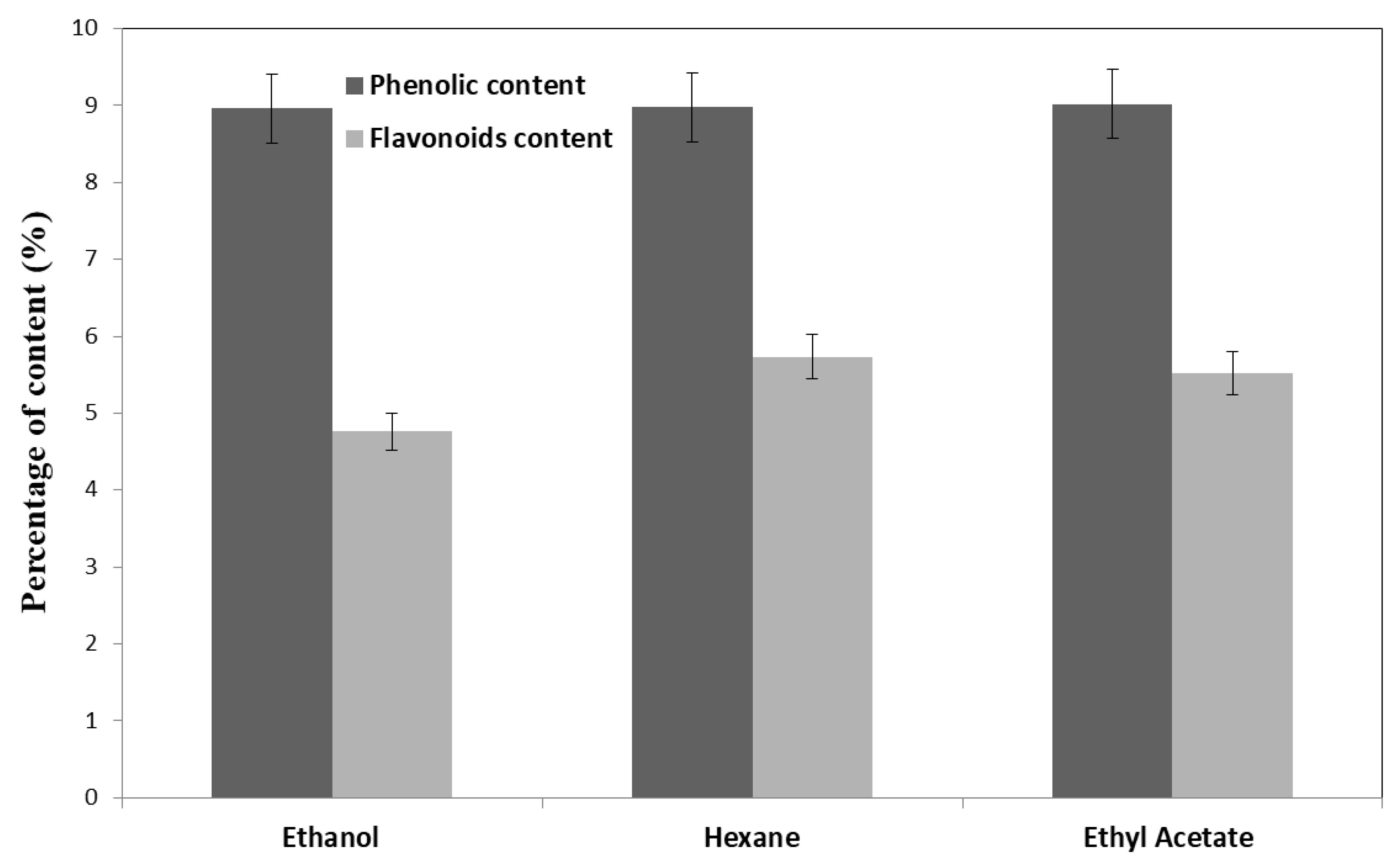
3.5. Phenolic and Flavonoid Contents of Echinops spinosissimus Extracts

Total phenols and flavonoids are considered to be the most important bioactive compounds in plants due to their ability to enhance antioxidant and antimicrobial activity. Typically, they are referred to as plant secondary metabolites and have at least one aromatic ring with a hydroxyl group [48]. Thus, it is very important to study these metabolites in E. spinosissimus extracts to understand the structure of the secondary metabolites of this plant. Three extracts were obtained, and the phenolic and flavonoid contents of each were examined. According to the data in Figure 3, the percentages of the extracts that were examined were close to each other in terms of their contents of phenols or flavonoids, reaching 9 and 5% for total phenols and flavonoids, respectively. This may be due to the ability of these extraction solvents to extract nearly the same amounts of the tested bioactive substances. However, the components of these extracts may be different; therefore, it is necessary to identify these components by using HPLC or GC-MS instruments. Additionally, Hegazy and co-authors reported that the total phenols in an Echinops spinosissimus methanolic extract was over 3 mg/g [49]. Polyphenols were studied in this work due to their antioxidant activity as radical-scavenging agents and possible helpful roles in human health, such as reducing cancer risk and cardiovascular disease [50].

3.6. Antioxidant Properties of Echinops spinosissimus Extracts

Analysis of Echinops spinosissimus extracts’ total antioxidant activity revealed that the ethyl acetate extract from the aerial parts had a high level of overall antioxidant capacity and free radical-scavenging abilities. The total antioxidant activity reached 87.72 and 161.66 % for DPPH and ABTS, respectively. Moreover, another extract (ethanol and hexane) also displayed antioxidant activity (Figure 4). This may be due to antioxidant phytochemicals such as phenols, flavonoids, cardiac glycosides, and alkaloids that were extracted from E. spinosissimus via the used solvents [51].

3.7. HPLC Characterization of Echinops spinosissimus Ethyl Acetate and Ethanol Extracts

An HPLC instrument was previously reported to identify phenolic and flavonolic compounds in plant extracts [9,52]. HPLC analysis of the Echinops spinosissimus ethyl acetate and ethanol extracts compared with the standards is represented in Figure 5. Additionally, the data collected from the HPLC results were used to quantitatively analyze the producing compounds from the different extracts as secondary metabolites, and these data were tabulated (Table 2). Protocatechuic acid, gallic acid, chlorogenic acid, p-hydroxybenzoic acid, syringic acid, caffeic acid, vanillic acid, sinapic acid, ferulic acid, p-coumaric acid, rosmarinic acid, rutin, cinnamic acid, apegnin-7-glycoside, quercetin, and kaempferol were detected as bioactive substances. Gallic acid has been linked to a number of positive effects, including antineoplastic, anti-inflammatory, and antioxidant characteristics. It has been found to have therapeutic effects in cardiovascular, metabolic, cognitive, and gastrointestinal diseases [53]. p-Coumaric acid is a phenolic acid that displays several biological activities, including anti-inflammatory, antioxidant, analgesic, and antimicrobial properties [54]. Protocatechuic acid, as a major metabolite of anthocyanin, has antioxidant, anti-inflammatory, antihyperglycemic, antiproliferative, and antiapoptotic properties [55]. For chlorogenic acid, it has been reported as having antioxidant, antibacterial, antitumor, and anti-inflammatory properties, as well as providing liver and kidney protection, protection of the nervous system, and regulation of sugar metabolism and lipid metabolism [56]. Additionally, anti-inflammatory, antioxidant, and anticarcinogenic effects are present in caffeic acid, such as phenolic acid and its derivatives [57]. In the case of vanillic acid, it has antioxidant, anti-inflammatory, and neuroprotective activity [58]. Cinnamic acid, as a natural aromatic carboxylic acid, exhibits many biological activities such as antioxidant, antimicrobial, anticancer, neuroprotective, anti-inflammatory, and antidiabetic properties [59]. With regard to rosmarinic acid activity, it has remarkable biological properties, including anticancer, antiviral, antioxidant, antibacterial, antiaging, cardioprotective, antidiabetic, hepatoprotective, antidepressant, nephroprotective, anti-inflammatory, and antiallergic properties [60]. Quercetin is a flavonoid compound that has numerous biological activities, including anti-inflammatory, antioxidant, radical-scavenging, antibacterial, gastroprotective, antiviral, and immune-modulatory properties [61].

3.8. GC-MS Analysis of Echinops spinosissimus Hexane Extract

Four main compounds, including hexadecanoic acid, octadecanoic or stearic acid, oleic acid, and linoleic acid, were detected by using GC-MS (Figure 6). The hexane extract of plants mainly produces bioactive fatty acids [62]. In the current study, one fatty acid with C16 (hexadecanoic acid) was detected and three fatty acids with C18 were produced. Hexadecanoic acid exhibited many biological activities such as hypocholesterolemic, antioxidant, pesticide, and nematicide properties [63]. The stearic acid biological activity has antibacterial and antifungal properties [64]. Oleic acid is a fatty acid that occurs naturally in various animal and vegetable fats and oils, and it has been found to have antibacterial activity. Linoleic acid inhibited the growth of different Gram-positive bacteria [65,66].

3.9. Antimicrobial Properties of Echinops spinosissimus Extracts

Three different extracts of Echinops spinosissimus were obtained using ethanol, hexane, and ethyl acetate solvents, and were used in this part of the experiment to evaluate their ability to inhibit the growth of the tested pathogens. The antimicrobial action of the tested extracts was tabulated and illustrated (Figure 7). The highest antibacterial properties were noted with L. monocytogenes. Additionally, the obtained extracts appeared to have high activity against Gram-positive pathogenic bacteria compared with Gram-negative bacteria. The antimicrobial activity of the extracts may be due to some bioactive secondary metabolites such as protocatechuic acid, chlorogenic acid, gallic acid, syringic acid, p-hydroxybenzoic acid, vanillic acid, rosmarinic acid, p-coumaric acid, rutin, cinnamic acid, quercetin, and kaempferol. These compounds were previously reported as antimicrobial agents after being extracted from plants [56]. Additionally, fatty acids in the hexane extract may respond to the antimicrobial properties of this extract [63].

4. Conclusions

Based on morphological and anatomical data, the plant collected from the northeast of Mecca was identified as Echinops spinosissimus. Findings from the study of its phytochemical properties showed that the best solvent assayed was ethyl acetate, as it produced high amounts of bioactive compounds such as phenols, flavonoids, and alkaloids. This extract showed high activity against Gram-positive pathogenic bacteria. The identification of extracts via HPLC and GC-MS led to the detection of protocatechuic acid, gallic acid, rutin, vanillic acid, quercetin, kaempferol, and hexadecanoic, stearic, oleic, and linoleic acids as bioactive substances. The ethyl acetate extract exhibited a high total antioxidant capability and free radical-scavenging properties.

Author Contributions

Conceptualization A.M.H. and L.M.A.M., methodology, A.M.H. and L.M.A.M., formal analysis, A.M.H. and L.M.A.M., data curation, A.M.H. and L.M.A.M., resources, L.M.A.M., writing—original draft preparation, A.M.H., writing and reviewing the manuscript, A.M.H. and L.M.A.M. All authors have read and agreed to the published version of the manuscript.

Funding

There was no external funding for this study.

Data Availability Statement

On request, any information relating to this text is available.

Acknowledgments

Grateful thanks are expressed to Osama M. Darwesh, National Research Centre, Egypt for the technical assistance in microbiological analyses.

Conflicts of Interest

The author declares no conflict of interest.

References

  1. Mozaffarian, V.; Ghahreman, A. Three new species of Echinops (Compositae, Cynareae) from Iran. Bot. J. Linn. Soc. 2002, 140, 181–186. [Google Scholar] [CrossRef]
  2. Özhatay, N.; Kültür, Ş.; Aslan, S. Check-list of Additional Taxa to the Supplement Flora of Turkey, IV. Turk. J. Bot. 2009, 33, 191–226. [Google Scholar] [CrossRef]
  3. Susanna, A.; Garcia-Jacas, N. The tribe Cardueae. In The Families and Genera of Vascular Plants. Flowering Plants, Eudicots, Asterales; Kadereit, J.W., Jeffrey, C., Eds.; Springer: Berlin/Heidelberg, Germany, 2007; Volume 8, pp. 135–158. [Google Scholar]
  4. Sánchez-Jiménez, I.; Lazkov, G.A.; Hidalgo, O.; Garnatje, T. Molecular systematics of Echinops L. (Asteraceae, Cynareae): A phylogeny based on ITS and trnL-trnF sequences with emphasis on sectional delimitation. Taxon 2010, 59, 698–708. [Google Scholar] [CrossRef]
  5. Mabberley, D.J. Mabberley’s Plant-Book, 4th ed.; Cambridge Univ. Press: Cambridge, UK, 2017. [Google Scholar]
  6. Khafagi, A.F. Taxonomic studies on some Compositae. Master’s Thesis, Faculty of science (Girls), Al-Azhar University, Cairo, Egypt, 1983. [Google Scholar]
  7. Bremer, K. Asteraceae: Cladistics and Classifications; Timber Press: Portland, OR, USA, 1994; 792p. [Google Scholar]
  8. Bouzabata, A.; Montoro, P.; Gil, K.A.; Piacente, S.; Youssef, F.S.; Al Musayeib, N.M.; Cordell, G.A.; Ashour, M.L.; Tuberoso, C.I.G. HR-LC-ESI-Orbitrap-MS-Based Metabolic Profiling Coupled with Chemometrics for the Discrimination of Different Echinops spinosus Organs and Evaluation of Their Antioxidant Activity. Antioxidants 2022, 11, 453. [Google Scholar] [CrossRef]
  9. Khedher, O.; Rigane, G.; Riguene, H.; Salem, R.B.; Moussaoui, Y. Phenolic profile (HPLC-UV) analysis and biological activities of two organic extracts from Echinops spinosissimus Turra roots growing in Tunisia. Nat. Prod. Res. 2021, 35, 5786–5793. [Google Scholar] [CrossRef] [PubMed]
  10. Mohamed, A.A.; Ali, S.I.; Darwesh, O.M.; El-Hallouty, S.M.; Sameeh, M.Y. Chemical Compositions, Potential Cytotoxic and Antimicrobial Activities of Nitraria retusa Methanolic Extract Sub-fractions. Int. J. Toxicol. Pharmacol. Res. 2015, 7, 204–212. [Google Scholar]
  11. Khalid, K.A.; Darwesh, O.M.; Ahmed, A.M.A. Peel Essential Oils of Citrus Types and Their Antimicrobial Activities in Response to Various Growth Locations. J. Ess. Oil. Bear. Plants 2021, 24, 480–499. [Google Scholar] [CrossRef]
  12. Eweys, A.S.; Zhao, Y.S.; Darwesh, O.M. Improving the antioxidant and anticancer potential of Cinnamomum cassia via fermentation with Lactobacillus plantarum. Biotechnol. Rep. 2022, 36, e00768. [Google Scholar] [CrossRef]
  13. Darwesh, O.M.; Matter, I.A.; Eida, M.F.; Moawad, H.; Oh, Y. Influence of Nitrogen Source and Growth Phase on Extracellular Biosynthesis of Silver Nanoparticles Using Cultural Filtrates of Scenedesmus obliquus. Appl. Sci. 2019, 9, 1465. [Google Scholar] [CrossRef] [Green Version]
  14. Darwesh, O.M.; Mahmoud, R.H.; Abdo, S.M.; Marrez, D.A. Isolation of Haematococcus lacustris as source of novel anti-multi-antibiotic resistant microbes agents; fractionation and identification of bioactive compounds. Biotechnol. Rep. 2022, 35, e00753. [Google Scholar] [CrossRef]
  15. Mourad, R.; Helaly, F.; Darwesh, O.M.; Sawy, S.E. Antimicrobial and physicomechanical natures of silver nanoparticles incorporated into silicone- hydrogel films. Contact Lens Anterior Eye 2019, 42, 325–333. [Google Scholar] [CrossRef] [PubMed]
  16. Abdelhameed, R.M.; Darwesh, O.M.; El-Shahat, M. Synthesis of arylidene hydrazinylpyrido[2,3-d]pyrimidin-4-ones as potent anti-microbial agents. Heliyon 2020, 6, e04956. [Google Scholar] [CrossRef] [PubMed]
  17. Mourad, R.M.; Darwesh, O.M.; Abdel-Hakim, A. Enhancing physico-mechanical and antibacterial properties of natural rubber using synthesized Ag-SiO2 nanoparticles. Int. J. Biol. Macromol. 2020, 164, 3243–3249. [Google Scholar] [CrossRef] [PubMed]
  18. El-Baz, F.K.; Mahmoud, K.; El-Senousy, W.M.; Darwesh, O.M.; El Gohary, A.E. Antiviral—Antimicrobial and Schistosomicidal Activities of Eucalyptus camaldulensis Essential Oils. Int. J. Pharm. Sci. Rev. Res. 2015, 31, 262–268. [Google Scholar]
  19. Sadek, Z.I.; Abdel-Rahman, M.A.; Azab, M.S.; Darwesh, O.M.; Hassan, M.S. Microbiological evaluation of infant foods quality and molecular detection of Bacillus cereus toxins relating genes. Toxicol. Rep. 2018, 5, 871–877. [Google Scholar] [CrossRef]
  20. El-Sofany, W.I.; Flefel, E.M.; Darwesh, O.M.; El-Shahat, M. Boosting the antimicrobial performance based on new fused spirothiazolidine framework analogs. J. Iran. Chem. Soc. 2022, 19, 4223–4236. [Google Scholar] [CrossRef]
  21. Darwesh, O.M.; El-Maraghy, S.H.; Abdel-Rahman, H.M.; Zaghloul, R.A. Improvement of paper wastes conversion to bioethanol using novel cellulose degrading fungal isolate. Fuel 2020, 262, 116518. [Google Scholar] [CrossRef]
  22. Zhao, Y.S.; Eweys, A.S.; Zhang, J.Y.; Zhu, Y.; Bai, J.; Darwesh, O.M.; Zhang, H.B.; Xiao, X. Fermentation Affects the Antioxidant Activity of Plant-Based Food Material through the Release and Production of Bioactive Components. Antioxidants 2021, 10, 2004. [Google Scholar] [CrossRef]
  23. Darwesh, O.M.; Eweys, A.S.; Zhao, Y.S.; Matter, I.A. Application of environmental-safe fermentation with Saccharomyces cerevisiae for increasing the cinnamon biological activities. Bioresour. Bioprocess. 2023, 10, 12. [Google Scholar] [CrossRef]
  24. El-Shanshoury, A.R.; Darwesh, O.M.; Sabae, S.Z.; Awadallah, O.A.; Hassan, S.H. Bio-manufacturing of selenium nanoparticles by Bacillus subtilis isolated from Qarun Lake and evaluation their activity for water remediation. Biointerf. Res. Appl. Chem. 2020, 10, 5834–5842. [Google Scholar] [CrossRef]
  25. Punt, W.; Hoen, P.P.; Blackmore, S.; Nilsson, S.; Le Thomas, A. Glossary of pollen and spore terminology. Rev. Palaeobot. Palynol. 2007, 143, 1–81. [Google Scholar] [CrossRef]
  26. Grant-Downton, R. Pollen terminology, an illustrated handbook. Ann. Bot. 2010, 105, viii–ix. [Google Scholar] [CrossRef] [Green Version]
  27. Bobrov, E.G. Echinops L. In Flora of the USSR; Shishkin, B.K., Bobrov, E.G., Eds.; Academy of Sciences of the USSR, Moscow & Leningrad: Saint Petersburg, Russia, 1962; Volume 27, pp. 1–54. [Google Scholar]
  28. Youssef, F.S.; Ovidi, E.; Musayeib, N.M.A.; Ashour, M.L. Morphology, Anatomy and Secondary Metabolites Investigations of Premna odorata Blanco and Evaluation of Its Anti-Tuberculosis Activity Using In Vitro and In Silico Studies. Plants 2021, 10, 1953. [Google Scholar] [CrossRef] [PubMed]
  29. Labib, R.M.; Youssef, F.S.; Ashour, M.L.; Abdel-Daim, M.M.; Ross, S.A. Chemical Composition of Pinus roxburghii Bark Volatile Oil and Validation of Its Anti-Inflammatory Activity Using Molecular Modelling and Bleomycin-Induced Inflammation in Albino Mice. Molecules 2017, 22, 1384. [Google Scholar] [CrossRef] [Green Version]
  30. Darwesh, O.M.; Elshahawy, I.E. Silver nanoparticles inactivate sclerotial formation in controlling white rot disease in onion and garlic caused by the soil borne fungus Stromatinia cepivora. Eur. J. Plant Patho. 2021, 160, 917–934. [Google Scholar] [CrossRef]
  31. Bouzabata, A.; Mahomoodally, F.; Tuberoso, C. Ethnopharmacognosy of Echinops spinosus L. in North Africa: A mini review. J. Compl. Med. Res. 2018, 8, 40–52. [Google Scholar] [CrossRef] [Green Version]
  32. Khan, M.S.; Yusufzai, S.K.; Rafatullah, M.; Sarjadi, M.S.; Razlan, M. Determination of total phenolic content, total flavonoid content and antioxidant activity of various organic crude extracts of Licuala spinosa leaves from Sabah Malaysia. ASM Sci. J. 2018, 11, 53–58. [Google Scholar]
  33. Abdel Rahman, S.M.; Abd-Ellatif, S.A.; Deraz, S.F.; Khalil, A.A. Antibacterial activity of some wild medicinal plants collected from western Mediterranean coast, Egypt: Natural alternatives for infectious disease treatment. African J. Biotechnol. 2011, 10, 10733–10743. [Google Scholar] [CrossRef]
  34. Sultan, Y.Y.; Ali, M.A.; Darwesh, O.M.; Embaby, M.A.; Marrez, D.A. Influence of Nitrogen Source in Culture Media on Antimicrobial Activity of Microcoleus lacustris and Oscillatoria rubescens. Res. J. Pharm. Biol. Chem. Sci. 2016, 7, 1444–1452. [Google Scholar]
  35. Darwesh, O.M.; Barakat, K.M.; Mattar, M.Z.; Sabae, S.Z.; Hassan, S.H. Production of antimicrobial blue green pigment Pyocyanin by marine Pseudomonas aeruginosa. Biointerf. Res. Appl. Chem. 2019, 9, 4334–4339. [Google Scholar] [CrossRef]
  36. Bitew, H.; Hymete, A. The Genus Echinops: Phytochemistry and Biological Activities: A Review. Front. Pharmacol. 2019, 10, 1234. [Google Scholar] [CrossRef] [PubMed] [Green Version]
  37. SMEGI, Saudi & Middle East Green Initiatives. 2022. Available online: https://www.greeninitiatives.gov.sa/about-mgi/mgi-targets/planting-trees/plant-trees-across-the-middle-east (accessed on 1 May 2022).
  38. Milan, P.; Hayashi, A.H.; Appezzato-da-Glória, B. Comparative leaf morphology and anatomy of three Asteraceae species. Brazilian Arc. Biol. Tech. 2006, 49, 135–144. [Google Scholar] [CrossRef] [Green Version]
  39. Kim, J.H.; Kim, T.J.; Kim, H.J.; Cho, C.W.; Kim, S.J.; Cho, H.S.; Kim, K.T.; Kang, J.S. A new high-performance liquid chromatographic method for the quality control of bioconverted Mori Folium extracts with appropriate marker compounds related to antidiabetes. J. Anal. Sci. Technol. 2021, 12, 2. [Google Scholar] [CrossRef]
  40. Rahman, A.H.; Islam, A.K.; Rahman, M.M. An anatomical investigation on Asteraceae family at Rajshahi Division, Bangladesh. Int. J. BioSci. 2013, 3, 13–23. [Google Scholar]
  41. Ekeke, C.; Mensah, S.I. Comparative anatomy of midrib and its significance in the taxonomy of the family Asteraceae from Nigeria. J. Plant. Sci. 2015, 10, 200–205. [Google Scholar] [CrossRef] [Green Version]
  42. Dickison, W.C. Integrative Plant Anatomy; Academic Press: New York, NY, USA, 2000; p. 534. [Google Scholar]
  43. Larcher, W. Ecofisiologia Vegetal; Rima Artes e Textos: São Carlos, Brazil, 2000; p. 531. [Google Scholar]
  44. Bukovac, M.J. Sorption of organic compounds by plant cuticles. Weed Sci. 1990, 38, 289–298. [Google Scholar] [CrossRef]
  45. Zhang, M.; Fan, S.; Hao, M.; Hou, H.; Zheng, H.; Darwesh, O.M. Improving the production of fungal exopolysaccharides with application of repeated batch fermentation technology coupling with foam separation in the presence of surfactant. Proc. Biochem. 2021, 100, 82–89. [Google Scholar] [CrossRef]
  46. Mozaffarian, V. A taxonomic survey of Echinops L. Tribe Echinopeae (Asteraceae) in Iran: 14 new species and diagnostic keys. Iran. J. Bot. 2006, 11, 197–239. [Google Scholar]
  47. Das, D.; Mukherjee, S.K. Diversity of cypselar features in seven species of the tribe Lactuceae (Asteraceae). J. Econ. Taxon. Bot. 2008, 32, 282–297. [Google Scholar]
  48. Shamso, E.M.; Hosny, A.H.; Ahmed, D.; Shaltout, K. Achene Characteristics of Some Taxa of Asteraceae from the Northwestern Mediterranean Coast of Egypt. Egypt J. Bot. 2021, 61, 1–31. [Google Scholar] [CrossRef]
  49. Tungmunnithum, D.; Thongboonyou, A.; Pholboon, A.; Yangsabai, A. Flavonoids and Other Phenolic Compounds from Medicinal Plants for Pharmaceutical and Medical Aspects: An Overview. Medicines 2018, 5, 93. [Google Scholar] [CrossRef] [PubMed]
  50. Hegazy, M.G.; Emam, M.A.; Khattab, H.I.; Helal, N.M. Biological activity of Echinops spinosus on inhibition of paracetamol-induced renal inflammation. Biochem. Cell. Biol. 2019, 97, 176–186. [Google Scholar] [CrossRef] [PubMed]
  51. Rudrapal, M.; Khairnar, S.J.; Khan, J.; Dukhyil, A.; Ansari, M.A.; Alomary, M.N.; Alshabrmi, F.M.; Palai, S.; Deb Prashanta, K.; Devi, R. Dietary Polyphenols and Their Role in Oxidative Stress-Induced Human Diseases: Insights into Protective Effects, Antioxidant Potentials and Mechanism(s) of Action. Front. Pharmacol. 2022, 13, 283. [Google Scholar] [CrossRef] [PubMed]
  52. Mujeeb, F.; Bajpai, P.; Pathak, N. Phytochemical evaluation, antimicrobial activity, and determination of bioactive components from leaves of Aegle marmelos. Biomed Res. Int. 2014, 2014, 497606. [Google Scholar] [CrossRef] [PubMed] [Green Version]
  53. Zitouni-Nourine, S.H.; Belyagoubi-Benhammou, N.; El-Houaria Zitouni-Haouar, F.; Douahi, O.; Chenafi, F.; Fetati, H.; Chabane Sari, S.; Benmahieddine, A.; Zaoui, C.; Mekaouche, F.Z.N.; et al. Echinops spinosissimus Turra Root Methanolic Extract: Characterization of the Bioactive Components and Relative Wound Healing, Antimicrobial and Antioxidant Properties. Plants 2022, 11, 3440. [Google Scholar] [CrossRef] [PubMed]
  54. Kahkeshani, N.; Farzaei, F.; Fotouhi, M.; Alavi, S.S.; Bahramsoltani, R.; Naseri, R.; Momtaz, S.; Abbasabadi, Z.; Rahimi, R.; Farzaei, M.H.; et al. Pharmacological effects of gallic acid in health and diseases: A mechanistic review. Iran. J. Basic Med. Sci. 2019, 22, 225–237. [Google Scholar] [CrossRef]
  55. Aldaba Muruato, L.R.; Ventura Juárez, J.; Perez Hernandez, A.M.; Hernández Morales, A.; Muñoz Ortega, M.H.; Martínez Hernández, S.L.; Alvarado Sánchez, B.; Macías Pérez, J.R. Therapeutic perspectives of p coumaric acid: Anti necrotic, anti cholestatic and anti amoebic activities. World Acad. Sci. J. 2021, 47. [Google Scholar] [CrossRef]
  56. Semaming, Y.; Pannengpetch, P.; Chattipakorn, S.C.; Chattipakorn, N. Pharmacological properties of protocatechuic Acid and its potential roles as complementary medicine. Evid. Based Compl. Alternat. Med. 2015, 2015, 593902. [Google Scholar] [CrossRef] [Green Version]
  57. Wang, L.; Pan, X.; Jiang, L.; Chu, Y.; Gao, S.; Jiang, X.; Zhang, Y.; Chen, Y.; Luo, S.; Peng, C. The Biological Activity Mechanism of Chlorogenic Acid and Its Applications in Food Industry: A Review. Front. Nutr. 2022, 9, 943911. [Google Scholar] [CrossRef]
  58. Espíndola, K.M.; Ferreira, R.G.; Narvaez, L.E.; Silva Rosario, A.C.; da Silva, A.H.; Silva, A.G.; Vieira, A.P.; Monteiro, M.C. Chemical and Pharmacological Aspects of Caffeic Acid and Its Activity in Hepatocarcinoma. Front. Oncol. 2019, 21, 541. [Google Scholar] [CrossRef] [Green Version]
  59. Ullah, R.; Ikram, M.; Park, T.J.; Ahmad, R.; Saeed, K.; Alam, S.I.; Rehman, I.U.; Khan, A.; Khan, I.; Jo, M.G.; et al. Vanillic Acid, a Bioactive Phenolic Compound, Counteracts LPS-Induced Neurotoxicity by Regulating c-Jun N-Terminal Kinase in Mouse Brain. Int. J. Mol. Sci. 2020, 22, 361. [Google Scholar] [CrossRef] [PubMed]
  60. Ruwizhi, N.; Aderibigbe, B.A. Cinnamic Acid Derivatives and Their Biological Efficacy. Int. J. Mol. Sci. 2020, 21, 5712. [Google Scholar] [CrossRef] [PubMed]
  61. Nadeem, M.; Imran, M.; Aslam Gondal, T.; Imran, A.; Shahbaz, M.; Muhammad Amir, R.; Wasim Sajid, M.; Qaisrani, T.B.; Atif, M.; Hussain, G.; et al. Therapeutic Potential of Rosmarinic Acid: A Comprehensive Review. Appl. Sci. 2019, 9, 3139. [Google Scholar] [CrossRef] [Green Version]
  62. Kim, J.K.; Park, S.U. Quercetin and its role in biological functions: An updated review. EXCLI J. 2018, 17, 856–863. [Google Scholar] [CrossRef] [PubMed]
  63. Kaseke, T.; Opara, U.L.; Fawole, O.A. Fatty acid composition, bioactive phytochemicals, antioxidant properties and oxidative stability of edible fruit seed oil: Effect of preharvest and processing factors. Heliyon 2020, 6, e04962. [Google Scholar] [CrossRef]
  64. Mazumder, K.; Nabila, A.; Aktar, A.; Farahnaky, A. Bioactive Variability and In Vitro and In Vivo Antioxidant Activity of Unprocessed and Processed Flour of Nine Cultivars of Australian lupin Species: A Comprehensive Substantiation. Antioxidants 2020, 9, 282. [Google Scholar] [CrossRef] [PubMed] [Green Version]
  65. Agoramoorthy, G.; Chandrasekaran, M.; Venkatesalu, V.; Hsu, M.J. Antibacterial and Antifungal Activities of Fatty Acid Methyl Esters of the Blind-Your-Eye Mangrove from India. Brazilian J. Microbiol. 2007, 38, 739–742. [Google Scholar] [CrossRef] [Green Version]
  66. Dilika, F.; Bremner, P.D.; Meyer, J.J. Antibacterial activity of linoleic and oleic acids isolated from Helichrysum pedunculatum: A plant used during circumcision rites. Fitoterapia 2000, 71, 450–452. [Google Scholar] [CrossRef]
Figure 1. Map and information of the collected samples’ locations.
Agronomy 13 00573 g001
Figure 2. Macro- and microphotographs of the studied taxa: (a). Whole plant (bd). Stem cross-section (e,f). Lamina vertical section (gi). Pollen morphological characters (LM and SEM) (jl). Achene morphological characters (LM and SEM). P—pith; Pa.—palisade layer; St. c.—stone cell.
Agronomy 13 00573 g002
Figure 3. Total phenol and flavonoid content (%) of ethanolic, hexanolic, and ethyl acetate Echinops spinosissimus extracts.
Agronomy 13 00573 g003
Figure 4. Antioxidant activity of the different extracts of Echinops spinosissimus.
Agronomy 13 00573 g004
Figure 5. Chromatogram for the HPLC analysis of standards (a), ethyl acetate extract (b), and ethanol extract (c) of Echinops spinosissimus.
Agronomy 13 00573 g005
Figure 6. GC-MS analysis of Echinops spinosissimus hexane extract.
Agronomy 13 00573 g006
Figure 7. Antimicrobial properties of Echinops spinosissimus extracts at concentration of 100 mg/mL. Abbreviations: list.—Listeria monocytogenes; St.—Staphylococcus aureus; E.c.—E. coli; Sal., Salmonella typhi; C.A—Candida albicans; H—hexane extract; E—ethanol extract; EA—ethyl acetate.
Agronomy 13 00573 g007
Table 1. Phytochemical quantitative analysis of Echinops spinosissimus extracts.
ExtractsAlkaloids (mg/g)Soluble Sugar (mg/g)Total Proteins (mg/g)Total Lipids (mg/g)Tannins (µg/g)Saponins (µg/g)
Ethanol30.90 b5.033 b115.00 b35.13 c0.510 b225.33 c
Hexane30.87 b4.333 c109.33 c44.67 a0.507 b245.33 b
Ethyl acetate35.00 a6.900 a124.67 a38.00 b0.723 a295.67 a
LSD0.5280.2191.61020.8830.0303.399
Means accompanied by the same letter are statistically not different at p < 0.05 (LSD test). In the same column, a is the greatest value and c is the lowest value.
Table 2. Phenolic profile of Echinops spinosissimus ethyl acetate and ethanol extracts analyzed via HPLC.
CompoundsEthyl Acetate (µg/g)Ethanol (µg/g)
Gallic acid24.077.71
Protocatechuic acid197.4151.05
p-Hydroxybenzoic acid135.6346.09
Chlorogenic acid3582.255167.99
Caffeic acid481.53155.92
Syringic acid405.45148.30
Vanillic acid9.1120.24
Ferulic acid81.8138.53
Sinapic acid42.280.00
p-Coumaric acid7725.944342.51
Rutin318.851050.18
Rosmarinic acid21,004.6513,077.16
Apegnin-7-glycoside113.7969.29
Cinnamic acid19.7445.24
Quercetin2980.631544.26
Kaempferol328.64401.76
Disclaimer/Publisher’s Note: The statements, opinions and data contained in all publications are solely those of the individual author(s) and contributor(s) and not of MDPI and/or the editor(s). MDPI and/or the editor(s) disclaim responsibility for any injury to people or property resulting from any ideas, methods, instructions or products referred to in the content.

