
Evaluation of the Effect of ‘Candidatus Liberibacter Solanacearum’ Haplotypes in Tobacco Infection
 , and
, and 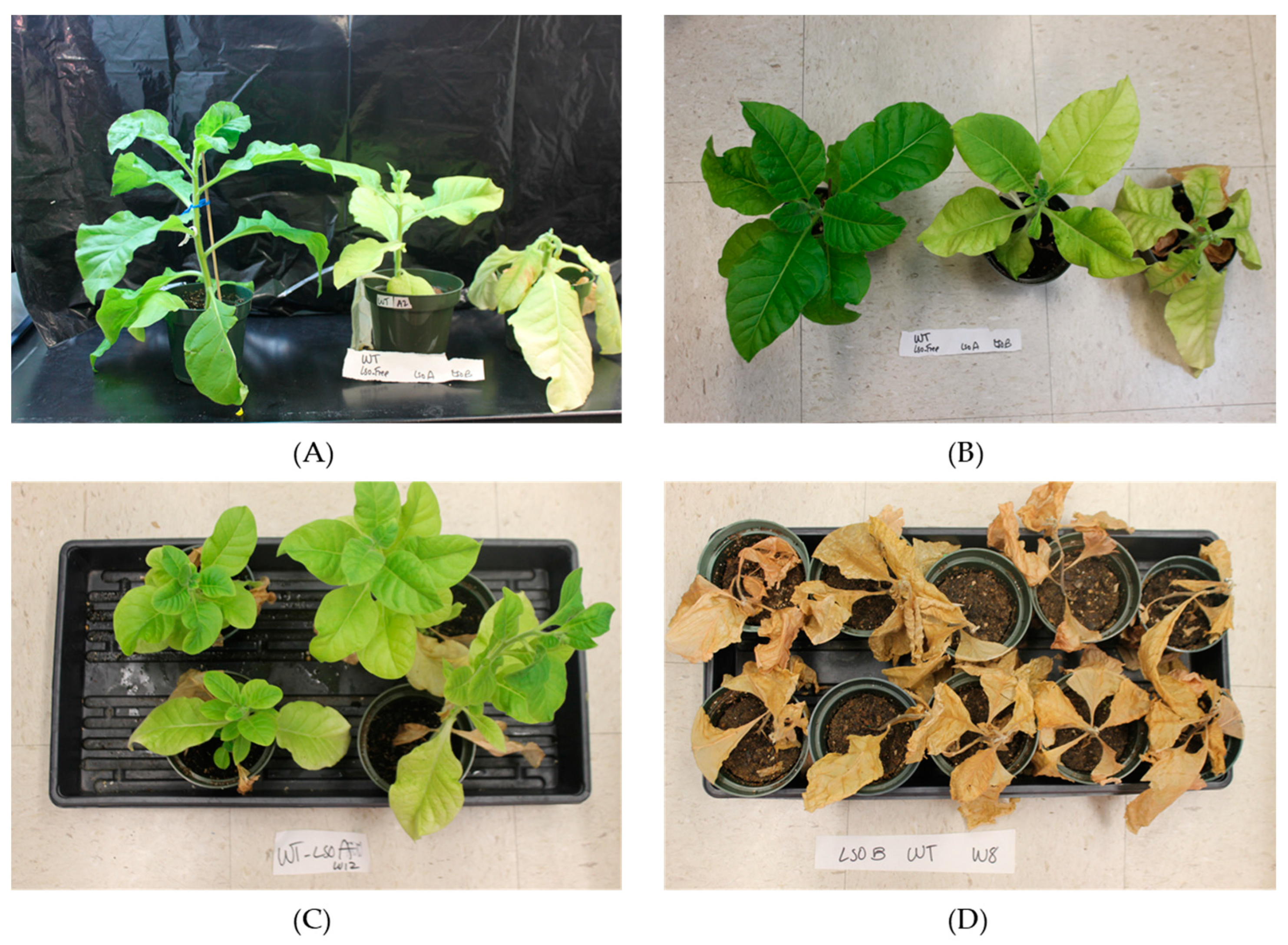
Abstract
:1. Introduction
2. Materials and Methods
2.1. Insects
2.2. Plant Source
2.3. Plant Infection
2.4. Symptom Progression and Lso Quantification in Tobacco Plants
2.5. Gene Expression Analysis
2.6. Quantification of SA and Other Metabolites
2.7. Data Analysis
3. Results
3.1. Symptoms in Tobacco Associated with ‘Candidatus Liberibacter Solanacearum’ Haplotypes A and B Infection
3.2. Lso Quantification in Tobacco Plants
3.3. PR1a Transcript Expression
3.4. Quantification of Salicylic Acid and Other Metabolites
4. Discussion
Author Contributions
Funding
Institutional Review Board Statement
Informed Consent Statement
Data Availability Statement
Conflicts of Interest
References
- Munyaneza, J.E.; Crosslin, J.M.; Upton, J.E. Association of Bactericera cockerelli (Homoptera: Psyllidae) with “zebra chip,” a new potato disease in southwestern United States and Mexico. J. Econ. Entomol. 2007, 100, 656–663. [Google Scholar] [CrossRef] [PubMed]
- Greenway, G.A.; Rondon, S. Economic impacts of zebra chip in Idaho, Oregon, and Washington. Am. J. Potato Res. 2018, 95, 362–367. [Google Scholar] [CrossRef]
- Wang, N.; Pierson, E.A.; Setubal, J.C.; Xu, J.; Levy, J.G.; Zhang, Y.Z.; Li, J.Y.; Rangel, L.T.; Martins, J. The Candidatus Liberibacter-host interface: Insights into pathogenesis mechanisms and disease control. Annu. Rev. Phytopathol. 2017, 55, 451–482. [Google Scholar] [CrossRef] [PubMed]
- Gottwald, T.R. Current epidemiological understanding of citrus Huanglongbing. Annu. Rev. Phytopathol. 2010, 48, 119–139. [Google Scholar] [CrossRef] [Green Version]
- Bové, J.M. Huanglongbing: A destructive, newly-emerging, century-old disease of citrus. J. Plant Pathol. 2006, 88, 7–37. [Google Scholar]
- Thapa, S.P.; De Francesco, A.; Trinh, J.; Gurung, F.B.; Pang, Z.; Vidalakis, G.; Wang, N.; Ancona, V.; Ma, W.; Coaker, G. Genome-wide analyses of Liberibacter species provides insights into evolution, phylogenetic relationships, and virulence factors. Mol. Plant Pathol. 2020, 21, 716–731. [Google Scholar] [CrossRef] [Green Version]
- Wen, A.; Johnson, C.; Gudmestad, N.C. Development of a PCR assay for the rapid detection and differentiation of ‘Candidatus Liberibacter solanacearum’ haplotypes and their spatiotemporal distribution in the United States. Am. J. Potato Res. 2013, 90, 229–236. [Google Scholar] [CrossRef]
- Wen, A.; Mallik, I.; Alvarado, V.Y.; Pasche, J.S.; Wang, X.; Li, W.; Levy, L.; Lin, H.; Scholthof, H.B.; Mirkov, T.E.; et al. Detection, distribution, and genetic variability of ‘Candidatus Liberibacter’ species associated with zebra complex disease of potato in North America. Plant Dis. 2009, 93, 1102–1115. [Google Scholar] [CrossRef] [Green Version]
- Lin, H.; Islam, M.S.; Bai, Y.; Wen, A.; Lan, S.; Gudmestad, N.C.; Civerolo, E.L. Genetic diversity of ‘Candidatus Liberibacter solanacearum’ strains in the United States and Mexico revealed by simple sequence repeat markers. Eur. J. Plant Pathol. 2012, 132, 297–308. [Google Scholar] [CrossRef] [Green Version]
- Lin, H.; Gudmestad, N.C. Aspects of pathogen genomics, diversity, epidemiology, vector dynamics, and disease management for a newly emerged disease of potato: Zebra chip. Phytopathology 2013, 103, 524–537. [Google Scholar] [CrossRef] [Green Version]
- Swisher Grimm, K.D.; Garczynski, S.F. Identification of a new haplotype of ‘Candidatus Liberibacter solanacearum’ in Solanum tuberosum. Plant Dis. 2019, 103, 468–474. [Google Scholar] [CrossRef] [PubMed] [Green Version]
- Grimm, K.D.S.; Horton, D.R.; Lewis, T.M.; Garczynski, S.F.; Jensen, A.S.; Charlton, B.A. Identification of three new ‘Candidatus Liberibacter solanacearum’ haplotypes in four psyllid species (Hemiptera: Psylloidea). Sci. Rep. 2022, 12, 20618. [Google Scholar] [CrossRef] [PubMed]
- Alfaro-Fernández, A.; Siverio, F.; Cebrián, M.C.; Villaescusa, F.J.; Font, M.I. ‘Candidatus Liberibacter solanacearum’ associated with Bactericera trigonica-affected carrots in the Canary Islands. Plant Dis. 2012, 96, 581. [Google Scholar] [CrossRef] [PubMed]
- Mawassi, M.; Dror, O.; Bar-Joseph, M.; Piasetzky, A.; Sjölund, J.; Levitzky, N.; Shoshana, N.; Meslenin, L.; Haviv, S.; Porat, C. ‘Candidatus Liberibacter solanacearum’ is tightly associated with carrot yellows symptoms in Israel and transmitted by the prevalent psyllid vector Bactericera trigonica. Phytopathology 2018, 108, 1056–1066. [Google Scholar] [CrossRef] [Green Version]
- Munyaneza, J.E.; Fisher, T.W.; Sengoda, V.G.; Garczynski, S.F.; Nissinen, A.; Lemmetty, A. Association of ‘Candidatus Liberibacter solanacearum’ with the psyllid, Trioza apicalis (Hemiptera: Triozidae) in Europe. J. Econ. Entomol. 2010, 103, 1060–1070. [Google Scholar] [CrossRef] [PubMed]
- Munyaneza, J.E.; Fisher, T.W.; Sengoda, V.G.; Garczynski, S.F.; Nissinen, A.; Lemmetty, A. First report of “Candidatus Liberibacter solanacearum” associated with psyllid-affected carrots in Europe. Plant Dis. 2010, 94, 639. [Google Scholar] [CrossRef]
- Swisher Grimm, K.D.; Mustafa, T.; Cooper, W.R.; Munyaneza, J.E. Growth and yield performance of Solanum tuberosum grown from seed potatoes infected with ‘Candidatus Liberibacter solanacearum’ haplotypes A and B. Plant Dis. 2020, 104, 688–693. [Google Scholar] [CrossRef]
- Harrison, K.; Tamborindeguy, C.; Rondon, S.I.; Levy, J.G. Effects of ‘Candidatus Liberibacter solanacearum’ haplotype on Atlantic potato tuber emergence rate in South Texas. Am. J. Potato Res. 2020, 97, 489–496. [Google Scholar] [CrossRef]
- Harrison, K.; Tamborindeguy, C.; Scheuring, D.C.; Herrera, A.M.; Silva, A.; Badillo-Vargas, I.E.; Miller, J.C.; Levy, J.G. Differences in zebra chip severity between ‘Candidatus Liberibacter solanacearum’ haplotypes in Texas. Am. J. Potato Res. 2019, 96, 86–93. [Google Scholar] [CrossRef]
- Mendoza Herrera, A.; Levy, J.; Harrison, K.; Yao, J.; Ibanez, F.; Tamborindeguy, C. Infection by ‘Candidatus Liberibacter solanacearum’ haplotypes A and B in Solanum lycopersicum ‘Moneymaker’. Plant Dis. 2018, 102, 2009–2015. [Google Scholar] [CrossRef] [Green Version]
- Harrison, K.; Levy, J.G.; Tamborindeguy, C. Effects of ‘Candidatus Liberibacter solanacearum’ haplotypes A and B on tomato gene expression and geotropism. BMC Plant Biol. 2022, 22, 156. [Google Scholar] [CrossRef] [PubMed]
- Munyaneza, J.E.; Sengoda, V.G.; Aguilar, E.; Bextine, B.; McCue, K.F. First Report of “Candidatus Liberibacter solanacearum” Associated with Psyllid-Infested Tobacco in Nicaragua. Plant Dis. 2013, 97, 1244. [Google Scholar] [CrossRef] [PubMed]
- Aguilar, E.; Sengoda, V.G.; Bextine, B.; McCue, K.F.; Munyaneza, J.E. First report of “Candidatus Liberibacter solanacearum” on tobacco in Honduras. Plant Dis. 2013, 97, 1376. [Google Scholar] [CrossRef] [PubMed]
- Yao, J.; Saenkham, P.; Levy, J.; Ibanez, F.; Noroy, C.; Mendoza, A.; Huot, O.; Meyer, D.F.; Tamborindeguy, C. Interactions ‘Candidatus Liberibacter solanacearum’—Bactericera cockerelli: Haplotype effect on vector fitness and gene expression analyses. Front. Cell Infect. Microbiol. 2016, 6, 62. [Google Scholar] [CrossRef] [Green Version]
- Swisher, K.D.; Munyaneza, J.E.; Crosslin, J.M. High resolution melting analysis of the cytochrome oxidase I gene identifies three haplotypes of the potato psyllid in the United States. Environ. Entomol. 2012, 41, 1019–1028. [Google Scholar] [CrossRef]
- Swisher, K.D.; Henne, D.C.; Crosslin, J.M. Identification of a fourth haplotype of Bactericera cockerelli (Hemiptera: Triozidae) in the United States. J. Insect Sci. 2014, 14, 161. [Google Scholar] [CrossRef] [Green Version]
- Levy, J.; Ravindran, A.; Gross, D.; Tamborindeguy, C.; Pierson, E. Translocation of ‘Candidatus Liberibacter solanacearum’, the zebra chip pathogen, in potato and tomato. Phytopathology 2011, 101, 1285–1291. [Google Scholar] [CrossRef] [Green Version]
- Li, W.; Abad, J.A.; French-Monar, R.D.; Rascoe, J.; Wen, A.; Gudmestad, N.C.; Secor, G.A.; Lee, M.; Duan, Y.; Levy, L. Multiplex real-time PCR for detection, identification and quantification of ‘Candidatus Liberibacter solanacearum’ in potato plants with zebra chip. J. Microbiol. Methods 2009, 78, 59–65. [Google Scholar] [CrossRef] [Green Version]
- Huot, O.B.; Levy, J.G.; Tamborindeguy, C. Global gene regulation in tomato plant (Solanum lycopersicum) responding to vector (Bactericera cockerelli) feeding and pathogen (‘Candidatus Liberibacter solanacearum’) infection. Plant Mol. Biol. 2018, 97, 57–72. [Google Scholar] [CrossRef]
- Wang, K.-D.; Borrego, E.J.; Kenerley, C.M.; Kolomiets, M.V. Oxylipins other than jasmonic acid are xylem-resident signals regulating systemic resistance induced by Trichoderma virens in maize. Plant Cell 2019, 32, 166–185. [Google Scholar] [CrossRef]
- RStudio. RStudio: Integrated Development for R; RStudio Inc.: Boston, MA, USA, 2015; Available online: http://www.rstudio.com (accessed on 6 July 2021).
- Mangiafico, S.S. Summary and Analysis of Extension Program Evaluation in R; Rutgers Cooperative Extension: New Brunswick, NJ, USA, 2016. [Google Scholar]
- Prost, I.; Dhondt, S.; Rothe, G.; Vicente, J.; Rodriguez, M.J.; Kift, N.; Carbonne, F.; Griffiths, G.; Esquerré-Tugayé, M.-T.; Rosahl, S. Evaluation of the antimicrobial activities of plant oxylipins supports their involvement in defense against pathogens. Plant Physiol. 2005, 139, 1902–1913. [Google Scholar] [CrossRef] [PubMed] [Green Version]
- Pitino, M.; Armstrong, C.M.; Cano, L.M.; Duan, Y. Transient expression of Candidatus Liberibacter asiaticus effector induces cell death in Nicotiana benthamiana. Front. Plant Sci. 2016, 7, 982. [Google Scholar] [CrossRef] [PubMed] [Green Version]
- Levy, J.G.; Gross, R.; Mendoza-Herrera, A.; Tang, X.; Babilonia, K.; Shan, L.; Kuhl, J.C.; Dibble, M.S.; Xiao, F.; Tamborindeguy, C. Lso-HPE1, an effector of ‘Candidatus Liberibacter solanacearum’, can repress plant immune response. Phytopathology 2020, 110, 648–655. [Google Scholar] [CrossRef] [PubMed]
- Clark, K.J.; Pang, Z.; Trinh, J.; Wang, N.; Ma, W. Sec-Delivered Effector 1 (SDE1) of ‘Candidatus Liberibacter asiaticus’ promotes citrus huanglongbing. Molecular Plant-Microbe Interactions® 2020, 33, 1394–1404. [Google Scholar] [CrossRef]
- Wang, Y.; Zhou, L.; Yu, X.; Stover, E.; Luo, F.; Duan, Y. Transcriptome profiling of huanglongbing (HLB) tolerant and susceptible citrus plants reveals the role of basal resistance in HLB tolerance. Front. Plant Sci. 2016, 7, 933. [Google Scholar] [CrossRef] [Green Version]
- Zhong, Y.; Cheng, C.-Z.; Jiang, N.-H.; Jiang, B.; Zhang, Y.-Y.; Wu, B.; Hu, M.-L.; Zeng, J.-W.; Yan, H.-X.; Yi, G.-J.; et al. Comparative transcriptome and iTRAQ proteome analyses of citrus root responses to Candidatus Liberibacter asiaticus infection. PLoS ONE 2015, 10, e0126973. [Google Scholar] [CrossRef] [Green Version]
- Nwugo, C.C.; Duan, Y.; Lin, H. Study on citrus response to huanglongbing highlights a down-regulation of defense-related proteins in lemon plants upon ‘Ca. Liberibacter asiaticus’ infection. PLoS ONE 2013, 8, e67442. [Google Scholar] [CrossRef]
- Rawat, N.; Kiran, S.P.; Du, D.; Gmitter, F.G., Jr.; Deng, Z. Comprehensive meta-analysis, co-expression, and miRNA nested network analysis identifies gene candidates in citrus against Huanglongbing disease. BMC Plant Biol. 2015, 15, 184. [Google Scholar] [CrossRef] [Green Version]
- Albrecht, U.; Bowman, K.D. Transcriptional response of susceptible and tolerant citrus to infection with Candidatus Liberibacter asiaticus. Plant Sci. 2012, 185, 118–130. [Google Scholar] [CrossRef]
- Aritua, V.; Achor, D.; Gmitter, F.G.; Albrigo, G.; Wang, N. Transcriptional and microscopic analyses of citrus stem and root responses to Candidatus Liberibacter asiaticus infection. PLoS ONE 2013, 8, e73742. [Google Scholar] [CrossRef] [Green Version]
- Levy, J.G.; Mendoza, A.; Miller, J.C.; Tamborindeguy, C.; Pierson, E.A. Global gene expression in two potato cultivars in response to ‘Candidatus Liberibacter solanacearum’ infection. BMC Genom. 2017, 18, 960. [Google Scholar] [CrossRef] [PubMed]
- Macho, A.P.; Zipfel, C. Targeting of plant pattern recognition receptor-triggered immunity by bacterial type-III secretion system effectors. Curr. Opin. Microbiol. 2015, 23, 14–22. [Google Scholar] [CrossRef] [PubMed]
- Prasad, S.; Xu, J.; Zhang, Y.; Wang, N. SEC-translocon dependent extracytoplasmic proteins of Candidatus Liberibacter asiaticus. Front. Microbiol. 2016, 7, 1989. [Google Scholar] [CrossRef] [PubMed] [Green Version]
- Kan, C.-C.; Mendoza-Herrera, A.; Levy, J.; Hull, J.J.; Fabrick, J.A.; Tamborindeguy, C. HPE1, an effector from zebra chip pathogen interacts with tomato proteins and perturbs ubiquitinated protein accumulation. Int. J. Mol. Sci. 2021, 22, 9003. [Google Scholar] [CrossRef]
- Ravindran, A.; Saenkham, P.; Levy, J.G.; Tamborindeguy, C.; Lin, H.; Gross, D.; Pierson, E.A. Characterization of the serralysin-like gene of ‘Ca. Liberibacter solanacearum’ associated with potato zebra chip disease. Phytopathology 2017, 108, 327–335. [Google Scholar] [CrossRef] [Green Version]
- Reyes Caldas, P.A.; Zhu, J.; Breakspear, A.; Thapa, S.P.; Toruño, T.Y.; Perilla-Henao, L.M.; Casteel, C.; Faulkner, C.R.; Coaker, G. Effectors from a bacterial vector-borne pathogen exhibit diverse subcellular localization, expression profiles and manipulation of plant defense. Mol. Plant Microbe Interact. 2022, in press. [CrossRef]
- Ibanez, F.; Suh, J.H.; Wang, Y.; Stelinski, L.L. Long-term, sustained feeding by Asian citrus psyllid disrupts salicylic acid homeostasis in sweet orange. BMC Plant Biol. 2019, 19, 493. [Google Scholar] [CrossRef] [Green Version]
- Park, S.W.; Kaimoyo, E.; Kumar, D.; Mosher, S.; Klessig, D.F. Methyl salicylate is a critical mobile signal for plant systemic acquired resistance. Science 2007, 318, 113–116. [Google Scholar] [CrossRef]
- Vlot, A.C.; Dempsey, D.A.; Klessig, D.F. Salicylic Acid, a multifaceted hormone to combat disease. Annu. Rev. Phytopathol. 2009, 47, 177–206. [Google Scholar] [CrossRef] [Green Version]
- An, C.; Mou, Z. Salicylic acid and its function in plant immunity. J. Integr. Plant Biol. 2011, 53, 412–428. [Google Scholar] [CrossRef]
- Gorman, Z.; Christensen, S.A.; Yan, Y.; He, Y.; Borrego, E.; Kolomiets, M.V. Green leaf volatiles and jasmonic acid enhance susceptibility to anthracnose diseases caused by Colletotrichum graminicola in maize. Mol. Plant Pathol. 2020, 21, 702–715. [Google Scholar] [CrossRef] [PubMed] [Green Version]
- Li, J.; Pang, Z.; Trivedi, P.; Zhou, X.; Ying, X.; Jia, H.; Wang, N. ‘Candidatus Liberibacter asiaticus’ encodes a functional salicylic acid (SA) hydroxylase that degrades SA to suppress plant defenses. Mol. Plant Microbe Interact. 2017, 30, 620–630. [Google Scholar] [CrossRef] [PubMed] [Green Version]
- Li, S.; Liu, J.; Liu, H.; Qiu, R.; Gao, Y.; Duan, A. Role of hydraulic signal and ABA in decrease of leaf stomatal and mesophyll conductance in soil drought-stressed tomato. Front. Plant Sci. 2021, 12. [Google Scholar] [CrossRef]


Disclaimer/Publisher’s Note: The statements, opinions and data contained in all publications are solely those of the individual author(s) and contributor(s) and not of MDPI and/or the editor(s). MDPI and/or the editor(s) disclaim responsibility for any injury to people or property resulting from any ideas, methods, instructions or products referred to in the content. |
© 2023 by the authors. Licensee MDPI, Basel, Switzerland. This article is an open access article distributed under the terms and conditions of the Creative Commons Attribution (CC BY) license (https://creativecommons.org/licenses/by/4.0/).
Share and Cite
Levy, J.G.; Mendoza-Herrera, A.; Merchant, N.; Berg-Falloure, K.M.; Kolomiets, M.V.; Tamborindeguy, C. Evaluation of the Effect of ‘Candidatus Liberibacter Solanacearum’ Haplotypes in Tobacco Infection. Agronomy 2023, 13, 569. https://doi.org/10.3390/agronomy13020569
Levy JG, Mendoza-Herrera A, Merchant N, Berg-Falloure KM, Kolomiets MV, Tamborindeguy C. Evaluation of the Effect of ‘Candidatus Liberibacter Solanacearum’ Haplotypes in Tobacco Infection. Agronomy. 2023; 13(2):569. https://doi.org/10.3390/agronomy13020569
Chicago/Turabian StyleLevy, Julien G., Azucena Mendoza-Herrera, Naveed Merchant, Katherine M. Berg-Falloure, Michael V. Kolomiets, and Cecilia Tamborindeguy. 2023. "Evaluation of the Effect of ‘Candidatus Liberibacter Solanacearum’ Haplotypes in Tobacco Infection" Agronomy 13, no. 2: 569. https://doi.org/10.3390/agronomy13020569