Communication

Proteomic Analysis of Ginseng (Panax ginseng C. A. Meyer) Fluid Proteins under Salt Stress

1
Department of Plant Bioscience, Life and Industry Convergence Research Institute, Pusan National University, Miryang 50463, Korea
2
College of General Education, Kookmin University, Seoul 02707, Korea
3
Department of Herbal Crop Research, Rural Development Administration, Eumseong 27709, Korea
4
Department of Life Science and Envirmental Biochemistry, Pusan National University, Miryang 50463, Korea
*
Authors to whom correspondence should be addressed.
These authors contributed equally to this work.
Agronomy 2022, 12(9), 2048; https://doi.org/10.3390/agronomy12092048
Submission received: 22 July 2022 / Revised: 24 August 2022 / Accepted: 25 August 2022 / Published: 28 August 2022
(This article belongs to the Topic Plant Responses and Tolerance to Salinity Stress)

Abstract

:
Ginseng (Panax ginseng C. A. Meyer), due to its relatively longer cultivation time, is often exposed to environmental stresses such as heat, salt, and drought. Particularly, salt-stress-derived oxidative damages greatly affect photosynthetic efficiency and consequently cause reduction of growth, development, and yield of ginseng. Thus, efforts have been made to understand the salt-stress-induced changes at proteome levels; however, the overall understanding of possible salt-responsive proteins in ginseng is still limited because of their low-abundance. A growing body of evidence suggests that plants secrete various low-abundant proteins localized in the intra- and extracellular spaces during stress conditions, and those proteins may have a key role for salt tolerance. Therefore, here, we report the ginseng fluids proteome to identify the potential salt-responsive proteins. This approach led to the identification of 261 secreted fluid proteins, and functional categorization revealed that identified proteins were majorly associated with photosynthesis, protein synthesis, cell binding, and various metabolisms. Further validation using qRT-PCR analysis showed similar expression profiles of heat-shock protein 70, glycosyl hydrolase 17, and fructose-bisphosphate aldolase class-I with proteome results. Overall, ginseng fluid proteomic analysis successfully identified the potential salt-responsive proteins, which might be helpful for understanding of salt-tolerance mechanisms in ginseng.

Graphical Abstract

1. Introduction

Ginseng (Panax ginseng C. A. Meyer) has been a backbone of the Korean and Chinese traditional medical systems and has long been used to promote health and for the treatment of various diseases [1]. Therefore, extensive research has been conducted worldwide to understand the medicinal properties of ginseng and to increase the concentration of its active compounds [2]. Recently, a few studies have reported that the associated medicinal benefits of ginseng are attributed to saponins, protopanaxadiols, and protopanaxatriols, collectively known as ginsenosides [3]. These active ingredients have been demonstrated to have various therapeutic potentials as anti-cancer and anti-inflammatory agents and in the treatment of neurological disorders and cardiovascular disease, among others [4]. Owing to its pharmacological and medicinal properties, the annual demand for ginseng is increasing. However, ginseng requires, over the long period that is cultivated for growth, placement under artificial shade for 4 to 6 years for sufficient accumulation of ginsenosides, during which it is exposed to multiple stressors including salt, which can significantly limits its productivity [5,6].
The optimum soil salinity for ginseng growth is relatively low (0.5 dS/m), and, thus, ginseng is considered a salt-sensitive crop [7]. Therefore, the stable production of ginseng is difficult because of salt-induced damage caused by rapid changes in climate and the use of high amounts of chemical fertilizers. When soil salinity is high, crops are severely damaged because of the increased osmotic potential and increased concentrations of ions and toxins [8]. High salinity also leads to the inhibition of various enzyme activities involved in metabolic pathways, the reduction in carbon-use efficacy, and the breakdown of protein/membrane structures [9,10,11]. Recently, several comparative proteomic analyses have been performed to identify and understand the potential salt-responsive proteins in response to salt stress in ginseng [12,13]. Those studies have shown that various proteins involved in photosynthesis, detoxification, and defense, among others, were differentially modulated during salt-stress conditions in ginseng [12,13]. Nonetheless, those studies ended up with the repeated identification of the high-abundant proteins, as well as the understanding of the molecular mechanism and role of salt-stress-responsive proteins in ginseng is currently elusive.
Recently, a few studies suggested that plant fluids containing sap and apoplastic fluid proteins localized in intra- and extracellular spaces were secreted by external stress such as biotic and abiotic stress conditions, and those secreted proteins have an important role for various physiological processes including water and nutrient trafficking, defense, and development, among others [14,15,16]. Therefore, plant-fluid proteomics has recently become one of the major fields that is defining plant biology during development and various environmental stress conditions [15,17]. The vascular system, through sap and apoplastic fluids, is a transporter of water, nutrients, and developmental and stress signals [14,16]. The major advantage of plant-fluid proteome analysis is that it is free from high-abundance intracellular proteins that hinder the identification of stress-specific markers [15,17]. Therefore, attempts have previously been made to analyze the fluid proteome in Solanum lycopersicum [18], Brassica oleracea [19,20], Glycine max [21], and grapevine [22], among others. In addition, Fernadez-Garcia et al. 2011 reported that salt-stress-responsive proteins were identified through the analysis of the sap proteome of Brassica oleracea, and, in particular, proteins involved in xylem differentiation and ligation under salt-stress conditions were identified as markers of salt stress [23].
In this study, we carried out fluid proteomic analyses through the collection of secreted fluid proteins from ginseng shoots, which are in direct contact with the salts, to decipher the proteome alteration and identify the potential salt-responsive proteins. The cultured fluids were collected after salt-stress treatment at 24, 72, and 120 h and used for the shotgun proteomic analysis. This is the first report of ginseng fluid proteome analysis, which will aid further understandings of salt-stress responses and the identification of potential salt-responsive proteins in ginseng.

2. Materials and Methods

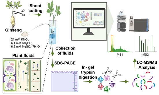
2.1. Plant Materials and Protein Extraction Using Ginseng Fluids and Leaves

The salt-resistant ginseng cultivar “Cheonryang” was obtained from the National Institute of Horticultural and Herbal Science, Rural Development Administration (RDA), Eumseong-gun, South Korea (36° N, 127° E). Five-year-old ginseng plants were grown in a greenhouse at an average temperature of 22.5 ± 2.5 °C and average humidity of 50 ± 10%. Salt stress was induced as described by [13]. For the collection of fluid proteome samples, the cut ends of ginseng shoots were kept in hydroponic solution (5 dS/m of salt concentration, 21 mM KNO3, 6.1 mM KH2PO4, and 6.2 mM MgSO4·7H2O) and the culture solution chosen as the culture fluid and harvested at 0, 24, 72, and 120 h, respectively. Sequentially, ginseng leaves were harvested for validation of the abundance profile of the antioxidant enzyme using Western blot analysis.
For the isolation of the total protein from ginseng leaves, we carried out TCA/Acetone precipitation combined with a methanol/chloroform washing method as followed in previous studies [13]. Moreover, for the extraction of fluid proteins from ginseng cultured fluids, 15 mL of cultured fluids were mixed with an equal volume of tris-saturated phenol (15 mL, pH 7.6) and centrifuged at 12,000× g for 10 min at 4 °C. The phenol phase was transferred to a new centrifuge tube. Then, four volumes of methanol containing 0.1 M ammonium acetate each were added to the phenol phase, mixed thoroughly, and precipitated at −20 °C for 1 h. After centrifugation at 12,000× g for 5 min at 4 °C, the pellet was washed thrice with methanol containing 0.1 M ammonium acetate and twice with ice-cold 80% (v/v) acetone containing 0.07% (v/v) β–mercaptoethanol. Finally, the pellet was suspended in 80% acetone and stored at −20 °C until further analysis.

2.2. Validation of Physiological Changes Using Ginseng Leaves and Culture Fluids and Western Blot

To determine the photosynthetic efficiency of ginseng leaves during exposure to salt-stress conditions, chlorophyll fluorescence imaging was performed using the saturation pulse method using an image fluorometer, as described previously (Handy FluorCam, Photo System Instruments, Drasov, Czech Republic) [24]. The relative changes of H2O2 in the culture fluids were quantified using luminol (Sigma-Aldrich, St. Louis, MO, USA) and horseradish peroxidase (HRP; Thermo Fisher Scientific, Waltham, MA, USA), as previously described [25]. Stock solutions of luminol (3.4 mg/mL) and HRP (2 mg/mL) were prepared. Culture fluids (35 μL) were mixed with luminal (100 μL) and HRP (15 μL) stock solutions and loaded onto a 96-well plate. The luminescence-detection assay was performed over 30 min with a signal integration time of 1 s using the Glomax® multi-detection system (Promega, Madison, WI, USA). For comparison of H2O2 accumulation in the fluid samples, the chemiluminescence intensities detected at 30 min were used.
In addition, for validation of salt-stress-derived changes of antioxidant enzymes in ginseng leaves, a Western blot analysis was carried out using the superoxide dismutase (SOD) antibody. For production of the SOD antibody, a protein sequence was obtained, and the genes for the respective proteins were cloned in a pQE30 expression vector system, as described in a previous study [26]. Subsequently, for antibody generation, the recombinant protein was purified according to the manufacturer’s instruction (Qiagen, Valencia, CA, USA), and polyclonal antibodies were generated by immunizing the rabbits. Finally, the antibody was then purified using the antigen–antibody interaction method as followed by a previous report [26]. For Western blot analysis, ginseng leaves proteins were resolved on SDS-PAGE and transferred to a PVDF membrane. Membrane was incubated with the SOD antibody (1:667) and anti-rabbit IgG conjugated with horseradish peroxidase (1:6667) as used for primary and secondary antibodies, respectively. Finally, the signal was detected using EzWestLumi plus kit (ATTO Corporation, Tokyo, Japan).

2.3. Shotgun Proteomic Analysis and Data Processing

A shotgun proteomic analysis using fluid protein samples was performed as described previously [27,28]. SDS-PAGE analyses of fluid proteins collected from the shoots of five-year-old ginseng in the ddH2O and the hydroponic solution, respectively, were carried out. Fluid proteins extracted from the five-year-old ginseng cultured with the hydroponic solution loaded on SDS-PAGE gels were cut out into five pieces and subjected to in-gel trypsin digestion (trypsin gold, MS grade, Promega, Madison, WI, USA) for LC-MS/MS analysis [29]. LC-MS/MS analysis was performed using a high-performance liquid chromatograph interfaced with a linear trap quadrupole-orbitrap (Thermo Fisher Scientific, Waltham, MA, USA) equipped with a nano-electrospray ionization (nESI) source and fitted with a fused silica emitter tip (New Objective, MA, USA), as described previously [30]. The acquired MS raw data were analyzed using MaxQuant (ver. 1.5.3.30) [31]. All the raw data were cross-referenced against the RNA-sequencing (RNA-seq) database using 14 tissues, 4 differently aged ginseng roots (SRP 066368, 215,879 sequences), and an RNA-seq (PAC-BIO) database developed in-house (SRA: SUB2796783, containing 135,317 sequences) [32,33]. Furthermore, raw data processing was carried out using the default precursor mass tolerance set by the Andromeda search engine, which is set at 20 ppm for the first search and 4.5 ppm for the main search. In addition, a maximum of two missed tryptic cleavages was allowed, and the carbamidomethylation of cysteine residues was specified as a fixed modification. For variable modifications, the acetylation of lysine residues and the oxidation of methionine residues were specified. A reverse-nonsense version of the original database was generated and used to determine the FDR, which was set to 1% for peptide identifications [31]. Raw data obtained from MS were deposited in the ProteomeXchange Consortium via the PRIDE partner repository with the dataset identifier PXD010322 [34]. Functional annotations of the identified proteins were performed using AgriGO v2 web-based software for gene-ontology (GO) enrichment analysis [35] and MapMan for metabolic pathway analysis [36].

2.4. Validation with Quantitative Real-Time PCR (qRT-PCR)

RNAs were extracted from an adventitious root exposed to 150 mM NaCl condition at different time points (0, 24, and 48 h) using an RNA isolation kit (Intron Biotechnology Inc., Seongnam, Korea) according to the manufacturer’s instructions. The total RNA was subjected to reverse transcription using the HelixCript First-Strand cDNA Synthesis Kit (NanoHelix, Korea). qRT-PCR was performed using the RealHelix™ qPCR kit (NanoHelix, Korea) and SYBR Green on a Bio-Rad CFX96™ thermal cycler (Bio-Rad Laboratories Inc., Hercules, CA, USA). The ratios of the transcript levels in the salt-stress-treated adventitious root samples at 0 h to 48 h were compared to the β-actin transcript levels in the corresponding samples. The primer sequences used for the qRT-PCR are listed in Table S1.

3. Results and Discussion

3.1. Salt-Stress-Triggered Accumulation of Reactive Oxygen Species in Ginseng

The osmotic stress caused by high soil salinity leads to the reduction in external water potential and water uptake by plants [37]. Previous studies have shown that osmotic stress, triggered by high salt concentrations, induces the generation of reactive oxygen species (ROS), such as superoxide (O2), hydroxyl radical (OH), H2O2, and singlet oxygen (1O2), which are known as major inhibitors of plant growth [38]. Moreover, the accumulation of ROS during the stress conditions induced the oxidative damage and resulted in the inhibition of various enzymes related to the photosynthetic activities in ginseng [37]. Therefore, the effects of salt stress on photosynthesis and ROS homeostasis were investigated. First, chlorophyll fluorescence imaging was conducted to determine the photosynthetic activity in the ginseng leaves after exposure to salt stress at 0, 24, 72, and 120 h, followed by the saturation pulse method [39]. The wilting and drying of leaf margins in ginseng plant were among the earliest visible symptoms of the salt stress [12,13]. In this result, we successfully confirmed the decrease of the photosynthetic activity by the measurement of chlorophyll fluorescence at the leaf margin at 120 h as compared with the 0, 24, and 72 h samples (chlorophyll fluorescence changed to red color) (Figure 1A). For measurement of the oxidative burst in the ginseng fluids, H2O2 was detected using a luminescence assay. We successfully observed 114-, 281-, and 633-fold increases of H2O2 in the cultured fluid after treatment with salt stress at 24, 72, and 120 h, respectively, with that at 0 h (Figure 1B). Moreover, the Western blot analysis revealed salt-stress-induced abundance of superoxide dismutase (SOD), a salt-stress marker protein in plants [40] (Figure 1C). The effect of salt stress was clearly visible at the time points selected for analysis, and, thus, these samples were used further for the high-throughput proteome analysis of the ginseng fluid proteins.

3.2. Extraction of Fluid Protein from Ginseng and SDS-PAGE Analysis

To analyze the changes in the fluid proteome in response to salt stress, the five-year-old salt-tolerant ginseng “Cheonryang” was used. The major bottle-necks in the extraction of fluid proteins include the difficulty in determining the harvesting time and extraction steps [41,42]. Previous studies showed that xylem sap proteins are constantly produced over a 28 h period in soybeans regardless of stress conditions; moreover, various stress resulted in variations of the xylem composition such as nitrate concentration and pH in Zea mays [43]. In addition, a few studies demonstrated that the successful extraction of sap from a few plants by the cut end of the petiole or stem; however, the major disadvantage of those methods is that they work only on several selected cultivars [44]. On the other hand, Tetyuk et al. 2013 reported a sap-protein-extraction method using EDTA and distilled water that allowed the exudation of sap proteins from the cut ends of the plants [44]. Interestingly, this simple method for the isolation of sap proteins was applicable to many herbaceous and woody plant species and can extract large amounts of sap proteins for further downstream analysis. In this method, the cut plants were first immersed in EDTA for 1 h to prevent the sealing of phloem followed by transferring the plants into water for the enrichment of sap proteins [44]. Following this method, in this study, we used distilled water for the enrichment of fluid proteins, including sap and apoplastic proteins, during salt-stress condition, in which the cut ends of the ginseng shoots were immersed in a salt solution over 0, 24, 72, and 120 h, and the secreted fluid proteins were collected. A SDS-PAGE analysis was carried out for validating the extraction efficiency of the used method, which revealed a clear separation of fluid proteins after treatment with salt stress at 72 and 120 h. However, fluid proteins were not observed in the 0 and 24 h samples, probably because of their low amounts. The higher amounts of fluid proteins observed at 72 h and 120 h could be because of the salt-stress-induced accumulation of ROS that led to the membrane peroxidation and allowed the higher secretion of proteins from the cut ends (Figure S1). In contrast, the proteins from the ginseng shoot cultured with ddH2O did not show the secretion of enough amount of fluid proteins for SDS-PAGE during 0 to 120 h, indicating that some of salt-responsive proteins were specifically secreted in ginseng when harvested under high salt conditions (Figure 2).

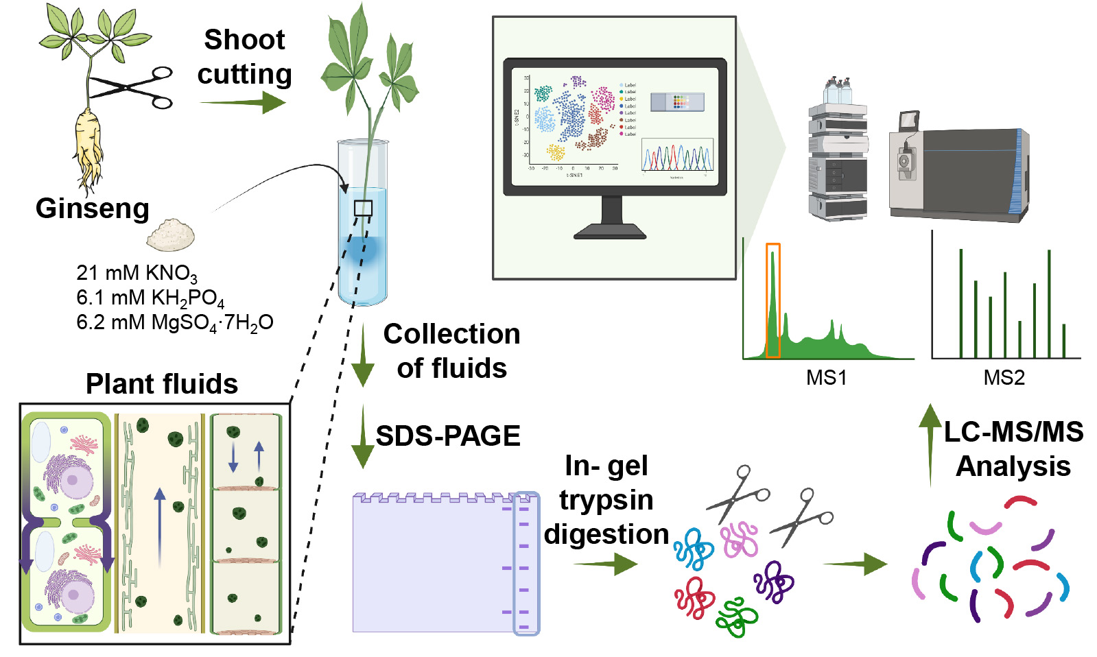
3.3. Application of Shotgun Proteomics for Identification of Salt-Responsive Proteins

The 120 h salt lane of the SDS-PAGE gel was directly subjected to in-gel trypsin digestion and LC-MS/MS analysis for the identification of salt-responsive proteins in ginseng. A total 216 protein were identified, of which 7 proteins were commonly available in both databases. Moreover, 158 and 96 proteins were specifically identified in the RNA-seq and PAC-BIO database, respectively (Tables S2 and S3). Further functional characterization of identified proteins, carried out using the GO enrichment analysis, revealed that the proteins with catalytic activity (46%), structural molecule activity (16%), and ATPase-coupled ion transmembrane transporter activity (3%) were dominant in the molecular function category (Figure 3A and Table S4). Moreover, response to stimulus (42%) and nitrogen-compound metabolic process (26 %) were the most prominent terms in the biological process category (Figure 3B and Table S4). Responses to stimulus were divided into response to stress (33%), response to abiotic stimulus (27%), response to heat (9%), response to oxidative stress (7%), and response to cold (11%), (Table S4). In addition, 20 proteins were involved in the catabolic process (11%), 12 proteins involved in protein folding (7%), and 11 proteins involved in ribonucleoprotein complex biogenesis (6%) (Figure 3B).
Further MapMan functional characterization revealed that proteins related to glycolysis (11 proteins), lipid metabolism (7 proteins), and amino-acid metabolism (6 proteins) were mainly annotated in the metabolism category (Figure 4). Moreover, in the cell function category, 23, 19, 13, and 7 proteins were related to protein synthesis, biotic and abiotic stress, cell, and redox, respectively (Figure 4).

3.4. Inhibition of Photosynthesis by Salt Stress in Ginseng

Photosynthesis is an important physiological process that is highly affected by changes in environmental conditions [45]. A salt-stress-induced reduction in photosynthetic efficiency is a well-known phenomenon [46,47]; thus, in this study, multiple proteins associated with photosynthesis were identified. For instance, ribulose-1,5-bisphosphate carboxylase-oxygenase (RuBisCO) and RuBisCO activase (RCA) were observed in the culture fluid in response to salt stress (Figure 5, Tables S2 and S3). RuBisCO is a key enzyme of the Calvin cycle involved in the fixation of carbon dioxide (CO2), while RCA is a member of the ATPases associated with the diverse cellular activity protein family, which has various chaperone-like functions. The major function of RCA is to remove inhibitory sugars from the active site of RuBisCO, and the decreased abundance of these two proteins highlights the decreased rate of photosynthesis under salt-stress conditions in ginseng [48]. A previous study showed that salinity stress leads to an oxidative burst, which subsequently leads to the degradation of the small subunits of RuBisCO and RCA [49]. Fructose bisphosphate aldolose (FBPA) is an enzyme associated with glycolysis, gluconeogenesis, and the Calvin cycle; its decreased abundance in response to salt stress has already been reported in various plants including wheat [50]. The secretion of these three photosynthesis-related proteins in the culture fluid of ginseng was predicted to be an outcome of the inhibition of carbon fixation due to salt stress. Furthermore, we identified various other photosynthesis-related proteins in the culture fluid, including oxygen-evolving enhancer (OEE), PS II oxygen-evolving complex 1 (PsbO), PS II subunit P (PsbP), PS I subunit D (PsaD), chlorophyll a-b binding protein (CAB), and manganese-stabilizing protein (MSP) (Tables S2 and S3). MSP is an element comprising the photosystem in vascular plants and playing a role in maintaining the structural stability of photosystem II (PSII) [51]. Similar to MSP, the specific role of PsbO in maintaining PSII stability and plant growth was reported previously [52]. In addition, the PSII oxygen-evolving complex (OEC), which consists of OEE1 and 2 proteins, and PsaD, which is a small extrinsic polypeptide of the PSI reaction center complex, were reported previously [53]. Several reports have shown an increased abundance of OEE proteins during salt-stress conditions in plants; moreover, these proteins play a role in repairing the damage caused by dissociation and maintaining a putative function of OEC in plants [54]. In parallel with the results obtained by chlorophyll fluorescence imaging analysis, these results clearly showed an inhibition of photosynthesis in ginseng plants in response to salt stress.

3.5. Accumulation of Energy Metabolism Related Proteins during Salt Stress

In this study, we identified nine proteins associated with energy metabolism (glycolysis) in ginseng, including phosphoglucomutase, FBPA, triosephosphate isomerase, glyceraldehyde 3-phosphate dehydrogenase (GAPDH), phosphoglycerate kinase, enolase, ATP synthase subunit, alcohol dehydrogenase, and malate dehydrogenase in the culture fluid of 5Y-120H ginseng shoot (Figure 5). In plants, the glycolytic enzymes are involved in the production energy and intermediates of various sugars required for the biosynthesis of other metabolites, such as amino acids, many of which function as compatible solutes that participate in salt-stress tolerance [55,56]. A previous study reported an increased abundance of glycolytic enzymes in rice and Aeluropus lagopoides, while a decrease in the abundance of enzymes was observed in soybeans during salt stress [57,58,59]. In addition, glyoxalase, which functions in the detoxification of methylglyoxal was identified in the culture fluids from ginseng shoot. The accumulation of methylglyoxal, a byproduct of triosephosphate in glycolysis, triggers glyoxalase activation in plants as a tolerance response to stress conditions [60]. Thus, the accumulation of energy-metabolism-related proteins, especially glycolysis, plays important roles in energy production and osmotic balance and may have a positive correlation with salt-stress tolerance in ginseng.

3.6. Activation of ROS Scavenging Enzymes by Oxidative Stress

Stomatal closure triggered by salt stress inhibits the CO2 fixation and induces the oxidative stress by generating ROS [61]. In this study, we successfully identified various ROS-scavenging enzymes, such as Cu/Zn SOD, Mg SOD, Glutathione peroxidase (GPX), peroxidase, and catalase (CAT) 1, 2, and 3 in the culture fluids from the ginseng shoot (Figure 5, Tables S2 and S3). Antioxidant enzymes such as SODs, CATs, and peroxidases are important components of ROS scavenging in plant systems [62]. The SODs involved in the glutathione-ascorbate cycle protect the plant cells by converting O⁻₂ into H2O2 and/or O2 [63]. During this cycle, the produced H2O2 is scavenged by CATs through the decomposition of H2O2 into water and molecular oxygen, while the peroxidases oxidize the phenolic compounds and antioxidants to decompose H2O2 in plants [64]. The GPX pathway is a major enzymatic defense mechanism against oxidative membrane damage that uses reduced glutathione or other reducing equivalents to convert H2O2 to the corresponding ∙OH compounds [65]. Moreover, the up-regulation of various antioxidant enzymes such as peroxidases, Mg SOD, and Cu/Zn SOD enzymes in salt-tolerant crops was reported in a previous report [66], and the accumulation of these enzymes in ginseng suggests activation of the tolerance response against salt-stress-induced oxidative stress.

3.7. Identification of Proteins Related to Various Metabolism

In this study, various chaperones were also identified (Figure 5). The function of heat-shock proteins (Hsps), such as Hsp17.6 II, Hsp70, Hsp81-2, and Hsp90.1, and the TCP-1/cpn60 chaperonin is required for protein folding, assembly, translocation, and degradation, and their role in the stress responses are well established in plants [67]. Hsp70 functions in cooperation with Hsp100 and caseinolytic proteases ClpB or ClpB3 for the removal or repair of misfolded proteins, which has been extensively studied in Arabidopsis, previously [68,69]. In particular, previous studies have shown that Hsp70 is up-regulated to activate defense mechanisms against various stress conditions and the overexpression of this protein elevated tolerance in plants during salt stress [70]. Therefore, the secretion of these chaperones in ginseng fluid suggests a key role for these proteins in the refolding of salt-stress-damaged proteins in ginseng.
Moreover, several proteins related to protein metabolism, defense and stress response, and cell composition ,including glycosyl hydrolase 17 (GH17, β-1,3-Glucanases), peptidyl-prolyl cis-trans isomerase (PPIase), actin, and tubulin, were also identified in this study (Figure 5). Among these, the physiological roles of the GH family proteins have been studied in plants using various molecular genetics approaches [71]. These proteins are involved in the biosynthesis and remodulation of glycans, mobilization of storage reserves, and defense response, among others [71,72]. Particularly, GH17 belongs to pathogenesis-related protein-2 (PR-2) and catalyzes the endo-type hydrolytic cleavage of the 1,3-β-D-glucosidic linkages in β-1,3-glucans. Regulation of this protein, both at the expression and activity levels, has been reported in response to viral and bacterial pathogen infections [71]. In addition, some salt-responsive GH family proteins, especially GH1, have been reported in Arabidopsis [73]. Here, we successfully identified GH17 proteins in ginseng fluids; however, the distinct function of GH17 proteins during salt stress in ginseng is largely elusive. PPIase catalyzes the cis–trans isomerization of proline imidic peptide bonds in oligopeptides to accelerate protein folding [74]. Moreover, the diverse functions of PPIase have been implicated in photosynthesis [75], growth and development [76], redox homeostasis [77], and stress responses [78,79]. Similar to GH family proteins, the overexpression of the PPIase gene in Arabidopsis resulted in the tolerance of various abiotic stresses such as salt, drought, and heat, while this protein promoted plant growth under normal conditions [80].
In addition, the proteins related to cytoskeletal components, such as actin and tubulin, were also found to accumulate in the culture fluid (Figure 5). The dynamic function of cytoskeleton networks is important for cell division, cell movement, cell morphogenesis, and cell signal transduction in plant [81]. Moreover, the depolymerization and reorganization of the cytoskeleton, such as active filaments and microtubules, were reported to play a pivotal role in salt-stress tolerance in Arabidopsis [81,82], and the re-organization of the cells’ structural components may play an important role in salt-stress tolerance in ginseng.

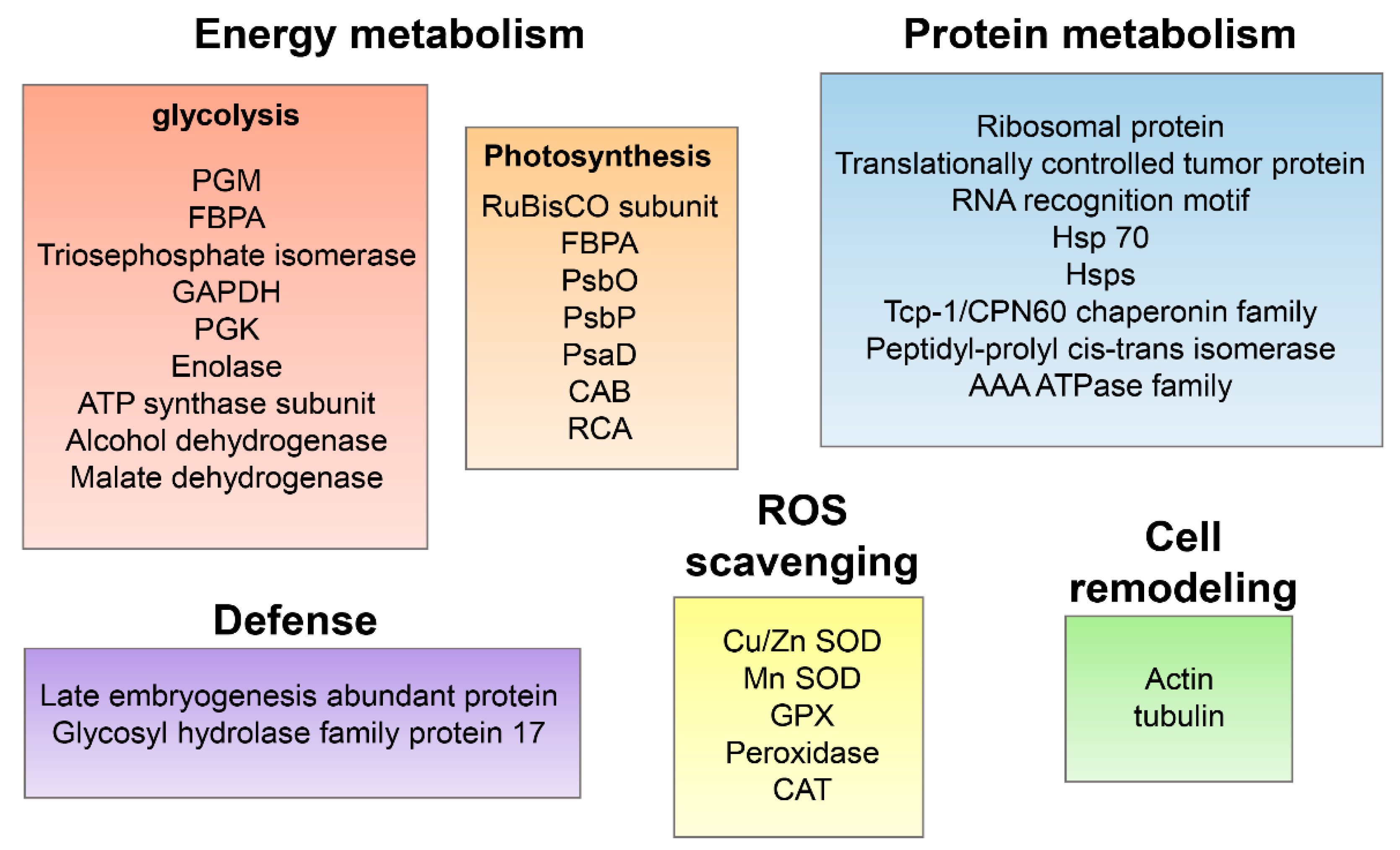
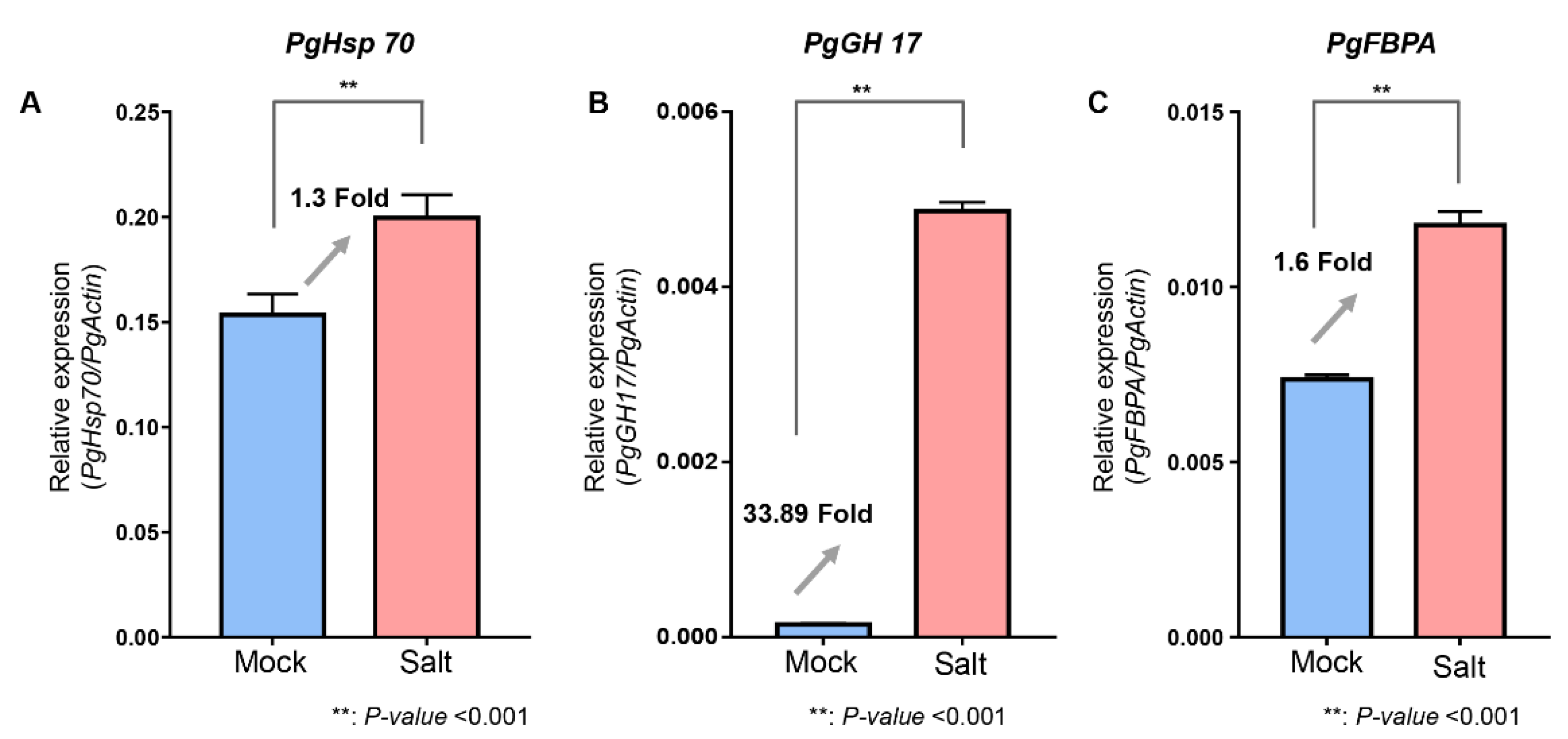
3.8. Validation of Identified Proteins Using qRT-PCR Analysis

To validate the expression profile of the identified proteins during salt stress, we performed a qRT-PCR analysis using RNA isolated from salt-treated ginseng root. A total of three proteins including Hsp70, GH17, and FBPA were selected from the fluid proteome analysis to determine their expression at the transcript level (Figure 6). In parallel with their corresponding proteins, 1.3-, 33.89-, and 1.6-fold increases were observed in Hsp70, GH17, and FBPA, respectively, against salt stress in ginseng roots (Figure 6). However, further investigation is required to determine their role in salt-stress tolerance in ginseng.

4. Conclusions

Salt stress is a major environmental stress that affects plant growth and causes severe damage to ginseng plants. Recently, a few studies have investigated the effects of salt stress to identify the key proteins responsible for salt-stress tolerance in plants; however, few studies have been conducted to define the salt-responsive mechanism in ginseng. Therefore, to expand our understanding of the salt-stress responses and investigate the salt-responsive proteins in ginseng, we employed a shotgun proteomic approach using fluid proteins collected from the cut ends of five-year-old ginseng shoots. This approach resulted in the identification of various photosynthesis-related enzymes with decreased abundance, while the accumulation of various other proteins associated with ROS scavenging, energy metabolism, and protein metabolism was also observed in the culture fluids of ginseng. Moreover, the expression levels of three proteins Hsp70, GH17, and FBPA were also confirmed to show parallel expression profiles with corresponding proteins in ginseng roots. Overall, this study not only provides a simple method for extraction of fluid proteins, but also identified potential salt-responsive proteins in ginseng. Moreover, this study will be helpful for the further understanding of the molecular mechanism of salt-stress tolerance and improving the salt tolerance of salt-sensitive ginseng cultivars by next-generation breeding programs.

Supplementary Materials

The following supporting information can be downloaded at: https://www.mdpi.com/article/10.3390/agronomy12092048/s1, Figure S1: SDS-PAGE showing profile of secreted fluid protein in each sample. Five-year-old (5Y)-120 h sample showing clear separation of fluid protein, and this line further applied shotgun proteomic analysis for identification of proteins. Table S1: List of primers used in this study, Table S2: List of identified proteins by RNA-seq database, Table S3: List of identified proteins by PAC-BIO database. Table S4: Gene ontology enrichment analysis of identified proteins

Author Contributions

Conceptualization, S.W.K. and I.-H.J.; Data curation, J.-Y.J.; Funding acquisition, S.T.K.; Investigation, J.-Y.J.; Methodology, J.-Y.J., S.W.K., W.J. and K.-H.B.; Validation, J.-Y.J.; Writing—original draft, J.-Y.J. and C.W.M.; Writing—review and editing, C.W.M., R.G. and Y.-J.K. All authors have read and agreed to the published version of the manuscript.

Funding

This research was funded by the Cooperative Research Program for Agriculture Science and Technology, Rural Development Administration, Republic of Korea, grant number PJ01492201.

Institutional Review Board Statement

Not applicable.

Informed Consent Statement

Not applicable.

Data Availability Statement

The mass spectrometry proteomics data ertr deposited to the ProteomeXchange Consortium via the PRIDE partner repository with the dataset identifier PXD010322 [33].

Conflicts of Interest

The authors declare no conflict of interest.

References

  1. Kumar, H.; Song, S.-Y.; More, S.V.; Kang, S.-M.; Kim, B.-W.; Kim, I.-S.; Choi, D.-K. Traditional korean east asian medicines and herbal formulations for cognitive impairment. Molecules 2013, 18, 14670–14693. [Google Scholar] [CrossRef] [PubMed]
  2. Sofowora, A.; Ogunbodede, E.; Onayade, A. The role and place of medicinal plants in the strategies for disease prevention. Afr. J. Tradit. Complement. Altern. Med. 2013, 10, 210–229. [Google Scholar] [CrossRef] [PubMed]
  3. Ratan, Z.A.; Haidere, M.F.; Hong, Y.H.; Park, S.H.; Lee, J.-O.; Lee, J.; Cho, J.Y. Pharmacological potential of ginseng and its major component ginsenosides. J. Ginseng Res. 2021, 45, 199–210. [Google Scholar] [CrossRef]
  4. Lee, C.H.; Kim, J.-H. A review on the medicinal potentials of ginseng and ginsenosides on cardiovascular siseases. J. Ginseng Res. 2014, 38, 161–166. [Google Scholar] [CrossRef]
  5. Cho, D.-H.; Park, K.-J.; Yu, Y.-H.; Oh, S.-H.; Lee, H.-S. Root-rot development of 2-year old ginseng (Panax ginseng C. A. Meyer) caused by Cylindrocarpon destructans (zinssm.) scholten in the continuous cultivation field. J. Ginseng Res. 1995, 19, 175–180. [Google Scholar]
  6. Kim, Y.-S.; Park, C.-S.; Lee, D.-Y.; Lee, J.-S.; Lee, S.-H.; In, J.-G.; Hong, T.-K. Phenological growth stages of korean ginseng (Panax ginseng) according to the extended BBCH scale. J. Ginseng Res. 2021, 45, 527–534. [Google Scholar] [CrossRef] [PubMed]
  7. Lee, O.; Sathiyaraj, G.; Kim, Y.-J.; In, J.-G.; Kwon, W.-S.; Kim, J.-H.; Yang, D.-C. Defense genes induced by pathogens and abiotic stresses in Panax ginseng C. A. Meyer. J. Ginseng Res. 2011, 35, 1–11. [Google Scholar] [CrossRef]
  8. Isayenkov, S.V.; Maathuis, F.J.M. Plant salinity stress: Many unanswered questions remain. Front. Plant Sci. 2019, 10, 80. [Google Scholar] [CrossRef]
  9. Onyekachi, O.G.; Boniface, O.O.; Gemlack, N.F.; Nicholas, N. The effect of climate change on abiotic plant stress: A review. In Biotic and Abiotic Stresses in Plants; Oliverira, A.D., Ed.; IntechOpen: London, UK, 2019; p. 17. [Google Scholar] [CrossRef]
  10. Howard, R.J.; Mendelssohn, I. A salinity as a constraint on growth of oligohaline marsh macrophytes species variation in stress tolerance. Am. J. Bot. 1999, 86, 785–794. [Google Scholar] [CrossRef]
  11. Riyazuddin, R.; Verma, R.; Singh, K.; Nisha, N.; Keisham, M.; Bhati, K.K.; Kim, S.T.; Gupta, R. Ethylene: A master regulator of salinity stress tolerance in plants. Biomolules 2020, 10, 959. [Google Scholar] [CrossRef]
  12. Kim, S.T.; Bae, D.-W.; Lee, K.; Hwang, J.E.; Bang, K.-H.; Kim, Y.-C.; Kim, O.-T.; Yoo, N.H.; Kang, K.Y.; Hyun, D.Y.; et al. Proteomic analysis of korean ginseng (Panax ginseng C. A. Meyer) following exposure to salt stress. J. Plant Biotechnol. 2008, 35, 185–193. [Google Scholar] [CrossRef]
  13. Kim, S.W.; Min, C.W.; Gupta, R.; Jo, I.H.; Bang, K.H.; Kim, Y.-C.; Kim, K.-H.; Kim, S.T. Proteomics analysis of early salt-responsive proteins in ginseng (Panax Ginseng C. A. Meyer) leaves. Korean J. Med. Crop Sci. 2014, 22, 398–404. [Google Scholar] [CrossRef]
  14. Rodríguez-Celma, J.; Ceballos-Laita, L.; Grusak, M.A.; Abadía, J.; López-Millán, A.-F. Plant fluid proteomics: Delving into the xylem sap, phloem sap and apoplastic fluid proteomes. Biochim. Biophys. Acta-Proteins Proteomics 2016, 1864, 991–1002. [Google Scholar] [CrossRef] [PubMed]
  15. Carella, P.; Wilson, D.C.; Kempthorne, C.J.; Cameron, R.K. Vascular sap proteomics: Providing insight into long-distance signaling during stress. Front. Plant Sci. 2016, 7, 651. [Google Scholar] [CrossRef] [PubMed]
  16. Sattelmacher, B.; Mühling, K.H.; Pennewiß, K. The apoplast-Its significance for the nutrition of higher plants. J. Plant Nutr. Soil. Sci. 1998, 161, 485–498. [Google Scholar] [CrossRef]
  17. Jorrin-Novo, J.V. Plant proteomics methods and protocols. In Plant Proteomics: Methods and Protocols, 2nd ed.; Jorrin-Novo, J.V., Komatsu, S., Weckwerth, W., Wienkoop, S., Eds.; Human Press: Totowa, NJ, USA, 2014; Volume 1072, pp. 3–13. [Google Scholar] [CrossRef]
  18. Rep, M.; Dekker, H.L.; Vossen, J.H.; de Boer, A.D.; Houterman, P.M.; Speijer, D.; Back, J.W.; de Koster, C.G.; Cornelissen, B.J.C. Mass spectrometric identification of isoforms of PR proteins in xylem sap of fungus-infected tomato. Plant Physiol. 2002, 130, 904–917. [Google Scholar] [CrossRef]
  19. Gawehns, F.; Ma, L.; Bruning, O.; Houterman, P.M.; Boeren, S.; Cornelissen, B.J.C.; Rep, M.; Takken, F.L.W. The effector repertoire of Fusarium oxysporum determines the tomato xylem proteome composition following infection. Front. Plant Sci. 2015, 6, 967. [Google Scholar] [CrossRef]
  20. Pu, Z.; Ino, Y.; Kimura, Y.; Tago, A.; Shimizu, M.; Natsume, S.; Sano, Y.; Fujimoto, R.; Kaneko, K.; Shea, D.J.; et al. Changes in the proteome of xylem sap in Brassica oleracea in response to Fusarium oxysporum stress. Front. Plant Sci. 2016, 7, 31. [Google Scholar] [CrossRef]
  21. Abeysekara, N.S.; Bhattacharyya, M.K. Analyses of the xylem sap proteomes identified candidate Fusarium virguliforme proteinacious toxins. PLoS ONE 2014, 9, e93667. [Google Scholar] [CrossRef] [Green Version]
  22. Figueiredo, J.; Cavaco, A.R.; Guerra-Guimarães, L.; Leclercq, C.; Renaut, J.; Cunha, J.; Eiras-Dias, J.; Cordeiro, C.; Matos, A.R.; Sousa Silva, M.; et al. An apoplastic fluid extraction method for the characterization of grapevine leaves proteome and metabolome from a single sample. Physiol. Plant. 2021, 171, 343–357. [Google Scholar] [CrossRef]
  23. Fernandez-Garcia, N.; Hernandez, M.; Casado-Vela, J.; Bru, R.; Elortza, F.; Hedden, P.; Olmos, E. Changes to the proteome and targeted metabolites of xylem sap in Brassica oleracea in response to salt stress. Plant. Cell Environ. 2011, 34, 821–836. [Google Scholar] [CrossRef]
  24. Nedbal, L.; Soukupová, J.; Kaftan, D.; Whitmarsh, J.; Trtílek, M. Kinetic imaging of chlorophyll fluorescence using modulated light. Photosynth. Res. 2000, 66, 3–12. [Google Scholar] [CrossRef]
  25. Meng, Q.; Gupta, R.; Min, C.W.; Kim, J.; Kramer, K.; Wang, Y.; Park, S.R.; Finkemeier, I.; Kim, S.T. A proteomic insight into the MSP1 and flg22 induced signaling in Oryza sativa leaves. J. Proteom. 2019, 196, 120–130. [Google Scholar] [CrossRef]
  26. Kim, S.T.; Wang, Y.; Kang, S.Y.; Kim, S.G.; Rakwal, R.; Kim, Y.C.; Kang, K.Y. Developing rice embryo proteomics reveals essential role for embryonic proteins in regulation of seed germination. J. Proteome Res. 2009, 8, 3598–3605. [Google Scholar] [CrossRef]
  27. Kim, Y.J.; Wang, Y.; Gupta, R.; Kim, S.W.; Min, C.W.; Kim, Y.C.; Park, K.H.; Agrawal, G.K.; Rakwal, R.; Choung, M.G.; et al. Protamine sulfate precipitation method depletes abundant plant seed-storage proteins: A case study on legume plants. Proteomics 2015, 15, 1760–1764. [Google Scholar] [CrossRef]
  28. Gupta, R.; Lee, S.J.; Min, C.W.; Kim, S.W.; Park, K.-H.; Bae, D.-W.; Lee, B.W.; Agrawal, G.K.; Rakwal, R.; Kim, S.T. Coupling of gel-based 2-DE and 1-DE shotgun proteomics approaches to dig deep into the leaf senescence proteome of Glycine max. J. Proteom. 2016, 148, 65–74. [Google Scholar] [CrossRef]
  29. Gupta, R.; Min, C.W.; Kim, S.W.; Wang, Y.; Agrawal, G.K.; Rakwal, R.; Kim, S.G.; Lee, B.W.; Ko, J.M.; Baek, I.Y.; et al. Comparative investigation of seed coats of brown- versus yellow-colored soybean seeds using an integrated proteomics and metabolomics approach. Proteomics 2015, 15, 1706–1716. [Google Scholar] [CrossRef]
  30. Kim, S.W.; Gupta, R.; Lee, S.H.; Min, C.W.; Agrawal, G.K.; Rakwal, R.; Kim, J.B.; Jo, I.H.; Park, S.-Y.; Kim, J.K.; et al. An integrated biochemical, proteomics, and metabolomics approach for supporting medicinal value of Panax ginseng fruits. Front. Plant Sci. 2016, 7, 994. [Google Scholar] [CrossRef]
  31. Tyanova, S.; Temu, T.; Cox, J. The MaxQuant computational platform for mass spectrometry-based shotgun proteomics. Nat. Protoc. 2016, 11, 2301–2319. [Google Scholar] [CrossRef]
  32. Jo, I.H.; Lee, J.; Hong, C.E.; Lee, D.J.; Bae, W.; Park, S.G.; Ahn, Y.J.; Kim, Y.C.; Kim, J.U.; Lee, J.W.; et al. Isoform sequencing provides a more comprehensive view of the Panax ginseng transcriptome. Genes 2017, 8, 228. [Google Scholar] [CrossRef]
  33. Wang, K.; Jiang, S.; Sun, C.; Lin, Y.; Yin, R.; Wang, Y.; Zhang, M. The spatial and temporal transcriptomic landscapes of ginseng, Panax ginseng C. A. Meyer. Sci. Rep. 2016, 5, 18283. [Google Scholar] [CrossRef]
  34. Vizcaíno, J.A.; Csordas, A.; Noemi, D.-T.; Dianes, J.A.; Griss, J.; Lavidas, I.; Mayer, G.; Yasset, P.-R.; Reisinger, F.; Ternent, T. 2016 Update of the PRIDE database and its related tools. Nucleic Acids Res. 2016, 44, D447–D456. [Google Scholar] [CrossRef]
  35. Tian, T.; Liu, Y.; Yan, H.; You, Q.; Yi, X.; Du, Z.; Xu, W.; Su, Z. AgriGO v2.0: A GO analysis toolkit for the agricultural community, 2017 Update. Nucleic Acids Res. 2017, 45, W122–W129. [Google Scholar] [CrossRef]
  36. Usadel, B.; Poree, F.; Nagel, A.; Lohse, M.; Czedik-Eysenberg, A.; Stitt, M. A guide to using MapMan to visualize and compare omics data in plants: A case study in the crop species, maize. Plant Cell Environ. 2009, 32, 1211–1229. [Google Scholar] [CrossRef]
  37. Chinnusamy, V.; Zhu, J.; Zhu, J.-K. Salt stress signaling and mechanisms of plant salt tolerance. In Genetic Engineering: Principles and Methods; Setlow, J.K., Ed.; Springer Nature: Berlin, Germany, 2006; pp. 141–177. [Google Scholar] [CrossRef]
  38. Lee, D.H.; Kim, Y.S.; Lee, C.B. The inductive responses of the antioxidant enzymes by salt stress in the rice (Oryza Sativa L.). J. Plant Physiol. 2001, 158, 737–745. [Google Scholar] [CrossRef]
  39. Quick, W.P.; Horton, P.; Walker, D.A. Studies on the induction of chlorophyll fluorescence in barley protoplasts. I. factors affecting the observation of oscillations in the yield of chlorophyll fluorescence and the rate of oxygen evolution. Proc. R. Soc. Lond. Ser. B. Biol. Sci. 1984, 220, 361–370. [Google Scholar] [CrossRef]
  40. Berwal, M.K.; Ram, C. Superoxide dismutase: A stable biochemical marker for abiotic stress tolerance in higher plants. In Biotic and Abiotic Stresses in Plants; Oliverira, A.D., Ed.; IntechOpen: London, UK, 2019. [Google Scholar] [CrossRef]
  41. Ligat, L.; Lauber, E.; Albenne, C.; Clemente, H.S.; Valot, B.; Zivy, M.; Pont-Lezica, R.; Arlat, M.; Jamet, E. Analysis of the xylem sap proteome of Brassica oleracea reveals a high content in secreted proteins. Proteomics 2011, 11, 1798–1813. [Google Scholar] [CrossRef] [Green Version]
  42. Djordjevic, M.A.; Oakes, M.; Li, D.X.; Hwang, C.H.; Hocart, C.H.; Gresshoff, P.M. The Glycine max xylem sap and apoplast proteome. J. Proteome Res. 2007, 6, 3771–3779. [Google Scholar] [CrossRef]
  43. Bahrun, A.; Jensen, C.R.; Asch, F.; Mogensen, V.O. Drought-induced changes in xylem pH, ionic composition, and ABA concentration act as early signals in field-grown maize (Zea mays L.). J. Exp. Bot. 2002, 53, 251–263. [Google Scholar] [CrossRef]
  44. Tetyuk, O.; Benning, U.F.; Hoffmann-Benning, S. Collection and analysis of arabidopsis phloem exudates using the EDTA-facilitated method. JoVE 2013, 80, e51111. [Google Scholar] [CrossRef]
  45. Tanaka, A.; Makino, A. Photosynthetic research in plant science. Plant Cell Physiol. 2009, 50, 681–683. [Google Scholar] [CrossRef]
  46. Sobhanian, H.; Aghaei, K.; Komatsu, S. Changes in the plant proteome resulting from salt stress: Toward the creation of salt-tolerant crops? J. Proteom. 2011, 74, 1323–1337. [Google Scholar] [CrossRef] [PubMed]
  47. Helena, H.; František, H.; Jaroslava, M.; Kamil, K. Effects of salt stress on water status, photosynthesis and chlorophyll fluorescence of rocket. Plant Soil Environ. 2017, 63, 362–367. [Google Scholar] [CrossRef]
  48. Xiong, J.; Sun, Y.; Yang, Q.; Tian, H.; Zhang, H.; Liu, Y.; Chen, M. Proteomic analysis of early salt stress responsive proteins in alfalfa roots and shoots. Proteome Sci. 2017, 15, 19. [Google Scholar] [CrossRef] [PubMed]
  49. Meng, F.; Luo, Q.; Wang, Q.; Zhang, X.; Qi, Z.; Xu, F.; Lei, X.; Cao, Y.; Chow, W.S.; Sun, G. Physiological and proteomic responses to salt stress in chloroplasts of diploid and tetraploid black locust (Robinia pseudoacacia L.). Sci. Rep. 2016, 6, 23098. [Google Scholar] [CrossRef]
  50. Lu, W.; Tang, X.; Huo, Y.; Xu, R.; Qi, S.; Huang, J.; Zheng, C.; Wu, C. Identification and characterization of fructose 1,6-bisphosphate aldolase genes in arabidopsis reveal a gene family with diverse responses to abiotic stresses. Gene 2012, 503, 65–74. [Google Scholar] [CrossRef]
  51. Lydakis-Simantiris, N.; Hutchison, R.S.; Betts, S.D.; Barry, B.A.; Yocum, C.F. Manganese stabilizing protein of photosystem II is a thermostable, natively unfolded polypeptide. Biochemistry 1999, 38, 404–414. [Google Scholar] [CrossRef]
  52. Lundin, B.; Hansson, M.; Schoefs, B.; Vener, A.V.; Spetea, C. The arabidopsis PsbO2 protein regulates dephosphorylation and turnover of the photosystem ii reaction centre D1 protein. Plant J. 2007, 49, 528–539. [Google Scholar] [CrossRef]
  53. Ettinger, W.F.; Theg, S.M. Physiologically active chloroplasts contain pools of unassembled extrinsic proteins of the photosynthetic oxygen-evolving enzyme complex in the thylakoid lumen. J. Cell Biol. 1991, 115, 321–328. [Google Scholar] [CrossRef]
  54. Razavizadeh, R.; Ehsanpour, A.A.; Ahsan, N.; Komatsu, S. Proteome analysis of tobacco leaves under salt stress. Peptides 2009, 30, 1651–1659. [Google Scholar] [CrossRef]
  55. Givan, C.V. Evolving concepts in plant glycolysis: Two centuries of progress. Biol. Rev. 1999, 74, 277–309. [Google Scholar] [CrossRef]
  56. Li, C.; Sun, J.; Qi, X.; Liu, L. NaCl stress impact on the key enzymes in glycolysis from Lactobacillus bulgaricus during freeze-drying. Braz. J. Microbiol. 2015, 46, 1193–1199. [Google Scholar] [CrossRef] [PubMed]
  57. Sobhanian, H.; Motamed, N.; Jazii, F.R.; Nakamura, T.; Komatsu, S. Salt stress induced differential proteome and metabolome response in the shoots of Aeluropus lagopoides (poaceae), a halophyte C4 plant. J. Proteome Res. 2010, 9, 2882–2897. [Google Scholar] [CrossRef] [PubMed]
  58. Chitteti, B.R.; Peng, Z. Proteome and phosphoproteome differential expression under salinity stress in rice (Oryza Sativa) Roots. J. Proteome Res. 2007, 6, 1718–1727. [Google Scholar] [CrossRef]
  59. Sobhanian, H.; Razavizadeh, R.; Nanjo, Y.; Ehsanpour, A.; Jazii, F.; Motamed, N.; Komatsu, S. Proteome analysis of soybean leaves, hypocotyls and roots under salt stress. Proteome Sci. 2010, 8, 19. [Google Scholar] [CrossRef]
  60. Kaur, C.; Singla-Pareek, S.L.; Sopory, S.K. Glyoxalase and methylglyoxal as biomarkers for plant stress tolerance. Crit. Rev. Plant Sci. 2014, 33, 429–456. [Google Scholar] [CrossRef]
  61. Gossett, D.R.; Milhollon, E.P.; Lucas, M.C. Antioxidant response to NaCl stress in salt-tolerant and salt sensitive cultivars of cotton. Crop Sci. 1994, 34, 706–714. [Google Scholar] [CrossRef]
  62. Das, K.; Roychoudhury, A. Reactive oxygen species (ROS) and response of antioxidants as ros-scavengers during environmental stress in plants. Front. Environ. Sci. 2014, 2, 53. [Google Scholar] [CrossRef]
  63. Hasanuzzaman, M.; Bhuyan, M.H.M.B.; Anee, T.I.; Parvin, K.; Nahar, K.; Mahmud, J.A.; Fujita, M. Regulation of ascorbate-glutathione pathway in mitigating oxidative damage in plants under abiotic stress. Antioxidants 2019, 8, 384. [Google Scholar] [CrossRef]
  64. Smirnoff, N.; Arnaud, D. Hydrogen peroxide metabolism and functions in plants. New Phytol. 2019, 221, 1197–1214. [Google Scholar] [CrossRef]
  65. Yoshimura, K.; Miyao, K.; Gaber, A.; Takeda, T.; Kanaboshi, H.; Miyasaka, H.; Shigeoka, S. Enhancement of stress tolerance in transgenic tobacco plants overexpressing chlamydomonas glutathione peroxidase in chloroplasts or cytosol. Plant J. 2004, 37, 21–33. [Google Scholar] [CrossRef]
  66. Meloni, D.A.; Oliva, M.A.; Martinez, C.A.; Cambraia, J. Photosynthesis and activity of superoxide dismutase, peroxidase and glutathione reductase in cotton under salt stress. Environ. Exp. Bot. 2003, 49, 69–76. [Google Scholar] [CrossRef]
  67. Park, C.-J.; Seo, Y.-S. Heat shock proteins: A review of the molecular chaperones for plant immunity. plant Pathol. J. 2015, 31, 323–333. [Google Scholar] [CrossRef] [PubMed]
  68. Mogk, A.; Kummer, E.; Bukau, B. Cooperation of Hsp70 and Hsp100 chaperone machines in protein disaggregation. Front. Mol. Biosci. 2015, 2, 22. [Google Scholar] [CrossRef] [PubMed]
  69. Parcerisa, I.L.; Rosano, G.L.; Ceccarelli, E.A. Biochemical characterization of ClpB3, a Chloroplastic disaggregase from Arabidopsis thaliana. Plant Mol. Biol. 2020, 104, 451–465. [Google Scholar] [CrossRef]
  70. Wang, W.; Vinocur, B.; Shoseyov, O.; Altman, A. Role of plant heat-shock proteins and molecular chaperones in the abiotic stress response. Trends Plant Sci. 2004, 9, 244–252. [Google Scholar] [CrossRef]
  71. Minic, Z. Physiological roles of plant glycoside hydrolases. Planta 2007, 227, 723. [Google Scholar] [CrossRef]
  72. Leubner-Metzger, G. Functions and regulation of β-1,3-glucanases during seed germination, dormancy release and after-ripening. Seed Sci. Res. 2003, 2003 13, 17–34. [Google Scholar] [CrossRef]
  73. Cao, Y.-Y.; Yang, J.-F.; Liu, T.-Y.; Su, Z.-F.; Zhu, F.-Y.; Chen, M.-X.; Fan, T.; Ye, N.-H.; Feng, Z.; Wang, L.-J.; et al. A phylogenetically informed comparison of GH1 hydrolases between arabidopsis and rice response to stressors. Front. Plant Sci. 2017, 8, 350. [Google Scholar] [CrossRef]
  74. Gottschalk, M.; Dolgener, E.; Xoconostle-Cázares, B.; Lucas, W.J.; Komor, E.; Schobert, C. Ricinuscommunis cyclophilin: Functional characterisation of a sieve tube protein involved in protein folding. Planta 2008, 228, 687. [Google Scholar] [CrossRef]
  75. Aigen, F.; Zengyong, H.; Sun, C.H.; Amparo, L.; Buchanan, B.B.; Sheng, L. A chloroplast cyclophilin functions in the assembly and maintenance of photosystem II in Arabidopsis thaliana. Proc. Natl. Acad. Sci. USA. 2007, 104, 15947–15952. [Google Scholar] [CrossRef]
  76. Kang, B.; Zhang, Z.; Wang, L.; Zheng, L.; Mao, W.; Li, M.; Wu, Y.; Wu, P.; Mo, X. OsCYP2, a chaperone involved in degradation of auxin-responsive proteins, plays crucial roles in rice lateral root initiation. Plant J. 2013, 74, 86–97. [Google Scholar] [CrossRef] [PubMed]
  77. Shapiguzov, A.; Edvardsson, A.; Vener, A.V. Profound redox sensitivity of peptidyl-prolyl isomerase activity in Arabidopsis thylakoid lumen. FEBS Lett. 2006, 580, 3671–3676. [Google Scholar] [CrossRef]
  78. Sekhar, K.; Priyanka, B.; Reddy, V.D.; Rao, K.V. Isolation and characterization of a pigeonpea cyclophilin (CcCYP) gene, and its over-expression in arabidopsis confers multiple abiotic stress tolerance. Plant. Cell Environ. 2010, 33, 1324–1338. [Google Scholar] [CrossRef]
  79. Min, C.W.; Lee, S.H.; Cheon, Y.E.; Han, W.Y.; Ko, J.M.; Kang, H.W.; Kim, Y.C.; Agrawal, G.K.; Rakwal, R.; Gupta, R.; et al. In-depth proteomic analysis of Glycine max seeds during controlled deterioration treatment reveals a shift in seed metabolism. J. Proteomics 2017, 169, 125–135. [Google Scholar] [CrossRef] [PubMed]
  80. Alavilli, H.; Lee, H.; Park, M.; Yun, D.-J.; Lee, B. Enhanced multiple stress tolerance in Arabidopsis by overexpression of the polar moss peptidyl prolyl isomerase FKBP12 gene. Plant Cell Rep. 2018, 37, 453–465. [Google Scholar] [CrossRef] [PubMed]
  81. Wang, C.; Zhang, L.-J.; Huang, R.-D. Cytoskeleton and plant salt stress tolerance. Plant Signal. Behav. 2011, 6, 29–31. [Google Scholar] [CrossRef] [Green Version]
  82. Wang, C.; Zhang, L.; Yuan, M.; Ge, Y.; Liu, Y.; Fan, J.; Ruan, Y.; Cui, Z.; Tong, S.; Zhang, S. The microfilament cytoskeleton plays a vital role in salt and osmotic stress tolerance in Arabidopsis. Plant Biol. 2010, 12, 70–78. [Google Scholar] [CrossRef]
Figure 1. Analysis of salt-stress-induced changes in ginseng. (A) Chlorophyll fluorescence measurement at different timepoints following salt-stress treatment in ginseng leaves. (B) H2O2 measurement in the harvested culture fluid at different timepoints of salt-stress treatment. (C) SDS-PAGE showing changes in ginseng proteins in response to salt stress and the Western blot of salt-marker superoxide dismutase. Different letters of each sample indicated statistically significant (p-value < 0.0001).
Agronomy 12 02048 g001
Figure 2. SDS-PAGE analysis of salt-stress-responsive fluid proteins extracted from ginseng cultured solution.
Agronomy 12 02048 g002
Figure 3. Functional characterization of identified proteins involved using GO-enrichment analysis. Bar charts showing functional categories of the 182 classified hits in molecular function (A) and biological process (B) categories using AgriGO v2.
Agronomy 12 02048 g003
Figure 4. Functional classification of the fluid proteins using MapMan software. Identified fluid proteins were annotated as metabolism and cell-function categories.
Agronomy 12 02048 g004
Figure 5. Salt-responsive proteins identified from fluid proteomic analysis. Identified proteins were classified according to the categories of energy metabolism, protein metabolism, cell reconstruction, ROS scavenging, and defense.
Agronomy 12 02048 g005
Figure 6. Bar charts showing the expression level of three genes, including Hsp70 (A), GH 17 (B), and FBPA (C), in ginseng roots after salt treatment at 48 h by qRT-PCR analysis. Asterisk mark (**) indicated statistically significant differences in each sample (p-value < 0.001).
Agronomy 12 02048 g006
Publisher’s Note: MDPI stays neutral with regard to jurisdictional claims in published maps and institutional affiliations.

