Article

Effect of Chitosan Nanoparticles (CS-NPs) on In Vitro Regeneration Response and Production of Potato virus Y (PVY)-Free Plants of Potato

1
Department of Natural Resources, Faculty of African Postgraduate Studies, Cairo University, Giza 12613, Egypt
2
Virus and Phytoplasma Research Department, Agricultural Research Center, Giza 12613, Egypt
3
Department of Plant Pathology, Faculty of Agriculture, Cairo University, Giza 12613, Egypt
*
Author to whom correspondence should be addressed.
Agronomy 2022, 12(11), 2901; https://doi.org/10.3390/agronomy12112901
Submission received: 30 October 2022 / Revised: 16 November 2022 / Accepted: 18 November 2022 / Published: 20 November 2022
(This article belongs to the Special Issue Pests, Pesticides and Food Safety in a Changing Climate)

Abstract

:
Potato virus Y (PVY) causes serious loss in the yield and quality of potatoes. The effect of chitosan nanoparticles (CS-NPs) on the regeneration response and production of PVY-free plants under in vitro conditions was studied. Murashige and Skoog (MS) medium supplemented with 0.1 mg L−1 Gibberellic acid (GA3), 0.1 mg L−1 Naphthalene acetic acid (NAA) and 500 mg L−1 malt extract was used for regeneration of plantlets from sprouts. Double Antibody Sandwich Enzyme-Linked Immunosorbent Assay (DAS-ELISA) and Reverse Transcript Polymerase Chain Reaction (RT-PCR) was used for virus indexing of the mother plant and in vitro-regenerated plantlets. Explants of PVY positive potato plants were cultured on same medium amended with 100, 200, 250, and 300 mg L−1 of (CS-NPs). Shoot regeneration decreased from 100 to 200 mg L−1 as the concentrations of antiviral (CS-NPs) up to 250 mg L−1. It was decreased with the increase in the concentration of the antiviral (CS-NPs) up to 300 mg L−1. Antiviral (CS-NPs) at the concentration of 250 mg L−1 showed a positive effect on shoot regeneration. In vitro-regenerated plantlets were virus free and tested negative in both ELISA and RT-PCR. The level of 100 mg L−1 of (CS_NPs) produced 38.8% PVY-free plants and 30.50% of cultures showed shoot regeneration. The level of 200 mg L−1 of (CS-NPs) produced 49.6% PVY-free plants while 46.8% of cultures showed shoot regeneration. The level of 250 mg L−1 of (CS-NPs) was the most effective and produced 100.0% PVY-free plants and 100.0% of cultures showed shoot regeneration. Histopathological changes simultaneously with elucidation of resistance and growth enhancement were evident in the treated plants with (CS-NPs) than those untreated control. In conclusion, (CS-NPs) treatment is an effective tool to produce PVY-free sprouts explants and has potential for producing virus-free planting material for the potato industry.

1. Introduction

The potato had been first domesticated in the region of southern Peru and northwestern Bolivia [1] then it was introduced to Europe from the Americas by the Spanish in the second half of the 16th century [2].
Potato (Solanum tuberosum L.) belongs to Solanaceae family, and it is a highly nutritious, easily digestible, wholesome food that contains carbohydrates, proteins, minerals, vitamins, and as a high-quality dietary fiber, after rice, wheat, and maize, potatoes are the world’s most important non-cereal food crop [3]. Researchers have demonstrated that 1.9–2.3 billion people obtain about 60–70% of their iron and zinc from potato [4]. In Egypt, potato is an important crop with 5.1 million tons produced in 2018 [5]. According to the Food and Agriculture Organization (FAO) statistical database, it makes the country the sixth-largest potato exporter in the world. Many countries rely on Egypt’s exports of potatoes, which in 2021 was approximately 734 thousand tons [6].
During the last twenty years, potato and tomato have been successfully used as model plants to investigate the induction of defense pathways after exposure to fungal, bacterial, viral and abiotic molecules, showing different mechanisms of resistance [7]. Many pathogens such as bacteria, fungi, viruses, insects, and nematodes attack potatoes causing significant losses for potato producers throughout the world. Viral diseases are considered the major constraint in many countries, resulting in production losses [8]. Potato virus Y (PVY) is the most economically important virus affecting potato production worldwide [9]. Potato virus Y (PVY) was the most prevalent virus found by Double Antibody Sandwich Enzyme-Linked Immunosorbent Assay [10]. Mixed infection of PVY with other viruses can increase the severity and virus accumulation [11], for example, Potato virus X (PVX) in potato and tomato plants developed mild local lesions and varying degrees of leaf deformation and mosaic symptoms, whereas these symptoms can be more severe when the infection is mixed with PVY (PVX + PVY) [12]. The fact that PVY is transmitted by 65 different aphid species in a non-persistent manner makes its control and prevention an ongoing challenge [13]. PVY is an aphid-borne virus that causes yield losses and tuber quality defects in commercial potato crops, while in seed crops, PVY infection increases the risk of the seed lot being downgraded or rejected from certification [14]. When temperatures increase, vectors become abundant, and the incidence of virus epidemics increases [15]. This increase in insect vectors and virus disease incidence, combined with the fact that virus-tested seed systems are weak or entirely absent explains the importance of potato virus diseases in the developing world which was estimated to account for 50% or more of the potential total loss in potato yield according to Karasev et al. [16]. PVY is a member of the Potyvirus genus that belongs to the family Potyviridae. It is a complex of strains that have developed through recombination and accumulation of mutations, allowing the virus to successfully adapt to new potato cultivars [17]. Some genetically modified strains of PVY cause similar necrotic symptoms known as “potato tuber ringspot disease” that caused heavy economic loss in potato production [18,19].
Plant virus diseases seem nearly impossible to control, however, several practical attempts are made to control them and manage their existence in the crop [20]. The efficacy of thermotherapy, chemotherapy, and cryotherapy treatments to eliminate single and mixed Potato virus S (PVS), Potato virus A (PVA), and Potato virus M (PVM) infections from three potato cultivars were studied individually and in combination [21]. Many researchers aimed at controlling pathogens using an alternative biological control strategy depending on a clean agriculture system. Recently, sustainable agricultural practices provide better promise. However, these methods have taken precedence over chemical-based methods of pathogen control and pest restriction. The adoption of biological control is the most important, crucial technique that is currently in the forefront of this area. Over several decades, various biocontrol strategies were carried out and exhibited great success. Different methods of plant-pathogen biocontrol employ a range of microorganisms and their byproducts. Microbiome management and engineering, phage cocktails, genetically modified biocontrol agents and microbial volatilome are available strategies to sustainable agricultural practices [22]. The use of myco-synthesized AgNPs (MS-AgNPs) has promising antimicrobial capabilities and might be beneficial for an extensive array of biological applications [23]. Nanotechnology has the potential to revolutionize agricultural systems, biomedicine, environmental engineering, safety and security, water resources, energy conversion, and numerous other areas [24]. However, the real impact of nanomaterials on plants depends on their composition, concentration, size, surface charge, and physical and chemical properties, besides the susceptibility of the plant species. A wide range of potential applications of nanotechnology has been investigated in agriculture, leading to intense research at both academic and industrial levels. This cutting-edge technology has a significant influence on many different application areas such as plant disease management, nanofertilizers and enhancement of plant adaptive potential to external stress [25]. Nanoparticles have been taken into consideration in both industry and agriculture. Engineered nanomaterials (ENMs) are being used in more applications, which cause them to be released into the environment and agricultural areas [26]. The larger applications in agriculture of nanoparticles are possible and preferred due to their unique features that include a very high surface area to volume ratio, higher reactivity, surface potential, tunable physical/chemical properties, and molecular manipulation [27,28]. The selection of desirable nanomaterials for application in the field may be critical non-toxic, biocompatible, and biodegradable [29].
Many studies stressed the positive effect of nanomaterials foliar application [30]. Attractive strategies to deal with the PVY problem include the development of new antiviral nanoparticles and using tissue culture techniques to facilitate the propagation of virus-free materials to limit viral transmission [31]. Both approaches of antiviral nanoparticles and tissue culture technique are essential and eventually a combination of the two techniques will be the most effective strategy in controlling the PVY epidemic by diminishing the incidence of plant-to-plant transmission events [32]. Meristem culture has been successfully used with micro propagation to create disease-free potato seeds [33,34]. Nanomaterials have been effectively used in vitro and in vivo to control plant viruses [35]. The objectives of this study were to test the effect of chitosan nanoparticles (CS-NPs) at different concentrations on the eradication of PVY affecting Spunta potato cultivar either cultivated using conventional tissue culture methods or cultivated in open field or greenhouse. Additionally, the present study was aimed to investigate the effect of (CS-NPs) on shoot regeneration response and elimination of PVY from potato. In vitro-produced plantlets have been tested by DAS-ELISA and RT-PCR for the presence/absence of PVY.

2. Materials and Methods

2.1. Selection of the PVY-Infected Plants

Potato plants Solanum tuberosum L.Spunta cv., naturally infected with PVY confirmed by DAS-ELISA and RT-PCR, were used as a source of sprouts for in vitro initiation and virus eradication therapy.

2.2. PVY Indexing by DAS-ELISA

DAS-ELISA assay was used for detecting PVY in potato leaves collected in spring season (April), according to the protocol described by Clark MF, Adams [36], which was modified by Domınguez et al. [37], using commercial kits produced by Agritist (Italy). About 5 to 8 fresh collected leaves from potato plants were placed in plastic bags, labeled, and brought to the laboratory in ice chests. Antigen samples were prepared by grinding 0.5 g of leaves tissue in 5 mL of extraction buffer using a Kleco tissue homogenizer (Garcia Machines). DAS-ELISA was essentially performed by coating the wells of the microtiter plates with 200 µL of diluted polyclonal Immunoglobulin G (1:200) in coating buffer made by dissolving 1.59 g sodium carbonate (Na2CO3) and 2.93 g sodium bicarbonate (NaHCO3) in distilled water to form pH 9.6 solution. The buffer solution was incubated for 4 h at 37 °C. The plates were washed 4 times by washing buffer (8.0 g sodium chloride (NaCl), 2.9 g sodium phosphate (Na2HPO4-12H2O), 0.2 g potassium phosphate (KH2PO4), 0.2 g potassium chloride (KCL), and 0.5 mL Tween 20) and dissolved in distilled water and diluted to 1 L, pH 7.4. Leaves sap was prepared by the homogenization of the fresh parts in the extraction buffer (Conjugate/Sample buffer) which consisted of 20 g polyvinyl (viscosity k10-k40), 2 g bovine serum albumin, 0.1 g NaN3 dissolved in distilled water, diluted to 1 L and the pH was adjusted to 9.8 using 1 N HCl. Then, in each well of substrate, 200 µL (1 mg mL 4-nitro phenyl phosphate di-Na-salt) in substrate buffer (97 mL ethanolamine, 0.2 g MgCl2-6H2O, dissolved in distilled water pH 9.8) were added. Positive and negative controls presented with the commercial kits of Agritist, were added to the plates to determine the healthy background. The enzymatic reaction was monitored at 405 nm, after incubation at 37 °C for 1 and 2 h by ELISA reader device (BIOTEC). Samples were considered positive when the mean optical density (OD) values were greater than two times the values of the negative controls.

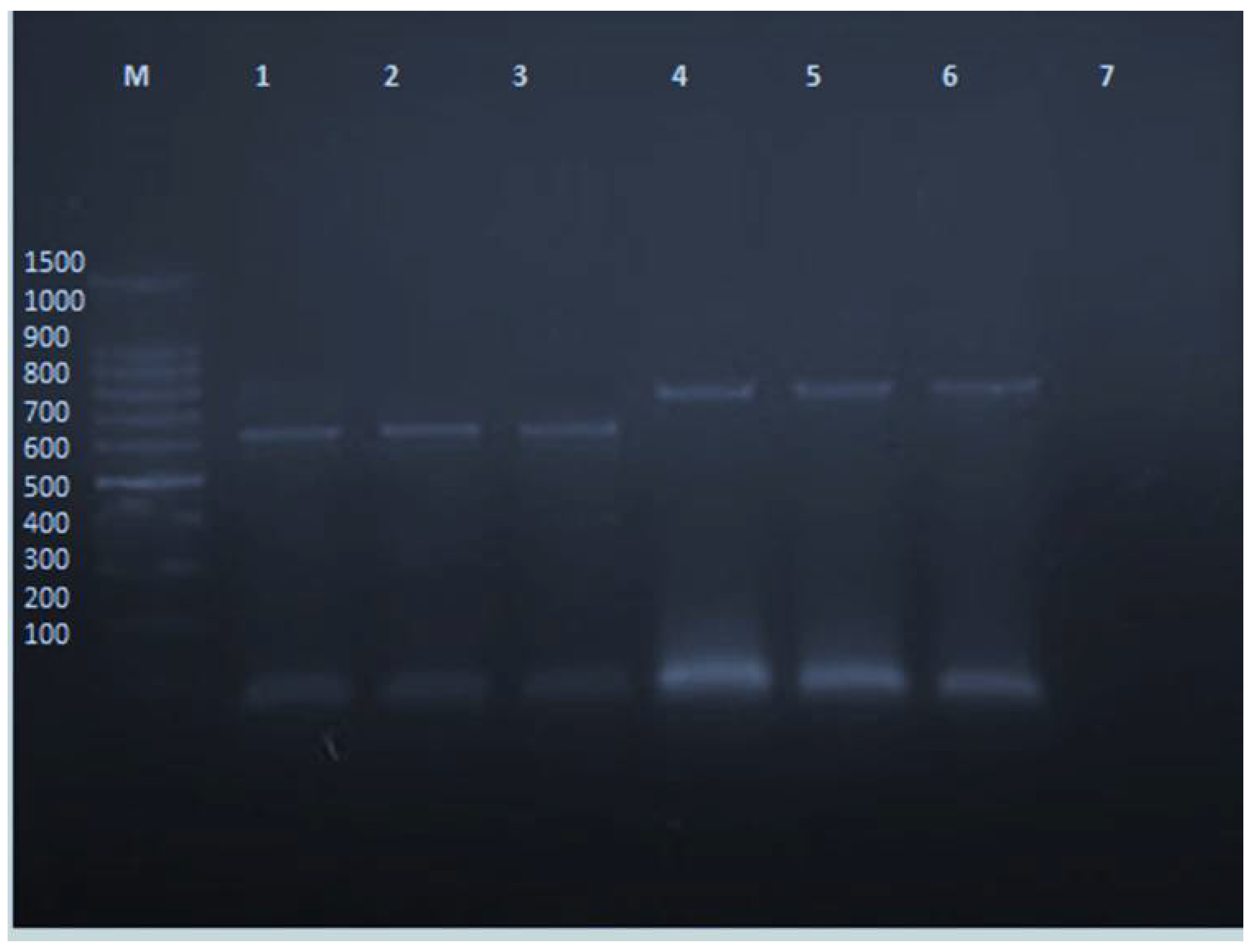
2.3. PVY Indexing by RT-PCR

Total RNA was extracted from leaves collected from PVY-infected potato plants grown in the field confirmed previously by DAS-ELISA test, using RNeasy Plant Mini kit (QIAGEN cat# 74903) following the manufacturer’s instructions. The sequences of the primers used to produce amplification of 825 bp in Rt-PCR were the upstream (5′TCAAGGATCCGCAAATGACACAATTGATGCAGG-3′) and downstream (5′-AGAGAGAATTCATCACATGTTCTTGACTCC3′) [38]. One hundred mg of fresh infected leaves were ground to a fine powder after freezing in liquid nitrogen. The powdered samples were transferred to sterile microfuge tubes. A 450 µL of buffer RLC was added (the exact composition of buffer RLC is confidential and is a proprietary component of RNeasy Kits. RLC comprising high concentration of guanidine hydrochloride), vortexed vigorously and incubated at 56 °C for 3 min. These lysates were applied to the QIAshredder spin column (lilac) sitting in a 2 mL collection tube and centrifuged for 2 min at maximum speed (rpm). The flow-through fraction was transferred from QIAshredder to a new tube without disturbing the cell-debris pellet in the collection tube, then 0.5 mL of ethanol (96–100%) was added to the cleared lysate and mixed well.
RT-PCR was carried out on RNA preparations with Reverse-iT™ One-Step RT-PCR Kit (ABgene®UK). This allows RT and amplification to be performed sequentially in the same tube. In particular, 2.5 µL of target RNA was mixed with 12.5 µL 2x RT-PCR Master mix containing 1.25 U/50µL (Taq Polymerase), 1.5 mM MgCl2, 0.2 mM each dNTPs, 10µM specific forward and reverse primers, 0.5 µL Reverse-iT™, RTase Blend (50 U/µL), RNase/DNase-free and distilled water to a final reaction volume of 25 µL. Synthesis of cDNA was performed at 47 °C for 30 min, followed by denaturation at 94 °C for 2 min. In thin-walled PCR tubes containing 5 µL of 10x PCR buffer (160 mM (NH4)2 SO4, 670 mM Tris-HCl pH 8.8, 0.1% Tween-20, 25 mM MgCl2), 1 µL of 10 mM dNTPs, 1 µL each of primer Pr-1F and PR-1R and 2.5 units of Taq DNA polymerase, nuclease-free water was added to make a volume of 45 µL. Five µL from the total DNA was added to PCR mixture and amplified with the following cycling parameters (denaturation at 94 °C for 30 s). Primer annealing at 55 °C for 30 s and extension at 72 °C for 30 s) for 30 cycles, with a final extension at 72 °C for 5 min and cooling to 4 °C. Amplified products were detected by agarose gel electrophoresis. Five-microliter aliquots of PCR products were analyzed on 1.7% agarose gels (6 × 8 cm), in TBE buffer (89 mM Tris-HCl, 89 mM boric acid, 2.5 mM EDTA, pH 8.5) at 120 volts. 100 bp DNA molecular weight markers (ABgene, UK) were used to determine the size of PCR products. Gels were stained with ethidium bromide 10 µg mL and visualized by UV illumination (Bio-Rad) [39]. About 75 min were required for running agarose gels and staining with ethidium bromide.

2.4. Chitosan (CS-NPs) Preparation

2.4.1. Characterization of Chitosan Nanoparticles

Chitosan nanoparticles preparation was characterized in the Central Laboratory of nanotechnology and advanced materials, at agricultural research center, Giza, Egypt.

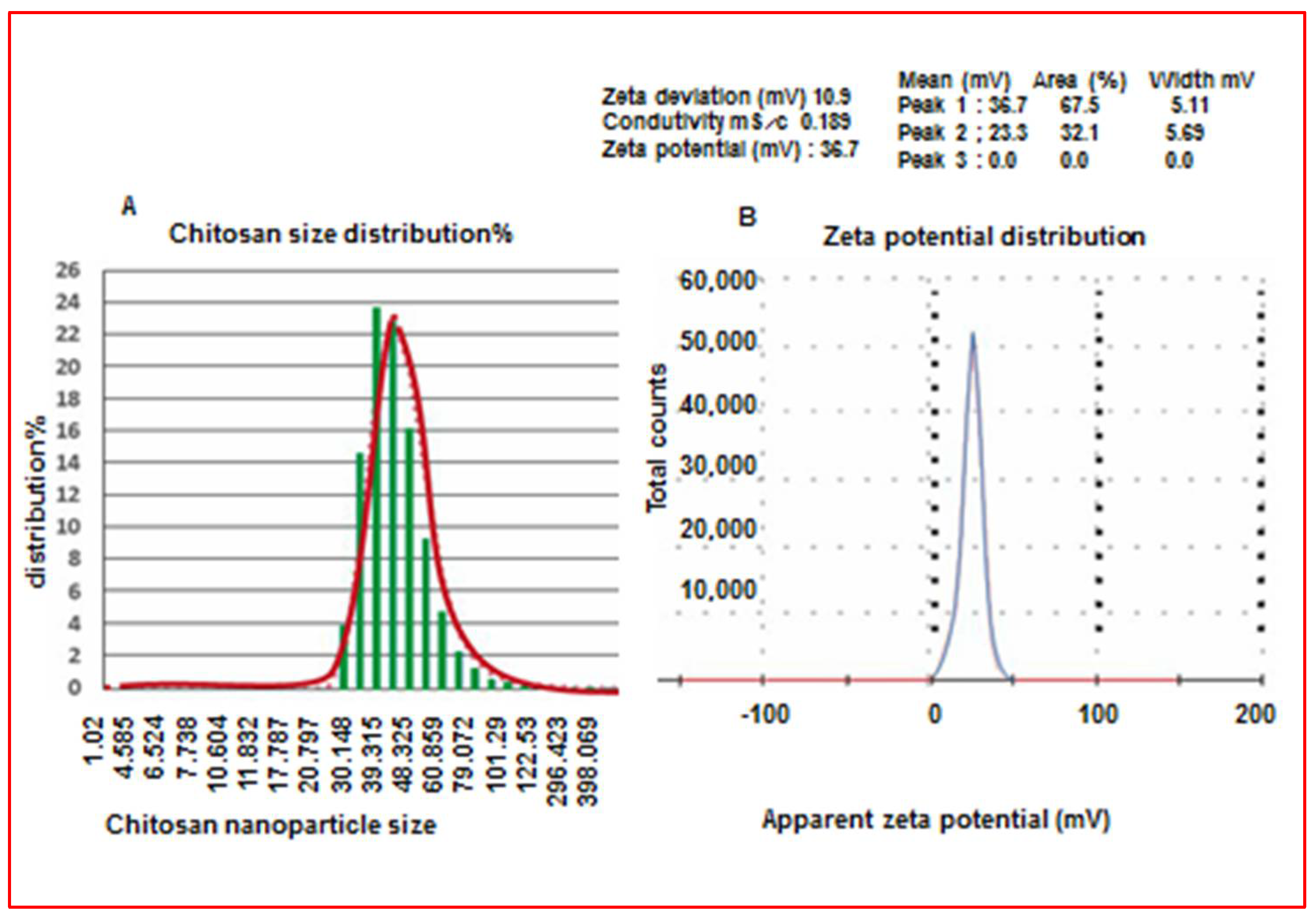
2.4.2. Determination of Nanoparticle Size

The particle size distribution and zeta potential of synthesized nanomaterials have been measured by Dynamic Light Scattering (DLS) analysis on a Zetasizer nano (ZS) instrument (Malvern, UK). The solution of the colloidal nanoparticles was diluted in 1 mL of distilled water and then placed in a disposable glass cuvette to analysis [40].

2.4.3. X-ray Diffraction (XRD) Measurement

X-ray diffraction (XRD) is a popular characterization technique for nanomaterials characterization which introduce important phase identification information for engineered nanoparticles. The physic-chemical crystalline nature of constructed nanomaterials had been conformed using an X-ray diffractometer (X’ per PRO, Panalytical, The Netherlands) operated with a Cu K radiation tube (=1.54 A°) at 40 kV. Then the colloidal nanoparticles solution was centrifuged at 16,000× g for 30 min at 4 °C for powder yield, then the precipitated nanoparticles were air dried after that bombarded by X-ray for phase analysis [41].

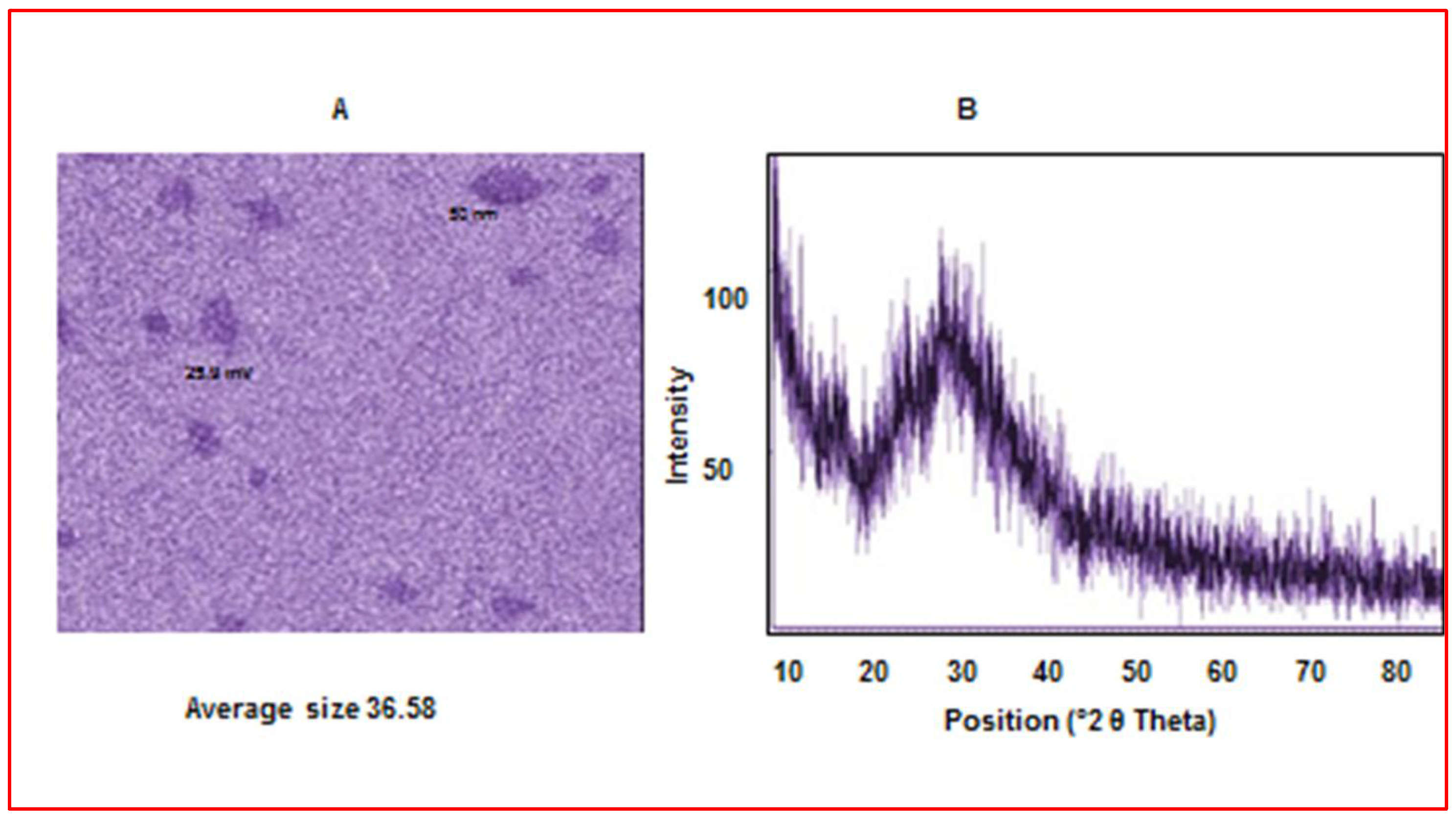
2.4.4. The Morphology of the Prepared Nanoparticles

The actual shape and size of the prepared nanoparticles have been performed by a high-resolution transmission electron microscope (HR-TEM) (Tecnai G2, FEI, and the Netherlands). The suspension of the aqueous chitosan (CS-NPs) was prepared and then sonicated for 10 min in an ultra-sonicator (SB-120DTN, Taiwan) before imaging. Then the prepared particles were deposited from the dilute aqueous solution on to 200 mesh-coper grids with the support of a 10 nm thick carbon film. Then the grids were placed in a double title grid holder. The images were captured using an Eagle CCD camera at an accelerating age of 200 kV.
The solution of chitosan was prepared chemically by reducing the molecular weight of chitosan (LMW, Techno-gene, Egypt) using the ion-gelation concept and sodium tripolyphosphate (STPP) as a reducing agent according to [42]. Then, 0.5 mg mL−1 of chitosan powder was dissolved in 1% acetic acid and left for 30 min at room temperature under vigorous stirring. To synthesize chitosan nanoparticles, 0.7 mg mL−1 of the reducing agent sodium tripolyphosphate (STPP) was dissolved separately in distilled water, and then 500 mL of chitosan was mixed with 160 mL of STTP under vortex or stirring for one hour. Chitosan nanoparticles (CS-NPs) at different concentrations were added to the culture medium in which in vitro shoots of PVY-infected potato plants were grown. To decrease or eliminate microorganisms in the MS medium, 1 w/w TiO2 NPs was used in the tissue culture media [43].

2.5. In Vitro Initiation and Stock Cultures

Potato (Solanum tuberosum L.) cv. Spunta sprouts explants were collected from potato tubers originated from PVY-infected plants, as confirmed by DAS-ELISA and RT-PCR indexing methods. These sprouts were used as initiators or starters propagating materials (Figure 1). Sprouts explants were washed with sterilized distilled water and were surface sterilized with 70% ethanol and 0.2% Mercuric chloride (HgCl2) for 1–2 min. Shoots were finally rinsed with sterilized distilled water for 3–5 min before inoculation on Murashige and Skoog (MS) medium [44] supplemented with 30 g L−1 sucrose, 6 g L−1 agar at pH 5.7 (standardized culture medium). The prepared medium was kept at the storage room for 3 days before the cultivation stage to make sure that there was no contamination in the medium. Plantlets were sub-cultured once every 30 days and kept in laminar air flow hood under controlled conditions on MS multiplication medium composed of MS supplemented with 30 g L−1 sucrose, 0.75 mL L−1 benzyl adenine (BAP), 0.1 mL L−1 kinetin, 2.5 mg L−1 gibberellic acid (GA3), 6 g L agar at pH 5.7. Culture room maintained under optimum light (1–10 K. Lux) with a temperature of 25 ± 2 °C and 70% relative humidity under 40 µmole m-2 s-1 cool white fluorescent light for 16 h daily. After 4 weeks of culture, regenerated plantlets were sub-cultured in 250 mL culture jar for complete plantlet development. The developed shoots were removed from the culture vessels and divided into sections containing three to four nodes. The nodal segments were put in 250 mL culture jar horizontally onto fresh multiplication medium. That medium has the same ingredients as described in the previous step. This procedure repeated every 3 to 4 weeks.

2.6. CS-NPs Treatments for PVY Eradication

After 3 subcultures shoot segments (5 cm in length) were collected from 4-week-old stock cultures and inoculated on MS standardized culture medium supplemented with different concentrations (100, 200, 250 and 300 mg L−1) of CS-NPs to evaluate its effect on shoot regeneration response, shoot proliferation, plantlets growth and PVY eradication. The pH was adjusted at 5.7 before autoclaving the media at 121 °C for 20 min. Chitosan treatments were filtered, sterilized, and incorporated in the autoclaved medium. Cultures were grown at the same conditions as described for stock cultures.

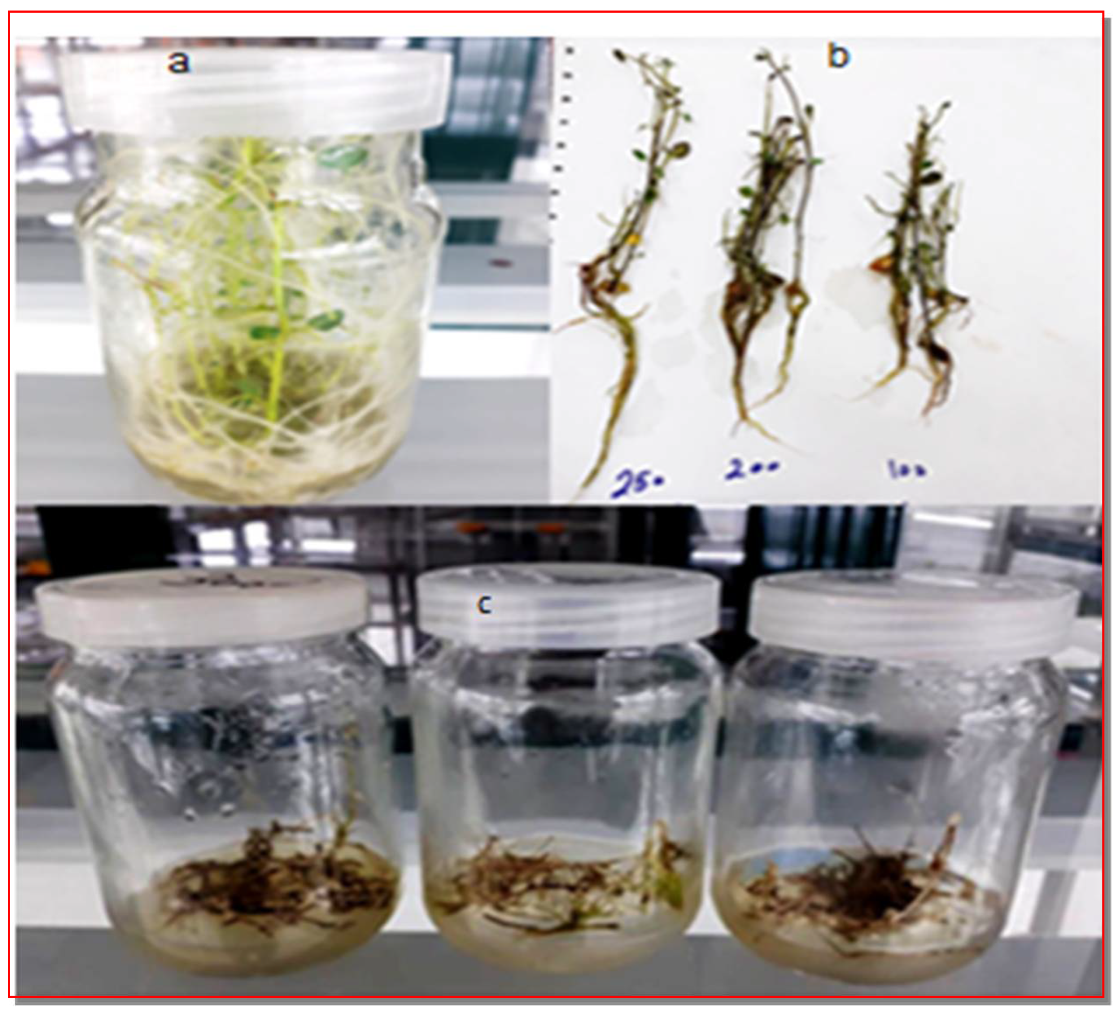
2.7. In Vitro Rooting Induction, Hardening and Acclimation

After 4 weeks of culture, the developed shoots were removed from the culture vessels. Individual shoots were cultured on the MS standardized culture rooting medium supplemented with 4.4 g MS, 30 g L−1 sucrose, 6 g L−1 agar and 0.1 mg L−1 α-naphthalene acetic acid (NAA) at pH 5.7 at a density of 8 shoots per 250 mL culture jar. After inoculation, cultures were incubated in culture room maintained under optimum light (1–10 K. Lux), 25 ± 2 °C and 70% relative humidity. Light was provided by 40 µmole m−2 s−1 cool white fluorescent for 16 h daily. After 4 weeks, plantlets of Potato (Solanum tuberosum L.) cv. Spunta (about 10 cm in length with well-developed root system) were taken out gently from each jar and washed with running tap water to remove traces of agar that may prevent the absorption of nutrients from the acclimatization culture substrates, then treated with liquid fungicide (2% dithane M-45) by lightly dipping the root base for 2–5 min. Plantlets were transferred to plastic pots 10 × 15 cm containing a mixture of peat moss, sand, and vermiculite at the ratio of 3:1:1. High humidity conditions of 95% were maintained around plantlets by covering them with clean transparent polythene bags having small holes for air circulation at 22 to 25 °C and 1000 lux illumination. After 10 days, the plantlets were exposed for 30–40 min daily. After another 10 days, the plantlets were exposed completely to the natural conditions and transferred to the earthen pots containing field soil only. After proper hardening for 30 days, plants were transferred to greenhouse and indexed for presence/absence of PVY virus by DAS-ELISA and RT-PCR.

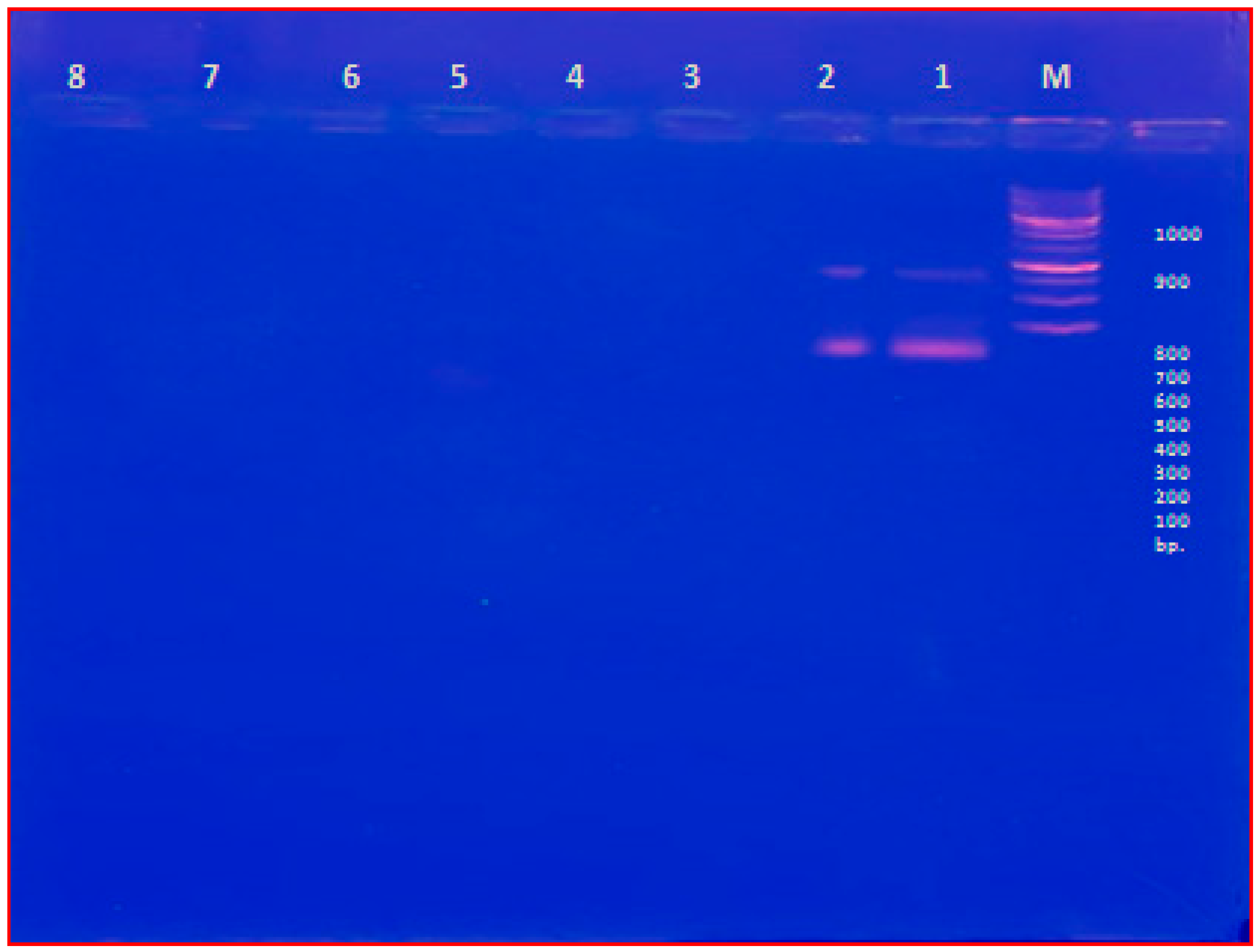
2.8. PVY Detection after CS-NPs Treatment

After 30 days of acclimatization, leaves were collected from treated potato plants and controls and tested for the PVY infection using DAS-ELISA and RT-PCR using the procedures described in “Section 2.1” and “Section 2.2”.

2.9. Statistical Analysis

The effect of Chitosan (CS-NPs) treatments and shoot regeneration responses were subjected to one-way analysis of variance (ANOVA) test. When significant differences were obtained, means were separated using Tukey’s HSD (honestly significant difference test) [45].

2.10. Histopathological Studies

A comparative anatomical study was carried out on the leaves of treated plants and control after 7 days of treatments. Small pieces were taken from the midrib region of the 4th upper apical leaf on the main stem. These pieces were killed, fixed in FAA (10 mL formalin, 5 mL glacial acetic acid and 85 mL ethyl alcohol 70%), then washed in 50% ethyl alcohol, dehydrated in a series of ethyl alcohols (70, 90, 95 and 100%), and infiltrated in xylene embedded in paraffin wax with a melting point 60–63 °C. Sections were made at 15–17 µm thick using rotary microtome, mounted on glass slides and stained with aqueous Safranin O (1%) and Fast Green (0.1% in 95% ethanol), as described by Ruzin, et al. [46]. Four sections were examined microscopically to detect histological manifestations of noticeable responses resulted from treatments. Counts and measurements (µ) were taken using a micrometer eye piece. Averages of 4 slides/treatment readings were calculated. Number of epidermal hairs was counted in 720 µ in the middle of the epidermis. Sections were examined with SEIWA OPTICAL light microscope (using a 10x lens) and photographed by Genius P931 digital camera using Image Manager 50 program. Various measurements were performed on microscopic images.

3. Results

Shoots regenerated on standard MS medium was successful from single node cuttings and gave high percentage of shoot regeneration (90.97% sprouts showed regeneration). Experiments of different (CS-NPs) treatments were further applied on same culture medium. Regenerated shoots when transferred to fresh medium containing antiviral (CS-NPs) in culture jar developed into complete plantlets (Figure 2).

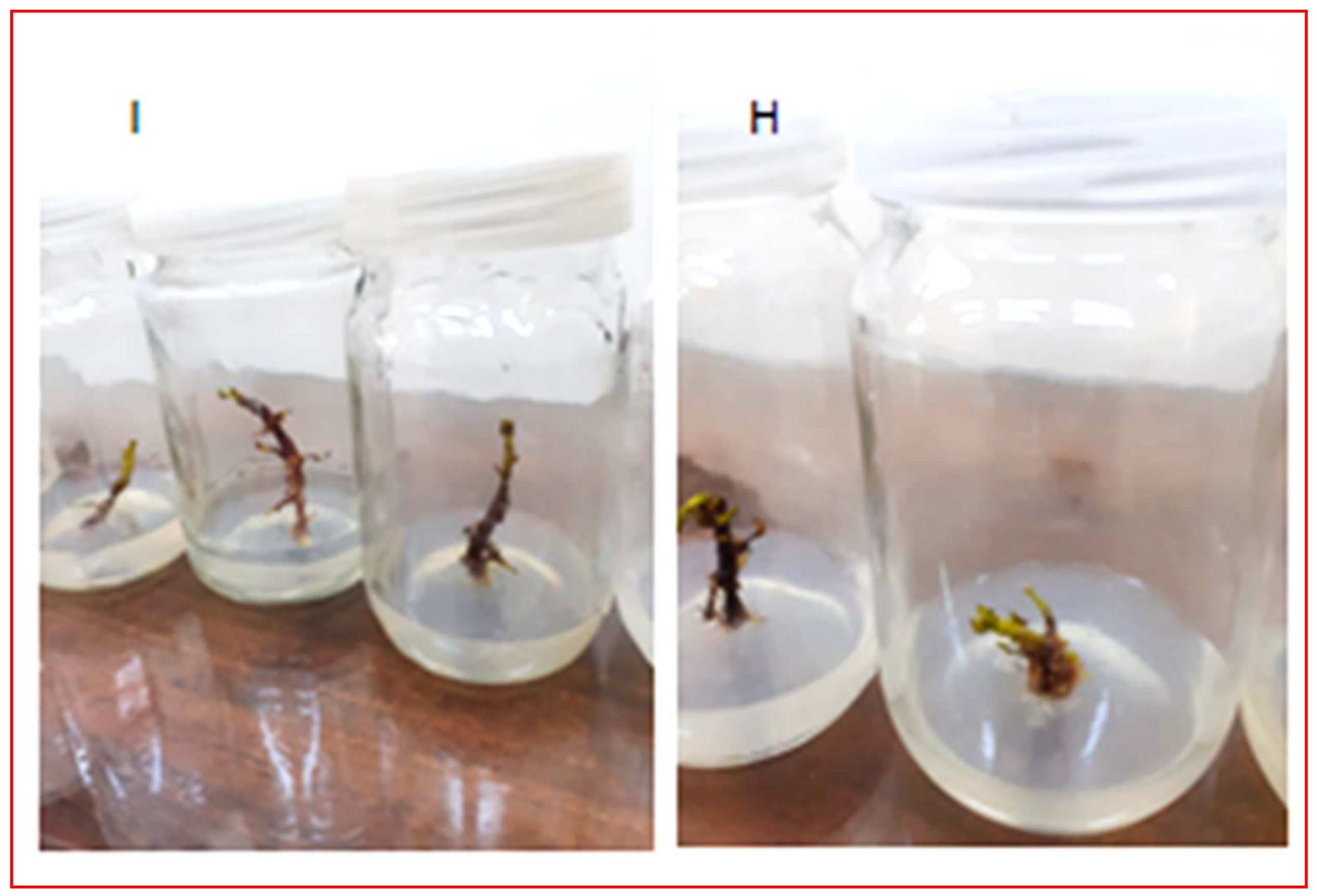
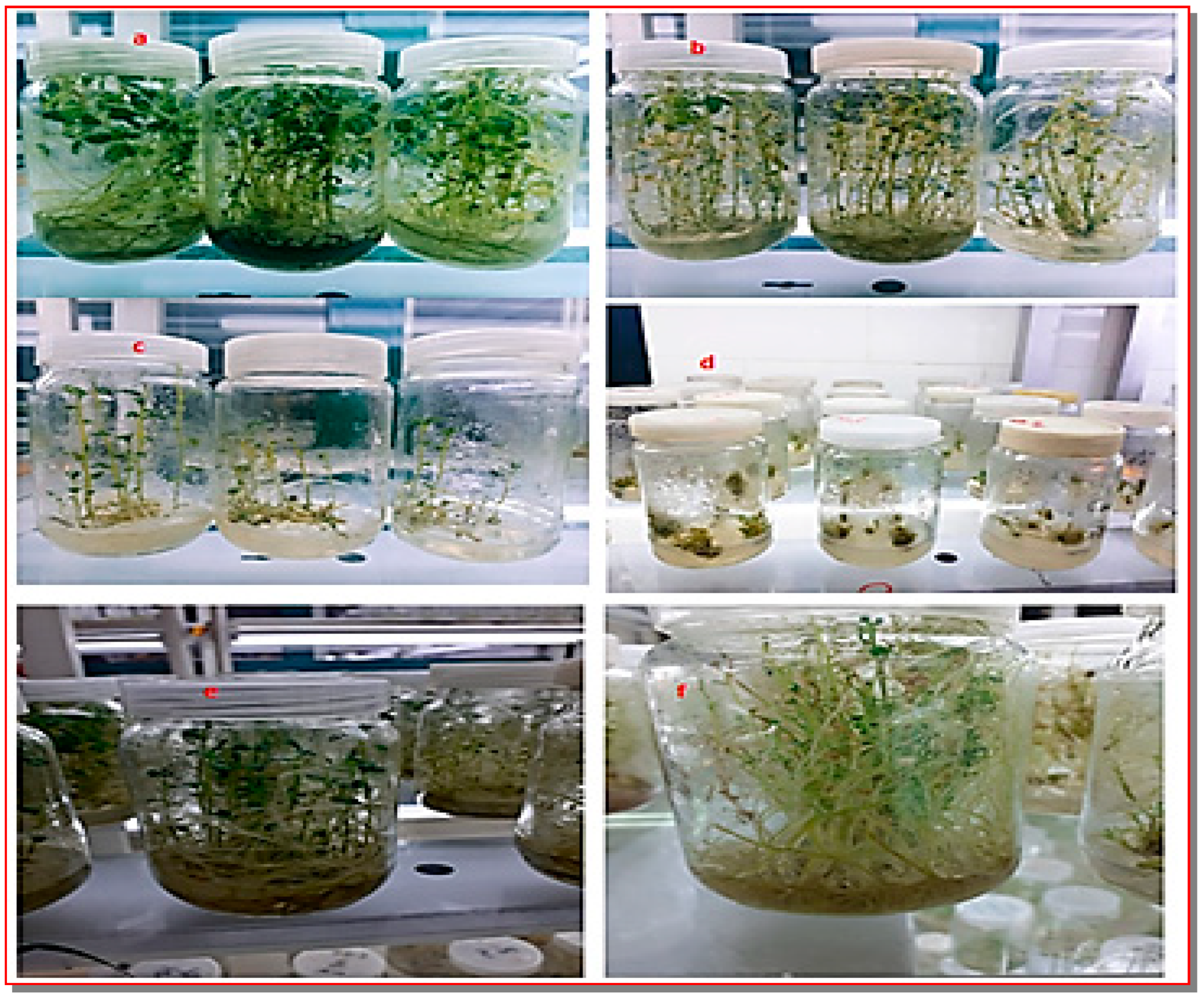
3.1. Effect of Chitosan (CS-NPs) Antiviral on Regeneration Response and Virus Elimination

Characterization of Chitosan Nanoparticles (CS-NPs)

The zeta potential of chitosan nanoparticles revealed a positive surface area charge with 25.9 mV. The synthesized chitosan nanoparticles indicted an average hydrodynamic size distribution of 34.39 nm (20.69 to 60.76 nm). X-ray diffraction (XRD) analysis was used to confirm the pseudospherical structure of chitosan nanoparticles. The amorphism fingerprint nature of chitosan nanoparticles was represented by the formation of a hump shape from 10 to 30 2θ angle. A TEM micrograph illustrated the pseudospherical shape of chitosan nanoparticles and the average size of nanoparticle was evaluated to be 36.58 nm (Figure 3 and Figure 4).
Comparing the effect of different concentrations of (CS-NPs) on regeneration response and production of PVY-free plants indicated that increasing the concentration of (CS-NPs) from 100 to 250 mg L−1 in the culture medium resulted in positive effects on shoot regeneration (Table 1 and Figure 5). Shoots regenerated on medium containing 200 mg L−1 (CS-NPs) showed higher growth and developed into complete plantlets. Excellent growth and regeneration shoots were obtained at 250 mg L−1 (Figure 6a,b). At the highest concentration of (CS-NPs) (300 mg L−1), shoot regeneration and growth were inhibited due to their phytotoxic effect (Figure 6c). Even after proliferation of axillary meristem from sprouts, shoot growth was determined by estimating the average of 10 shoot length measurements in cm, and the shoot growth was slow as compared to untreated cultures. High phytotoxicity and no shoot regeneration response were observed when treated with 300 mg L−1 (CS-NPs).
DAS-ELISA and RT-PCR results showed that (CS-NPs) produced high regeneration response with high frequencies of PVY-free plants. Electrophoresis of RT-PCR product showed an amplification of 825 bp DNA fragment in PVY-infected plants and no such amplification was obtained in PVY-free potato plantlets (Figure 7). For the confirmation of the virus, amplified band was extracted, sequenced, and analyzed. The sequence obtained had confirmed its homology with portion of coat protein gene of PVY. While indexing, eight samples of the tested negative plantlets in DAS-ELISA at the 100 mg L−1 (CS-NPs) were positive using RT-PCR. Plantlets which were tested negative in DAS-ELISA and RT-PCR were only considered virus free.
Results confirmed that (CS-NPs) was effective in the elimination of PVY from potato Sprouts of shoots under in vitro conditions. Shoot regeneration response was high with (CS-NPs) at the levels of 100 mg L−1 and 200 mg L−1 (4.69 and 6.11%, respectively). Furthermore, regeneration response and number of virus-free plants produced 38.78% PVY free plants with 30.50% cultures showing shoot regeneration at the level of 100 mg L−1 (CS NPs), and 100 % PVY-free plants with 46.80% cultures showing shoot regeneration at the level of 200 mg L−1. Regeneration response of explants was the highest at the level of 250 mg L−1.

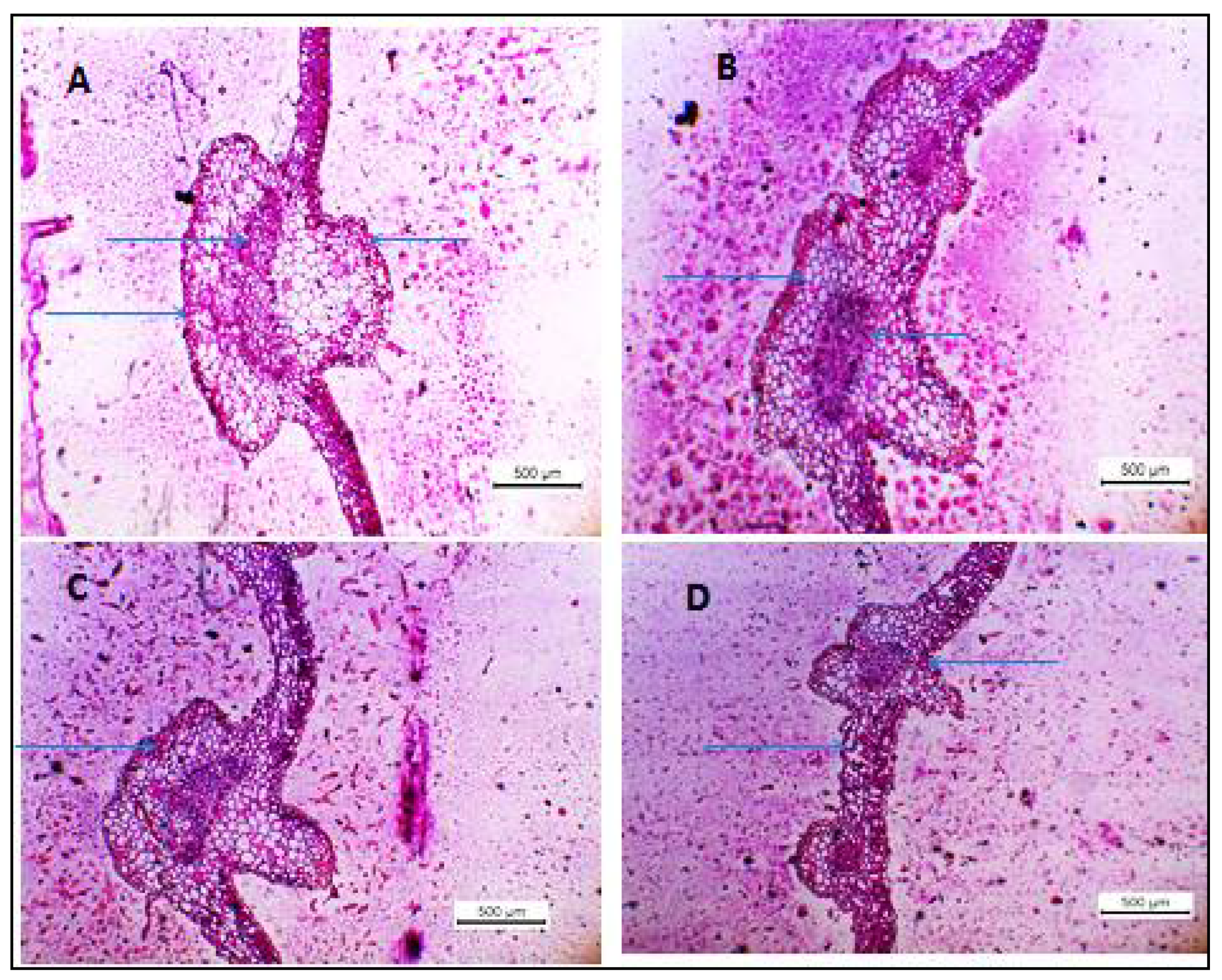
3.2. Histopathological Changes

The leaves’ tissues as evidence of the resistant reaction were elicited after 7 days of nanoparticles treatment. When treated with (CS-NPs), tissues alterations were observed in fixed tissues after 7 days of treatment. Progressive increasing in lignin accumulation in epidermal cells, number of hairs, thickness of blade, number of xylem arms and phloem layers (Table 2 and Table 3 and Figure 8) was observed. Tissue-shrinkage, intense staining, and precipitation of lignin in sub stomatal cavity, folding in mesophyll cell, and layering of cell wall while the remains of host palisade cell walls were noticed.

4. Discussion

The antiviral (CS-NPs) treatment eliminated PVY from in vitro-grown potato sprouts after 4 weeks of incubation on culture medium supplemented with (CS-NPs). Shoot regeneration response from potato sprouts grown on culture medium containing 300 mg L−1 of (CS-NPs) was low when compared with that of untreated control. This might be due to slow growth and phytotoxic effects of (CS-NPs) when used in high concentration. Previous studies indicated that antiviral chemicals in chemotherapy caused phytotoxic effect [47,48]. Symptoms of sprouts phytotoxicity (Figure 8) were commonly observed when concentrations of (CS-NPs) were increased from 250 to 300 mg L−1. Similar results were obtained with ribavirin treatments [49,50]. Meanwhile, ribavirin showed phytotoxic effect on axillary bud tips during PVY and (PVS) control of potato plants, high phytotoxicity and very low shoot regeneration response (%) was observed by using 30 mg L−1 of Ribavirin in the culture medium. Regeneration response and shoot growth from axillary bud of nodal segment on medium containing 5–30 mg L−1 of ribavirin was compared after 24 days of inoculation. Nodal explants cultured on 25 and 30 mg L−1 of ribavirin showed slow growth with very weak shoots when compared with those regenerated on lower concentrations [51,52]. In our study, among the tested concentrations of (CS-NPs), 300 mg L−1 resulted in the highest phytotoxic effects and the lowest shoot regeneration rate.
Regenerated plants from sprouts were acclimatized, grown in the greenhouse for 2 months and then tested using DAS-ELISA and RT-PCR for the detection of PVY as they are reliable viral indexing methods [48,52,53]. Results of DAS-ELISA and RT-PCR showed that (CS-NPs) was effective to produce PVY-free potato plants. Chitosan at 250 mg L−1 produced 100% PVY-free plants with excellent percentage of shoot regeneration (90.97%) as compared to other tested concentrations. Chitosan has been successfully used against potato virus PVY. Among the tested chitosan concentrations, the highest tested concentration, i.e., 300 mg L−1 was effective in the elimination of PVY from potato sprouts due to the phytotoxic effects on the explants.
Our results indicated that 100% of the plantlets were free of PVY under (CS-NPs) treatment as indicated by RT-PCR indexing. The highest tested concentration of 300 mg L−1 was less effective in the production of PVY-free potato plants as was noticed at 100 mg L−1. Similar results were reported for silver nanoparticles (AgNPs) which was an effective tool for preventing bean yellow mosaic virus (BYMV) disease in faba bean plants [54]. Chitosan inhibits multiplication of virus in the treated tissues. Such inhibition is a factor of virus type, plant, and chitosan concentration similar to ribavirin effect [53]. Chitosan may inhibit on the viral nucleic acid synthesis [55]. It could the virus replication processes. Since viruses do not move from old tissue to newly growing ones, they become virus free [49]. Chitosan stops the synthesis of new viral particles through the inhibition of the viral RNA synthesis [56]. The efficiency of chitosan in the eradication of virus depends on its interaction with viral particles in the plant tissues, replication process and synthesis enzymes. Our results proved that using high concentrations of (CS-NPs) (250 mg L−1) during in vitro regeneration of plantlets from sprouts explants could efficiently produce PVY-free plants.
There are a few reports concerning using of chitosan-based nanomaterials for controlling phytoviruses. In this concern, foliar spray of chitosan nanoparticles on Nicotiana glutinosa plants reduced disease severity causing by alpha mosaic virus (AMV), significantly decreased virus accumulation levels and enhanced the transcriptional levels of antioxidants enzymes, POD, PR1 and PAL [57]. However, Chitosan, a natural polymer, has been discovered to be an effective biotic elicitor in plants, promoting systemic resistance development [58]. Chitosan is a well-known biocontrol agent and abundant dual-effect natural polymer due to its non-toxic, biodegradable, and biocompatible properties [30]. Chitosan treatment might prevent microbial pathogens from developing and sporulating by breaking their cell membranes and blocking numerous biochemical processes during the plant-pathogen interaction, resulting in unique defensive responses in host plants. Because of the difficulties and specific requirements of producing viruses, chitosan’s antiviral activity (and derivatives) has received little study.
However, some information concerning chitosan’s antiviral effect can be discovered in the literature, and various research has indicated that chitosan protects plants against viral infection [27,28].
Primarily chitosan-based total nanocarriers display many advantageous properties, consisting of nanoscale dimensions, excessive surface area to volume ratio, in addition to the ability to tailor the surface charge and contact to the targeting ligands. Moreover, the chitosan matrix is incorporated in antiviral drugs, proteins, peptides, nucleic acids, or even whole inactivated viruses. Our research suggested that chitosan nano-applications enhanced plant defense mechanisms against virus infection and had a dual role in controlling plant viral infection and disease expression. Our results demonstrated that chitosan nano-applications prevented the dwarfness of the plants induced by the virus, by decreasing the virus infectivity, and increasing growth parameters. The mechanism of chitosan in promoting plant growth was attributed to its ability to induce some physiological processes such as nutrient uptake, photosynthesis, and cell division as well as plant growth regulators.
Histopathological changes (as indicator for resistance) in leaf tissues sprayed before 7 days with NPs and pre-virus inoculation were examined. Progressive increasing in lignin accumulation in epidermal cells, number of hairs, thickness of blade, number of xylem arms and phloem layers. The alterations included tissue-shrinkage, intense staining, and precipitation of lignin in sub stomatal cavity, mesophyll cell showing folding and layering of cell wall and remains of host palisade cell walls. Histopathological changes simultaneity with elicitation of resistance was tend toward growth enhancement in the treated plants with (CS-NPs) than untreated control.

5. Conclusions

In vitro regeneration rate and plantlet growth were improved as a result of the increase in the concentrations of chitosan nanoparticles from 100 to 250 mg L−1, while at the concentration of 300 mg L−1 phytotoxicity occured. The comparison among different concentrations of (CS-NPs) on in vitro regeneration response and production of virus-free plants indicated that the concentration of 250 mg L−1 was found to be effective. This research developed an effective way to eradicate PVY from infected potato plants by using sprouts explants. This technique would be helpful in the production of PVY free potato germplasm and seeds, Histopathological changes simultaneously with elucidation of resistance tend to enhance the growth in the treated plants with tested (CS-NPs) than untreated control.

Author Contributions

Conceptualization, M.S.; data curation, S.E.; formal analysis, S.E.; methodology, T.E.; project administration, M.S. and T.E.; supervision, M.M.; visualization, M.M.; writing—original draft, T.E. and M.M.; writing—review & editing, M.S. and A.G. All authors have read and agreed to the published version of the manuscript.

Funding

This research received no external funding.

Data Availability Statement

Not applicable.

Conflicts of Interest

The authors declare no conflict of interest.

References

  1. Spooner, M.; Mclean, K.; Ramsay, G.; Waugh, R.; Bryan, J. A single Domestication for Potato Based on Multilocus Amplified Fragment Length Polymorphism Genotyping. Proc. Natl. Acad. Sci. USA 2011, 41, 14694–14699. [Google Scholar] [CrossRef] [PubMed] [Green Version]
  2. Hawkes, G.; Javier, F. The Potato in Spain During the late 16th century. Econ. Bot. 1992, 46, 86–97. [Google Scholar] [CrossRef]
  3. Russ, A.M. Causes of Flowering of Long-day Potato Species Under Short-day and Cold-night Conditions. J. Plant Physiol. 2002, 49, 465–469. [Google Scholar]
  4. Myers, S.S.; Zanobetti, A.; Kloog, I.; Huybers, P.; Leakey, A.D.; Bloom, A.J.; Carlisle, E.; Dietterich, L.H.; Fitzgerald, G.; Hasegawa, T.; et al. Increasing CO2 Threatens Human Nutrition. Nat. Hasegawa 2014, 510, 139–142. [Google Scholar] [CrossRef] [PubMed] [Green Version]
  5. Central Agency for Public Mobilization and Statistics. Estimates by Governorate 1/1/2018. Available online: www.capmas.gov.eg (accessed on 2 November 2018).
  6. Food and Agriculture Organization. Crop Water Information; Food and Agriculture Organization of the United Nations. Available online: https://www.fao.org/land-water/databases-and-software/crop-information/en/ (accessed on 7 November 2021).
  7. Saberi Riseh, R.; Gholizadeh Vazvani, M.; Ebrahimi-Zarandi, M.; Skorik, Y.A. Alginate-Induced Disease Resistance in Plants. Polymers 2022, 14, 661. [Google Scholar] [CrossRef]
  8. Tamisier, L.; Szadkowski, M.; Girardot, G.; Djian-Caporalino, C.; Palloix, A.; Hirsch, J. Concurrent Evolution of Resistance and Tolerance to Potato virus Y in Capsicum annuum Revealed by genome-wide association. Mol. Plant Pathol. 2022, 23, 254–264. [Google Scholar] [CrossRef]
  9. Sergio, P.; Piero, R.; Maurizio, C. Yield Losses in Virus-infected Crops. Arch. Phytopathol. Plant Prot. 1996, 30, 283–296. [Google Scholar]
  10. Kreuze, J.; Souza-Dias, J.; Jeevalatha, A.; Figueira, A.; Valkonen, J.; Jones, R. Viral Diseases in Potato. In The Potato Crop; Springer: Berlin, Germany, 2020; pp. 389–430. [Google Scholar]
  11. Abbas, M.F.; Hameed, S.; Rauf, A.; Nosheen, Q.; Ghani, A.; Qadir, A.; Zakia, S. Incidence of Six Viruses in Potato Growing Areas of Pakistan. Pak. J. Phytopathol. Zakia 2012, 24, 44–47. [Google Scholar]
  12. Liang, Z.; Dickison, V.; Singh, M.; Xiong, X.; Nie, X. Studies of Tomato Plants in Response to Infections with PVX and Different PVY Isolates Reveal a Remarkable PVX-PVYNTN Synergism and Diverse Expression Profiles of Genes Involved in Different Pathways. Eur. J. Plant Pathol. 2016, 144, 55–71. [Google Scholar] [CrossRef]
  13. Lacomme, C.; Pickup, J.; Fox, A.; Glais, L.; Dupuis, B.; Steinger, T.; Rolot, J.L.; Valkonen, J.; Kruger, K.; Nie, X.; et al. Transmission and Epidemiology of Potato virus Y; Springer International Publishing: New York, NY, USA, 2017; pp. 141–176. [Google Scholar]
  14. Dupuis, B.; Bragard, C.; Carnegie, S.; Kerr, J.; Glais, L.; Singh, M.; Lacomme, C. Potato virus Y: Control, Management and Seed Certification Programmers. In Potato virus Y: Biodiversity, Pathogenicity, Epidemiology and Management; Springer: Berlin, Germany, 2017; pp. 177–220. [Google Scholar]
  15. Harahagazwe, D.; Condori, B.; Barreda, C.; Bararyenya, A.; Byarugaba, A.A.; Kude, D.A.; Lung’aho, C.; Martinho, C.; Mbiri, D.; Nasona, B.; et al. How Big is the Potato (Solanum tuberosum L.) Yield Gap in Sub-Saharan Africa and why? A participatory approach. Open Agric. 2018, 3, 180–189. [Google Scholar] [CrossRef]
  16. Karasev, A.V.; Gray, S. Continuous and Emerging Challenges of Potato virus Y in potato. Annu. Rev. Phytopathol. 2013, 51, 571–586. [Google Scholar] [CrossRef]
  17. Kehoe, M.; Jones, R. Improving Potato virus Y Strain Nomenclature: Lessons from comparing isolates obtained over a 73-year period. Plant Pathol. 2016, 65, 322–333. [Google Scholar] [CrossRef]
  18. Fuentes, S.; Gibbs, A.; Adams, I.P.; Wilson, C.; Botermans, M.; Fox, A. Potato virus A isolates from Three Continents: Their bio-logical properties, phylogenetics, and prehistory. Am. Phytopathol. Soc. 2021, 111, 217–226. [Google Scholar] [CrossRef]
  19. Houben, S.; Braber, H.; Blom-Zandstra, M.; Anten, N. Current Potato Production in Algeria; Stichting Wageningen Research, Wageningen Plant Research, Business Unit Plant: Wageningen, The Netherlands, 2017; 51p. [Google Scholar]
  20. Gong, H.; Clement, I.; Leonce, D. Major In Vitro Techniques for Potato Virus Elimination and Post Eradication Detection Methods. A Review. Am. J. Potato Res. 2019, 96, 379–389. [Google Scholar] [CrossRef] [Green Version]
  21. Bettoni, J.C.; Mathew, L.; Pathirana, R.; Wiedow, C.; Hunter, D.A.; McLachlan, A.; Khan, S.; Tang, J.; Nadarajan, J. Eradication of Potato Virus S, Potato Virus A, and Potato Virus M From Infected in vitro-Grown Potato Shoots Using in vitro Therapies. Front. Plant Sci. 2022, 13, 1431. [Google Scholar] [CrossRef] [PubMed]
  22. Pandit, M.A.; Kumar, J.; Gulati, S.; Bhandari, N.; Mehta, P.; Katyal, R.; Rawat, C.D.; Mishra, V.; Kaur, J. Major Biological Control Strategies for Plant Pathogens. Pathogens 2022, 11, 273. [Google Scholar] [CrossRef]
  23. Saqib, S.; Faryad, S.; Afridi, M.I.; Arshad, B.; Younas, M.; Naeem, M.; Zaman, W.; Ullah, F.; Nisar, M.; Ali, S.; et al. Bimetallic Assembled Silver Nanoparticles Impregnated in Aspergillus fumigatus Extract Damage the Bacterial Membrane Surface and Release Cellular Contents. Coatings 2022, 12, 1505. [Google Scholar] [CrossRef]
  24. Hoang, N.H.; Thanh, T.; Sangpueak, R.; Treekoon, J.; Saengchan, C.; Thepbandit, W.; Papathoti, N.K.; Kamkaew, A.; Buensanteai, N. Chitosan Nanoparticles-Based Ionic Gelation Method: A Promising Candidate for Plant Disease Management. Polymers 2022, 14, 662. [Google Scholar] [CrossRef] [PubMed]
  25. Behl, T.; Kaur, I.; Sehgal, A.; Singh, S.; Sharma, N.; Bhatia, S.; Al-Harrasi, A.; Bungau, S. The Dichotomy of Nanotechnology as The Cutting Edge of Agriculture: Nano-farming as an asset versus nanotoxicity. Chemosphere 2022, 288, 132–533. [Google Scholar] [CrossRef] [PubMed]
  26. Wu, H.; Li, Z. Nano-enabled Agriculture: How Do Nanoparticles Cross Barriers in Plants. Plant Commun. 2022, 3, 1100346. [Google Scholar] [CrossRef] [PubMed]
  27. Chen, H.; Yada, R.Y. International Conference on Food and Agriculture Applications of Nanotechnologies, NanoAgri, São Pedro, SP, Brazil. Trends Food Sci. Technol. 2011, 22, 583–584. [Google Scholar] [CrossRef]
  28. Parisi, C.; Vigani, M.; Rodríguez-Cerezo, E. Agricultural Nanotechnologies: What Are The Current Possibilities. Nano Today 2015, 10, 124–127. [Google Scholar] [CrossRef]
  29. Su, S.; Kang, P.M. Systemic Review of Biodegradable Nanomaterials in Nanomedicine. Nanomaterials 2020, 10, 656. [Google Scholar] [CrossRef] [PubMed]
  30. Hong, J.; Wang, C.; Wagner, D.C.; Jorge, L.; Torresdey, G.; He, F.; Cyren, M. Foliar Application of Nanoparticles: Mechanisms of Absorption, Transfer, and Multiple Impacts. Environ. Sci. Nano Rico 2021, 8, 1196–1210. [Google Scholar] [CrossRef]
  31. Hegazi, S.; Yousef, A.; Mohamed, A.; Allatif, A.; Shaban, T.; Kamal, M. Effect of Silver Nanoparticles, Medium Composition and Growth Regulators on in vitro propagation of Picual Olive Cultivar. Egypt. J. Chem. 2021, 64, 6961–6969. [Google Scholar]
  32. El-Kosary, S.; Allatif, A.; Stino, R.; Hassan, M.; Kinawy, A. Effect Of Silver Nanoparticles On Micropropagation Of Date Palm (Phoenix Dactylifera L, Cv. Sewi and Medjool). Plant Arch. 2020, 2, 9701. [Google Scholar]
  33. Badoni, A.; Chauhan, J. Potato Seed Production Of Cultivar Kufri Himalini, in vitro. Health Environ. Res. Online 2010, 1, 7–10. [Google Scholar]
  34. Singh, C.R. Review On Problems And its Remedy In Plant Tissue Culture. Asian J. Biol. Sci. 2018, 11, 165–172. [Google Scholar] [CrossRef] [Green Version]
  35. Yasmine, R.; Ahmad, J.; Qamar, S.; Qureshi, M.I. Engineered Nanomaterials for Sustainable Agricultural Production, Soil Improvement and Stress Management. In Plant Biology, Sustainability and Climate Change; Elsevier: Amsterdam, The Netherlands, 2023; pp. 495–512. [Google Scholar] [CrossRef]
  36. Clark, M.F.; Adams, A.N. Characteristics of The Microplate Method of Enzyme-Linked Immunosorbent Assay for The Detection of Plant Viruses. J. Gen. Virol. 1977, 34, 475–483. [Google Scholar] [CrossRef]
  37. Dominguez, S.; Aparicio, F.; Sánchez-Navarro, J.A.; Pallás, V.; Garcia-Brunton, J.; Cano, A. Studies on the Incidence of Ilarviruses and Apple Chlorotic Leafspot Trichovirus in Apricot Trees in the Murcia Region (Spain) using serological and molecular hybridization methods. Acta Hortic. 1997, 472, 203–210. [Google Scholar] [CrossRef]
  38. Shalaby, A.A.; Nakhla, M.K.; Soliman, A.M.; Mazyad, H.M.; Hadidi, A.; Maxwell, D.P. Development of a Highly Sensitive Multiplex Reverstrans-cription-Polymerase Chain Reaction (m-RT-PCR) Method for Detection of Three Potato Viruses in a Single Reaction and nested PCR. Arab J. Biotech. 2002, 5, 275–286. [Google Scholar]
  39. Sambrook, J.; Fritsch, E.F.; Maniatis, T. Molecular Cloning: A Laboratory Manual; Cold Spring Harbor Laboratory Press: Cold Spring Harbor, NY, USA, 1987; p. 1546. [Google Scholar]
  40. Murdock, R.C.; Braydich-Stolle, L.; Schrand, A.M.; Schlager, J.J.; Hussain, S.M. Characterization of Nanomaterial Dispersion in Solution Prior to In Vitro Exposure Using Dynamic Light Scattering Technique. Toxicol. Sci. 2008, 101, 239–253. [Google Scholar] [CrossRef] [PubMed] [Green Version]
  41. Phanjom, P.; Zoremi, E.; Mazumder, J.; Saha, M.; Baruah, S.B. Green Synthesis of Silver Nanoparticles Using Leaf Extract of Myrica esculenta. Int. J. NanoSci. Nanotechnol. 2012, 3, 73–79. [Google Scholar]
  42. Masarudin, M.J.; Cutts, S.M.; Evison, B.J.; Phillips, D.R.; Pigram, P.J. Factors Determining the Stability, Size Distribution, and Cellular Accumulation of Small, Monodisperse Chitosan Nanoparticlies as Candidate Vectors for Anticancer Drug Delivery: Application to the passive encapsulation of [14C]- doxorubicin. Nanotechnol. Sci. Appl. 2015, 8, 67–80. [Google Scholar] [CrossRef] [Green Version]
  43. Safavi, K. Effect of Titanium Dioxide Nanoparticles in Plant Tissue Culture Media for Enhance Resistance to Bacterial Activity. Bull. Environ. Pharmacol. Life Sci. 2014, 3, 163–166. [Google Scholar]
  44. Murashige, T.; Skoog, F. A Revised Medium for Rapid Growth and Bio Assays with Tobacco Tissue Cultures. Physiol. Plantarum. 1962, 15, 473–497. [Google Scholar] [CrossRef]
  45. Meyers, L.; Grossen, N.; Theory, P.; Design, W.H.; Freeman, S.F. Analysis of independent group designs. Am. Psychol. Assoc. 1974, 237–252. [Google Scholar]
  46. Ruzin, S.E. Staining Techniques. In Plant Microtechnique and Microscopy; Oxford University Press: Oxford, UK, 1999; pp. 87–116. [Google Scholar]
  47. Hansen, A.; Lane, W. Elimination of Apple Chlorotic Leafspot Virus from Apple Shoot Cultures by Ribavirin. Plant Dis. 1985, 69, 134–135. [Google Scholar]
  48. Sharma, S.; Rani, G.; Zaidi, A.A.; Hallan, V.; Nagpal, A.; Virk, G.S. Production of Indian Citrus Ringspot Virus-free Plants of Kinnow mandarin (Citrus nobilis Lour × C. deliciosa Tenora) employing chemotherapy coupled with shoot tip grafting. J. Plant Biotechnol. 2007, 8, 1–8. [Google Scholar]
  49. Simpkins, I.; Walkey, D.; Neely, H. Chemical Suppression of Virus in Cultured Plant Tissues. Ann. Appl. Biol. 1981, 9, 161–169. [Google Scholar] [CrossRef]
  50. Klein, R.; Livingston, C. Eradication of potato viruses X and S from potato shoot-tip cultures with ribavirin. Phytopathology 1983, 73, 1049–1050. [Google Scholar] [CrossRef]
  51. Wambugu, F.; Secor, G.; Gudmestad, N.J. Eradication of Potato virus Y and S from Potato by Chemotherapy of Cultured Axillary Bud Tips. Am. Potato Springer 1985, 62, 667–672. [Google Scholar] [CrossRef]
  52. Toussaint, A.; Kummert, J.; Maroquin, C.; Lebrun, A.; Roggemans, J. Tissue Culture o. Use of Virazole to Eradicate odontoglossum Ringspot Virus from In Vitro Cultures of Cymbidium Sw. In Plant Cell, Tissue and Organ Culture; Springer: Berlin, Germany, 1993; Volume 32, pp. 303–309. [Google Scholar]
  53. Ram, R.; Verma, N.; Singh, A.; Singh, L.; Hallan, V.; Zaidi, A. Indexing and Production of Virus-Rree Chrysanthemums. Biol. Plant Springer 2005, 49, 149–152. [Google Scholar] [CrossRef]
  54. Ahmed, G.; Mia, Z.; Mahmoud, A.; Tarek, S. Silver Nanoparticles as A Highly Viricidal Agent to Deter Plant-Infecting Viruses and Disrupt their Acquisition and Transmissibility by Vector Aphid. Res. Sq. 2021, 1–20. [Google Scholar]
  55. Fraser, R.; Gerwitz, A. Effects of 2-α-hydroxybenzylbenzimidazole on Tobacco Mosaic Virus and Host RNA Synthesis in Tobacco Leaf Discs and plants. Plant Sci. Lett. 1984, 34, 111–117. [Google Scholar] [CrossRef]
  56. Chirkov, S. The antiviral activity of chitosan. J. Appl. Biochem. Microbiol. 2002, 38, 1–8. [Google Scholar] [CrossRef]
  57. Abdelkhalek, A.; Qari, S.H.; Abu-Saied, M.A.A.-R.; Khalil, A.M.; Younes, H.A.; Nehela, Y.; Behiry, S.I. Chitosan Nanoparticles Inactivate Alfalfa Mosaic Virus Replication and Boost Innate Immunity in Nicotiana glutinosa Plants. Plants 2021, 10, 2701. [Google Scholar] [CrossRef]
  58. Davydova, V.N.; Nagorskaya, V.I.; Gorbach, A.A.; Kalitnik, A.V.; Reunov, T.F.; Solov’Eva, I.M. Applied Biochemistry Ermak, and Microbiology’Chitosan antiviral activity: Dependence on structure and depolymerization method. Appl. Biochem. 2011, 47, 103–108. [Google Scholar] [CrossRef]
Figure 1. Sprouts of PVY infected potato explants (Solanum tuberosum L.) cv. Spunta (I) showing shoot regeneration on MS medium supplemented with different concentrations of Chitosan (CS-NPs). Healthy explants (H).
Agronomy 12 02901 g001
Figure 2. Plantlets regenerated from mature sprouts on culture medium containing different Chitosan (CS-NPs) concentrations. Excellent shoot regeneration, shoot proliferation, shoot length, and shoot growth on MS medium supplemented with 250 mg L of (CS-NPs) (a). Very good shoot regeneration, shoot proliferation, shoot length, and shoot growth on MS medium supplemented with 200 mg L of (CS-NPs) (b). Slow and weak shoot regeneration, shoot proliferation, shoot length, and shoot growth on MS medium supplemented with 100 mg L of (CS-NPs) (c). Poor shoot regeneration, shoot proliferation, shoot length, and shoot growth on MS medium supplemented with 300 mg L of (CS-NPs) (d). Good shoot regeneration, shoot proliferation, shoot length, and shoot growth on MS medium without treatment (Healthy control) (e). Weak shoot regeneration, shoot proliferation, shoot length, and shoot growth on MS medium without treatment (infected control) (f).
Agronomy 12 02901 g002
Figure 3. Characterization of physic chemical synthesized chitosan nanoparticles (CS-NPs). (A) chitosan particle size distribution histogram with average size of 34.39 nm. (B) Zeta potential charge measurement revealed a positive surface charge with 25.9 mV.
Agronomy 12 02901 g003
Figure 4. Characterization of physic chemical synthesized chitosan nanoparticles (CS-NPs). (A) X-ray diffraction (XRD) pattern indicating a morphism nature of chitosan particle. (B) TEM histogram showing chitosan nanoparticles (CS-NPs) a pseudospherical shape with average size of 36.59.
Agronomy 12 02901 g004
Figure 5. Agarose gel electrophoresis of RT-PCR products of in vitro regenerated plantlets after (CS-NPs) treatment. M 100 bp DNA ladder (Lanes 1 positive control, Lane 2: 100 mg L−1, Lanes 3 and 4: 200 mg L−1, Lanes 5 and 6: 250 mg L−1, Lane 7: 300 mg L−1, Lane 8: negative control.
Agronomy 12 02901 g005
Figure 6. (a) In vitro rooted potato plantlets Spunta cv. ready for acclimatization stage in the greenhouse. (b) different length of roots with (CS-NPs) treatments. (c) high phytotoxicity and very low shoot regeneration as response to higher concentration of chitosan nanoparticles at (300 mg L−1) on the explants in culture media.
Agronomy 12 02901 g006
Figure 7. PCR analysis using 1.7% Agarose gel electrophoresis showing the amplified fragment of Potato virus Y (PVY) coat protein gene from potato tissues. Specific PVY cDNA band 825 bp by using specific PVY primers. Lanes 1, 2 and 3 (PLRV) while Lanes 4, 5 and 6 PVY infected potato samples and Lane 7 is healthy potato sample. Lane M is the marker (Biometric) 100 bp DNA Ladder, the size of the cDNA fragment is indicated on the presence of PVY at 825 bp.
Agronomy 12 02901 g007
Figure 8. Anatomical alterations in potato leaves treated with nanoparticles; (100× and D 60×). (A) healthy, (B) precipitation of lignin in sub stomatal cavity, (C) mesophyll cells showing folding and layering of cell walls, (D) tissue showing intense staining and increasing number of xylem vessels.
Agronomy 12 02901 g008
Table 1. Effect of different concentrations of antiviral chitosan (CS-NPs) on shoot regeneration and the production of PVY-free potato plants (Solanum tuberosum L.) cv. Spunta from infected potato sprouts.
Concentrations of (CS-NPs) in Growth Medium mg L−1Proliferation Percent Shoot RegenerationPlant Length, cm ± SDDAS-ELISART-PCR
Untreated (Control)1.78 ± 0.30 *90.96 ± 1.423.99 ± 0.40 cm0.654+
3000.0010.41 ± 1.20 c-0.236 a
2506.11 ± 0.2690.97 ± 1.41 a12.40 ± 0.38 *0.256 a
2004.69 ± 0.2487.49 ± 1.22 a11.96 ± 0.18 *0.564 b
1003.69 ± 0.3043.05 ± 1.85 b6.6 ± 0.01 *0.595 b+
* Means ± SE (Standard Error). Means within columns with the same letter are not significantly different using Tukey’s HSD test at p < 0.01.
Table 2. Anatomical properties of potato leaves treated with chitosan (CS-NPs).
TreatmentHairCuticle ThicknessEpidermal ThicknessPalisade TissueCollenchyma TissueSpongy Tissue
Healthy control145.6 a *3 b1 c7.0 b5.5 c4.5 b9.7 c45.8 d1 f6 d110.2 f2 d58.8 f72.3 d4 f
Chitosan (CS-NPs)250.7 a6 b4 c9.0 b6.3 c17.4 b11.3 c76.5 d1 f6 d190.6 f3 d70.0 f95.2 d5 f
* thickness (μm), a: length (μm), b: lower epidermis (μm), c: upper epidermis (μm), d: thickness f: number.
Table 3. Vascular bundle and xylem (μm) properties of potato leaves treated with chitosan (CS-NPs).
TreatmentMid. Vein ThicknessVasc. Bundle Length Vasc. Bundle WidthEx. Phlo. ThicknessIntr. Phlo. ThicknessXylem ThicknessNo. Xylem VesselThickness of Major Xylem
Healthy control7745.2215.2460.635.339.4120.41235.2
Chitosan (CS-NPs)980.6225.6560.455.548.3132.62045.3
Publisher’s Note: MDPI stays neutral with regard to jurisdictional claims in published maps and institutional affiliations.

