Article

The Effect of TIBA and NPA on Shoot Regeneration of Cannabis sativa L. Epicotyl Explants

Department of Biotechnology, Institute of Natural Fibres & Medicinal Plants—National Research Institute, Wojska Polskiego 71b, 60-630 Poznań, Poland
*
Author to whom correspondence should be addressed.
Agronomy 2022, 12(1), 104; https://doi.org/10.3390/agronomy12010104
Submission received: 27 November 2021 / Revised: 17 December 2021 / Accepted: 29 December 2021 / Published: 31 December 2021
(This article belongs to the Section Crop Breeding and Genetics)

Abstract

:
Industrial hemp (Cannabis sativa L., family Cannabaceae) is a multi-purpose crop, used in the production of food, nutraceuticals, cosmetics and medicines. Therefore, development of new varieties with specific chemical profiles is necessary. In vitro culture methods could be complementary to conventional breeding and a useful tool for large-scale propagation. Strong apical dominance is considered as one of the factors contributing to the recalcitrance of industrial hemp in shoot proliferation. In this study, we tested the polar transport inhibitors N-1-naphtylphtalamic acid (NPA) and 2,3,5-triiodobenzoic acid (TIBA) to enhance shoot regeneration as the result of suppression of apical dominance and to develop in vitro protocols for Diana, Finola and Fedora 17 cultivars. Shoot tips derived from epicotyls were cultivated on Murashige and Skoog medium (MS) supplemented with meta-topolin (mT) and NPA, and also thidiazuron (TDZ) with a combination of TIBA and NPA. The results showed that the combination of TDZ with NPA (1–5 mg L−1) and TDZ with TIBA (0.5–2.5 mg L−1) increased the response of explants and the multiplication rate, but the effect was genotype-dependent and malformations were observed. To optimize the developed protocol, a two-step procedure with shortened time of exposure to inhibitors and reduced concentrations of them was applied. Shoots were rooted on media containing indole-3-butyric acid (IBA) and then successfully acclimatized. The obtained results will be useful in micropropagation of recalcitrant industrial hemp varieties.

1. Introduction

Industrial hemp (Cannabis sativa L., family Cannabaceae) is a multi-purpose crop used for the production of fiber, seeds, oil, essential oil, composites, drugs, supplements, cosmetics, etc. [1,2,3]. This multiple use of hemp raw material generates the need to develop new varieties adapted to current demands. In the past, industrial hemp was mainly bred for fiber, but for the last decade, there has been a growing demand for hemp products such as “natural foods”, drugs, and diet supplements derived from its content in cannabinoids and terpenes. Research on new applications of hemp plants is still ongoing, and the potential perspectives of medical and industrial use of hemp are increasing. Considering that the demand for standardized plant material has been constantly increasing, development of new or improved varieties with a specific chemical profile is necessary.
Traditionally, hemp has been cultivated and propagated by seeds. Conventional breeding is a time- and labor-consuming process. New germplasm, knowledge, and breeding techniques are required to design and develop new hemp varieties with specific features. In this respect, plant in vitro cultures and clonal propagation techniques can be useful for large-scale propagation of the selected elite clones [4,5]. It may enable shortening the time of the breeding process and/or large-scale production of homogeneous plant material. Regeneration protocols are essential for most in vitro techniques employed in plant breeding and in crop improvement using genetic transformation. However, there is still a lack of efficient and reproducible in vitro protocols for hemp. Cultivar- and genotype-dependent responses of explants and the low regeneration rate are the main limitations for hemp in vitro propagation.
So far, in vitro protocols developed from leaf [6], axillary nodal explants [7,8], cotyledons [9], shoot tips [10], and epicotyls [11] have been published. A high multiplication rate (12–13 shoots per explant) was achieved using Murashige and Skoog medium [12] supplemented with thidiazuron (TDZ) [7] and meta-topolin (mT) [8]. It should be highlighted that the aforementioned reports have been devoted to a single high THC yielding clone (MX) of marijuana. In contrast, much less efficient protocols have been developed for fiber-type varieties [4,9,10,11,13]. The average multiplication rate expressed as shoots per explant was usually in the range of around 2–3 [4,9,10]. A low multiplication rate and problems with initiation of multi-shoot cultures have been reported as results of the low branching tendency and high degree of apical dominance [4,13]. Strong apical dominance is characteristic for fiber-type hemp. It was a favorable and economic feature because taller plants had longer fibers and could grow with higher sowing density. Therefore, plants characterized by strong apical dominance have been cultivated and selected in the breeding process.
Apical dominance is a phenomenon in plants where the main shoot dominates and inhibits the outgrowth of axillary buds, which are in a dormant state [14]. Bud outgrowth is controlled by a complex network of endogenous hormones involving auxin, strigolactones and cytokinins. Although the mechanisms of apical dominance are not fully understood, there is a consensus that sugar level and auxin play a key role in it [15,16]. Shoot tip keeps control under branching through auxin, which is synthesized in young leaves in the apical meristem and transported down within the stem in the polar auxin pathway. Auxin may inhibit lateral buds via different mechanisms: firstly, suppressing flow of auxins from lateral buds competing for access to the polar auxin pathway [17] and secondly by hormonal interaction between strigolactones and cytokinins [18,19,20]. The outgrowth of axillary buds depends on the ratio of these plant hormones. Auxin cannot enter the buds, but regulates bud outgrowth or dormancy via strigolactones and cytokinins, which are mobile within the stem. Strigolactones inhibit, whereas cytokinins promote, bud growth. Auxin can upregulate (strigolactones) and downregulate (cytokinins) biosynthetic genes mediated mainly by transcription factors localized in buds [17,19,21].
Suppression of apical dominance by removing shoot tips was effective in shoot regeneration in C. sativa var. Epsilon 68 [4] and Piper sarmentosum [22]. Smýkalová and colleagues [13] used the combination of an auxin antagonist (PEO-IAA) and the cytokinin derivate 6-benzyloamino-9-(-tetrahydroxypyranyl) purine (BAP9THP) for shoot induction from isolated apical meristems of the USO 31 cultivar. This led to the successful formation of multi-shoot cultures and a higher multiplication rate (4.4).
Considering that suppression of auxin was a necessary step in the development of multi-shoot cultures, the use of auxin polar transport inhibitors could be effective in breaking apical dominance and in shoot multiplication. There are several synthetic inhibitors that affect auxin flux, the most common being N-1-naphtylphtalamic acid (NPA) and 2,3,5-triiodobenzoic acid (TIBA). Both inhibitors are widely applied in studies on polar auxin transport mechanisms as well as in studies on in vitro shoot regeneration, reported in Acer trees [23], Alnus glutinosa [24], Morus alba [25], Rosa hybrida [26], Cucumis sativus [27] and Citrus limon [28].
Therefore, the aim of this study was to verify whether NPA and TIBA promote shoot multiplication in hemp and also to develop an in vitro protocol for Diana, Finola and Fedora varieties. We found that the combination of TDZ with inhibitors had a positive effect on explant response and shoot regeneration, but the effect was genotype-dependent and induced unfavorable morphological changes of explants. In the next step, we developed a two-step procedure to optimize the time and treatment and inhibitor concentration for each hemp cultivar. The obtained regenerants were rooted on medium with indole-3-butyric acid (IBA) and successfully acclimatized. This study demonstrated for the first time that polar auxin transport inhibitors used for suppression of apical dominance could be effective in shoot organogenesis in industrial hemp cultivars.

2. Materials and Methods

2.1. Plant Material

The following criteria for cultivar selection were adopted: different genetic background and genotypic expression (monoecious/dioecious) as well as commercial availability of seeds on the market. Seeds were obtained from the Fibrous Plants Gene Bank of the Institute of Natural Fibers and Medicinal Plants (INF&MP NRI; Poznań, Poland). The basic breeding information on hemp varieties is summarized in Table 1.

2.2. Establishment of In Vitro Cultures and Shoot Proliferation

Seeds were surface-sterilized in 70% (v/v) ethanol for 1 min, followed by immersion in commercial bleach (ACE, Fater SpA, Pescara, Italy) solution in deionized water (3:1) with two drops of Tween 20 for 20 min and then washed 3–4 times in autoclaved deionized water. Sterilized seeds were germinated on petri dishes containing half-strength (1/2) MS medium [12], 15 g L−1 sucrose, and 8.5 g L−1 agar (Bacto Agar, Becton, Dickinson and Company, Sparks, Le Pont de Claix, France), and cultivated in the dark for 3 days, then for the next 7 days at 25 ± 2 °C with a 16 h photoperiod under a photosynthetic flux of 120 μ mol m2 s−1 (daylight fluorescent tubes).
After germination, shoot tips of epicotyls (±0.5 cm) were dissected from 7-day-old seedlings (Figure 1a) and put on full MS medium supplemented with meta-topolin (mT) at a concentration of 1.0 mg L−1 and NPA (0.0, 1.0, 2.5, 5.0, 10.0 mg L−1) (Table 2). In a separate experiment, shoot tips were put on MS medium supplemented with TDZ (0.5 mg L−1) and inhibitor: NPA (0.0, 1.0, 2.5, 5.0, 10.0 mg L−1) or TIBA (0.0, 0.25, 0.5, 2.5.0, 5.0, 12, 5 mg L−1) (Table 3). Shoot tips were placed vertically on the medium and cultured separately in glass jars (100 mL). For each treatment, 15 explants were planted for each treatment and done at least in triplicate. Explants producing shoots and the number of shoots on explants were counted after 3 weeks of culture, except for the optimization experiments when the duration of exposure to the inhibitors was shortened to 1–2 weeks (Table 4, Table 5 and Table 6). Additionally, the percentage of malformed and callusing explants was calculated for the treatments. Shoot regeneration and rooting proceeded at 25 °C ± 2 °C with a 16 h photoperiod under a photosynthetic flux of 120 μ mol m2 s−1 (daylight fluorescent tubes).
All the chemicals were supplied by Merck (Sigma-Aldrich products, Irvine, UK), except: Bacto Agar (BD, Becton, Dickinson and Company, Sparks, Le Pont de Claix, France), sucrose (POCH S.A., Gliwice, Poland), NPA and mT (DUCHEFA BIOCHEMIE B.V., Haarlem, The Netherlands). NPA was dissolved in 1 M KOH to prepare stock solution (50 mg L−1) and TIBA in 96% ethanol to prepare 100 µM stock solution and stored at 4 °C. NPA and TIBA were added to autoclaved sterile filtered media (0.22 μm; Corning, Glendale, CA, USA). The pH of all the media was adjusted to 5.7 before autoclaving at 121 °C at 0.1 MPa for 20 min.

2.3. Rooting and Acclimatization

In vitro regenerated shoots were subcultured individually into glass jars containing the following rooting medium: half-strength (1/2) MS medium with indole-3-butyric acid (IBA, 0.5 mg L−1). After 3 weeks of culture, the number of shoots producing roots and the number of regenerated roots and leaves were counted and the height of shoots was measured for each regenerant. Additionally, callusing explants were counted.
Well-rooted shoots were removed from the vessels and washed under running water, then place in the pots with sterilized soil (autoclaved mixture of soil and sand 2:1), covered with glass cups and grown at 22 ± 2 °C (18/6 photoperiod, 120 μ mol m2 s−1). After 2 weeks, the cups were removed and plants were progressively exposed to the environmental humidity (60–70%). The percentage of acclimatized plants was calculated after 4 weeks of planting into soil.

2.4. Statistical Analysis

The results were expressed as a percentage (%) and the mean of the measurements and reported as mean ± SD (standard deviation). The data were statistically analyzed using one-way analysis of variance (ANOVA), and the statistical significance was determined applying Duncan’s post hoc test as well as Fisher’s test. The significance level was 0.05. All the statistical analysis was performed using STATISTICA 10 (StatSoft Inc., Krakow, Poland, 2011).

3. Results

3.1. Effect of Auxin Polar Transport Inhibitors on Shoot Regeneration and Response of Explants

In the experiment with mT, for the variant without inhibitor, the number of shoots per explant as well as the response of the explants was relatively low (Table 2). However, when NPA (1–2.5 mg L−1) was added to the medium, the response of explants was slightly increased for Diana and Fedora 17 varieties, and only in the case of Finola var. was it fourfold (41.3%). The increase in the number of shoots per explant was statistically significant only for Finola var. Moreover, the explants exposed to NPA concentration above 2.5 mg L−1 reacted with yellowing of the lower leaves. About 10–11% plants of Finola var. cultivated on medium with mT (including control without inhibitor) flowered regardless of NPA concentration. (Figure 1f).
The effect of a combination of TDZ with inhibitors was more effective (Table 3). The response of explants was moderate for the variant with only TDZ, i.e., about 50–70% of explants were able to induce shoots. When the shoot tips were exposed to the combination of TDZ and inhibitors (in both cases NPA and TIBA), the percentage of explants inducing shoots and the number of shoots per explant were increased for all varieties (Table 3). Results recorded for NPA and TIBA treatments were similar, but the maximal response of explants was higher (86–91%) for NPA than for TIBA (71–81%). The multiplication rates ranged from 2.0 to 2.6 shoots per explant for medium supplemented with only TDZ, but after the treatment with inhibitor, it maximally increased to 3.0–3.4 dependently on hemp cultivar. It should be noted that inhibitors were less effective for Finola explants compared to Diana or Fedora 17.
The effect of genotype on explant response and mean number of shoots was observed for both inhibitor treatments. The highest explant frequency (89.1%) as well as the highest number of shoots per explant (3.1) was recorded for the Diana variety at lower NPA concentration (1.0 mg L−1), whereas for Fedora 17, explants at a higher concentration of 2.5 mg L−1 of NPA.
The combination of TDZ with TIBA (0.25–2.5 mg L−1) was also effective in shoot regeneration in the tested hemp varieties. Generally, the percentage of responding explants was higher (71–81%) compared to medium without inhibitors (56–69%). Also, the number of shoots per explant was similar in values to variants with NPA, i.e., about one shoot more in relation to the variant of the medium without TIBA. An effect of genotype on shoot regeneration was also noted. The highest numbers of shoots per explant (2.9 and 3.0 respectively) for Fedora 17 and Finola var. were recorded at 1.0 µM of TIBA, whereas for Diana (3.4) they were noted at the higher concentration of 2.5 mg L−1 of TIBA.
Shoot regeneration (Figure 1b–e) was initiated from the existing axillary buds and there was no observed de novo shoot regeneration from somatic or callus tissues. TIBA as well as NPA hampered callus formation, but NPA was more effective. Even at the highest concentration of TIBA (12. mg L−1), about 50% of explants formed callus irrespectively of the hemp variety. As was observed before, TIBA also induced morphological changes including: vitrification, elongated shape of the leaves and petioles, yellowing and necrosis of lower leaves. The effect was dose-dependent, and up to 30–40% of explants showed morphological changes at the higher (5 and 12.5 mg L−1) concentrations (Figure 1g). TIBA at a concentration of 25 mg L−1 caused the death of some Fedora and Finola explants (4% and 11%. respectively) in a preliminary study (data not shown).

3.2. Optimization Treatments for Fedora 17, Finola and Diana Varieties

In the optimization experiments, the duration of exposure to the inhibitor was shortened and the concentration of TIBA and TDZ was decreased to avoid morphological changes (Table 4, Table 5 and Table 6). Four variant treatments were applied for the Fedora 17 cultivar (Table 4).
The best results (3.1 shoots per explant and 84.8% of responding explants) were achieved for the third variant. Time of exposure to TIBA (0.5 mg L−1) was shortened to 2 weeks and then the regenerating shoot tips were transferred to medium with TDZ at a concentration of 0.5 mg L−1 for one week. The number of shoots per explants was comparable with the results (3.1 vs. 3.4) obtained for higher (2.5 mg L−1) TIBA concentration in the previous experiment (Table 3), but the percentage of malformed explants was lower. It was one third of the previously obtained results for three-week exposure. The percentage of malformed shoots was very similar to all treatments and ranged from 9.7% to 13.3%.
In the case of Finola explants, time of exposure to the inhibitor was maximally shortened to one week in all tested variants due to the greater sensitivity of this variety (Table 5). Despite this short exposure time, comparable or even better results (90% response rate) compared to the 3-week incubation of explants were obtained for variants 2 and 3. However, the malformation rate was similar for all the variants and was about 20%, but it was significantly lower compared to the 30–40% noted for the previous experiment.
For Diana var., two variants with TIBA and NPA were tested with two-week exposure time for both variants (Table 6). Better results were obtained for TIBA treatment (3.3 shoots per explant), but a lower malformation rate was recorded for the NPA variant.

3.3. Rooting and Acclimatization of In Vitro Regenerated Shoots

The shoots obtained on media with NPA and TIBA were isolated and transferred into rooting medium (1/2 MS) supplemented with IBA (0.5 mg L−1) (Figure 1h). The effects of hemp genotype on rooting rate, shoot length and the mean number of leaves are presented in Table 7.
The rooting rate for the tested hemp verities ranged from 23.9% (Diana) to 46.7% (Fedora 17). Concerning the rooting rate (46.7%) and the mean number of roots per plant (6.3), the best results were recorded for Fedora 17 var. The mean plant height was rather low and similar for all tested genotypes. Significant differences in the percentage of callusing explants (shoots) were identified between varieties. The lowest result (6.7%) was recorded for Fedora 17, whereas for Diana and Finola varieties the callusing rate was much higher and was over 30%.
All the rooted plants were successfully acclimatized and 100% of them survived the hardening process (Figure 1i). The plants acclimatized easily and did not show any morphological changes. The whole cycle from explant to acclimatized plants lasted 9–10 weeks including: shoot regeneration (2–3 weeks), rooting (3 weeks), acclimatization (4 weeks). In order to verify the proper and vigorous growth, the acclimatized plants were grown for two additional months under greenhouse conditions up to flowering and developing seeds, which proved their full functionality.

4. Discussion

In vitro shoot regeneration can significantly contribute to the improvement of hemp recalcitrance and complement the conventional breeding through large-scale micropropagation of the selected elite genotypes. Unfortunately, the most efficient micropropagation protocols were developed for a single high-THC genotype of C. sativa [6,7,8]. Developed protocols are not applicable to the fibrous genotypes or even to the commercially available drug-type genotypes [29,30]. Therefore, regeneration protocols that are efficient and better suited to the fibrous cultivars are needed.
Strong apical dominance together with the genotype-dependent response of explants and the low multiplication rate were identified as the main obstacles to successful development of an in vitro multiplication system for hemp. Enhanced shoot multiplication by suppression of apical dominance by removal of shoot tips was reported previously in hemp [4]. Decapitated hemp plants (Epsilon 68 var.) regenerated shoots from lateral buds and thus enabled multiplication via shoot tips and nodal cuttings without the use of cytokinins. In the present study, we replaced the physical method by use of the auxin polar flow inhibitors NPA and TIBA. Our main purpose was to verify whether the auxin polar flux inhibitors are able to enhance shoot regeneration as the result of suppression of the apical dominance in shoot tips derived from epicotyls.
The choice of explant type and the selection of growth regulators (TDZ and mT) as well as inhibitors were based on the analysis of the literature and the results of the preliminary studies. TDZ has been mainly used for adventitious shoot bud proliferation. Effective induction of axillary shoots under the influence of TDZ in C. sativa explants was reported previously [7,9,31,32]. Meta-topolin is a less frequently tested regulator, but it was also successfully used in shoot multiplication [8,33]. Unfortunately, all of the reports concern drug-type cultivars. Moreover, the high multiplication rate (13.44 shoots per explant) reported by Lata and colleagues [8] was not repeatable in other studies [29]. In this study, mT and TDZ were used, but mT was less effective than TDZ (Table 2 and Table 3), so TDZ was chosen for the further experiments.
The effect of genotype was notable in the response of explants recorded for both regulators as well as inhibitor treatments. The effects of hemp genotype on shoot regeneration and the response of explants have been reported previously [11,29,34,35]. In the report published by Gálan-Ávila et al. (2020), who also tested fibrous cultivars, the genotypic factor also significantly influenced the response of explants and multiplication rate [11].
In contrast to mT, TDZ combined with NPA or TIBA resulted in the promotion of shoot multiplication and in the increased response of explants (Table 3). The best response of explants (80–90%) and multiplication rates (3 shoots per explant) were recorded for the combination of TDZ and 2.5 mg L−1 of NPA as well as TDZ and TIBA at a concentration range from 0.5–2.5 mg L−1 (equivalent 1–5 µM). This is consistent with previous reports [24,26,27,28] that revealed a promoting effect of TIBA and NPA on shoot regeneration at a similar concentration range. Shukla and colleagues [27] found that a combination of BAP and TIBA (1–2 µM) enhanced explants’ response and direct shoot regeneration in Cucumis sativus. The highest regeneration efficiency, 64–55% compared to 33–35% (medium with no TIBA), was achieved for both Cucumis cultivars. Comparable results were obtained in this study, in terms of the frequency of explant response. However, the multiplication rate (3.0–3.2) was lower than that reported in the literature for different plant species [24,26,27,28]. The multiplication rate was usually enhanced twofold after treatment with inhibitors. In Citrus explants, NPA (20 mg L−1) enhanced shoot organogenesis from 3.93 to 7.48 and from 0.79 to 1.97 shoots per explant depending on Citrus cultivar [33]. Enhanced shoot regeneration (8 vs. 3.5 shoots per explant) was noted for Alnus glutinosa shoot tips treated with TIBA and NPA (1–3 µM) [24]. Enhancement of shoot multiplication was correlated with higher inhibitor concentration [24,26] and dependent on genotype [27,28]. The effect of genotype on the response of explants was also observed in this study. The differences may result from the different levels of endogenous auxins; therefore, different concentrations of inhibitors were needed to suppress the effect of auxin and promote shoot regeneration. The effect of inhibitors can be explained by the correction of the auxin: cytokinin ratio required for optimal shoot proliferation [26,28]. Generally, the effect of inhibitors on shoot regeneration was similar, but some differences between NPA and TIBA were noted (Table 3). TIBA application resulted in a slightly less frequent response of explants. The greatest differences were noted in callus formation of explants. NPA was more effective than TIBA in this aspect, but none of the tested inhibitors completely suppressed callusing. Suppression or inhibition of callus formation has been reported previously [23,25,26,27]. The effect was dose- and genotype-dependent, as in the present study. Callus formation was hampered by increasing concentrations of NPA and TIBA, but to a varying degree in tested varieties. For example, callusing of Finola explants was drastically reduced from 91% to 10% by NPA even at the lowest concentration. Fedora 17 and Diana explants were less reactive to NPA at the same concentration.
Apart from the promotional effect on shoot regeneration, inhibitors also had unfavorable effects on explants. In this study, senescence of leaves, vitrification and malformation were observed for both inhibitor treatments, especially at higher concentrations. Unfavorable effects of auxin flux inhibitors were reported previously [36]. It was found that TIBA and 2-(1-pyrenoyl) benzoic acid (PBA) affect actin dynamics and inhibit subcellular vesicle trafficking in plant, yeast, and mammalian cells [37,38]. Inhibitors such as TIBA and NPA interfere with basipetal polar transport of auxin and change auxin distribution, and, in consequence, perturb plant growth and development. Vitrification and hyperhydration as effects of TIBA were also observed in Alnus glutinosa shoots [24]. Apart from inhibitors’ activity, malformations could have also been caused by TDZ. TDZ may cause different undesired anomalies or disorders at the cell, tissue, and organ levels. Such anomalies as hyperhydricity, abnormal leaf morphology fasciation, and reduced elongation of roots were well documented [39]. Reducing the time of explants’ exposure to TDZ and decreasing the TDZ concentration in the induction media are recommended [39] to minimize the adverse effects of this regulator on shoot morphology. Therefore, in the further optimization experiments, we reduced the time of inhibition and TDZ concentration in a two-step procedure (
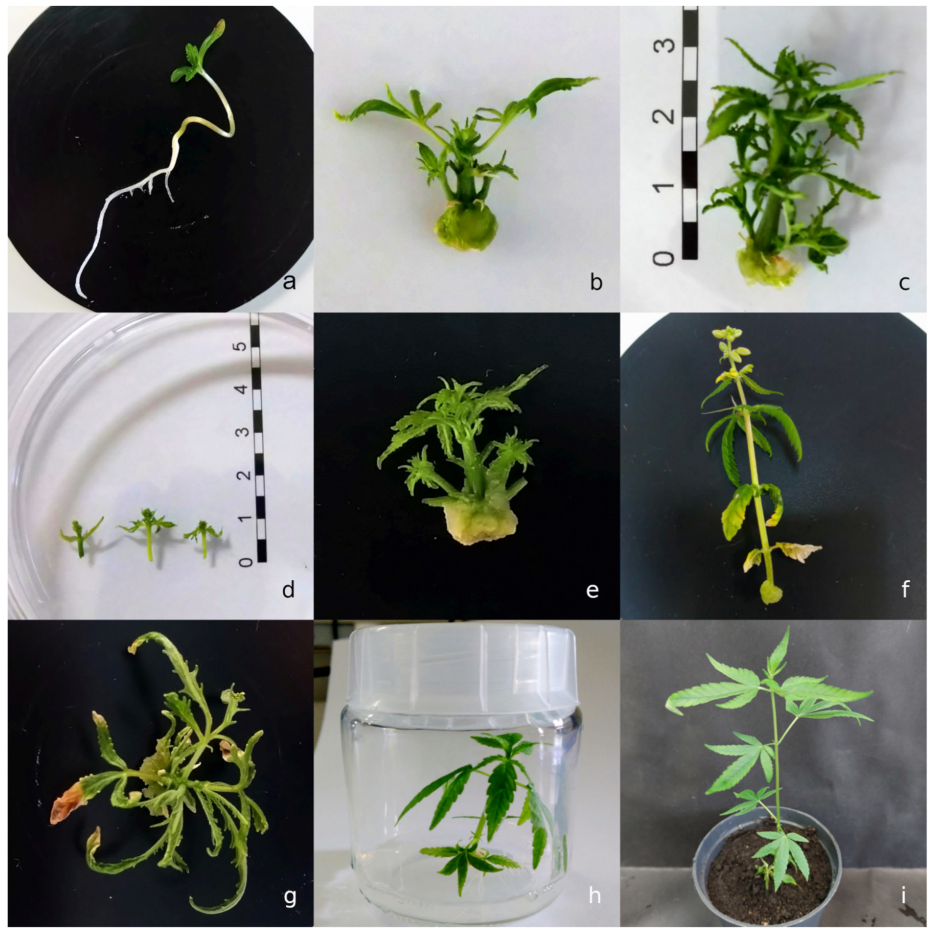
Table 3). Reduced TIBA concentration (1 µM and 0.5 µM) and shortened time of exposure resulted in lower malformation rates (13%, 22% and 14.7% respectively for Fedora 17, Finola and Diana) and relatively high response and multiplication rates (3.1, 3.7 and 3.3). In turn, lowered TDZ concentration usually resulted in lower numbers of shoots per explant (Table 4 and Table 5). Therefore, to reach a compromise between a satisfactory multiplication rate and lack of or a low malformation rate, additional tests for a given hemp variety are required.
After three weeks of culture, all shoots were rooted on the same rooting medium containing the same concentration of auxin (IBA) to compare the effect of hemp genotype. IBA has been tested and recommended for hemp rooting previously [6,40], whereas in another study [4] no significant difference was found in rooting rates between media supplemented with IBA and IAA. In the present study, relatively low rooting rates were recorded for Finola or Diana var. Fedora plants were characterized by the highest rooting rate (46.7%) and number of roots per plant (4.6). These results are comparable with rooting rates of 44% and 50% reported by Monthony et al. [33] and Smýkalová et al. [13], respectively. However, higher values of 74.6% [4] and lower values of 18% [11] were recorded for rooting rates of fibrous hemp. It is worth noting that the effect of genotype plays a crucial role in hemp rooting, as was confirmed in this study. On the other hand, a prolonged effect of polar auxin flux inhibition on the poor rooting cannot be excluded. It is known that polar auxin transport inhibitors can completely suppress rooting and affect morphogenesis and development of roots [36,41]. However, another explanation should be considered: the extensive callusing of explants. The best rooting rate was achieved for poor callusing (6.7%) Fedora 17 explants, whereas callusing rates recorded for Diana and Finola were significantly higher (32–33%). Additional tests are needed, using different rooting media and different auxins, to match the appropriate rooting medium to a given hemp variety. Regardless, the rooted plants acclimatized easily and showed no morphological changes. In this study, the whole cycle lasted 63–70 days. In alternative procedures, shoot regeneration, rooting and acclimatization of plants took from 56–63 days [9,10] to 66–70 days [4,7]. Further optimization of the rooting and acclimatization steps could significantly shorten the whole procedure.
Plants transferred under greenhouse conditions flowered and developed seeds, which proved their full functionality. Therefore, we can conclude that our study showed that by using auxin polar transport inhibitors such as NPA and TIBA it is possible to increase the efficiency of shoot regeneration. However, other factors, predominantly the genotype effect, influence shoot hemp micropropagation.

5. Conclusions

This study demonstrated for the first time that auxin polar transport inhibitors NPA and TIBA had a promotional effect on shoot regeneration from hemp epicotyl explants. Both inhibitors increased the response of explants and multiplication rate, but the effect was dose- and genotype-dependent. Side effects of the inhibitors such as senescence of leaves, vitrification and malformations were also observed. Therefore, we recommend adjusting the concentration and time of exposure to the inhibitor for a given genotype. We suggest the culture of explants in a two-step procedure as more suitable than long-term exposure of explants on media with inhibitors. The developed protocol enables direct shoot regeneration of fibrous hemp and obtaining the plantlets within 9–10 weeks. Further studies should include a wider pool of Cannabis genotypes and focus on developing rooting media. The results obtained in this study will be useful in micropropagation of recalcitrant hemp varieties and can contribute to the development of a tissue culture system suitable for fibrous type hemp.

Author Contributions

Conceptualization, M.D.; methodology, M.D. and M.S.; software, M.D. and M.S.; investigation, M.D.; data curation, M.D.; writing—original draft preparation, M.D.; writing—review and editing, M.D.; visualization, M.D.; project administration, M.S.; funding acquisition, M.S. All authors have read and agreed to the published version of the manuscript.

Funding

This research was funded by the Polish Ministry of Agriculture and Rural Development, resolution of the Council of Ministers No: 171/2017.

Institutional Review Board Statement

Not applicable.

Informed Consent Statement

Not applicable.

Data Availability Statement

All data sets are available upon reasonable request from the corresponding author.

Acknowledgments

The authors would like to thank Karolina Wielgus and Małgorzata Górska-Paukszta as well as Grażyna Mańkowska for providing hemp seeds from the collection of the Fibrous Plants Gene Bank of INF&MP NRI.

Conflicts of Interest

The authors declare no conflict of interest.

References

  1. Shen, P.; Gao, Z.; Fang, B.; Rao, J.; Chen, B. Ferreting out the secrets of industrial hemp protein as emerging functional food ingredients. Trends Food Sci. Technol. 2021, 112, 1–15. [Google Scholar] [CrossRef]
  2. Zhao, J.; Xu, Y.; Wang, W.; Griffin, J.; Roozeboom, K.; Wang, D. Bioconversion of industrial hemp biomass for bioethanol production: A review. Fuel 2020, 281, 118725. [Google Scholar] [CrossRef]
  3. Duque Schumacher, A.G.; Pequito, S.; Pazour, J. Industrial Hemp Fiber: A Sustainable and Economical Alternative to Cotton. J. Clean. Prod. 2020, 268, 122180. [Google Scholar] [CrossRef]
  4. Wróbel, T.; Dreger, M.; Wielgus, K.; Słomski, R. Modified Nodal Cuttings and Shoot Tips Protocol for Rapid Regeneration of Cannabis sativa L. J. Nat. Fibers 2020. [Google Scholar] [CrossRef]
  5. Monthony, A.S.; Page, S.R.; Hesami, M.; Jones, A.M.P. The Past, Present and Future of Cannabis sativa Tissue Culture. Plants 2021, 10, 185. [Google Scholar] [CrossRef]
  6. Lata, H.; Chandra, S.; Khan, I.; ElSohly, M.A. High Frequency Plant Regeneration from Leaf Derived Callus of High delta 9-Tetrahydrocannabinol Yielding Cannabis sativa L. Planta Med. 2010, 76, 1629–1633. [Google Scholar] [CrossRef]
  7. Lata, H.; Chandra, S.; Khan, I.; ElSohly, M.A. Thidiazuron-induced high-frequency direct shoot organogenesis of Cannabis sativa L. In Vitro Cell. Dev. Biol. Plant 2009, 45, 12–19. [Google Scholar] [CrossRef]
  8. Lata, H.; Chandra, S.; Techen, N.; Khan, I.A.; ElSohly, M.A. In vitro mass propagation of Cannabis sativa L.: A protocol refinement using novel aromatic cytokinin meta-topolin and the assessment of eco-physiological, biochemical and genetic fidelity of micropropagated plants. J. Appl. Res. Med. Aromat. Plants 2016, 3, 18–26. [Google Scholar] [CrossRef]
  9. Chaohua, C.; Gonggu, Z.; Lining, Z.; Chunsheng, G.; Qing, T.; Jianhua, C.; Xinbo, G.; Dingxiang, P.; Jianguang, S. A rapid shoot regeneration protocol from the cotyledons of hemp (Cannabis sativa L.). Ind. Crops Prod. 2016, 83, 61–65. [Google Scholar] [CrossRef]
  10. Wang, R.; He, L.S.; Xia, B.; Tong, J.F.; Li, N.; Peng, F. A micropropagation system for cloning of hemp (Cannabis sativa L.) by shoot tip culture. Pak. J. Bot. 2009, 41, 603–608. [Google Scholar]
  11. Galán-Ávila, A.; García-Fortea, E.; Prohens, J.; Herraiz, F.J. Development of a direct In Vitro plant regeneration protocol from Cannabis sativa L. Seedling explants: Developmental morphology of shoot regeneration and ploidy level of regenerated plants. Front. Plant Sci. 2020, 11, 645. [Google Scholar] [CrossRef] [PubMed]
  12. Murashige, T.; Skoog, F. A revised medium for rapid growth and bioassays wit tobacco tissue cultures. Physiol. Plant. 1962, 15, 473–497. [Google Scholar] [CrossRef]
  13. Smýkalová, I.; Vrbová, M.; Cvečková, M.; Plačková, L.; Žukauskaitė, A.; Zatloukal, M.; Hrdlicka, J.; Plíhalová, L.; Doležal, K.; Griga, M. The effects of novel synthetic cytokinin derivatives and endogenous cytokinins on the In Vitro growth responses of hemp (Cannabis sativa L.) explants. Plant Cell Tissue Organ Cult. 2019, 139, 381–394. [Google Scholar] [CrossRef]
  14. Barbier, F.F.; Dun, E.A.; Beveridge, C.A. Apical dominance. Curr. Biol. 2017, 11, R853–R909. [Google Scholar] [CrossRef] [PubMed] [Green Version]
  15. Kebrom, T.H. A Growing Stem Inhibits Bud Outgrowth—The Overlooked Theory of Apical Dominance. Front. Plant Sci. 2017, 8, 1874. [Google Scholar] [CrossRef] [PubMed]
  16. Bertheloot, J.; Barbier, F.; Boudon, F.; Perez-Garcia, M.D.; Péron, T.; Citerne, S.; Dun, E.; Beveridge, C.A.; Godin, C.; Sakr, S. Sugar availability suppresses the auxin-induced strigolactone pathway to promote bud outgrowth. New Phytol. 2020, 225, 866–879. [Google Scholar] [CrossRef] [Green Version]
  17. Prusinkiewicz, P.; Crawford, S.; Smith, R.S.; Ljung, K.; Bennett, T.; Ongaro, V.; Leyser, O. Control of bud activation by an auxin transport switch. Proc. Natl. Acad. Sci. USA 2009, 106, 17431–17436. [Google Scholar] [CrossRef] [Green Version]
  18. Cheng, Z.J.; Wang, L.; Sun, W.; Zhang, Y.; Zhou, C.; Su, Y.H.; Li, W.; Sun, T.T.; Zhao, X.Y.; Li, X.G.; et al. Pattern of auxin and cytokinin responses for shoot meristem induction results from the regulation of cytokinin biosynthesis by AUXIN RESPONSE FACTOR3. Plant Physiol. 2013, 161, 240–251. [Google Scholar] [CrossRef] [Green Version]
  19. Luisi, A.; Lorenzi, R.; Source, C. Strigolactone may interact with gibberellin to control apical dominance in pea (Pisum sativum). Plant Growth Regul. 2011, 65, 415–419. [Google Scholar] [CrossRef]
  20. Li, M.; Wei, Q.; Xiao, Y.; Peng, F. The effect of auxin and strigolactone on ATP/ADP isopentenyltransferase expression and the regulation of apical dominance in peach. Plant Cell Rep. 2018, 37, 1693–1705. [Google Scholar] [CrossRef]
  21. Müller, D.; Waldie, T.; Miyawaki, K.; To, J.P.; Melnyk, C.W.; Kieber, J.J.; Kakimoto, T.; Leyser, O. Cytokinin is required for escape but not release from auxin mediated apical dominance. Plant J. 2015, 82, 874–886. [Google Scholar] [CrossRef] [PubMed] [Green Version]
  22. Stephin, S.; Gangaprasad, A.; Mathew, S.P.; Muthukrishnan, S. Enhanced In Vitro Shoot Multiplication of Piper sarmentosum by Suppression of Apical Dominance. Proc. Natl. Acad. Sci. India Sect. B Biol. Sci. 2020, 90, 87–94. [Google Scholar] [CrossRef]
  23. Marks, T.R.; Simpson, S.E. Factors affecting shoot development in apically dominant Acer cultivars in vitro. J. Hortic. Sci. 1994, 69, 543–551. [Google Scholar] [CrossRef]
  24. Lall, S.; Mandegaran, Z.; Roberts, A.V. Shoot multiplication in cultures of mature Alnus glutinosa. Plant Cell Tissue Organ Cult. 2005, 83, 347–350. [Google Scholar] [CrossRef]
  25. Bhau, B.S.; Wakhlu, A.K. Effect of genotype, explant type and growth regulators on organogenesis in Morus alba. Plant Cell Tissue Organ Cult. 2001, 66, 25–29. [Google Scholar] [CrossRef]
  26. Sing, S.K.; Syamal, M.M. Anti-auxin enhance Rosa hybrida L. micropropagation. Biol. Plant. 2000, 43, 279–281. [Google Scholar] [CrossRef]
  27. Shukla, P.S.; Das, A.K.; Jha, B.; Agarwal, P.K. High-frequency in vitro shoot regeneration in Cucumis sativus by inhibition of endogenous auxin. Vitro Cell. Dev. Biol. Plant 2014, 50, 729–737. [Google Scholar] [CrossRef]
  28. Hu, W.; Fagundez, S.; Katin-Grazzini, L.; Li, Y.; Li, W.; Chen, Y.; Wang, X.; Deng, Z.; Shenxi, X.; McAvoy, R.J.; et al. Endogenous auxin and its manipulation influence in vitro shoot organogenesis of citrus epicotyl explants. Hortic. Res. 2017, 4, 17071. [Google Scholar] [CrossRef] [Green Version]
  29. Mestinšek-Mubi, Š.; Svetik, S.; Flajšman, M.; Murovec, J. In vitro tissue culture and genetic analysis of two high-CBD medical cannabis (Cannabis sativa L.) breeding lines. Genetika 2020, 52, 925–941. [Google Scholar] [CrossRef]
  30. Monthony, A.S.; Kyne, S.T.; Grainger, C.M.; Jones, A.M.P. Recalcitrance of Cannabis sativa to de novo regeneration; a multi-genotype replication study. PLoS ONE 2021, 16, e0235525. [Google Scholar] [CrossRef]
  31. Piunno, K.; Golenia, G.; Boudko, E.A.; Downey, C.; Jones, A.M.P. Regeneration of shoots from immature and mature inflorescences of Cannabis sativa. Can. J. Plant Sci. 2019, 99, 556–559. [Google Scholar] [CrossRef]
  32. Page, S.R.G.; Monthony, A.S.; Jones, A.M.P. Basal media optimization for the micropropagation and callogenesis of Cannabis sativa L. bioRxiv 2020, 1, 1–23. [Google Scholar] [CrossRef] [Green Version]
  33. Monthony, A.S.; Bagheri, S.; Zheng, Y.; Jones, A.M.P. Flower power: Floral reversion as a viable alternative to nodal micropropagation in Cannabis sativa. Vitro Cell. Dev. Biol. Plant 2021. [Google Scholar] [CrossRef]
  34. Slusarkiewicz-Jarzina, A.; Ponitka, A.; Kaczmarek, Z. Influence of cultivar, explant source and plant growth regulator on callus induction and plant regeneration of Cannabis sativa L. Acta Biol. Crac. Ser. Bot. 2005, 47, 145–151. [Google Scholar]
  35. Wielgus, K.; Luwańska, A.; Lassocinski, W.; Kaczmarek, Z. Estimation of Cannabis sativa L. tissue culture conditions essential for callus induction and plant regeneration. J. Nat. Fibers 2008, 5, 199–207. [Google Scholar] [CrossRef]
  36. Ueda, J.; Saniewski, M.; Miyamoto, K. Auxin, One Major Plant Hormone. In Bioactive Compounds in Agricultural Soils; Szajdak, L., Ed.; Springer: Cham, Switzerland, 2016. [Google Scholar] [CrossRef]
  37. Dhonukshe, P.; Grigoriev, I.; Fischer, R.; Tominaga, M.; Robinson, D.G.; Hašek, J.; Paciorek, T.; Petrášek, J.; Seifertová, D.; Tejos, R.; et al. Auxin transport inhibitors impair vesicle motility and actin cytoskeleton dynamics in diverse eukaryotes. Proc. Natl. Acad. Sci. USA 2008, 105, 4489–4494. [Google Scholar] [CrossRef] [PubMed] [Green Version]
  38. Zhu, J.; Bailly, A.; Zwiewka, M.; Sovero, V.; Di Donato, M.; Ge, P.; Oehri, J.; Aryal, B.; Hao, P.; Linnert, M.; et al. TWISTED DWARF1 Mediates the Action of Auxin Transport Inhibitors on Actin Cytoskeleton Dynamics. Plant Cell 2016, 28, 930–948. [Google Scholar] [CrossRef] [PubMed] [Green Version]
  39. Novikova, T.I.; Zaytseva, Y.G. Some Approaches to Overcome TDZ-Induced Abnormalities. In Thidiazuron: From Urea Derivative to Plant Growth Regulator; Naseem, A., Mohammad, F., Eds.; Springer Nature Singapore Pte Ltd.: Singapore, 2018; pp. 80–93. [Google Scholar] [CrossRef]
  40. Movahedi, M.; Ghasemi-Omran, V.-O.; Torabi, S. The effect of different concentrations of TDZ and BA on In Vitro regeneration of Iranian cannabis (Cannabis sativa) using cotyledon and epicotyl explants. J. Plant Mol. Breed. 2015, 3, 20–27. [Google Scholar] [CrossRef]
  41. Saniewski, M.; Góraj, J.; Węgrzynowicz-Lesiak, E.; Miyamoto, K.; Ueda, J. Differential effects of auxin polar transport inhibitors on rooting in some Crassulaceae species. Acta Agrobot. 2014, 67, 85–92. [Google Scholar] [CrossRef] [Green Version]
Figure 1. (a) Hemp seedling (Fedora 17 var.); (b) shoot regeneration (Diana var.) on medium with TDZ and TIBA (5.0 mg L−1); (c) shoot regeneration (Finola var.) on medium with TDZ and TIBA (1.0 mg L−1); (d) the regenerated shoots (var. Finola); (e) shoot regeneration (Fedora var.) on medium TDZ with NPA (2.5 mg L−1); (f) flowering Finola explant on medium with mT and NPA (10 mg L−1); (g) adverse morphological changes after exposition exposure to high TIBA concentration 12.5 mg L−1); (h) regenerated and rooted plant (Fedora var.); (i) the acclimatized plant.
Agronomy 12 00104 g001
Table 1. Breeding information on tested hemp varieties.
VarietyOriginPurposeGenotypic ExpressionHeight at Maturity (cm)Vegetative Cycle (Days)CBD Content (%)THC Content (%)
Fedora 17FranceGrain/CBD/FiberMonoecious200–250<1251.5–2.0<0.06
DianaRomaniaGrain/FiberMonoecious200–250<130>1.0<0.15
FinolaFinlandGrain/CBD/FiberDioecious150–200<1100.4–2.7<0.1
Table 2. Effect of NPA and genotype on shoot regeneration of C. sativa epicotyl explants after three weeks of culture on medium with mT.
Regulator (mg L−1)NPA (mg L−1)Responding Explants (%)Mean No. of Shoots per Explant
DianaFedora 17FinolaDianaFedora 17Finola
mT (1.0)0.051.1 a50.0 ab10.9 a2.1 ± 1.29 a2.3 ± 1.41 a1.2 ± 0.63 ab
1.053.2 a56.5 b41.3 b2.0 ± 0.95 a2.1 ± 1.14 ab1.9 ± 1.07 c
2.552.2 a52.2 ab29.2 b2.2 ± 1.28 a2.3 ± 1.34 a1.5 ± 0.92 a
5.053.2 a43.5 ab28.3 b2.1 ± 1.19 a1.9 ± 1.04 ab1.5 ± 0.86 ac
10.041.3 a34.8 a8.5 a2.0 ± 0.68 a1.6 ± 0.95 b1.1 ± 0.56 b
Values represent percentage (of responding explants) and mean ± SE of three replicates, each with 15 explants for treatment. Different lowercase letters in the same column indicate a significant difference according to Fisher’s test (responding explants) and Duncan’s test.
Table 3. Effect of auxin transport inhibitors NPA and TIBA and genotype on shoot regeneration of C. sativa epicotyl explants cultured after three weeks of culture on medium with TDZ.
Regulator (mg L−1)NPA (mg L−1)TIBA (mg L−1)Responding Explants (%)Mean No. of Shoots per ExplantCallusing Explants (%)
DianaFedora 17FinolaDianaFedora 17FinolaDianaFedora 17Finola
TDZ (0.5)0.00.063.8 abc56.3 ab68.9 ab2.4 ± 1.21 ab2.0 ± 1.10 a2.6 ± 1.25 ab80.9 a62.5 a91.1 a
1.0-89.1 d82.2 cd59.6 ad3.1 ± 1.13 cd2.8 ± 1.18 bc2.3 ± 1.12 ac56.5 b22.2 b8.5 b
2.5-80.4 ad91.3 d86.4 b2.9 ± 1.31 acd3.2 ± 1.15 b2.8 ± 0.96 ab26.1 c10.9 b4.6 b
5.0-76.6 ad71.7 acd75.5 ab3.0 ± 1.52 acd3.0 ± 1.49 b2.7 ± 1.16 ab27.7 c23.9 b4.1 b
10.0-48.9 b70.2 ac71.7 ab2.0 ± 1.14 b2.3 ± 0.99 ac2.4 ± 1.25 ac15.6 c10.6 b8.7 b
-0.25-75.0 acd68.9 ab-2.7 ± 1.45 bc2.9 ± 1.54 ab-81.3 c95.6 a
-0.571.1 abd80.0 cd71.1 ab3.0 ± 1.58 acd2.9 ± 1.27 b3.0 ± 1.61 b80.0 a62.2 a86.7 a
-2.572.9 ad80.9 cd71.1 ab3.4 ± 2.00 b2.8 ± 1.11 bc2.7 ± 1.38 ab66.7 ab57.2 a57.8 c
-5.066.7 abc75.6 acd44.4 cd2.5 ± 1.34 abc2.7 ± 1.18 bc2.0 ± 1.39 cd57.8 b48.9 a53.3 c
-12.551.1 bc42.6 b35.6 c2.1 ± 1.21 b1.9 ± 1.32 a1.5 ± 1.18 d46.7 b55.3 a46.4 d
Values represent percentage (of responding explants) and mean ± SE of three replicates, each with 15 explants for treatment. Different lowercase letters in the same column indicate a significant difference according to Fisher’s test (responding explants) and Duncan’s test.
Table 4. Effect of different treatment variants (inhibitors, TDZ concentration, time of cultivation) on shoot regeneration and malformation rate (%) of initial explants of C. sativa var. Fedora 17.
Variant TreatmentInhibitor (mg L−1)Time of Incubation (Week)TDZ (mg L−1)Mean No. of Shoots per ExplantResponse Rate (%)Malformation (%)
1TIBA (0.25)10.252.5 ± 1.00 a74.2 ab9.7
2TIBA (0.5)20.252.6 ± 1.13 ab73.3 ab10.0
3TIBA (0.5)20.503.1 ± 1.32 b84.8 b13.0
4NPA (2.5)10.252.1 ± 0.99 a60.0 a13.3
Values represent percentage (of responding explants) and mean ± SE of three replicates, each with 15 explants for treatment. Different lowercase letters in the same column indicate a significant difference according to Duncan’s test.
Table 5. Effect of different treatment variants (inhibitors and TDZ concentration) on shoot regeneration and malformation rate (%) of initial explants of C. sativa var. Finola.
Variant TreatmentInhibitor (mg L−1)TDZ (mg L−1)Mean No. of Shoots per ExplantResponse Rate (%)Malformation (%)
1TIBA (0.5)0.252.4 ± 1.41 a54.5 a21.2
2TIBA (0.5)0.503.7 ± 1.50 b90.3 b22.9
3NPA (2.5)0.253.0 ± 1.39 ab76.7 b20.0
Time of incubation on induction media was one week. Values represent percentage (of responding explants) and mean ± SE of three replicates, each with 15 explants for treatment. Different lowercase letters in the same column indicate a significant difference according to Duncan’s test.
Table 6. Effect of different treatment variant (inhibitors, TDZ concentration) on shoot regeneration and malformation rate (%) of initial explants of C. sativa var. Diana.
Variant TreatmentInhibitor (mg L−1)TDZ (mg L−1)Mean No. of Shoots per ExplantResponse Rate (%)Malformation (%)
1TIBA (0.25)0.53.3 ± 1.38 a85.3 a14.7
2NPA (1.0)0.52.4 ± 1.14 b64.5 b6.5
Time of incubation on induction media was two weeks. Values represent percentage (of responding explants) and mean ± SE of three replicates, each with 15 explants for treatment. Different lowercase letters in the same column indicate a significant difference according to Fisher’s test.
Table 7. Effect of genotype on rooting rate, mean number of roots and leaves per shoot, and callusing rate after three weeks of culture on rooting medium.
VarietyNRooting Rate (%)Mean Plant Height (cm)Mean No. of Roots per PlantMean No. of Leaves per PlantCallusing Explants (%)No. of Rooted Plants
Diana4623.9 a1.1 ± 0.66 a4.1 ± 2.70 a3.2 ± 1.48 a32.6 a11
Fedora 174546.7 b1.5 ± 1.30 a6.3 ± 3.99 b4.6 ± 2.36 b6.7 b21
Finola4438.6 ab1.4 ± 1.35 a4.4 ± 3.44 ab4.6 ± 1.52 b34.1 a17
Values represent percentage (of responding explants) and mean ± SE of three replicates, each with 15 explants for treatment. The mean number of roots per plant was calculated for the rooted plants; the number of them is presented in the last column. Different lowercase letters in the same column indicate a significant difference according to Duncan’s test.
Publisher’s Note: MDPI stays neutral with regard to jurisdictional claims in published maps and institutional affiliations.

