Article

The Effects of Hydrogen Peroxide on Plant Growth, Mineral Accumulation, as Well as Biological and Chemical Properties of Ficus deltoidea

by
Nik Nurnaeimah
1,
Nashriyah Mat
1,
Khamsah Suryati Mohd
1,
Noor Afiza Badaluddin
1,
Nornasuha Yusoff
1,
Mohammad Hailmi Sajili
1,
Khairil Mahmud
2,
Ahmad Faris Mohd Adnan
3 and
Mohammad Moneruzzaman Khandaker
1,*
1
School of Agriculture Science & Biotechnology, Faculty of Bioresources and Food Industry, Universiti Sultan Zainal Abidin, Besut Campus, Besut 22200, Terengganu, Malaysia
2
Department of Crop Science, Faculty of Agriculture, Universiti Putra Malaysia, Seri Kembangan 43400, Selangor, Malaysia
3
Institute of Biological Sciences, Faculty of Science, University of Malaya, Kuala Lumpur 50603, Malaysia
*
Author to whom correspondence should be addressed.
Agronomy 2020, 10(4), 599; https://doi.org/10.3390/agronomy10040599
Submission received: 27 January 2020 / Revised: 9 April 2020 / Accepted: 17 April 2020 / Published: 23 April 2020
(This article belongs to the Section Soil and Plant Nutrition)

Abstract

:
Hydrogen peroxide (H2O2) is defined as a reactive oxygen species (ROS), able to cause damage to a variety of cellular structures. On the other hand, recent work has demonstrated that H2O2 can also act as a potent signaling molecule that mediates various physiological and biochemical processes in plants. This study was carried out to investigate the effects of H2O2 on the growth, mineral nutrient accumulation, as well as the biologic and chemical properties of Ficus deltoidea var. deltoidea. F. deltoidea plants were spray-treated with 0- (control), 8-, 16-, 30- and 60-mM H2O2 under field conditions. Plant height, leaf area, chlorophyll content, net photosynthetic rate, stomatal conductance and quantum yield of the F. deltoidea plants significantly increased after treatment with 16 and 30-mM H2O2. The results indicate that 60-mM H2O2 increased the accumulation of arsenic, iron and sodium content in the leaves of F. deltoidea. On the other hand, 8-mM H2O2 significantly enhanced the accumulation of arsenic, iron, calcium and potassium content in the syconium of F. deltoidea plants. In addition, H2O2 treatment did not produce any significant effects on antimony and magnesium accumulation in the leaves or the syconium of F. deltoidea plants. The results show that the F. deltoidea plant has strong antidiabetic properties and its α-glucosidase activity increased in treated plants compared to standard acarbose. Hydrogen peroxide, particularly in concentrations of 16 and 30 mM, increased the antioxidant activity, total phenolic and flavonoid content and the vitexin and isovitexin content. There was a positive correlation between antioxidant activity with total phenol and total flavonoid content in H2O2-treated plants. The quantitative analysis by HPTLC indicates that the amount of vitexin and isovitexin increased with the higher concentrations of H2O2. From this study, it can be concluded that spraying 16 and 30-mM H2O2 once a week enhances growth, mineral accumulation and stimulates bioactive compounds of the F. deltoidea plants.

1. Introduction

Ficus deltoidea, is a medicinal herb from the Moraceae family, that can found in Malaysia, Africa, Thailand, Hawaii, Indonesia and the Southern Philippines [1,2] and, according to Badron et. al. [3], the Malaysian forest has approximately 16% (101) of the known species of Ficus. This plant can be found in abundance along the peat soils, beaches and in hilly forest up to 3000 m above sea level [4]. The species is cultivated in various parts of the world and is grown as a house plant in cooler regions [2]. It is a small plant up to 3 m tall, sometimes occurring as an epiphyte and exhibits low photosynthetic rates during its vegetative growth. Much research has been done to recognize the importance of this plant highlighting its antidiabetic [5,6], antinociceptive [7], antioxidant [8], antimicrobial [9] and anti-proliferative [10] uses. Historically, each part of this plant is known to provide benefits including reduced sugar levels in the blood [5], reduced blood pressure [10], contraction of the vagina after delivery [11], delayed menopause and also to reduce the risk of cancer [11,12,13]. At present, numerous plant-based sources are used by the pharmaceutical industry for the production of bioactive compounds or secondary metabolites [8]. The plant’s use as an alternative medicinal herb is gaining popularity with the sale of tea bags and capsules of F. deltoidea in the local market [5]. F. deltoidea contains 5 main active compounds including flavonoids [11], tannins, triterpenoids, proanthocyanins and phenols [14]. More than 20 classes of flavonoids have been reported present in the leaves of F. deltoidea displaying properties of high antioxidant activities [15]. Currently, two bioactive compounds suggested to be possessed by F. deltoidea which have α-glucosidase inhibition are vitexin and isovitexin [11]. To increase plant growth, mineral accumulation, as well as biologic and chemical properties of F. deltoidea, hydrogen peroxide (H2O2) was used as an alternative treatment to regulate the mechanism and role of H2O2 in the plant, with the hope of increasing the plant’s potential utility in herb production or research.
Hydrogen peroxide (H2O2) is defined as a reactive oxygen species (ROS) generated from molecular oxygen (O2) with relatively high stability and a long half-life. The controlled cellular production of H2O2 plays an important physiological role since the high cellular level of H2O2 can produce carcinogenic effects and induce cell death. Superoxide radical (O2) and H2O2 are produced during cell metabolism in plants and in normal high amounts in various cell compartments, especially chloroplasts, because chloroplasts are well equipped with defense enzymes against O2 and H2O2 [16]. Almeida et al. [17] stated that H2O2 is one of the ROS produced in plants under both biotic and abiotic conditions and plays a key role against O2 derived cell toxicity. H2O2 acts as a messenger molecule involved in adaptive signaling, triggering tolerance against various abiotic stresses at low concentrations, but at high concentrations it orchestrates programmed cell death [18]. Usually, abiotic stress, such as drought, will increase the production of ROS in the plant. Orabi et al. [19] stated that a lower level of treatment with H2O2 can have a significant positive effect on plant growth, growth regulators, antioxidant enzyme activity, fruit yield and quality of tomato. It has also been reported in a study by Khandaker et al. [20], that exogenous application of H2O2 increased the plant growth, physiological activities and biochemical properties of wax apple fruits.
To the best of our knowledge, there is little research about the effects of H2O2 on the mineral accumulation, as well as the biologic and chemical properties of Ficus deltoidea. What has been reported in the literature so far shows scarcely any information of F. deltoidea available. The main purpose of this study was to improve the plant performance by growth, photosynthesis, mineral accumulation and the biologic and chemical properties of F. deltoidea by using H2O2. To reach the aim of this study an experiment was carried out with these objectives; to identify the effects of H2O2 on the growth and photosynthesis of F. deltoidea; and to determine the influence of H2O2 on mineral absorption and biologic and chemical properties of F. deltoidea. The novel findings of this study will have a fundamental impact to improve the photosynthetic capacity and plant productivity, mineral accumulation, increase antioxidant activity, as well as improve the phytochemical properties of this plant.

2. Materials and Methodology

2.1. Plant Material and Experimental Site

The studies were carried out at a research plot of the Faculty of Bioresources and Food Industry, Universiti Sultan Zainal Abidin, Besut Campus, Besut, Terengganu, under a sunlight-proof shade house which covers an area of approximately 40 m2, between the months of January, 2015 to October, 2016. The mother plant of F. deltoidea var. deltoidea was obtained from Kampung Sungai Nibong, Batu Pahat, Johor, Malaysia. Forty (40) uniform eight-week-old F. deltoidea cuttings were made from the collected mother plant and transplanted into a growing medium containing 15 kg of BRIS soil, which contained rotten empty oil palm fruit bunches and organic manure. Approximately ten grams of nutrients (N: P: K, at a ratio of 10: 10: 10) per plant were applied 15-days before treatment application. The plants were watered daily with a sprinkler during the whole growth stage.
All the experiments, which were conducted in the field, were performed under the following normal prevailing conditions: temperature 21–30 °C, maximum PAR 500–1000 μE/m2/s and relative humidity of 60–90%. Plants were sprayed manually with approximately 20 mL of the solution once a week, using water as control (0-mM H2O2) and, 8 mM, 16 mM, 30 mM and 60-mM H2O2, using a hand sprayer. The H2O2 treatment was applied to the plants weekly from the vegetative growth stage to flowering stage. In this study, a completely randomized design was used to layout the pot experiment and each treatment had eight randomly distributed replicates.

2.2. Plant Physiology and Growth Parameters

Data for chlorophyll content was recorded by a Chlorophyll Content Meter (Model CCM 200; Opti-science, USA). Net photosynthetic rate and stomatal conductance were collected using the same equipment, C1-340 Handheld Photosynthesis System (CID Bio-Science, Camas, WA, USA). All of the necessary setup and calibration was followed in accordance with the C1–340 Handled Photosynthesis System manual (CID Bio-Science, USA). For chlorophyll fluorescence, the method described by Xia et al. [21] was used with a slight modification in data collection and equipment. Plant height was measured from the base of the plant, on the soil surface, to the tip of the highest branch point in centimeters. The matured leaves of each plant were selected, and the leaf area was measured using the Leaf Area Meter (Model Portable Laser CI-202, CID Bio-science, Camas, WA, USA) with replicate data recorded.

2.3. Mineral Accumulation

There were 7 kinds of minerals analyzed including arsenic (As), antimony (Sb), iron (Fe), magnesium (Mg), calcium (Ca), sodium (Na) and potassium (K). Two parts of the plant were selected, the leaf and the syconium. Fresh and healthy leaves and syconium were collected and cleaned with distilled water before being dried in the oven for 24 h at 60 °C. The dried samples were ground into a powder in a mortar and pestle. The samples were stored in an airtight bottle at the Malaysian Nuclear Agency until needed to carry out the experiment. The method used to analyze the mineral uptake was the Neutron Activation Analysis (NAA) as described by Nashriyah et al. [22].

2.4. Biologic and Chemical Properties

2.4.1. Sample Preparation Procedure

Five hundred grams of fresh leaves of F. deltoidea for each solvent were collected and cleaned with water, and then oven dried at 60 °C for a day then ground into a fine powder. The sample was extracted using three types of solvent, namely water, ethanol and chloroform. The hot aqueous extracts were prepared by boiling the powder samples in distilled water for 3 h. The combined suspension was filtered using Whatman filter paper no. 54, while the ethanolic and chloroform extracts were extracted using the maceration method (ratio 1:10, solid: liquid), the leaf powder was soaked with each solvent for 3 days at room temperature and the crude extracts were obtained using centrifugal evaporators (EZ 2 ELITE).

2.4.2. DPPH• Radical -Scavenging Activity, in Vitro α-Glucosidase Inhibitory, Total Phenolic and Total Flavonoid Content Assay

1,1-Diphenyl-2-picrylhydrazyl (DPPH•) radical scavenging activity was performed by the method described by Aktel et al. [23] with some modifications. For each determination, the stock solution (5 mg/mL) was diluted to a dilution series, 0 (Blank)–500 µg/mL with dimethyl sulfoxide. An aliquot of each dilution (25 µL) and dimethyl sulfoxide (25 µL) were added to each well and mixed with a methanolic solution of DPPH• (200 µL, 0.1 mM). The mixtures were incubated at 37 °C in the dark for 30 min. The absorbance was measured at 517 nm. The concentration of the sample necessary to scavenge DPPH• concentration by 50% was obtained by interpolation from linear regression analysis and denoted as IC50 value (µg/ mL). All determinations were carried out in triplicate. Ascorbic acid, gallic acid and quercetin were used as reference compounds.
The antidiabetic assay was determined by in vitro α-glucosidase inhibitory assay. In this assay, the method by Choo et al. [24] was followed with slight modifications and α-glucosidase enzyme derived from Bacillus stearothermophilus (Sigma-Aldrich, USA) was used. For each assay, 10 µL of the test sample at various concentrations was mixed with 50 µL of 0.1 M phosphate buffer (pH 7.0) and 25 µL of α-glucosidase solution (0.2 Unit/mL) in each well of a 96 well microplate. After incubation at 37 °C for 10 min, 25 µL of 0.5 mM 4-nitrophenyl α-D-glucopyranade (pNPG) was added to start the reaction, then the mixture was incubated at 37 °C for 30 min. After the incubation time had finished, 100 µL of 0.2 M sodium bicarbonate (Na2CO3) was added to each well to terminate the reaction. The absorbance was read at 410 nm. The percentage inhibition versus concentration of samples was plotted. The concentration of the sample necessary to scavenge α-glucosidase by 50% was obtained by interpolation from linear regression analysis and denoted IC50 value (µg/mL). All determinations were carried out in triplicate. Acarbose and quercetin were used as a reference compound.
The total soluble phenolic content in the extract was determined by using Folin-Ciocalteu reagent and gallic acid (3,4,5-trihydroxybenzoic acid) as a standard according to the method of Singleton and Rossi [25] with slight modifications on the standard concentration. A solution of 5 mg/mL of extract was diluted in methanol and a calibration of gallic acid standard solutions of 0.0625, 0.125, 0.25, 0.5, 1, 2.5 and 5 mg/mL were prepared. Twenty microliters of extract and each concentration of the gallic acid solution was pipetted into separate test tubes followed by the addition of 1.58 mL of distilled deionized water and 100 μL of Folin-Ciocalteu reagent. Subsequently, the test tubes were mixed thoroughly. After 8 min, 300 μL of 20% Na2CO3 solution was added. The mixture was then allowed to stand for 2 h. The absorbance of the solution was measured at 765 nm. All sample determinations were carried out in triplicate. The total phenolic content was calculated using the gallic acid calibration curve and the results were expressed as gallic acid equivalents (GAE) per g of dry extract.
The total flavonoid content in extracts was determined by using the aluminum chloride colorimetric method with quercetin as a standard [26]. A solution of 5-mg/mL of extract in methanol and solutions of 0.0078-, 0.0156-, 0.0313-, 0.0625-, 0.125-, 0.5-, 1-, 2.5- and 5-mg/mL of quercetin in methanol were prepared. Samples (500 μL) and each concentration of the quercetin solution were pipetted into separate test tubes followed by the addition of 0.1 mL of 10% (w/v) aluminum chloride solution, 0.1 mL of 1-M potassium acetate solution, 1.5 mL of methanol and 2.8 mL of distilled deionized water. The test tubes were thoroughly mixed. And after incubation at room temperature for 30 min, the absorbance of the reaction mixture was measured at 415 nm. All evaluation was carried out in triplicate. The amount of 10% aluminum chloride was substituted for the same amount of distilled water in a blank. The concentration of the total flavonoids was determined as mg of quercetin per g dry extract equivalent by using an equation obtained from the quercetin calibration curve.

2.4.3. High Performance Thin Layer Chromatography Fingerprint (HPTLC)

HPTLC was performed with some modifications from Mohd et al. [27] on the standard reference concentration. The sample extracts and standard were spotted on HPTLC plates precoated with silica gel F254 (layer thickness of 0.2 mm) (Merck) using TLC sampler ATS 4 Camag Linomat 5 model with the following settings; 7 tracks on a 20 cm × 10 cm plate. The samples were spotted in the form of narrow bands with a length of 8.0 mm from the bottom edge, 40 mm from the margin and 13.3 mm apart at a constant rate of 100 nL/seconds using a nitrogen aspirator. The migration distance was 8 cm with a migration time of 25 min. The development was performed in the automated developing chamber ADC 2 (CAMAG) with 5 min pre-drying, 1 min of preconditioning and mobile phase mixture of methanol: ethyl acetate: formic acid (0.01%). The densitometric analysis of the separated components was carried out using a Camag thin layer chromatography (TLC) scanner 3 (Camag, Switzerland) in the absorbance mode at 340 nm. The bands were scanned using deuterium and tungsten lamps and the scanning speed was maintained at 20 mm/seconds with a macro slit dimension of 8.00 mm × 0.2 mm. The integration of chromatograms was performed using the Camag TLC scanner system and win CATS software 1.4.6.2002 (CAMAG, Switzerland). For the quantitative analyses, a standard curve of vitexin and isovitexin were used as references.

2.5. Statistical Analysis

Statistical analysis was performed using SPSS 20 software. Repeated measures of ANOVA were used to analyze data from the plant physiological study. Other studies were subjected to one-way analysis of variance (ANOVA) using the general linear model and Tukey’s HSD-test to evaluate significant differences between treatments and the control plant at p = 0.05.

3. Results

3.1. Effects of Hydrogen Peroxide (H2O2) on Morphologic and Physiological Characteristics

The results show that a 30-mM H2O2 treatment produced the highest leaf chlorophyll content which was 17% higher than the control on the 12th week of observation. Table 1 also shows the significant effect of H2O2 on net photosynthetic rate and stomatal conductance of F. deltoidea plants. Among five treatments applied, 30-mM H2O2 increased the net photosynthetic rate 1.8-fold compared to the control treatment at the 12th week of the growth period. The control and 60-mM H2O2 treatments produced the lowest net photosynthetic rate.
As can be seen, the treatment with 30-mM H2O2 shows the highest leaf stomatal conductance at 11% higher than the control treatment. The treatment of H2O2 produced a significant effect on the quantum yield of F. deltoidea leaves (Table 1). However, all treatments produced higher values when compared to the control which were statistically different. Medium concentrations of applied H2O2 (16 and 30 mM) produced a significant effect on F. deltoidea plant height (Table 1). The plants treated with 16 and 30-mM H2O2 show the highest plant height when compared to the control plant. According to Table 1, there was no difference in leaf area between the treatments and control.

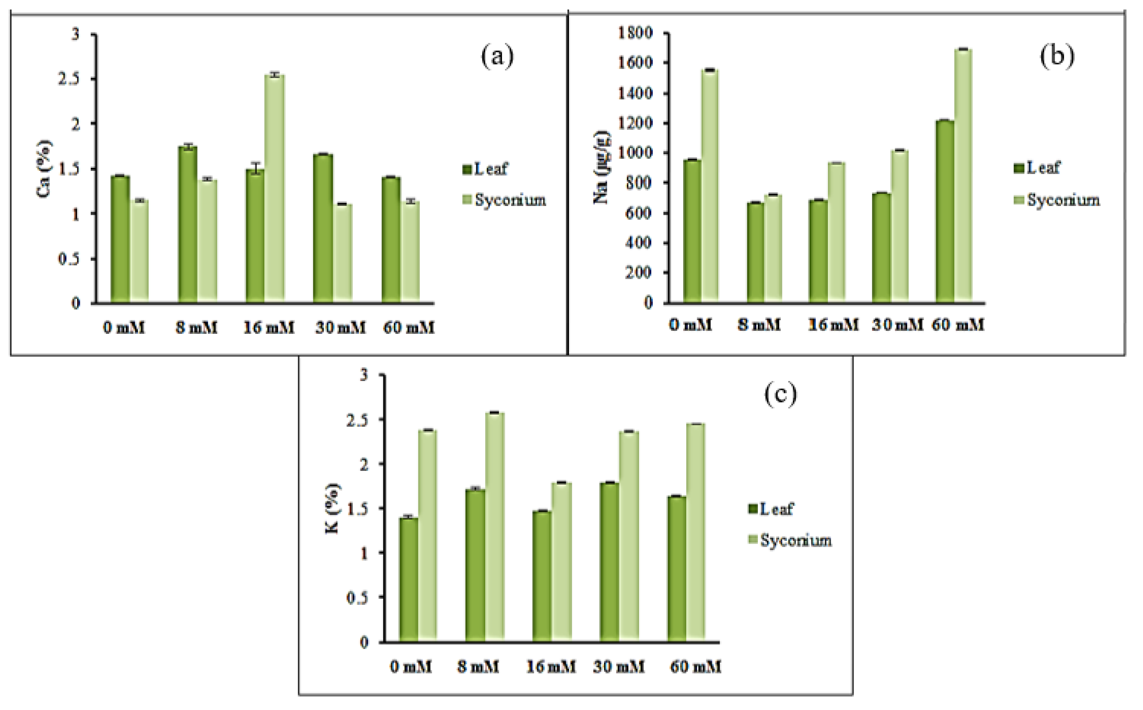
3.2. Mineral Accumulation

Figure 1 describes the arsenic (As) accumulation in the leaves and syconium. The intake in the leaves for the control plant was the lowest (0.51 ug/g) and the highest arsenic intake was in the plants treated with 60-mM H2O2 (0.64 ug/g). However, the As accumulation in the syconium was the highest (1.1 ug/g) in the 8 mM treated plants and the lowest were in the plants sprayed with 16, 30 and 60-mM H2O2 with values of 0.22, 0.20 and 0.21 ug/g, respectively. In the control plants, the arsenic accumulation was the same in leaves and syconium.
Figure 1b displays the antimony (Sb) accumulation in the leaves and syconium. A significant increase in Sb content was observed in the treated leaves compared to untreated plants (0.03 ug/g). The Sb accumulation in syconium shows different patterns, but there is no significant difference between the treated plants and untreated plants. However, there was a significant difference in Sb accumulation between treated leaves and syconium but there was no significant difference in untreated leaves and syconium.
Figure 1c shows the iron (Fe) uptake in leaves and syconium. The Fe accumulation in treated leaves significantly increased when compared with untreated leaves; the treatment of 60 mM produced the highest Fe accumulation (134.0 ug/g). A similar trend was also noticed in treated syconium when compared with untreated syconium. Although the trend of Fe accumulation in leaves and syconium was similar in the leaves and syconium of untreated plants, in treated plants with a lower concentration of H2O2 (8 mM), the Fe accumulation was higher in the syconium compared to the leaves. The difference between leaves and syconium in accumulating Fe disappeared in cases of a moderate concentration of H2O2 (16 mM), but at higher concentrations (30 and 60 mM) of H2O2, the Fe uptake increased in leaves more than in the syconium.
Figure 1d shows the magnesium (Mg) accumulation in leaves and syconium. The graph indicates that there was not much difference in Mg accumulation in the leaves between all treated plants and untreated plants. The highest Mg uptake was in untreated plants (0.38%) and the lowest value (0.34%) was in plants treated with 60 mM. That means the Mg uptake decreased with higher concentrations of H2O2. A similar trend of Mg uptake was noticed in the syconium, with the highest Mg uptake (0.40%) in untreated plants and the lowest was (0.30%) in 8 mM treated plants. However, the Mg uptake decreased with the lower and moderate concentrations of H2O2 (8 mM–30 mM) but increased when treated with high concentrations of H2O2. At the higher concentrations of H2O2, the Mg uptake was the same in leaves and syconium.
Figure 2 shows the calcium (Ca), sodium (Na) and potassium (K) accumulation in leaves and syconium. The highest Ca uptake in leaves was in plants treated with 8-mM H2O2 (1.75%), followed by 30 mM (1.66%) and the lowest was the control treatment (1.43%) (Figure 2a). The Ca uptake in syconium shows that the plants treated with 16 mM produced the highest accumulation (2.54%) and the lowest accumulation was in the plants treated with 30-mM H2O2 (1.11%). The Ca uptake in the treated plants with 16 mM shows a higher value in the syconium than the leaves.
Figure 2b describes the sodium (Na) accumulation in leaves and syconium. It shows that Na in the syconium was higher than in the leaves for all treated and untreated plants. Plants treated with 60 mM show the highest Na uptake in leaves and syconium (1220 and 1690 ug/g). The Na accumulation decreased in treated plants when low and moderate concentrations of H2O2 (8 mM–30 mM) were applied compared to untreated plants. However, there was a drastic increase of Na uptake in leaves and syconium when high concentrations (60 mM) were applied. Figure 2c shows the potassium (K) accumulation in leaves and syconium. The K uptake was higher in the syconium compared to the leaves. The graph indicates that all the treated plants show higher K uptake in leaves and syconium when compared to untreated plants.

3.3. Effect of Hydrogen Peroxide (H2O2) on Biologic and Chemical Properties

3.3.1. DPPH• Scavenging Activity

Table 2 indicates the IC50 value of DPPH•’s scavenging activity for untreated and treated plants of F. deltoidea with different solvent extractions. The lowest IC50 value (µg/mL) corresponds to the highest antioxidant activity. Gallic acid, ascorbic acid and quercetin were the standard compounds. When compared with standard compounds, all plant extracts show a lower amount of antioxidant activity for all three types of extract. For ethanolic extract, the plants treated with 16 mM show the highest antioxidant activity with a mean of IC50 25 µg/mL compared with the untreated plants. While for chloroform extract, the plants treated with 30 mM and 60 mM show the antioxidant activity at more than 50% with the IC50 201 µg/mL and 290 µg/mL, respectively (Table 2). For the aqueous extract, plants treated with 30 mM show the highest antioxidant activity (IC50 45 µg/mL) when compared with the untreated plants. From these results, it is obvious that the ethanolic extract produced the highest DPPH• scavenging activity when compared to the other extracts.

3.3.2. In Vitro α-Glucosidase Assay

Based on antioxidant data, in vitro α-glucosidase assay was performed by using a crude ethanolic extract only, with quercetin and acarbose as standard compounds. The results were determined by comparing IC50 values, which means the small value indicated higher α-glucosidase inhibitory activity. Based on Table 3, all values show significant differences among each other. Within the standard compounds, quercetin produced the highest α-glucosidase inhibitory activity with a value of IC50 12.50 µg/mL. However, acarbose showed the lowest glucosidase activity with IC50 value of 235 µg/mL. This means that the F. deltoidea plant has strong potential for antidiabetic properties. When the plant was treated with H2O2, there was an increase in α-glucosidase inhibition compared with the standard acarbose, but there were decreases in α-glucosidase activity when compared with the control plant. The ranking of α-glucosidase inhibition based on IC50 value are as follows: 25 µg/mL (control) > 40 µg/mL (30 mM) > 57 µg/mL (60 mM) > 60 µg/mL (8 mM) > 100 µg/mL (16 mM).

3.3.3. Total Phenolic Content

The total phenolic content assay was done in order to determine the effect of H2O2 on total phenolic production by the plant, and gallic acid was used as a standard compound by plotting the standard curve. The result is expressed by mg of GAE (gallic acid equivalents) per g of dry extract. The findings indicated that H2O2 enhanced the total phenolic content and it was differing from one solvent to another. For the ethanolic extract (Figure 3a) the treated plant with 16 and 30 mM showed the highest total phenolic content by 376.22 mg of GAE/g and 375.19 mg of GAE/g, followed by the control plant by 368.26 mg of GAE/g and the lowest was the plant treated with 16-mM H2O2 (264.05 mg of GAE/g).
Unlike the total phenolic content of chloroform extract (Figure 3b), the highest was plant treated with 16-mM H2O2 (62.48 mg of GAE/g), followed by the untreated plant and the plant treated with 60-mM H2O2. While the lowest total phenolic content was the 30 mM treated plant with a value of 37.51 mg of GAE/g.
On top of that, the plant treated with 8 mM showed the highest total phenolic content (Figure 3c) for aqueous extract with a value of 385.66 mg of GAE/g. The second highest was the plant treated with 30-mM H2O2 (345.22 mg of GAE/g); there was no significant difference between 16 mM and 60 mM. The lowest value of total phenolic content was 298.62 mg of GAE/g (control plant). From the results stated above, it is clear that the phenolic content was higher in ethanolic and aqueous extracts than in the chloroform extract. Therefore, H2O2 affected the phenolic content in F. deltoidea.

3.3.4. Total Flavonoid Content

For total flavonoid content assay, the experiment was carried out for three types of solvent extraction with quercetin as the standard reference used. The results were expressed in mg of quercetin equivalents per g dry extract (mg of QE/g). Figure 4a shows a significant difference of total flavonoid content in the ethanolic extract of all treatments. The result indicate that the plant treated with 60-mM had the highest total flavonoid content (147.94 mg of QE/g), followed by the plant treated with 8-mM H2O2 (129.61 mg of QE/g) and the lowest was the control with 94.92 mg of QE/g.
Total flavonoid content of chloroform extract is shown in Figure 4b. The result indicates that there was a significant difference between all untreated and treated plants. The plant treated with 60 mM showed the highest amount of total flavonoid content (13.56 mg of QE/g), followed by 30 mM (11.53 mg of QE/g). The lowest total flavonoid content was 5.55 mg of QE/g (control).
For aqueous extract (Figure 4c) results, 16-mM H2O2 treatment to the plant had the highest total flavonoid content with 168.81 mg of QE/g. This was followed by the plant treated with 8 mM (139.61 mg of QE/g) and 30 mM (139.20 mg of QE/g) and the lowest was the control plant (untreated) with 124.07 mg of QE/g. While comparing the effects of the three types of solvent extracts, it was obvious that the aqueous extract showed the highest total flavonoid content. As can be seen, all the treated plants show the highest total flavonoid content when compared to the untreated plants.
A high degree of positive correlation (r = 0.783 for ethanolic extract, r = 0.848 for chloroform extract and r = 0.695 for aqueous extract) was observed between the antioxidant activity and total phenolic content (Table 4). Similarly, a positive correlation (r = 0.870 for ethanolic extract, r = 0.886 for chloroform extract and r = 0.708 for aqueous extract) was observed between antioxidant activity and total flavonoid content. There was no positive relationship (r = 0.332) between the antioxidant activity and α-glucosidase activity of treated and untreated plants (Table 4).

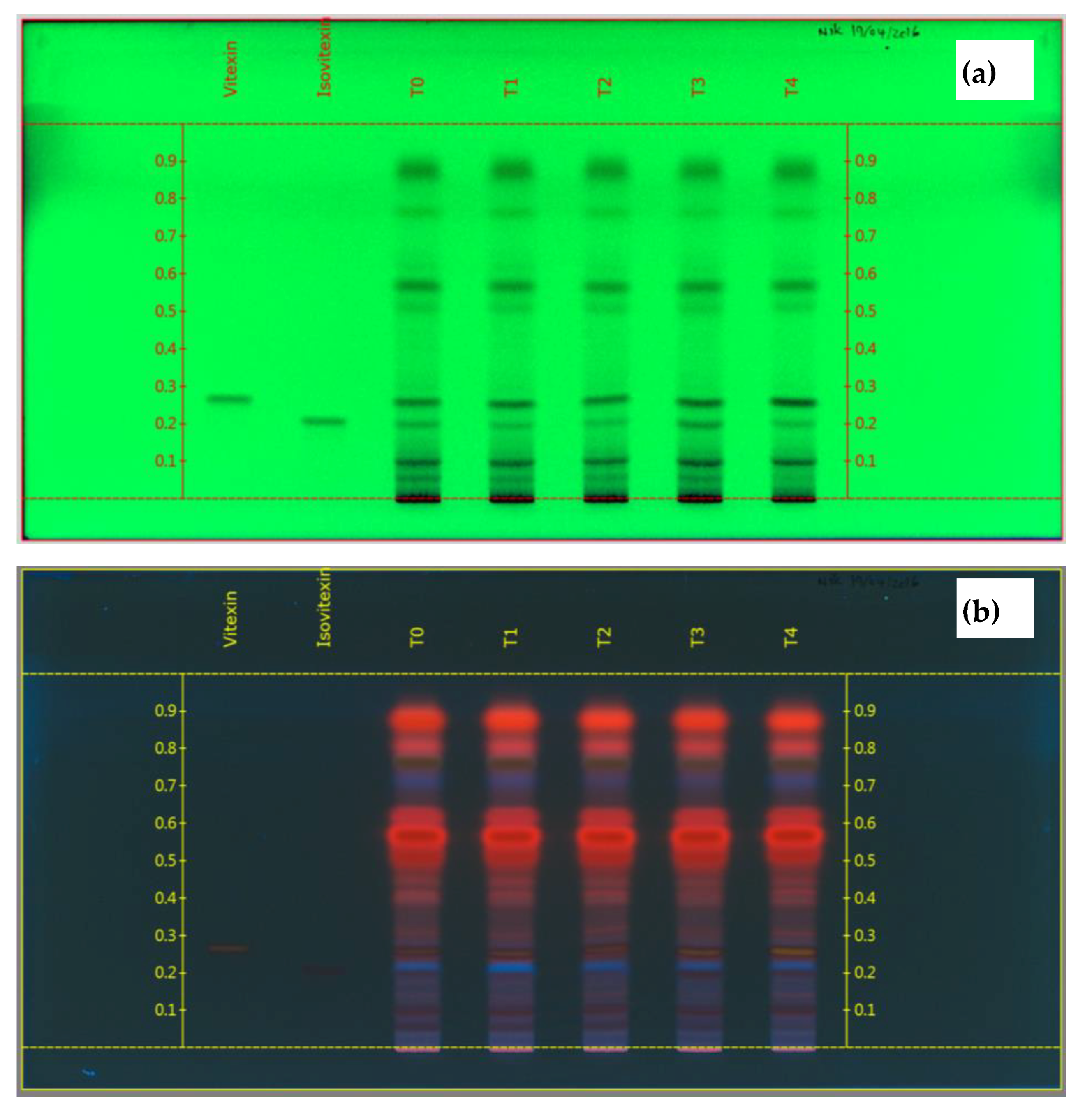
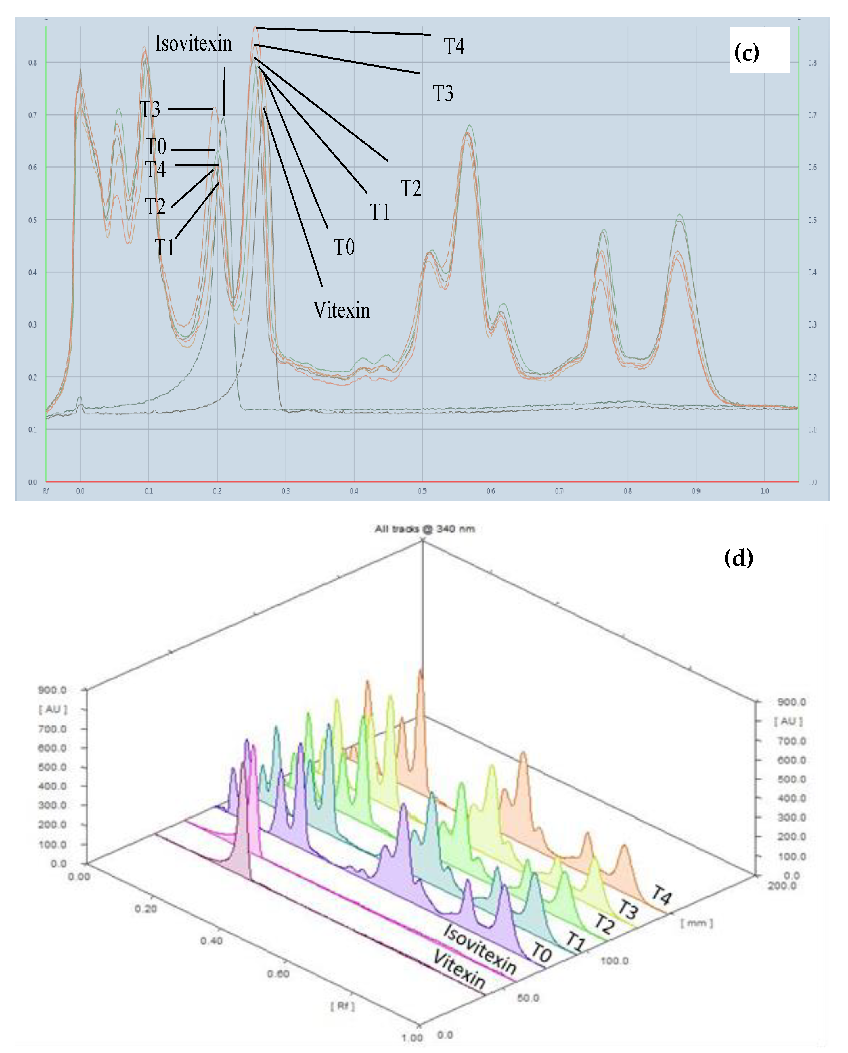
3.3.5. High Performance Thin Layer Chromatography (HPTLC) Fingerprint

Figure 5 shows the chromatogram of the F. deltoidea under different concentrations of H2O2. The results indicate that all of the treated and untreated plants show the same band position and color on the chromatogram under the 366 nm wavelength (Figure 5b). Figure 5a shows the chromatogram under a 254 nm wavelength, which indicates that all the plants have a compound of vitexin and isovitexin with dark bands occurring under the position of relative Rf 0.27 and 0.21.
In addition, the intensity of the compounds varied between treated and untreated plants. Figure 5c,d show the 2D and 3D densitogram of the HTPLC analyses. First, the vitexin compound in untreated plants (T0) shows the lowest band of intensities and the treated plants show a greater intensity of band color. It is also clear that the intensity is dependent on the treatment dose of the H2O2. Although unlikely, the isovitexin compounds appear as an opposite trend, i.e., 30 mM (T3) produced the strongest intensities and 8 mM (T1) the weakest. However, the untreated plants (T0) show the second strongest band of intensity after 30 mM.
The quantitative estimation of vitexin and isovitexin was performed using the HPTLC technique based on the peak densitometry measurement. The plotted standard curve was linear over a range of 100—200 ng (Figure 6) with the R2 values of 0.989 and 0.988 for vitexin and isovitexin, respectively. The results indicate that (Table 5) the highest vitexin was in plants treated with 60 mM (152.53 ng/ug and 0.31% in extract), while the lowest value, was 135.63 ng/ug in untreated plants with 0.27% extract. The results also confirmed that the vitexin content in plants was positively related to the concentrations of the H2O2 applied.
The quantitative estimate of isovitexin indicates that the highest value was due to an application of 30-mM H2O2 (72.44 ng/µg with 0.14% as extract) and the lowest was in the plants treated with 16 mM (39.93 ng/µg) (Table 5). However, there was not much difference between treated plants with 8 and 16 mM. In general, the amount of vitexin was higher than isovitexin in F. deltoidea plants.

4. Discussion

Our study indicates that applications of exogenous H2O2 significantly improved chlorophyll content in F. deltoidea plants. Kato and Shimizu [28] demonstrated that deterioration of the subcellular environment engendered by leaf senescence accelerates chlorophyll bleaching by a peroxidase-H2O2 catalyzed antiphon. The growth promoting chemical H2O2 may protect chloroplast ultrastructure to preserve photosynthetic pigments and stimulate photosynthesis. The results of this study show that H2O2 treatments increased the photosynthetic rate and stomatal conductance of F. deltoidea plants. This may be because the application of H2O2 increased the stomatal opening, CO2 concentration and accumulated photosynthetic pigments. H2O2 synthesized via several routes in the plant cells such as electron transport processes during photosynthesis and played a positive role in plant growth and development [29]. As a signal molecule, H2O2, may mediate a wide range of physiological processes such as stomatal movement, photosynthesis, photorespiration, senescence and the cell cycle and biochemical reactions during the plant’s growth and development [30]. Our study also demonstrates that the application of H2O2 significantly affects the photosynthetic yield of F. deltoidea plants.
The plant height of F. deltoidea increased significantly with H2O2 treatments. This may be due to the positive role of H2O2 in plant growth and development. Rodriguez et al. [31] stated in his study, that H2O2 played an important active role in plant growth, development and other physiological processes. It has been reported that H2O2 application induces the activity of sucrose phosphate synthase (SPS), an enzyme important in the formation of sucrose from triose phosphates during and after photosynthesis in rice seedlings at the transcriptional level [32]. It may be that the elevated levels of H2O2 positively regulate the metabolic and antioxidant enzyme activities in favor of plant growth and development. Niu and Liao [33] stated that H2O2 acts as a signaling molecule which regulates plant growth and development by modulating NO and Ca2+ signaling pathways. The leaf area of the treated plant was not affected by exogenous H2O2.
In our study, all mineral uptakes were significantly improved by the H2O2 treatments. It may be due to the exogenous applications of H2O2 producing a vigorous root system in the plants which increased the absorption area [34]. Plants may contain excessive amounts of heavy metals (As, Cd, Pb, Tl, Zn) in their tissues if grown on waste heaps since metal accumulation levels depend on plant species and metal identity [35].
Arsenic (As) uptake in the treated leaves of F. deltoidea increased with H2O2 treatments; it was higher in all treatments compared to the control plant. But, in the syconium, it was higher at lower concentrations of H2O2. This may be due to the accumulated arsenic which plays a role to improve the plant’s growth and development [36]. The increase of As uptake may also occur due to natural and anthropogenic inputs [37]. Plants grown in uncontaminated soil (0.01–1.5 mg/kg) accumulated arsenic at a lower rate but accumulation of arsenic increased more than the standard [38]. There is evidence that exposure of plants to inorganic arsenic results in the generation of reactive oxygen species (ROS) [39].
Our results confirmed that there were increases in Sb uptake in treated leaves but decreases in treated syconium compared to the control plant. However, the Sb uptake in treated plants was lower than the usual content in herbs such as parsley [40]. Thus, H2O2 application affected the As and Sb uptake but it did not lead to soil contamination since these two metalloids are found in very low accumulations in F. deltoidea plants. Antimony can readily be absorbed by the plants, as expected from the low mobile fraction of Sb in soils. The content observed in the plants is in the range of a natural reference value (below 1 mg/kg) [41].
The treated plants showed higher Fe uptake in leaves but a lower uptake in syconium. The results prove why the net photosynthesis rates and transpiration rates were higher from a dose of 30 mM since a higher Fe content in leaves regulates the accumulation of photosynthetic pigments and improves net photosynthetic rate. According to Briat et al. [42] the Fe level in edible plant tissues ranges from 6 to 360 mg/kg. Our results indicate the range of Fe levels in treated plants with 30 and 60 mM were 100 to 140 µg/g in the leaves.
The magnesium accumulation in leaves and syconium of the F. deltoidea was not significantly affected by H2O2. When looking at the net photosynthetic rate, it is evident that H2O2 affected the photosynthetic rate to some extent positively which may be related to the Mg content in the leaves as Mg is the main component of the chlorophyll molecule and the central atom in the tetrapyrrole ring of both chlorophyll molecules (Chl a and Chl b) in the chloroplasts that makes effective absorption of light possible for the photosynthetic carbon reduction reaction. Magnesium is also involved in CO2 assimilation reactions as the photophosphorylation reactions, that occur in the chloroplasts, are affected by Mg ions [43].
Calcium content increased in leaves with 8–30-mM H2O2 but decreased at a higher concentration (60 mM). It is to be noted that a suitable concentration of H2O2 triggers plant growth and development. H2O2 and Ca are recognized as signal molecules which induce plant tolerance to various biotic and abiotic stresses [18].
The Na uptake was low in 8–30 mM treated plants but higher in the 60 mM treatment of H2O2. According to Jia et al. [44], sodium concentrations of more than 1.0 mg/kg in plant tissues can hinder plant growth. The potassium uptake in syconium was higher than in the leaves and there was little difference between treated and untreated plants. However, the treated plants with 16-mM H2O2 showed the lowest potassium uptake.
All the treated plants showed antioxidant activity, but it was lower than the standard references; namely, ascorbic acid, gallic acid and quercetin that were used in this study. The results of the DPPH• scavenging assay indicate that H2O2 increased the antioxidant activity and it was in a dose dependent manner. It may be that the exogenous H2O2 increased the activity of antioxidant enzymes and antioxidant activity of plant cells. The reactive oxygen species (ROS) does harm to the cell membrane and makes cells lose their function. To respond to the damaging effects of ROS, the plant possesses antioxidant enzymes and also evolves a non-enzymatic antioxidant. Pre-treatment with H2O2 increases these antioxidant activities of plants and thereby meliorates the damage from chilling, salt, heat and osmotic exogenous [45]. Another study indicated that the induced stress tolerance was dependent on the dose of H2O2 applied. Moderate doses of H2O2 enhanced the antioxidant status and induced stress tolerance, while higher concentrations caused oxidative stress and symptoms resembling a hypersensitive response [46]. There were increases in antioxidant activity when treated with H2O2, the possible reason may be due to the properties of H2O2 as it is a highly reactive molecule and controls their degradation through the integrated antioxidant system [47]. Zhang et al. [45] determined in his study, that pre-treatments with H2O2 in cucumber seedling increases the antioxidant activities in low light stress leaves. In general, the over-generation of H2O2 does harm to the cell membrane and makes cells lose function; therefore, to respond to the damaging effects of H2O2, plants possess antioxidant enzymes and also evolve non-enzymatic antioxidant. In addition to that, our results indicate that ethanolic extract showed the highest value in the DPPH• scavenging assay, but according to Kraujalienė et al. [48], water extract of B. crassifolia and B. ornate were the most active radical scavengers in the DPPH• and ABTS assays. Therefore, the effect of solvent extracts depends on the plant species involved.
One of the properties of F. deltoidea is to have high α-glucosidase inhibitory activities as a potential as an antidiabetic agent. This was proven in our study when F. deltoidea showed higher in vitro α-glucosidase inhibitory activities compared with the standard (acarbose). The result was also supported by research from Misbah et al. [5], where the use of acarbose as the inhibitor of α-glucosidase and α-amylase did not show any sign of activation within the range of concentration tested, which stated little inhibition occurs. In addition, the result was supported by a study from Adam et al. [49] that suggested F. deltoidea has the potential to be developed as a future oral antidiabetic agent.
In the total flavonoid content assay, quercetin was used as a standard reference to plot the standard curve, this was because the antioxidant capacity of quercetin is higher than other phenolics [50]. Our results indicated that there was a significant change in total phenolic and flavonoid content in F. deltoidea when treated with H2O2. As we know, a flavonoid is a large group of phenolic secondary metabolites that are widespread among plants and are involved in many plant functions [51]. The possible reason there were changes in total phenolic and total flavonoid content, when treated with H2O2 compared to the control plant, is because it involves H2O2 as signal molecules in phenolic synthesis. This reason was supported by Zhang et al. [52] in his review, where H2O2, salicylic acid (SA) and nitric oxide (NO) were signaling molecules that have been demonstrated to contribute to phenolic synthesis. The second possible reason is that the plants developed specific antioxidative defense enzymes including peroxidase and polyphenol oxidase to control rapidly increasing ROS under various environmental stress conditions.
We observed a significant change in the total phenolic and flavonoid content in F. deltoidea when treated with H2O2. Solvent extracts such as the polar solvent (ethanol) has a higher content compared to semi polar (chloroform) where an increase in TPC and TFC significantly increase antioxidant activity. This was supported by Kamran et al. [9] where his recent study also indicated that semi polar solvent has higher antioxidant activity compared with polar solvent. Which was not surprising, as we know that flavonoids are a large group of phenolic secondary metabolites which are widespread among plants and are involved in many plant functions [49]. Khandaker et al. [20] also stated that H2O2 treatment elevated the level and the activity of antioxidant enzymes in wax apple fruits. Our results indicate a positive correlation between the antioxidant activity with total phenolic and flavonoid content but not correlated within α-glucosidase activity. This correlation may be slightly stronger for total phenolic compound due to the overestimating of total hydroxyl cinnamic acids. It is possible that the non-specificity of the Folin Ciacalteu method had caused some interference in the absorbance reading. This suggests the antioxidant activities were assisted by the phenolic content, while other polar plant components were possibly involved in antidiabetic properties [5].
One of the properties of F. deltoidea is to have high antidiabetic activity, which was proven in our study, since F. deltoidea shows higher in vitro α-glucosidase activities compared to the standard reference (acarbose), antidiabetic drug use as the inhibitor of α-glucosidase and α-amylase. F. deltoidea may have the potential for development as a future oral antidiabetic agent [49]. Although unlikely, the results of this study indicate that the treatment with H2O2 decreased the α-glucosidase activity. We performed high performance thin layer chromatography using isovitexin and vitexin as the standard compounds to prove whether H2O2 decreased the antidiabetic activity. It was observed that the H2O2 caused α-glucosidase inhibition in F. deltoidea. The H2O2 applications increased the vitexin compound in plants but only decreased isovitexin compounds in the plants when treated with 8, 16 and 60-mM H2O2 compared with the control plant. However, there was no major difference in quantification of isovitexin between the treated and untreated plants. Choo et al. [24] suggested the use of F. deltoidea as an antidiabetic since there exists both bioflavonoids (vitexin and isovitexin) in F. deltoidea plants. It was reported that the extract of F. deltoidea can potentially improve insulin sensitivity, suppress hepatic glucose output and enhance glucose uptake in type 2 diabetes mellitus through down-regulation of the protein tyrosine phosphatase 1B (PTP1B) [53]. They also stated the herb can be utilized as a useful remedy for alleviation of diabetes complications.

5. Conclusions

The main purpose of this study was to pinpoint the best way to enhance regulatory processes within the F. deltoidea by using H2O2. From the results, we can conclude that the application of H2O2 particularly 16 and 30 mM increased the leaf chlorophyll content, net photosynthetic rate, stomatal conductance and quantum yield of the F. deltoidea plants. In addition, exogenous H2O2 increased its mineral uptake including arsenic (As), antimony (Sb), iron (Fe), calcium (C), potassium (K) and sodium (N) in leaves and syconium. Additionally, the application of 16 and 30-mM H2O2 significantly increased the antioxidant activity, as well as the total phenolic and flavonoid content. The chemical fingerprint of the leaves of F. deltoidea var. deltoidea show that the content of vitexin and isovitexin were significantly increased with an increase in the concentration of H2O2. Our results also indicate that the plants treated with 30-mM H2O2 show the highest amount of isovitexin which has a wide range of positive pharmacological effects.

Author Contributions

Conceptualization, M.M.K.; writing—original draft, N.N. and M.M.K.; methodology and data curation, N.N. and M.M.K.; formal analysis, N.N.; validation, N.M., K.S.M., N.A.B., N.Y., M.H.S., K.M. and A.F.M.A.; resources, M.M.K. A.F.M.A., N.M. and K.S.M.; writing—review & editing, K.M., N.Y., M.H.S., K.S.M. and N.A.B.; Funding acquisition, M.M.K.; supervision, M.M.K. and N.M. All authors have read and agreed to the published version of the manuscript.

Funding

This research was funded by the Ministry of Education Malaysia from the Fundamental Research Grant Scheme, FRGS Grant No. FRGS/2/2014/SG03/UNISZA/02/01.

Acknowledgments

We are grateful to Anuar McAfee for his English editing and proof reading of this paper.

Conflicts of Interest

The authors declare that they have no conflict of interests.

References

  1. Hasham, R.; Choi, H.K.; Sarmidi, M.R.; Park, C.S. Protective effects of a Ficus deltoidea extract against UVB-induced photoageing in skin cells. Biotechnol. Bioprocess Eng. 2013, 18, 185–193. [Google Scholar] [CrossRef]
  2. Starr, F.; Starr, K.; Loope, L. Ficus deltoidea; United States Geological Survey—Biological Resources Division, Haleakala Field Station: Maui, HI, USA, 2003. [Google Scholar]
  3. Badron, U.H.; Talip, N.; Mohamad, A.L.; Affenddi, A.E.A.; Ahmad Juhari, A.A. Studies on leaf venation in selected taxa of the genus Ficus L. (Moraceae) in peninsular Malaysia. Trop. Life Sci. Res. 2014, 25, 111–125. [Google Scholar] [PubMed]
  4. Musa, Y. Variability in morphology and agronomy of mas cotek accessions found in Kelantan and Terengganu. Bul. Teknol. Tanam. 2005, 2, 35–48. [Google Scholar]
  5. Misbah, H.; Abdul Aziz, A.; Aminudin, N. Antidiabetic and antioxidant properties of F. deltoidea fruit extracts and fractions. BMC Complement. Altern. Med. 2013, 13, 118. [Google Scholar] [CrossRef] [PubMed] [Green Version]
  6. Draman, S.; Aris, M.A.M.; Razman, S.F.; Akter, U.; Azlina, H.; Norazlina, A.R.; Norazlansha, M. Mas cotek (Ficus deltoidea): A possible supplement for type II diabetes: (A pilot study). Pertanika J. Trop. Agric. Sci. 2012, 35, 93–102. [Google Scholar]
  7. Sulaiman, M.R.; Hussain, M.K.; Zakaria, Z.A. Evaluation of the antinociceptive activity of Ficus deltoidea aqueous extract. Fitoterapia 2008, 79, 557–561. [Google Scholar] [CrossRef]
  8. Haida, Z.; Syahida, A.; Ariff, S.M.; Hakiman, M. Factors Affecting Cell Biomass and Flavonoid Production of Ficus deltoidea var. kunstleri in Cell Suspension Culture System. Sci. Rep. 2009, 9, 9533. [Google Scholar] [CrossRef]
  9. Ashraf, K.; Halim, H.; Lim, S.M.; Ramasamy, K.; Sultan, S. In vitro antioxidant, antimicrobial and antiproliferative studies of four different extracts of Orthosiphon stamineus, Gynura procumbens and Ficus deltoidea. Saudi J. Biol. Sci. 2020, 27, 417–432. [Google Scholar] [CrossRef]
  10. Abolmaesoomi, M.; Abdul Aziz, A.; Mat Junit, S.; Mohd Ali, J. Ficus deltoidea: Effects of solvent polarity on antioxidant and anti-proliferative activities in breast and colon cancer cells. Eur. J. Integr. Med. 2019, 28, 57–67. [Google Scholar] [CrossRef]
  11. Nazarni Che Isa, M.; Ajit, A.; Naila, A.; Sulaiman, A.Z. Effect of microwave assisted hydro-distillation extraction on extracts of Ficus deltoidea. Mater. Today Proc. 2018, 5, 21772–21779. [Google Scholar] [CrossRef]
  12. Fasihuddin, B.A.; Din, L.B. Medicinal plants used by various Ethnic Groups in Sabah. In Proceedings of the The French Malaysian-Symposium on Natural Products, Kuala Lumpur, Malaysia, 24–25 July 2002. [Google Scholar]
  13. Armaghan, S.; Nahdzatul, S.M.; Zeyad, D.N.; Abdalrahim, F.A.A.; Amin, M.S.; Zhari, I. Antiangiogenic effect of Ficus deltoidea Jack standardised leaf extracts. Trop. J. Pharm. Res. 2014, 13, 761–768. [Google Scholar]
  14. Aris, S.R.S.; Mustafa, S.; Ahmat, N.; Jaafar, F.M.; Ahmad, R. Phenolic content and antioxidant activity of fruits of Ficus deltoidea var Angustifolia sp. Malay. J. Anal. Sci. 2009, 13, 146–150. [Google Scholar]
  15. Nurdiana, S.; Goh, Y.M.; Hafandi, A.; Dom, S.M.; Nur Syimal’ain, A.; Noor Syaffinaz, N.M.; Ebrahimi, M. Improvement of spatial learning and memory, cortical gyrification patterns and brain oxidative stress markers in diabetic rats treated with Ficus deltoidea leaf extract and vitexin. J. Trad. Complement. Med. 2018, 8, 190–202. [Google Scholar] [CrossRef]
  16. Oh, M.J.; Abdul, H.M.; Ngadiran, S.; Seo, Y.K.; Sarmidi, M.R.; Park, C.S. Ficus deltoidea extract exerted anti-melanogenic activity by preventing tyrosinase activity in vitro and by suppressing tyrosinase gene expression in B16F1 melanoma cells. Arch. Dermat. Res. 2011, 303, 161–170. [Google Scholar] [CrossRef] [Green Version]
  17. Cakmak, I.; Marschner, H. Effect of zinc nutritional-status on activities of superoxide radical and hydrogen-peroxide scavenging enzymes in bean-leaves. Plant Soil. 1993, 155, 127–130. [Google Scholar] [CrossRef]
  18. Almeida, J.M.; Fidalgo, F.; Confraria, A.; Santos, A.; Pires, H.; Santos, I. Effect of hydrogen peroxide on catalase gene expression, isoform activities and levels in leaves of potato sprayed with homobrassinolide and ultrastructural changes in mesophyll cells. Funct. Plant Biol. 2005, 32, 707–720. [Google Scholar] [CrossRef]
  19. Orabi, S.A.; Dawood, M.G.; Salman, S.R. Comparative study between the physiological role of hydrogen peroxide and salicylic acid in alleviating the harmful effect of low temperature on tomato plants grown under sand-ponic culture. Sci. Agric. 2015, 9, 49–59. [Google Scholar]
  20. Khandaker, M.M.; Boyce, A.N.; Osman, N. The influence of hydrogen peroxide on the growth, development and quality of wax apple (Syzygium samarangense, [Blume] Merrill & L.M. Perry var. jambu madu) fruits. Plant Physiol. Biochem. 2012, 53, 101–110. [Google Scholar] [CrossRef]
  21. Xia, X.-J.; Huang, L.-F.; Zhou, Y.-H.; Mao, W.-H.; Shi, K.; Wu, J.-X.; Asami, T.; Chen, Z.; Yu, J.-Q. Brassinosteroids promote photosynthesis and growth by enhancing activation of Rubisco and expression of photosynthetic genes in Cucumis sativus. Planta 2009, 230, 1185–1196. [Google Scholar] [CrossRef]
  22. Nashriyah, M.; Zaini, H.; Mazleha, M.; Abdul, K.W. Mineral uptake by taro (Colocasia esculenta) in swamp agroecosystem following gramaxone® (paraquat). J. Nuclear Relat. Technol. 2006, 3, 59–68. [Google Scholar]
  23. Akter, M.S.; Ahmed, M.; Eun, J.B. Solvent effects on antioxidant properties of persimmon (Diospyros kaki, L. cv Daebong) seeds. Int. J. Food. Sci. Technol. 2010, 45, 2258–2264. [Google Scholar] [CrossRef]
  24. Choo, C.Y.; Sulong, N.Y.; Man, F.; Wong, T.W. Vitexin and isovitexin from the Leaves of Ficus deltoidea with in-vivo -glucosidase inhibition. J. Ethnopharmacol. 2012, 142, 776–781. [Google Scholar] [CrossRef]
  25. Singleton, V.; Rossi, J.A.J. Colorimetry of total phenolic with phosphomolybdic-phosphotungstic acid reagents. Am. J. Enol. Viticult. 1965, 16, 144–158. [Google Scholar]
  26. Woisky, R.G.; Salatino, A. Analysis of propolis: Some parameters and procedures for chemical quality control. J. Apicult. Res. 1998, 37, 99–105. [Google Scholar] [CrossRef]
  27. Mohd, K.S.; Azemin, A.; Hamil, M.S.R.; Bakar, A.R.A.; Dharmaraj, S.; Hamdan, M.R.; Ismail, Z. Application of high performance thin layer chromatography and fourier transform Infrared profiling coupled with chemometrics for the differentiation of the varieties of Ficus deltoidea Jack. Asian J. Pharma Clin. Res. 2014, 7, 110–116. [Google Scholar]
  28. Kato, M.; Shimizu, S. Chlorophyll metabolism in higher plants. VII. Chlorophyll degradation in senescing tobacco leaves: Phenolic-dependent peroxidative degradation. Can. J. Bot. 1987, 65, 729–735. [Google Scholar] [CrossRef]
  29. Li, S.; Xue, L.; Xu, S.; Feng, H.; An, L. Hydrogen peroxide involvement in formation and development of adventitious roots in cucumber. Plant Growth Regul. 2007, 52, 173–180. [Google Scholar] [CrossRef]
  30. Deng, X.P.; Cheng, Y.J.; Wu, X.B.; Kwak, S.S.; Chen, W.; Egrinya, A. Exogenous hydrogen peroxide positively influences root growth and metabolism in leaves of sweet potato seedlings. Aust. J. Crop Sci. 2012, 6, 1572–1578. [Google Scholar]
  31. Rodriguez, A.A.; Grunberg, K.A.; Taleisnik, E.L. Reactive oxygen species in the elongation zone of maize meves are necessary for leaf extension. Plant Physiol. 2014, 129, 1627–1632. [Google Scholar] [CrossRef] [PubMed] [Green Version]
  32. Uchida, A.; Jagendorf, A.T.; Hibino, T.; Takabe, T.; Takabe, T. Effects of hydrogen peroxide and nitric oxide on both salt and heat stress tolerance in rice. Plant Sci. 2002, 163, 515–523. [Google Scholar] [CrossRef]
  33. Niu, L.; Liao, W. Hydrogen peroxide signaling in plant development and abiotic responses: Crosstalk with nitric oxide and calcium. Front. Plant Sci. 2016, 7, 230. [Google Scholar] [CrossRef] [PubMed] [Green Version]
  34. Hameed, A.; Farooq, S. Influence of exogenous application of hydrogen peroxide on root and seedling growth on wheat (Triticum aestivum L.). Int. J. Agric. Biol. 2004, 6, 366–369. [Google Scholar]
  35. Stefanowicz, A.M.; Stanek, M.; Woch, M.W.; Kapusta, P. High concentration of heavy metals in beech forest understory plants growing in waste heaps leaft by Zn-Pb ore mining. Geochem. Explor. 2016, 59, 157–162. [Google Scholar] [CrossRef]
  36. Han, Y.H.; Yang, G.M.; Fu, J.W.; Guan, D.X.; Chen, Y.; Ma, L.Q. Arsenic-induced plant growth of arsenic-hyper accumulator in Pteris vittata: Impact of arsenic and phosphate rock. Chemosphere 2016, 149, 366–372. [Google Scholar] [CrossRef] [PubMed]
  37. Dai, Y.; Lv, J.; Liu, K.; Zhao, X.; Cao, Y. Major controlling factors and prediction models for arsenic uptake from soil to wheat plants. J. Exotoxicol. Environ. Saf. 2016, 130, 256–262. [Google Scholar] [CrossRef] [PubMed]
  38. Farooq, M.; Islam, F.; Ali, B.; Najeeb, U.; Mao, B.; Gill, R.A.; Yan, G.; Siddique, K.H.M.; Zhou, W. Arsenic toxicity in plants: Cellular and molecular mechanisms of its transport and metabolism. Environ. Exp. Bot. 2016, 132, 42–52. [Google Scholar] [CrossRef]
  39. Pandey, N.; Bhatt, R. Role of soil associated Exiguobacterium in reducing arsenic toxicity and promoting plant growth in Vigna radiata. Eur. J. Soil Biol. 2016, 75, 142–150. [Google Scholar] [CrossRef]
  40. Pierart, A.; Shahid, M.; Séjalon-Delmas, N.; Dumat, C. Antimony bioavailability: Knowledge and research perspectives for sustainable agricultures. J. Hazard. Mat. 2015, 289, 219–234. [Google Scholar] [CrossRef] [Green Version]
  41. Pérez-Sirvent, C.; Martínez-Sánchez, M.J.; Martínez-López, S.; Bech, J.; Bolan, N. Distribution and bioaccumulation of arsenic and antimony in Dittrichia viscosa growing in mining-affected semiarid soils in southeast Spain. J. Geochem. Explor. 2012, 123, 128–135. [Google Scholar] [CrossRef]
  42. Briat, J.F.; Dubos, C.; Gaymard, F. Iron nutrition, biomass production, and plant product quality. Trends Plant Sci. 2015, 20, 33–40. [Google Scholar] [CrossRef]
  43. Tatagiba, S.D.; Rodrigues, F.A. Magnesium decreases the symptoms of leaf scald on rice leaves. Trop. Plant Pathol. 2016, 41, 132–137. [Google Scholar] [CrossRef]
  44. Jia, Y.; Kong, X.; Weiser, M.D.; Lv, Y.; Akbar, S.; Jia, X.; Tian, X. Sodium limits litter decomposition rates in a subtropical forest: Additional tests of the sodium ecosystem respiration hypothesis. Appl. Soil Ecol. 2015, 93, 98–104. [Google Scholar] [CrossRef]
  45. Zhang, X.L.; Jia, X.F.; Yu, B.; Gao, Y.; Bai, J.G. Exogenous hydrogen peroxide influences antioxidant enzyme activity and lipid peroxidation in cucumber leaves at low light. Sci. Hortic. 2011, 129, 656–662. [Google Scholar] [CrossRef]
  46. Hu, W.H.; Song, X.S.; Shi, K.; Xia, X.J.; Zhou, Y.H.; Yu, J.Q. Changes in electron transport, superoxide dismutase and ascorbate peroxidase isoenzymes in chloroplasts and mitochondria of cucumber leaves as influenced by chilling. Photosynthetica. 2008, 46, 581–588. [Google Scholar] [CrossRef]
  47. Sagrillo, M.R.; Garcia, L.F.M.; De Souza Filho, O.C.; Duarte, M.M.M.F.; Ribeiro, E.E.; Cadon, F.C.; Da Cruz, I.B.M. Tucum fruit extracts (Astrocaryum aculeatum Meyer) decrease cytotoxic effects of hydrogen peroxide on human lymphocytes. Food Chem. 2015, 173, 741–748. [Google Scholar] [CrossRef] [Green Version]
  48. Kraujalienė, V.; Pukalskas, A.; Kraujalis, P.; Venskutonis, P.R. Biorefining of Bergenia crassifolia L. roots and leaves by high pressure extraction methods and evaluation of antioxidant properties and main phytochemicals in extracts and plant material. Ind. Crop Prod. 2016, 89, 390–398. [Google Scholar] [CrossRef]
  49. Adam, Z.; Khamis, S.; Ismail, A.; Hamid, M. Ficus deltoidea: A potential alternative medicine for diabetes mellitus. Evid. Based Complementary Altern. Med. 2012, 2012, 632763. [Google Scholar] [CrossRef] [Green Version]
  50. Bi, X.; Zhang, J.; Chen, C.; Zhang, D.; Li, P.; Ma, F. Anthocyanin contributes more to hydrogen peroxide scavenging than other phenolics in apple peel. Food Chem. 2014, 152, 205–209. [Google Scholar] [CrossRef]
  51. Jaakola, L.; Määttä, K.; Pirttilä, A.M.; Törrönen, R.; Kärenlampi, S.; Hohtola, A. Expression of genes involved in anthocyanin biosynthesis in relation to anthocyanin, proanthocyanidin, and flavonol levels during bilberry fruit development. Plant Physiol. 2002, 130, 729–739. [Google Scholar] [CrossRef] [Green Version]
  52. Zhang, R.Q.; Zhu, H.H.; Zhao, H.Q.; Yao, Q. Arbuscular mycorrhizal fungal inoculation increases phenolic synthesis in clover roots via hydrogen peroxide, salicylic acid and nitric oxide signaling pathways. J. Plant Physiol. 2013, 170, 74–79. [Google Scholar] [CrossRef]
  53. Abdel-Rahman, R.F.; Ezzat, S.M.; Ogaly, H.A.; Abd-Elsalam, R.M.; Hessin, A.F.; Fekry, M.I.; Mansour, D.F.; Mohamed, S.O. Ficus deltoidea extract down-regulates protein tyrosine phosphatase 1 expression in a rat model of type 2 diabetes mellitus: A new insight into its antidiabetic mechanism. J. Nutr. Sci. 2020, 9, e2. [Google Scholar] [CrossRef] [PubMed] [Green Version]
Figure 1. The effect of H2O2 on (a) arsenic (As), (b) antimony (Sb), (c) iron (Fe), (d) magnesium (Mg) in leaf and syconium of Ficus deltoidea var deltoidea plant. Error bars indicate (± S. E).
Agronomy 10 00599 g001
Figure 2. The effect of H2O2 on (a) calcium (Ca), (b) sodium (Na), (c) potassium (K) in leaf and syconium of Ficus deltoidea var deltoidea plant. Error bars indicate (± S. E).
Agronomy 10 00599 g002
Figure 3. Effect of H2O2 on total phenolic content in the leaf of Ficus deltoidea var deltoidea in different solvent extraction, (a) ethanolic extract, (b) chloroform extract and (c) aqueous extract. Bars indicate (±S.E). Different letters represent the significance at the 5% level of HSD test.
Agronomy 10 00599 g003
Figure 4. Effect of H2O2 on total flavonoid content in leaf of Ficus deltoidea var deltoidea under different solvent extractions; (a) ethanolic extract, (b) chloroform extract and (c) aqueous extract. Bars indicate (±S. E). Different letters represent the significance at the 5% level of HSD test.
Agronomy 10 00599 g004
Figure 5. Visualization of the sample (T0, Control; T1, 8 mM; T2, 16 mM; T3, 30 mM; T4, 60 mM) with selected markers (Vitexin, Isovitexin) under 254 nm wavelength (a) and 366 nm wavelength (b), 2D densitogram (c) and 3D densitogram (d).
Agronomy 10 00599 g005aAgronomy 10 00599 g005b
Figure 6. Standard curve for vitexin (a) and isovitexin (b) of Ficus deltoidea var deltoidea.
Agronomy 10 00599 g006
Table 1. The effects of H2O2 on plant physiological characters and growth of Ficus deltoidea plants on the 12th week of observation. All the data are a mean of five replicates. Different letters in the same column represent the significant difference at 5% level. Chlorophyll content index (CCI); Variable fluorescence (Fv); Maximum fluorescence (Fm).
H2O2 (mM)Leaf Chlorophyll (CCI)Net Photosynthesis (μmol/m2/s)Stomatal Conductance (m2s/mol)Quantum Yield (Fv/Fm)Plant Height (cm)Leaf Area (cm2)
0 76 c2.5 b360 b0.77 b31 b7 a
8 80 b2.4 b320 b0.81 a32 b7 a
16 81 b3.6 a300 b0.80 a35 a8 a
30 89 a4.5 a400 a0.80 a38 a9 a
60 79 b2.2 b080 c0.79 a32 b8 a
Table 2. The IC50 (µg/mL) effect of different treatments of H2O2 on DPPH• scavenging activity of different extract of Ficus deltoidea plant leaf. Means (± S.E.) with same letter do not differ significantly according to Sheffer’s post hoc test at α = 0.05.
H2O2 (mM) IC50 (µg/mL)
Ethanolic Extract Chloroform ExtractAqueous Extract
0 76.33 ± 0.67 c-95.67 ± 0.88 c
8 86.33 ± 1.20 d-95.33 ± 1.20 c
16 25.00 ± 1.53 a-116.33 ± 0.67 d
30 61.67 ± 0.88 b201.67 ± 0.88 a45.00 ± 1.15 a
60 75.00 ± 1.15 c290.77 ± 0.67 b80.00 ± 0.218 b
Gallic Acid3.50 ± 0.253.50 ± 0.253.50 ± 0.25
Quercetin3.67 ± 0.083.67 ± 0.083.67 ± 0.08
Ascorbic Acid4.92 ± 0.084.92 ± 0.084.92 ± 0.08
Table 3. Effect of H2O2 on α-glucosidase activity (IC50 mg/mL) of Ficus deltoidea var deltoidea leaf. Means (S.E.) with same letter do not differ significantly according to the Sheffer’s post hoc test at α = 0.05.
H2O2 (mM)IC50 (µg/mL)
α-Glucosidase Activity
025.0 ± 0.77 b
860.0 ± 0.11 d
16100.0 ± 0.81 e
3040.0 ± 0.10 c
60
Quercetin
Acarbose
57.5 ± 0.67 g
12.5 ± 0.15 a
235.0 ± 0.66 f
Table 4. Regression equation, correlation coefficient (r) and coefficients of determination (R2) of antioxidant activity with total phenol, total flavonoid and α-glucosidase activity. EE = Ethanol extract, CE = Chloroform extract and AE = Aqueous extract.
Regression EquationCorrelation Coefficient (r)Coefficients of Determination (R2)
Relationship between antioxidant activity with total phenolic content
YEE = 1.4464x + 244.08r = 0.783R2 = 0.6138
YCE = −0.062x + 55.475r = 0.848R2 = 0.7189
YAE = 3.2971x + 20.955r = 0.695R2 = 0.4832
Relationship between antioxidant activity with total flavonoid content
YEE = 0.5116x + 105.07r = 0.870R2 = 0.7573
YCE = 0.0025x + 6.2605r = 0.886R2 = 0.7851
YAE = 0.8122x + 65.338r = 0.708R2 = 0.5017
Relationship between antioxidant activity with α-glucosidase activity
YEE = −0.2276x + 64.061r = 0.332R2 = 0.1105
Table 5. Quantification of vitexin and isovitexin under different concentrations of H2O2 in Ficus deltoidea var deltoidea plant. Means (± S.E.) with same letter do not differ significantly according to the Sheffer’s post hoc test at α = 0.05.
H2O2 (mM)Vitexin (ng/ µg)% in ExtractIsovitexin (ng/ µg)% in Extract
0 135.63 ± 0.09 e0.2750.93 ± 0.20 b0.10
8 138.30 ± 0.40 d0.2842.18 ± 0.22 d0.08
16 137.23 ± 0.23 c0.2739.93 ± 0.23 e0.08
30 144.27 ± 0.17 b0.2972.44 ± 0.17 a0.14
60 152.53 ± 0.19 a0.3144.90 ± 0.19 c0.09

