
Microbial Exopolysaccharide Composites in Biomedicine and Healthcare: Trends and Advances

 , , , ,
Abstract
:1. Introduction
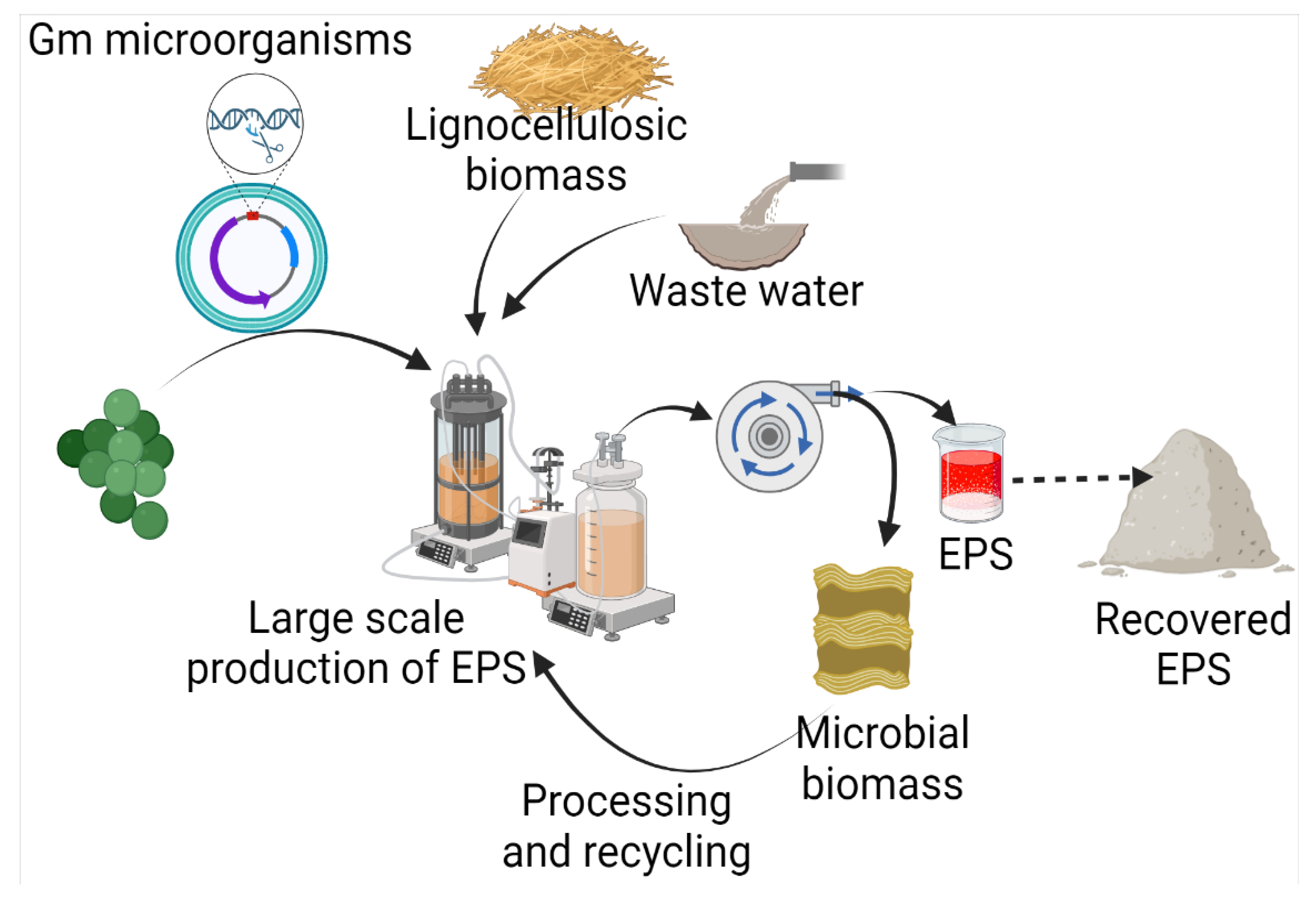
2. Microbes Producing Exopolysaccharides

| EPS | Organism | Substrate | Growth Conditions | Working Volume | EPS Yield | Key Achievements | Reference |
|---|---|---|---|---|---|---|---|
| Dextran (α-D-gluco pyranosyl moieties interlinked with α-(1,6) linkage and have α-(1,2)/α-(1,3)/α-(1,4) branching)  | Leuconostoc mesenteroides SF3 | 10% sucrose | Temp 25 °C; pH 6; incubation period 16 h; inoculum 24 h old, 10% inoculum with cell concentration of 108 cells/mL. | 100 mL | 23.8 ± 4 g/L | Water absorption capacity 361.8% ± 3.1; oil absorption capacity 212.0% ± 6.7; emulsion activity 58.3% ± 0.7. | [35] |
| Lactobacillus spp. | 15% sucrose | Temp 30 °C; pH 7; incubation period 24 h; inoculum 24 h old 4%, growth conditions aerobic. | 100 mL | 5.8 mg/mL | Lactobacillus strains were isolated from the human vagina and infant stool. | [36] | |
| Leuconostoc pseudomesenteroides DSM20193 | Brewers’ spent grain | Initial cell concentration 6.0 Log cfu/g; temp 25 °C; period 24 h. | 1000 g | 1.2 g/100 g | EPS production is accompanied by mannitol; no dextran production without a starter (commercial granulated sugar). | [37] | |
| Leuconostoc pseudomesenteroides XG5 | 100 g/L sucrose | Temp 25 °C; pH 7.0; mixing rate 20 rpm; time period 60 h; inoculum 2%. | 35 L | 26.02 g/L dextran | Protein content in EPS reduced when extracted with EDTA or NaOH+formal-dehyde. | [31] | |
| Weissella confusa A16 | Brewers’ spent grain | Initial cell concentration 6.0 log cfu/g; temp 25 °C; period 24 h. | 1000 g | 1.1 g/100 g | No mannitol production was observed, but a starter was required for EPS production. | [37] | |
| Curdlan (Type HOEPS, unbranched; molecular weight 5.3 × 10⁴–2 × 10⁶ Da; components glucosyl residues inter-connected with β-D-(1→3) bonds)  | Agrobacterium sp. IFO 13140 | 50 g/L | Synthetic medium; temp 30 °C; mixing rate 150 rpm; period 5 d; pH 7. | 100 mL | - | Water holding capacity and oil holding capacity 64% and 98% higher in comparison to commercial curdlan. | [38] |
| Bacillus cereus PR3 | 10% starch | Synthetic medium; period 96 h. | 100 mL | 20.88 g/L | Anti-oxidant activity increased with curdlan. | [39] | |
| Agrobacterium sp. ATCC 31749 | Asparagus spear bottom part juice | Synthetic medium; temp 30 °C; mixing rate 200 rpm; period 168 h. | 100 mL | 40.2 g/L | Curdlan production is higher with sucrose in comparison to mineral salt. | [40] | |
| Xanthan (Type HEEPS; components backbone made of D-glucose unit linked with β-1,4-glycosidic bonds and side chain trisaccharide; side chain comprised of mannose, glucuronic acid, and mannose, terminal mannose with pyruvic acid residues; molecular weight 2.0 × 10⁶–2.0 × 10⁷ Da)  | Xanthomonas campestris | 20 g/L glucose | Stainless steel supported biofilm reactor; period 27 h; synthetic medium; mixing rate 180 rpm. | 150 mL | 3.47 ± 0.71 g/L | Use of biofilm reactor increased the xanthan recovery. | [41] |
| Xanthomonas campestris | 20 g/L glucose | Polyethylene supported biofilm reactor; period 78.5 h; synthetic medium; mixing rate 180 rpm. | 150 mL | 3.21 ± 0.68 g/L | Biofilm reactor increased the glucose consumption. | [41] | |
| Gellan gum (Type HEEPS; components backbone made up of β-d-glucose, l-rhamnose, and d-glucuronic acid along with acetate and glycerate attached to glucose)  | Sphingomonas pseudosanguinis (Accession No. GI:724472387) | 80 g/L biodiesel-derived waste glycerol | pH 7; synthetic medium; temp 30 °C; mixing rate 200 rpm; inoculum 10%; period 7 days; 0.5 vvm. | 3 L | 51.6 g/L | At lower concentrations, glycerol is consumed completely at all pHs, but at a higher concentration, it is not exhausted completely. | [42] |
| Sphingomonas yabuuchiae (GI:724472388) | 52.6 g/L | [42] | |||||
| EPS Br42 was found to be a heteropolysaccharide consisting of glucose and galacturonic acid with a molecular weight of about 286 kDa. | Brevibacillus borstelensis M42 | 2% glucose | Period 60 h. | - | 1.88 g/L | Water-holding capacity 510 ± 0.35%, oil-holding 374 ± 0.12% and swelling capacities 146.6 ± 5.75%. | [43] |
| EPS K1T-9 (EPS type HEEPS; components glucose and galacturonic acid; molecular weight 207 kDa. | Neorhizobium urealyticum sp. nov. | Glucose 5 g/L | Zobell’s marine broth; pH 7; temp 28 °C; mixing rate 150 rpm; inoculum size 5 mL/100 mL; period 72 h. | Working volume 400 mL | 3.38 g/L | Water holding 356 ± 0.8%, oil holding 697 ± 1% (coconut oil); 317 ± 1.3% (olive oil), swelling capacity 200 ± 1.1%. | [44] |
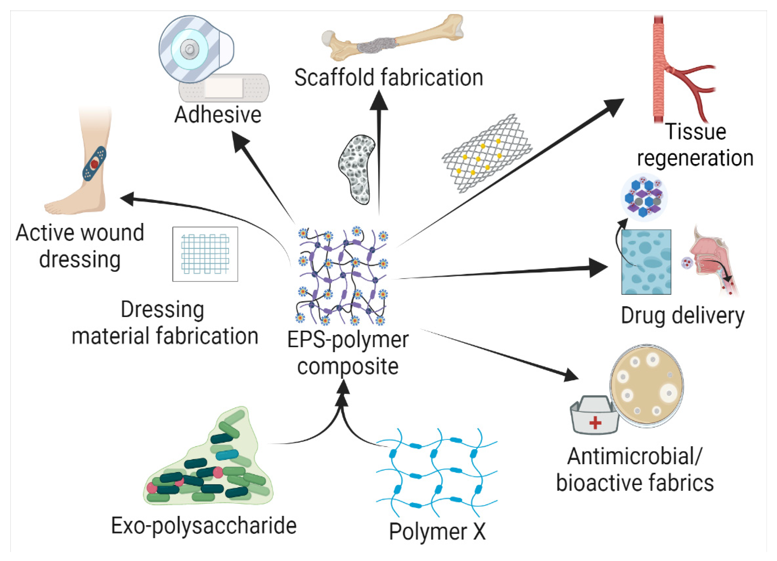
3. Microbial Exopolysaccharide Composites and Their Applications
3.1. Exopolysaccharide Composites with Natural Materials
3.1.1. Cellulose Composites with Natural Polymers
3.1.2. Dextran Composites with Natural Polymers
3.1.3. Xanthan Composites with Natural Polymers
3.1.4. Pullulan Composites with Natural Polymers
3.1.5. Levan Composites with Natural Polymers
3.1.6. Gellan Composites with Natural Polymers
3.2. Exopolysaccharide Composites with Synthetic Polymers
3.2.1. Dextran Composites with Synthetic Polymers
3.2.2. Cellulose Composite with Synthetic Polymers
3.2.3. Xanthan Composites with Synthetic Polymers
3.2.4. Gellan Gum/Levan Composite with Synthetic Polymers
3.2.5. Pullulan Composites with Synthetic Polymers
| EPS Composites and Derivatives | Product | Applications | Preparation | Composite Properties | References |
|---|---|---|---|---|---|
| Gelatin- penta methyl cyclo pentadienyl triphenylphosphine ruthenium chloride, and sodium persulfate | Hydrogel | Wound healing |
|
| [106] |
| Poly(vinyl alcohol)/Dextran-aldehyde | Hydrogel | Wound dressing |
|
| [107] |
| Gelatin-pullulan Composite Nanofibers | Nanofibers | Tissue engineering |
|
| [108] |
| P3HB4HB/(GE + PVA) | Scaffold | Tissue engineering |
|
| [109] |
| Polycaprolactone/gelatin | Scaffold | Diaphragmatic muscle reconstruction |
|
| [110] |
| Gellan gum-egg shell membrane | Hydrogel | Regeneration of retinal pigment epithelium |
|
| [111] |
| Hydroxyapatite-embedded levan | Hydrogel | Dermal filler improved collagen production and anti-wrinkle activity |
|
| [112] |
| Alginate-gelatin | Hydrogel | Biomedical applications in wound dressing |
|
| [113] |
| Hydroxyapatite-chitosan-based hydrogels biomaterials loaded with metronidazole. | Hydrogel | Controlled drug delivery |
|
| [114] |
| chitosan-gelatin scaffold loaded with aceclofenac | Scaffold | Controlled drug delivery |
|
| [115] |
| Curdlan-phosphorylated curdlan-ionic hydrogel-Metronidazole | Hydrogel | Controlled drug release |
|
| [116] |
| Hyaluronic acid-gelatin (0.5% HA-Ph + 5% gelatin-Ph) | Hydrogel | Adipose stem cells cultivation |
|
| [117] |
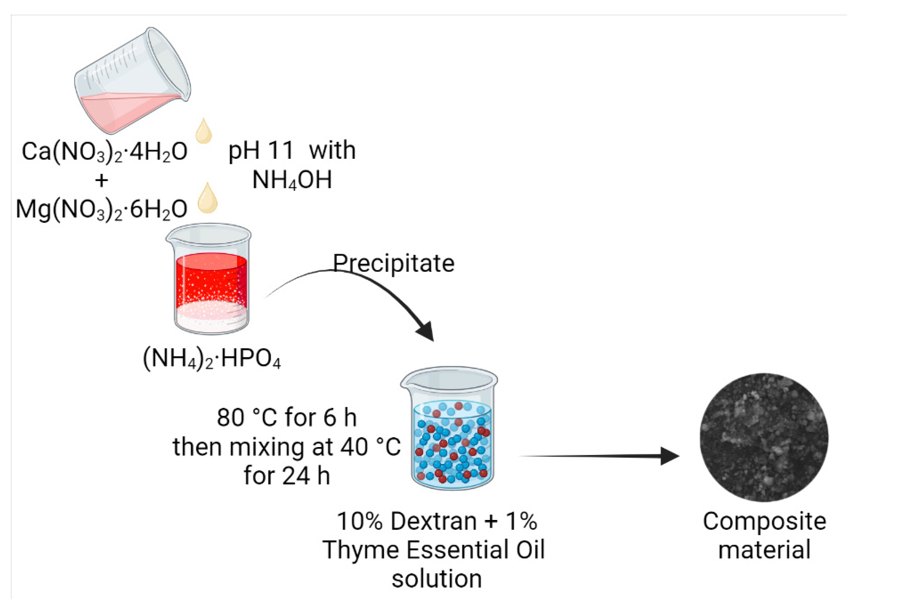
| Dextran-Thyme Magnesium-Doped Hydroxyapatite | Coating | Antimicrobial coating |
|
| [67] |
| Methacrylated gelatin-hyaluronic acid | Hydrogel scaffold | Tissue engineering |
|
| [118] |
| ‘Gelatin-hydroxy-phenyl propionic acid’- ‘hyaluronic acid tyramine’ | Polymer network | Retinal ganglion cells replacement therapy |
|
| [119] |
| Gellan Gum, Alginate and Nisin-Enriched Lipid Nanoparticles | Hydrogel | Wound recovery |
|
| [120] |
| Mg2+-Gellan Gum/Poly-Acrylamide | Hydrogel | Wound healing |
|
| [121] |
| Oxidized gellan gum + carboxy methyl chitosan | Hydrogel | Drug delivery and wound dressing |
|
| [122] |
| Gellan gum-alginate-calcium chloride | Hydrogel | Osteochondral repair |
|
| [123] |
4. Challenges and Future Prospective
4.1. Strain Selection
4.2. Downstream Processing
4.3. Composite-Forming Ability of EPS
4.4. Stability and Degradation Products of EPS Composites
4.5. Side Effects of Synthetic Polymers
5. Conclusions
Author Contributions
Funding
Institutional Review Board Statement
Informed Consent Statement
Acknowledgments
Conflicts of Interest
References
- Kaur, N.; Dey, P. Bacterial Exopolysaccharides as Emerging Bioactive Macromolecules: From Fundamentals to Applications. Res. Microbiol. 2022, 174, 104024. [Google Scholar] [CrossRef]
- Kuschmierz, L.; Meyer, M.; Bräsen, C.; Wingender, J.; Schmitz, O.J.; Siebers, B. Exopolysaccharide composition and size in Sulfolobus acidocaldarius biofilms. Front. Microbiol. 2022, 13, 982745. [Google Scholar] [CrossRef] [PubMed]
- Qi, M.; Zheng, C.; Wu, W.; Yu, G.; Wang, P. Exopolysaccharides from Marine Microbes: Source, Structure and Application. Mar. Drugs 2022, 20, 512. [Google Scholar] [CrossRef] [PubMed]
- Cázares-Vásquez, M.L.; Rodríguez-Herrera, R.; Aguilar-González, C.N.; Sáenz-Galindo, A.; Solanilla-Duque, J.F.; Contreras-Esquivel, J.C.; Flores-Gallegos, A.C. Microbial Exopolysaccharides in Traditional Mexican Fermented Beverages. Fermentation 2021, 7, 249. [Google Scholar] [CrossRef]
- Jeong, J.; Kim, Y.; Hu, Y.; Jung, S. Bacterial Succinoglycans: Structure, Physical Properties, and Applications. Polymers 2022, 14, 276. [Google Scholar] [CrossRef]
- Miao, K.H.; Guthmiller, K.B. Dextran; StatPearls Publishing: Treasure Island, FL, USA, 2022. [Google Scholar]
- Nwodo, U.U.; Green, E.; Okoh, A.I. Bacterial Exopolysaccharides: Functionality and Prospects. Int. J. Mol. Sci. 2012, 13, 14002–14015. [Google Scholar] [CrossRef] [Green Version]
- Ibrahim, H.A.H.; Abou Elhassayeb, H.E.; El-Sayed, W.M.M. Potential functions and applications of diverse microbial exopolysaccharides in marine environments. J. Genet. Eng. Biotechnol. 2022, 20, 151. [Google Scholar] [CrossRef] [PubMed]
- Gupta, P.; Diwan, B. Bacterial Exopolysaccharide mediated heavy metal removal: A Review on biosynthesis, mechanism and remediation strategies. Biotechnol. Rep. 2017, 13, 58–71. [Google Scholar] [CrossRef]
- Mohd Nadzir, M.; Nurhayati, R.W.; Idris, F.N.; Nguyen, M.H. Biomedical Applications of Bacterial Exopolysaccharides: A Review. Polymers 2021, 13, 530. [Google Scholar] [CrossRef]
- Díaz-Montes, E. Dextran: Sources, Structures, and Properties. Polysaccharides 2021, 2, 554–565. [Google Scholar] [CrossRef]
- Prasher, P.; Sharma, M.; Kumar Singh, S.; Gulati, M.; Kumar, D.; Gupta, G.; Kumar Chellappan, D.; Gregory George Oliver, B.; Wich, P.R.; Dua, K. Versatility of acetalated dextran in nanocarriers targeting respiratory diseases. Mater. Lett. 2022, 323, 132600. [Google Scholar] [CrossRef]
- Yahoum, M.M.; Toumi, S.; Tahraoui, H.; Lefnaoui, S.; Kebir, M.; Amrane, A.; Assadi, A.A.; Zhang, J.; Mouni, L. Formulation and Evaluation of Xanthan Gum Microspheres for the Sustained Release of Metformin Hydrochloride. Micromachines 2023, 14, 609. [Google Scholar] [CrossRef] [PubMed]
- Khare, P.; Chogale, M.; Kakade, P.; Patravale, V. Gellan gum-based in situ gelling ophthalmic nanosuspension of Posaconazole. Drug Deliv. Transl. Res. 2022, 12, 2920–2935. [Google Scholar] [CrossRef] [PubMed]
- Patel, J.; Maji, B.; Narayana Moorthy, N.S.H.; Maiti, S. Xanthan gum derivatives: Review of synthesis, properties and diverse applications. RSC Adv. 2020, 10, 27103–27136. [Google Scholar] [CrossRef]
- Dzionek, A.; Wojcieszyńska, D.; Guzik, U. Use of xanthan gum for whole cell immobilization and its impact in bioremediation—A review. Bioresour. Technol. 2022, 351, 126918. [Google Scholar] [CrossRef]
- Chaudhari, V.; Buttar, H.S.; Bagwe-Parab, S.; Tuli, H.S.; Vora, A.; Kaur, G. Therapeutic and Industrial Applications of Curdlan with Overview on Its Recent Patents. Front. Nutr. 2021, 8, 646988. [Google Scholar] [CrossRef]
- Di Mola, A.; Landi, M.R.; Massa, A.; D’Amora, U.; Guarino, V. Hyaluronic Acid in Biomedical Fields: New Trends from Chemistry to Biomaterial Applications. Int. J. Mol. Sci. 2022, 23, 14372. [Google Scholar] [CrossRef]
- Guo, L.; Chai, Y.; Zhou, F.; Wang, P. Preparation and Properties of Hyaluronic Acid Hydrogel Modified by L-cysteine Hydrochloride. IOP Conf. Ser. Earth Environ. Sci. 2021, 651, 042022. [Google Scholar] [CrossRef]
- Hussain, A.; Zia, K.M.; Tabasum, S.; Noreen, A.; Ali, M.; Iqbal, R.; Zuber, M. Blends and composites of exopolysaccharides; properties and applications: A review. Int. J. Biol. Macromol. 2017, 94, 10–27. [Google Scholar] [CrossRef]
- Xu, W.; Qian, J.; Zhang, Y.; Suo, A.; Cui, N.; Wang, J.; Yao, Y.; Wang, H. A double-network poly(Nε-acryloyl L-lysine)/hyaluronic acid hydrogel as a mimic of the breast tumor microenvironment. Acta Biomater. 2016, 33, 131–141. [Google Scholar] [CrossRef]
- Sieni, E.; Bazzolo, B.; Pieretti, F.; Zamuner, A.; Tasso, A.; Dettin, M.; Conconi, M.T. Breast cancer cells grown on hyaluronic acid-based scaffolds as 3D in vitro model for electroporation. Bioelectrochemistry 2020, 136, 107626. [Google Scholar] [CrossRef]
- Buhome, O.; Wongwattanakul, M.; Daduang, J.; Limpaiboon, T. 3D Silk Fibroin-Gelatin/Hyaluronic Acid/Heparan Sulfate Scaffold Enhances Expression of Stemness and EMT Markers in Cholangiocarcinoma. Vivo 2022, 36, 1155–1167. [Google Scholar] [CrossRef] [PubMed]
- Toullec, C.; Le Bideau, J.; Geoffroy, V.; Halgand, B.; Buchtova, N.; Molina-Peña, R.; Garcion, E.; Avril, S.; Sindji, L.; Dube, A.; et al. Curdlan–Chitosan Electrospun Fibers as Potential Scaffolds for Bone Regeneration. Polymers 2021, 13, 526. [Google Scholar] [CrossRef] [PubMed]
- Angelin, J.; Kavitha, M. Exopolysaccharides from probiotic bacteria and their health potential. Int. J. Biol. Macromol. 2020, 162, 853–865. [Google Scholar] [CrossRef] [PubMed]
- Zhang, Y.; Dai, X.; Jin, H.; Man, C.; Jiang, Y. The effect of optimized carbon source on the synthesis and composition of exopolysaccharides produced by Lactobacillus paracasei. J. Dairy Sci. 2021, 104, 4023–4032. [Google Scholar] [CrossRef] [PubMed]
- Gientka, I.; Bzducha-Wróbel, A.; Stasiak-Różańska, L.; Bednarska, A.A.; Błażejak, S. The exopolysaccharides biosynthesis by Candida yeast depends on carbon sources. Electron. J. Biotechnol. 2016, 22, 31–37. [Google Scholar] [CrossRef] [Green Version]
- Jin, H.; Jeong, Y.; Yoo, S.-H.; Johnston, T.V.; Ku, S.; Ji, G.E. Isolation and characterization of high exopolysaccharide-producing Weissella confusa VP30 from young children’s feces. Microb. Cell Factories 2019, 18, 110. [Google Scholar] [CrossRef] [Green Version]
- da Silva, J.A.; Cardoso, L.G.; de Jesus Assis, D.; Gomes, G.V.P.; Oliveira, M.B.P.P.; de Souza, C.O.; Druzian, J.I. Xanthan Gum Production by Xanthomonas campestris pv. Campestris IBSBF 1866 and 1867 from Lignocellulosic Agroindustrial Wastes. Appl. Biochem. Biotechnol. 2018, 186, 750–763. [Google Scholar] [CrossRef]
- Choi, I.S.; Ko, S.H.; Lee, M.E.; Kim, H.M.; Yang, J.E.; Jeong, S.-G.; Lee, K.H.; Chang, J.Y.; Kim, J.-C.; Park, H.W. Production, Characterization, and Antioxidant Activities of an Exopolysaccharide Extracted from Spent Media Wastewater after Leuconostoc mesenteroides WiKim32 Fermentation. ACS Omega 2021, 6, 8171–8178. [Google Scholar] [CrossRef]
- Pan, L.; Wang, Q.; Qu, L.; Liang, L.; Han, Y.; Wang, X.; Zhou, Z. Pilot-scale production of exopolysaccharide from Leuconostoc pseudomesenteroides XG5 and its application in set yogurt. J. Dairy Sci. 2022, 105, 1072–1083. [Google Scholar] [CrossRef]
- Bhatia, S.K.; Gurav, R.; Kim, B.; Kim, S.; Cho, D.-H.; Jung, H.; Kim, Y.-G.; Kim, J.-S.; Yang, Y.-H. Coproduction of exopolysaccharide and polyhydroxyalkanoates from Sphingobium yanoikuyae BBL01 using biochar pretreated plant biomass hydrolysate. Bioresour. Technol. 2022, 361, 127753. [Google Scholar] [CrossRef] [PubMed]
- Ahuja, V.; Kshirsagar, S.; Ghosh, P.; Sarkar, B.; Sutar, A.; More, S.; Dasgupta, D. Process development for detoxification of corncob hydrolysate using activated charcoal for xylitol production. J. Environ. Chem. Eng. 2022, 10, 107097. [Google Scholar] [CrossRef]
- Ahuja, V.; Bhatt, A.K.; Mehta, S.; Sharma, V.; Rathour, R.K. Sheetal Xylitol production by Pseudomonas gessardii VXlt-16 from sugarcane bagasse hydrolysate and cost analysis. Bioprocess Biosyst. Eng. 2022, 45, 1019–1031. [Google Scholar] [CrossRef] [PubMed]
- Yáñez-Fernández, J.; Herrera Ovando, M.G.; Patlán Ramírez, L.; Ramírez-Sotelo, G.; Guarin, C.A.; Castro-Rodríguez, D.C. Factorial Design to Optimize Dextran Production by the Native Strain Leuconostoc mesenteroides SF3. ACS Omega 2021, 6, 31203–31210. [Google Scholar] [CrossRef]
- Jumma Kareem, A.; Abdul Sattar Salman, J. Production of Dextran from Locally Lactobacillus spp. Isolates. Rep. Biochem. Mol. Biol. 2019, 8, 287–300. [Google Scholar]
- Koirala, P.; Maina, N.H.; Nihtilä, H.; Katina, K.; Coda, R. Brewers’ spent grain as substrate for dextran biosynthesis by Leuconostoc pseudomesenteroides DSM20193 and Weissella confusa A16. Microb. Cell Factories 2021, 20, 23. [Google Scholar] [CrossRef] [PubMed]
- Mangolim, C.S.; da Silva, T.T.; Fenelon, V.C.; Koga, L.N.; Ferreira, S.B.d.S.; Bruschi, M.L.; Matioli, G. Description of recovery method used for curdlan produced by Agrobacterium sp. IFO 13140 and its relation to the morphology and physicochemical and technological properties of the polysaccharide. PLoS ONE 2017, 12, e0171469. [Google Scholar] [CrossRef]
- Prakash, S.; Rajeswari, K.; Divya, P.; Ferlin, M.; Rajeshwari, C.T.; Vanavil, B. Optimization and production of curdlan gum using Bacillus cereus PR3 isolated from rhizosphere of leguminous plant. Prep. Biochem. Biotechnol. 2018, 48, 408–418. [Google Scholar] [CrossRef]
- Anane, R.F.; Sun, H.; Zhao, L.; Wang, L.; Lin, C.; Mao, Z. Improved curdlan production with discarded bottom parts of Asparagus spear. Microb. Cell Factories 2017, 16, 59. [Google Scholar] [CrossRef] [Green Version]
- Nejadmansouri, M.; Razmjooei, M.; Safdarianghomsheh, R.; Shad, E.; Delvigne, F.; Khalesi, M. Semi-continuous production of xanthan in biofilm reactor using Xanthomonas campestris. J. Biotechnol. 2021, 328, 1–11. [Google Scholar] [CrossRef]
- Raghunandan, K.; Kumar, A.; Kumar, S.; Permaul, K.; Singh, S. Production of gellan gum, an exopolysaccharide, from biodiesel-derived waste glycerol by Sphingomonas spp. 3 Biotech 2018, 8, 71. [Google Scholar] [CrossRef] [PubMed]
- Srivastava, N.; Kumari, S.; Kurmi, S.; Pinnaka, A.K.; Choudhury, A.R. Isolation, purification, and characterization of a novel exopolysaccharide isolated from marine bacteria Brevibacillus borstelensis M42. Arch. Microbiol. 2022, 204, 399. [Google Scholar] [CrossRef]
- Roychowdhury, R.; Srivastava, N.; Kumari, S.; Pinnaka, A.K.; Roy Choudhury, A. Isolation of an exopolysaccharide from a novel marine bacterium Neorhizobium urealyticum sp. nov. and its utilization in nanoemulsion formation for encapsulation and stabilization of astaxanthin. LWT 2021, 151, 112105. [Google Scholar] [CrossRef]
- Gangalla, R.; Sampath, G.; Beduru, S.; Sarika, K.; Kaveriyappan Govindarajan, R.; Ameen, F.; Alwakeel, S.; Thampu, R.K. Optimization and characterization of exopolysaccharide produced by Bacillus aerophilus rk1 and its in vitro antioxidant activities. J. King Saud Univ. Sci. 2021, 33, 101470. [Google Scholar] [CrossRef]
- Miller, G.L. Use of Dinitrosalicylic Acid Reagent for Determination of Reducing Sugar. Anal. Chem. 1959, 31, 426–428. [Google Scholar] [CrossRef]
- Bradford, M.M. A rapid and sensitive method for the quantitation of microgram quantities of protein utilizing the principle of protein-dye binding. Anal. Biochem. 1976, 72, 248–254. [Google Scholar] [CrossRef] [PubMed]
- Waterborg, J.H. The Lowry Method for Protein Quantitation. In The Protein Protocols Handbook; Walker, J.M., Ed.; Humana Press: Totowa, NJ, USA, 2009; pp. 7–10. ISBN 978-1-60327-474-6. [Google Scholar]
- Zaghloul, E.H.; Ibrahim, M.I.A. Production and Characterization of Exopolysaccharide from Newly Isolated Marine Probiotic Lactiplantibacillus plantarum EI6 with in vitro Wound Healing Activity. Front. Microbiol. 2022, 13, 903363. [Google Scholar] [CrossRef] [PubMed]
- Säwén, E.; Huttunen, E.; Zhang, X.; Yang, Z.; Widmalm, G. Structural analysis of the exopolysaccharide produced by Streptococcus thermophilus ST1 solely by NMR spectroscopy. J. Biomol. NMR 2010, 47, 125–134. [Google Scholar] [CrossRef]
- Piola, B.; Sabbatini, M.; Gino, S.; Invernizzi, M.; Renò, F. 3D Bioprinting of Gelatin–Xanthan Gum Composite Hydrogels for Growth of Human Skin Cells. Int. J. Mol. Sci. 2022, 23, 539. [Google Scholar] [CrossRef] [PubMed]
- Alves, A.; Miguel, S.P.; Araujo, A.R.T.S.; de Jesús Valle, M.J.; Sánchez Navarro, A.; Correia, I.J.; Ribeiro, M.P.; Coutinho, P. Xanthan Gum–Konjac Glucomannan Blend Hydrogel for Wound Healing. Polymers 2020, 12, 99. [Google Scholar] [CrossRef] [PubMed] [Green Version]
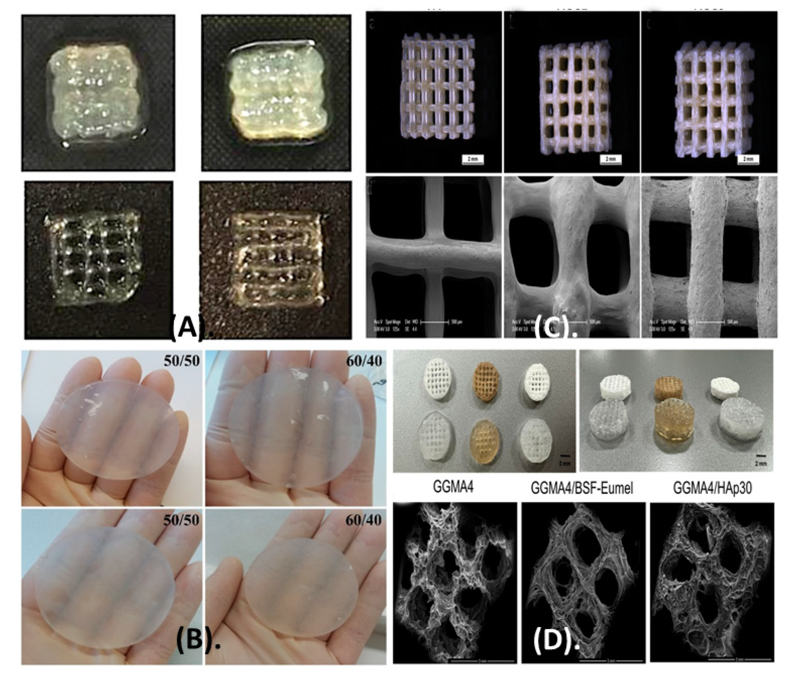
- Akkineni, A.R.; Ahlfeld, T.; Funk, A.; Waske, A.; Lode, A.; Gelinsky, M. Highly Concentrated Alginate-Gellan Gum Composites for 3D Plotting of Complex Tissue Engineering Scaffolds. Polymers 2016, 8, 170. [Google Scholar] [CrossRef] [PubMed] [Green Version]
- D’Amora, U.; Ronca, A.; Scialla, S.; Soriente, A.; Manini, P.; Phua, J.W.; Ottenheim, C.; Pezzella, A.; Calabrese, G.; Raucci, M.G.; et al. Bioactive Composite Methacrylated Gellan Gum for 3D-Printed Bone Tissue-Engineered Scaffolds. Nanomaterials 2023, 13, 772. [Google Scholar] [CrossRef]
- He, W.; Wang, X.; Hang, T.; Chen, J.; Wang, Z.; Mosselhy, D.A.; Xu, J.; Wang, S.; Zheng, Y. Fabrication of Cu2+-loaded phase-transited lysozyme nanofilm on bacterial cellulose: Antibacterial, anti-inflammatory, and pro-angiogenesis for bacteria-infected wound healing. Carbohydr. Polym. 2023, 309, 120681. [Google Scholar] [CrossRef] [PubMed]
- Biswas, A.; Ahmed, T.; Rana, M.R.; Hoque, M.M.; Ahmed, M.F.; Sharma, M.; Sridhar, K.; Ara, R.; Stephen Inbaraj, B. Fabrication and Characterization of ZnO Nanoparticles-Based Biocomposite Films Prepared Using Carboxymethyl Cellulose, Taro Mucilage, and Black Cumin Seed Oil for Evaluation of Antioxidant and Antimicrobial Activities. Agronomy 2023, 13, 147. [Google Scholar] [CrossRef]
- Ul-Islam, M.; Alhajaim, W.; Fatima, A.; Yasir, S.; Kamal, T.; Abbas, Y.; Khan, S.; Khan, A.H.; Manan, S.; Ullah, M.W.; et al. Development of low-cost bacterial cellulose-pomegranate peel extract-based antibacterial composite for potential biomedical applications. Int. J. Biol. Macromol. 2023, 231, 123269. [Google Scholar] [CrossRef] [PubMed]
- Lahiri, D.; Nag, M.; Dutta, B.; Dey, A.; Sarkar, T.; Pati, S.; Edinur, H.A.; Abdul Kari, Z.; Mohd Noor, N.H.; Ray, R.R. Bacterial Cellulose: Production, Characterization, and Application as Antimicrobial Agent. Int. J. Mol. Sci. 2021, 22, 12984. [Google Scholar] [CrossRef]
- Bai, F.-W.; Yang, S.; Ho, N.W.Y. 3.05—Fuel Ethanol Production from Lignocellulosic Biomass. In Comprehensive Biotechnology, 3rd ed.; Moo-Young, M., Ed.; Pergamon: Oxford, UK, 2019; pp. 49–65. ISBN 978-0-444-64047-5. [Google Scholar]
- Orlando, I.; Basnett, P.; Nigmatullin, R.; Wang, W.; Knowles, J.C.; Roy, I. Chemical Modification of Bacterial Cellulose for the Development of an Antibacterial Wound Dressing. Front. Bioeng. Biotechnol. 2020, 8, 557885. [Google Scholar] [CrossRef]
- Ojagh, S.M.A.; Vahabzadeh, F.; Karimi, A. Synthesis and characterization of bacterial cellulose-based composites for drug delivery. Carbohydr. Polym. 2021, 273, 118587. [Google Scholar] [CrossRef]
- Ma, N.; Cheung, D.Y.; Butcher, J.T. Incorporating nanocrystalline cellulose into a multifunctional hydrogel for heart valve tissue engineering applications. J. Biomed. Mater. Res. Part A 2022, 110, 76–91. [Google Scholar] [CrossRef]
- Zhu, Q.; Chen, X.; Liu, Z.; Li, Z.; Li, D.; Yan, H.; Lin, Q. Development of alginate-chitosan composite scaffold incorporation of bacterial cellulose for bone tissue engineering. Int. J. Polym. Mater. Polym. Biomater. 2023, 72, 296–307. [Google Scholar] [CrossRef]
- Balakrishnan, B.; Soman, D.; Payanam, U.; Laurent, A.; Labarre, D.; Jayakrishnan, A. A novel injectable tissue adhesive based on oxidized dextran and chitosan. Acta Biomater. 2017, 53, 343–354. [Google Scholar] [CrossRef]
- Andrabi, S.M.; Majumder, S.; Gupta, K.C.; Kumar, A. Dextran based amphiphilic nano-hybrid hydrogel system incorporated with curcumin and cerium oxide nanoparticles for wound healing. Colloids Surf. B Biointerfaces 2020, 195, 111263. [Google Scholar] [CrossRef]
- Ma, C.; Zhao, J.; Zhu, C.; Jiang, M.; Ma, P.; Mi, Y.; Fan, D. Oxidized dextran crosslinked polysaccharide/protein/polydopamine composite cryogels with multiple hemostatic efficacies for noncompressible hemorrhage and wound healing. Int. J. Biol. Macromol. 2022, 215, 675–690. [Google Scholar] [CrossRef]
- Iconaru, S.L.; Predoi, M.V.; Motelica-Heino, M.; Predoi, D.; Buton, N.; Megier, C.; Stan, G.E. Dextran-Thyme Magnesium-Doped Hydroxyapatite Composite Antimicrobial Coatings. Coatings 2020, 10, 57. [Google Scholar] [CrossRef] [Green Version]
- Sabit, H.; Abdel-Hakeem, M.; Shoala, T.; Abdel-Ghany, S.; Abdel-Latif, M.M.; Almulhim, J.; Mansy, M. Nanocarriers: A Reliable Tool for the Delivery of Anticancer Drugs. Pharmaceutics 2022, 14, 1566. [Google Scholar] [CrossRef] [PubMed]
- Rizvi, S.A.A.; Saleh, A.M. Applications of nanoparticle systems in drug delivery technology. Saudi Pharm. J. 2018, 26, 64–70. [Google Scholar] [CrossRef] [PubMed]
- Kumar, A.; Rao, K.M.; Han, S.S. Show more Development of sodium alginate-xanthan gum based nanocomposite scaffolds reinforced with cellulose nanocrystals and halloysite nanotubes. Polym. Test. 2017, 63, 214–225. [Google Scholar] [CrossRef]
- Feng, C.; Wang, F.; Xu, Z.; Sui, H.; Fang, Y.; Tang, X.; Shen, X. Characterization of Soybean Protein Adhesives Modified by Xanthan Gum. Coatings 2018, 8, 342. [Google Scholar] [CrossRef] [Green Version]
- Zia, I.; Jolly, R.; Mirza, S.; Umar, M.S.; Owais, M.; Shakir, M. Hydroxyapatite Nanoparticles Fortified Xanthan Gum–Chitosan Based Polyelectrolyte Complex Scaffolds for Supporting the Osteo-Friendly Environment. ACS Appl. Bio. Mater. 2020, 3, 7133–7146. [Google Scholar] [CrossRef]
- Neves, J.G.; Navarro da Rocha, D.; Lopes, C.C.; Barbosa, R.M.; Ferreira, L.F.; Westin, C.B.; Moraes, Â.M.; Calsa, B.; Santamaria, M., Jr.; Correr-Sobrinho, L.; et al. Calcium phosphates Chitosan-Xanthan composite scaffolds associated with mesenchymal stem cells for regenerative dentistry application. Ceram. Int. 2022, 48, 23088–23095. [Google Scholar] [CrossRef]
- Lochhead, R.Y. The Use of Polymers in Cosmetic Products. In Cosmetic Science and Technology; Elsevier: Amsterdam, The Netherlands, 2017; pp. 171–221. ISBN 978-0-12-802005-0. [Google Scholar]
- Nasrollahzadeh, M.; Sajjadi, M.; Nezafat, Z.; Shafiei, N. Chapter 3—Polysaccharide biopolymer chemistry. In Biopolymer-Based Metal Nanoparticle Chemistry for Sustainable Applications; Nasrollahzadeh, M., Ed.; Elsevier: Amsterdam, The Netherlands, 2021; pp. 45–105. ISBN 978-0-12-822108-2. [Google Scholar]
- Coltelli, M.-B.; Danti, S.; De Clerck, K.; Lazzeri, A.; Morganti, P. Pullulan for Advanced Sustainable Body- and Skin-Contact Applications. J. Funct. Biomater. 2020, 11, 20. [Google Scholar] [CrossRef] [PubMed] [Green Version]
- Chen, C.-T.; Chen, K.-I.; Chiang, H.-H.; Chen, Y.-K.; Cheng, K.-C. Improvement on Physical Properties of Pullulan Films by Novel Cross-Linking Strategy. J. Food Sci. 2017, 82, 108–117. [Google Scholar] [CrossRef] [PubMed]
- Amrita; Arora, A.; Sharma, P.; Katti, D.S. Pullulan-based composite scaffolds for bone tissue engineering: Improved osteoconductivity by pore wall mineralization. Carbohydr. Polym. 2015, 123, 180–189. [Google Scholar] [CrossRef]
- Cutiongco, M.F.A.; Tan, M.H.; Ng, M.Y.K.; Le Visage, C.; Yim, E.K.F. Composite pullulan–dextran polysaccharide scaffold with interfacial polyelectrolyte complexation fibers: A platform with enhanced cell interaction and spatial distribution. Acta Biomater. 2014, 10, 4410–4418. [Google Scholar] [CrossRef] [PubMed]
- Kicková, E.; Salmaso, S.; Mastrotto, F.; Caliceti, P.; Urtti, A. Pullulan Based Bioconjugates for Ocular Dexamethasone Delivery. Pharmaceutics 2021, 13, 791. [Google Scholar] [CrossRef]
- Chen, K.; Sivaraj, D.; Davitt, M.F.; Leeolou, M.C.; Henn, D.; Steele, S.R.; Huskins, S.L.; Trotsyuk, A.A.; Kussie, H.C.; Greco, A.H.; et al. Pullulan-Collagen hydrogel wound dressing promotes dermal remodelling and wound healing compared to commercially available collagen dressings. Wound Repair Regen. 2022, 30, 397–408. [Google Scholar] [CrossRef]
- Baron, R.I.; Duceac, I.A.; Morariu, S.; Bostănaru-Iliescu, A.-C.; Coseri, S. Hemostatic Cryogels Based on Oxidized Pullulan/Dopamine with Potential Use as Wound Dressings. Gels 2022, 8, 726. [Google Scholar] [CrossRef]
- Park, J.K.; Khan, T. 21—Other microbial polysaccharides: Pullulan, scleroglucan, elsinan, levan, alternant, dextran. In Handbook of Hydrocolloids, 2nd ed.; Phillips, G.O., Williams, P.A., Eds.; Woodhead Publishing Series in Food Science, Technology and Nutrition; Woodhead Publishing: Abington, PA, USA; Cambridge, UK, 2009; pp. 592–614. ISBN 978-1-84569-414-2. [Google Scholar]
- Kırtel, O.; Avşar, G.; Erkorkmaz, B.A.; Öner, E.T. Microbial Polysaccharides as Food Ingredients. In Microbial Production of Food Ingredients and Additives; Elsevier: Amsterdam, The Netherlands, 2017; pp. 347–383. ISBN 978-0-12-811520-6. [Google Scholar]
- Sezer, A.D.; Kazak, H.; Öner, E.T.; Akbuğa, J. Levan-based nanocarrier system for peptide and protein drug delivery: Optimization and influence of experimental parameters on the nanoparticle characteristics. Carbohydr. Polym. 2011, 84, 358–363. [Google Scholar] [CrossRef]
- Choi, W.I.; Hwang, Y.; Sahu, A.; Min, K.; Sung, D.; Tae, G.; Chang, J.H. An injectable and physical levan-based hydrogel as a dermal filler for soft tissue augmentation. Biomater. Sci. 2018, 6, 2627–2638. [Google Scholar] [CrossRef]
- Cinan, E.; Cesur, S.; Erginer Haskoylu, M.; Gunduz, O.; Toksoy Oner, E. Resveratrol-Loaded Levan Nanoparticles Produced by Electrohydrodynamic Atomization Technique. Nanomaterials 2021, 11, 2582. [Google Scholar] [CrossRef]
- Osman, A.; Lin, E.; Hwang, D.S. A sticky carbohydrate meets a mussel adhesive: Catechol-conjugated levan for hemostatic and wound healing applications. Carbohydr. Polym. 2023, 299, 120172. [Google Scholar] [CrossRef] [PubMed]
- Zheng, Y.; Liang, Y.; Zhang, D.; Sun, X.; Liang, L.; Li, J.; Liu, Y.-N. Gelatin-Based Hydrogels Blended with Gellan as an Injectable Wound Dressing. ACS Omega 2018, 3, 4766–4775. [Google Scholar] [CrossRef] [PubMed] [Green Version]
- Kim, S.; Jeon, G.Y.; Kim, S.E.; Choe, S.H.; Kim, S.J.; Seo, J.S.; Kang, T.W.; Song, J.E.; Khang, G. Injectable Hydrogel Based on Gellan Gum/Silk Sericin for Application as a Retinal Pigment Epithelium Cell Carrier. ACS Omega 2022, 7, 41331–41340. [Google Scholar] [CrossRef]
- Kenawy, E.-R.S.; Kamoun, E.A.; Eldin, M.S.; Soliman, H.M.A.; EL-Moslamy, S.H.; El-Fakharany, E.M.; Shokr, A.M. Electrospun PVA–Dextran Nanofibrous Scaffolds for Acceleration of Topical Wound Healing: Nanofiber Optimization, Characterization and In Vitro Assessment. Arab. J. Sci. Eng. 2023, 48, 205–222. [Google Scholar] [CrossRef]
- Aki, D.; Ulag, S.; Unal, S.; Sengor, M.; Ekren, N.; Lin, C.-C.; Yılmazer, H.; Ustundag, C.B.; Kalaskar, D.M.; Gunduz, O. 3D printing of PVA/hexagonal boron nitride/bacterial cellulose composite scaffolds for bone tissue engineering. Mater. Des. 2020, 196, 109094. [Google Scholar] [CrossRef]
- Zhang, M.; Huang, Y.; Pan, W.; Tong, X.; Zeng, Q.; Su, T.; Qi, X.; Shen, J. Polydopamine-incorporated dextran hydrogel drug carrier with tailorable structure for wound healing. Carbohydr. Polym. 2021, 253, 117213. [Google Scholar] [CrossRef]
- Wang, H.; Xia, H.; Xu, Z.; Natsuki, T.; Ni, Q.-Q. Effect of surface structure on the antithrombogenicity performance of poly(-caprolactone)-cellulose acetate small-diameter tubular scaffolds. Int. J. Biol. Macromol. 2023, 226, 132–142. [Google Scholar] [CrossRef] [PubMed]
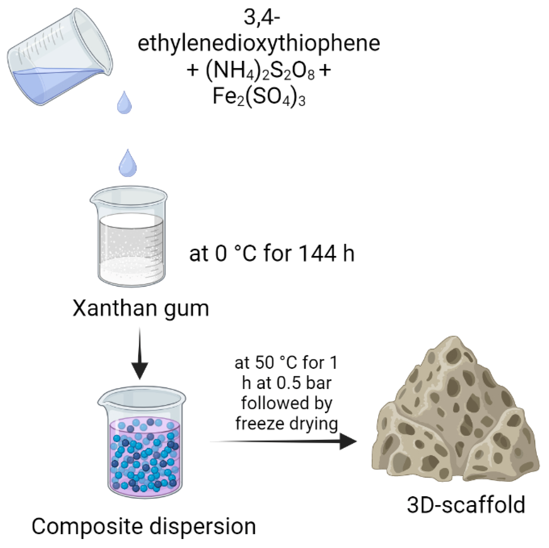
- del Agua, I.; Marina, S.; Pitsalidis, C.; Mantione, D.; Ferro, M.; Iandolo, D.; Sanchez-Sanchez, A.; Malliaras, G.G.; Owens, R.M.; Mecerreyes, D. Conducting Polymer Scaffolds Based on Poly(3,4-ethylenedioxythiophene) and Xanthan Gum for Live-Cell Monitoring. ACS Omega 2018, 3, 7424–7431. [Google Scholar] [CrossRef] [PubMed] [Green Version]
- Shawan, M.M.A.K.; Islam, N.; Aziz, S.; Khatun, N.; Sarker, S.R.; Hossain, M.; Hossan, T.; Morshed, M.; Sarkar, M.; Shakil, S.; et al. Fabrication of Xanthan gum: Gelatin (Xnt:Gel) Hybrid Composite Hydrogels for Evaluating Skin Wound Healing Efficacy. Mod. Appl. Sci. 2019, 13, 101–111. [Google Scholar] [CrossRef] [Green Version]
- Malik, N.S.; Ahmad, M.; Minhas, M.U.; Tulain, R.; Barkat, K.; Khalid, I.; Khalid, Q. Chitosan/Xanthan Gum Based Hydrogels as Potential Carrier for an Antiviral Drug: Fabrication, Characterization, and Safety Evaluation. Front. Chem. 2020, 8, 50. [Google Scholar] [CrossRef] [Green Version]
- Singh, S.; Nwabor, O.F.; Sukri, D.M.; Wunnoo, S.; Dumjun, K.; Lethongkam, S.; Kusolphat, P.; Hemtanon, N.; Klinprathum, K.; Sunghan, J.; et al. Poly (vinyl alcohol) copolymerized with xanthan gum/hypromellose/sodium carboxymethyl cellulose dermal dressings functionalized with biogenic nanostructured materials for antibacterial and wound healing application. Int. J. Biol. Macromol. 2022, 216, 235–250. [Google Scholar] [CrossRef] [PubMed]
- Avsar, G.; Agirbasli, D.; Agirbasli, M.A.; Gunduz, O.; Oner, E.T. Levan based fibrous scaffolds electrospun via co-axial and single-needle techniques for tissue engineering applications. Carbohydr. Polym. 2018, 193, 316–325. [Google Scholar] [CrossRef] [PubMed]
- Adrover, A.; Paolicelli, P.; Petralito, S.; Di Muzio, L.; Trilli, J.; Cesa, S.; Tho, I.; Casadei, M.A. Gellan Gum/Laponite Beads for the Modified Release of Drugs: Experimental and Modeling Study of Gastrointestinal Release. Pharmaceutics 2019, 11, 187. [Google Scholar] [CrossRef] [Green Version]
- Lin, C.-C.; Chiu, J.-Y. A novel γ-PGA composite gellan membrane containing glycerol for guided bone regeneration. Mater. Sci. Eng. C 2021, 118, 111404. [Google Scholar] [CrossRef]
- Yuan, H.; Zhong, W.; Wang, R.; Zhou, P.; Nie, Y.; Hu, W.; Tao, X.; Yang, P. Preparation of Cholesteryl-Modified Aminated Pullulan Nanoparticles to Evaluate Nanoparticle of Hydrophobic Degree on Drug Release and Cytotoxicity. J. Nanomater. 2020, 2020, e7171209. [Google Scholar] [CrossRef] [Green Version]
- Mommer, S.; Gehlen, D.; Akagi, T.; Akashi, M.; Keul, H.; Möller, M. Thiolactone-Functional Pullulan for in situ Forming Biogels. Biomacromolecules 2021, 22, 4262–4273. [Google Scholar] [CrossRef] [PubMed]
- Constantin, M.; Cosman, B.; Bercea, M.; Ailiesei, G.-L.; Fundueanu, G. Thermosensitive Poloxamer-graft-Carboxymethyl Pullulan: A Potential Injectable Hydrogel for Drug Delivery. Polymers 2021, 13, 3025. [Google Scholar] [CrossRef]
- Shahriari-Khalaji, M.; Hu, G.; Chen, L.; Cao, Z.; Andreeva, T.; Xiong, X.; Krastev, R.; Hong, F.F. Functionalization of Aminoalkylsilane-Grafted Bacterial Nanocellulose with ZnO-NPs-Doped Pullulan Electrospun Nanofibers for Multifunctional Wound Dressing. ACS Biomater. Sci. Eng. 2021, 7, 3933–3946. [Google Scholar] [CrossRef]
- Kushibiki, T.; Mayumi, Y.; Nakayama, E.; Azuma, R.; Ojima, K.; Horiguchi, A.; Ishihara, M. Photocrosslinked gelatin hydrogel improves wound healing and skin flap survival by the sustained release of basic fibroblast growth factor. Sci. Rep. 2021, 11, 23094. [Google Scholar] [CrossRef]
- Zheng, C.; Liu, C.; Chen, H.; Wang, N.; Liu, X.; Sun, G.; Qiao, W. Effective wound dressing based on Poly (vinyl alcohol)/Dextran-aldehyde composite hydrogel. Int. J. Biol. Macromol. 2019, 132, 1098–1105. [Google Scholar] [CrossRef]
- Wang, Y.; Guo, Z.; Qian, Y.; Zhang, Z.; Lyu, L.; Wang, Y.; Ye, F. Study on the Electrospinning of Gelatin/Pullulan Composite Nanofibers. Polymers 2019, 11, 1424. [Google Scholar] [CrossRef] [Green Version]
- Ma, M.-X.; Liu, Q.; Ye, C.; Grottkau, B.; Guo, B.; Song, Y.-F. Preparation of P3HB4HB/(Gelatin + PVA) Composite Scaffolds by Coaxial Electrospinning and Its Biocompatibility Evaluation. BioMed Res. Int. 2017, 2017, e9251806. [Google Scholar] [CrossRef] [PubMed] [Green Version]
- Navaei, T.; Milan, P.B.; Samadikuchaksaraei, A.; Davari, H.R.; Hardy, J.G.; Mozafari, M. Design and fabrication of polycaprolactone/gelatin composite scaffolds for diaphragmatic muscle reconstruction. J. Tissue Eng. Regen. Med. 2021, 15, 78–87. [Google Scholar] [CrossRef]
- Choi, J.; Lee, J.; Shin, M.E.; Been, S.; Lee, D.H.; Khang, G. Eggshell Membrane/Gellan Gum Composite Hydrogels with Increased Degradability, Biocompatibility, and Anti-Swelling Properties for Effective Regeneration of Retinal Pigment Epithelium. Polymers 2020, 12, 2941. [Google Scholar] [CrossRef] [PubMed]
- Hwang, Y.; Lee, J.S.; An, H.; Oh, H.; Sung, D.; Tae, G.; Choi, W.I. Hydroxyapatite-embedded levan composite hydrogel as an injectable dermal filler for considerable enhancement of biological efficacy. J. Ind. Eng. Chem. 2021, 104, 491–499. [Google Scholar] [CrossRef]
- Wang, Q.-Q.; Liu, Y.; Zhang, C.-J.; Zhang, C.; Zhu, P. Alginate/gelatin blended hydrogel fibers cross-linked by Ca2+ and oxidized starch: Preparation and properties. Mater. Sci. Eng. C 2019, 99, 1469–1476. [Google Scholar] [CrossRef] [PubMed]
- Heragh, B.K.; Javanshir, S.; Mahdavinia, G.R.; Naimi-Jamal, M.R. Development of pH-sensitive biomaterial-based nanocomposite for highly controlled drug release. Results Mater. 2022, 16, 100324. [Google Scholar] [CrossRef]
- Rajput, I.B.; Tareen, F.K.; Khan, A.U.; Ahmed, N.; Khan, M.F.A.; Shah, K.U.; Rahdar, A.; Díez-Pascual, A.M. Fabrication and in vitro evaluation of chitosan-gelatin based aceclofenac loaded scaffold. Int. J. Biol. Macromol. 2023, 224, 223–232. [Google Scholar] [CrossRef]
- Suflet, D.M.; Popescu, I.; Prisacaru, A.I.; Pelin, I.M. Synthesis and characterization of curdlan–phosphorylated curdlan based hydrogels for drug release. Int. J. Polym. Mater. Polym. Biomater. 2021, 70, 870–879. [Google Scholar] [CrossRef]
- Sakai, S.; Ohi, H.; Taya, M. Gelatin/Hyaluronic Acid Content in Hydrogels Obtained through Blue Light-Induced Gelation Affects Hydrogel Properties and Adipose Stem Cell Behaviors. Biomolecules 2019, 9, 342. [Google Scholar] [CrossRef] [Green Version]
- Velasco-Rodriguez, B.; Diaz-Vidal, T.; Rosales-Rivera, L.C.; García-González, C.A.; Alvarez-Lorenzo, C.; Al-Modlej, A.; Domínguez-Arca, V.; Prieto, G.; Barbosa, S.; Soltero Martínez, J.F.A.; et al. Hybrid Methacrylated Gelatin and Hyaluronic Acid Hydrogel Scaffolds. Preparation and Systematic Characterization for Prospective Tissue Engineering Applications. Int. J. Mol. Sci. 2021, 22, 6758. [Google Scholar] [CrossRef] [PubMed]
- Dromel, P.C.; Singh, D.; Andres, E.; Likes, M.; Kurisawa, M.; Alexander-Katz, A.; Spector, M.; Young, M. A bioinspired gelatin-hyaluronic acid-based hybrid interpenetrating network for the enhancement of retinal ganglion cells replacement therapy. Npj Regen. Med. 2021, 6, 85. [Google Scholar] [CrossRef] [PubMed]
- Reczyńska-Kolman, K.; Hartman, K.; Kwiecień, K.; Brzychczy-Włoch, M.; Pamuła, E. Composites Based on Gellan Gum, Alginate and Nisin-Enriched Lipid Nanoparticles for the Treatment of Infected Wounds. Int. J. Mol. Sci. 2022, 23, 321. [Google Scholar] [CrossRef]
- Li, W.; Jian, X.; Zou, Y.; Wu, L.; Huang, H.; Li, H.; Hu, D.; Yu, B. The Fabrication of a Gellan Gum-Based Hydrogel Loaded with Magnesium Ions for the Synergistic Promotion of Skin Wound Healing. Front. Bioeng. Biotechnol. 2021, 9, 709679. [Google Scholar] [CrossRef]
- Zhang, X.; Pan, Y.; Li, S.; Xing, L.; Du, S.; Yuan, G.; Li, J.; Zhou, T.; Xiong, D.; Tan, H.; et al. Doubly crosslinked biodegradable hydrogels based on gellan gum and chitosan for drug delivery and wound dressing. Int. J. Biol. Macromol. 2020, 164, 2204–2214. [Google Scholar] [CrossRef] [PubMed]
- Xing, J.; Peng, X.; Li, A.; Chen, M.; Ding, Y.; Xu, X.; Yu, P.; Xie, J.; Li, J. Gellan gum/alginate-based Ca-enriched acellular bilayer hydrogel with robust interface bonding for effective osteochondral repair. Carbohydr. Polym. 2021, 270, 118382. [Google Scholar] [CrossRef] [PubMed]
- May, T.; Herold, A.; Heinrich, D.C.; Heuschkel, I.; Sendrowski, H. Exopolysaccharide Production Microorganisms and Uses Thereof. WO2023036938A1, 16 March 2023. [Google Scholar]
- Picken, S.J.; Zlopasa, J.; Binneveld, R.A.G.; Böttger, W.O.J. Modification of Biopolymers Using Polyols and Polyacids. WO2023038519A1, 16 March 2023. [Google Scholar]
- Tsai, Y.-C.; Wu, C.-C.; Huang, C.-L.; Hsu, C.-C. Method for Treating Sleeping Disorders with Exopolysaccharides. WO2023025235, 2 March 2023. [Google Scholar]
- May, T.; Heinrich, D.C.; Herold, A.; Stierl, R. Paenibacillus Strains Producing Low Amounts of Exopolysaccarides. WO2023020880A1, 23 February 2023. [Google Scholar]
- Гомбоева, С.В.; Улаханова, Л.А.; Цыренов, В.Ж.; Vladimirovna, G.S.; Alekseevna, U.L.; Zhigzhitovich, T.V. Paenibacillus polymyxa Strain Essutm-2—Producer of Exopolysaccharides. RU2782953, 7 November 2022. [Google Scholar]
- Colliec-Jouault, S.; Sinquin, C.; Garcia, J.M.; Heymann, D.; Zykwinska, A. Low-Molecular-Weight he800 Exopolysaccharide Derivatives with Anti-Cancer Properties and Uses Thereof. WO2023275343, 5 January 2023. [Google Scholar]
- Гомбоева, С.В.; Улаханова, Л.А.; Цыренов, В.Ж.; Vladimirovna, G.S.; Alekseevna, U.L.; Zhigzhitovich, T.V. Paenibacillus polymyxa Vsgutu-1 Strain—Producer of Exopolysaccharides. RU2784088, 19 September 2022. [Google Scholar]
- Domínguez, P.J.A.; Narváez, M.J.M.; Chowdhury, P.S. Bacterial Cellulose Biopolymer and Method of Obtainment. WO2023014213, 9 February 2023. [Google Scholar]
- Peng, J.; Zhao, J.; Yang, F.; Liang, J.; Zhang, K.; Chen, G. Method for Separating and Screening Exopolysaccharide-Producing Lactic Acid Bacteria. CN115572690A, 6 January 2023. [Google Scholar]
- Hu, J. Soothing and Repairing Composition and Application Thereof in Cosmetics. CN115554197A, 3 January 2023. [Google Scholar]
- Fu, Y.; Mao, D.; Bian, X.; Wang, X.; Zhang, Y. Preparation Method of Chlorella Pyrenoidosa Exopolysaccharide with Antitumor Activity. CN115505610A, 23 December 2022. [Google Scholar]
- Srivastava, N.; Choudhury, A.R. Microbial Polysaccharide-Based Nanoformulations for Nutraceutical Delivery. ACS Omega 2022, 7, 40724–40739. [Google Scholar] [CrossRef]
- Theuer, R.; Augustine, T.; Kishter, L.; Lanspa, R. Petition to Add Pullulan to the National List at §205.605(a) as an Allowed Nonsynthetic Ingredient in Tablets and Capsules for Dietary Supplements Labeled “Made with Organic (Specified Ingredients or Food Group(s)); United States Department of Agriculture: Washington, DC, USA, 2013.
- Costa, O.Y.A.; Raaijmakers, J.M.; Kuramae, E.E. Microbial Extracellular Polymeric Substances: Ecological Function and Impact on Soil Aggregation. Front. Microbiol. 2018, 9, 1636. [Google Scholar] [CrossRef] [PubMed] [Green Version]
- Brandi, J.; Cheri, S.; Manfredi, M.; Di Carlo, C.; Vita Vanella, V.; Federici, F.; Bombiero, E.; Bazaj, A.; Rizzi, E.; Manna, L.; et al. Exploring the wound healing, anti-inflammatory, anti-pathogenic and proteomic effects of lactic acid bacteria on keratinocytes. Sci. Rep. 2020, 10, 11572. [Google Scholar] [CrossRef]
- Becker, A. Challenges and perspectives in combinatorial assembly of novel exopolysaccharide biosynthesis pathways. Front. Microbiol. 2015, 6, 687. [Google Scholar] [CrossRef] [Green Version]
- Schmid, J.; Sieber, V.; Rehm, B. Bacterial exopolysaccharides: Biosynthesis pathways and engineering strategies. Front. Microbiol. 2015, 6, 496. [Google Scholar] [CrossRef] [PubMed] [Green Version]
- Mishra, A.; Jha, B. Microbial Exopolysaccharides. In The Prokaryotes; Rosenberg, E., DeLong, E.F., Lory, S., Stackebrandt, E., Thompson, F., Eds.; Springer: Berlin/Heidelberg, Germany, 2013; pp. 179–192. ISBN 978-3-642-31330-1. [Google Scholar]
- Akdeniz Oktay, B.; Bozdemir, M.T.; Ozbas, Z.Y. Optimization of hazelnut husk medium for pullulan production by a domestic A. pullulans strain. Prep. Biochem. Biotechnol. 2022, 53, 317–330. [Google Scholar] [CrossRef] [PubMed]
- Mirzaee, H.; Khodaiyan, F.; Kennedy, J.F.; Hosseini, S.S. Production, Optimization and Characterization of Pullulan from Sesame Seed Oil Cake as a New Substrate by Aureobasidium pullulans; Carbohydrate Polymer Technologies and Applications: Amsterdam, The Netherlands, 2020; Volume 1. [Google Scholar] [CrossRef]
- He, C.; Zhang, X.; Zhang, Z.; Wang, C.; Wang, D.; Wei, G. Whole-crop biorefinery of corn biomass for pullulan production by Aureobasidium pullulans. Bioresour. Technol. 2023, 370, 128517. [Google Scholar] [CrossRef]
- Aftab, M.N.; Iqbal, I.; Riaz, F.; Karadag, A.; Tabatabaei, M. Different Pretreatment Methods of Lignocellulosic Biomass for Use in Biofuel Production; IntechOpen: London, UK, 2019; ISBN 978-1-78923-988-1. [Google Scholar]
- Kant Bhatia, S.; Ahuja, V.; Chandel, N.; Gurav, R.; Kant Bhatia, R.; Govarthanan, M.; Kumar Tyagi, V.; Kumar, V.; Pugazendhi, A.; Rajesh Banu, J.; et al. Advances in algal biomass pretreatment and its valorisation into biochemical and bioenergy by the microbial processes. Bioresour. Technol. 2022, 358, 127437. [Google Scholar] [CrossRef]
- Santos, F.L.; de Amorim, G.M. Biotechnological challenges and perspectives of using exopolysaccharides. J. Anal. Pharm. Res. 2018, 7, 264–266. [Google Scholar] [CrossRef] [Green Version]
- Singh, S.; Datta, S.; Narayanan, K.B.; Rajnish, K.N. Bacterial exo-polysaccharides in biofilms: Role in antimicrobial resistance and treatments. J. Genet. Eng. Biotechnol. 2021, 19, 140. [Google Scholar] [CrossRef] [PubMed]
- Xu, R.-Z.; Cao, J.-S.; Feng, G.; Luo, J.-Y.; Wu, Y.; Ni, B.-J.; Fang, F. Modeling molecular structure and behavior of microbial extracellular polymeric substances through interacting-particle reaction dynamics. Chem. Eng. J. Adv. 2021, 8, 100154. [Google Scholar] [CrossRef]
- Zou, P.; Yao, J.; Cui, Y.-N.; Zhao, T.; Che, J.; Yang, M.; Li, Z.; Gao, C. Advances in Cellulose-Based Hydrogels for Biomedical Engineering: A Review Summary. Gels 2022, 8, 364. [Google Scholar] [CrossRef]
- Alheib, O.; da Silva, L.P.; da Silva Morais, A.; Mesquita, K.A.; Pirraco, R.P.; Reis, R.L.; Correlo, V.M. Injectable laminin-biofunctionalized gellan gum hydrogels loaded with myoblasts for skeletal muscle regeneration. Acta Biomater. 2022, 143, 282–294. [Google Scholar] [CrossRef]
- McClatchy, D.B.; Martínez-Bartolomé, S.; Gao, Y.; Lavallée-Adam, M.; Yates, J.R. Quantitative analysis of global protein stability rates in tissues. Sci. Rep. 2020, 10, 15983. [Google Scholar] [CrossRef]
- Hu, Y.; Zheng, Q.; Zhang, S.; Noll, L.; Wanek, W. Significant release and microbial utilization of amino sugars and d-amino acid enantiomers from microbial cell wall decomposition in soils. Soil Biol. Biochem. 2018, 123, 115–125. [Google Scholar] [CrossRef] [PubMed]
- Beier, S.; Bertilsson, S. Bacterial chitin degradation—Mechanisms and ecophysiological strategies. Front. Microbiol. 2013, 4, 149. [Google Scholar] [CrossRef] [PubMed] [Green Version]
- Maitz, M.F. Applications of synthetic polymers in clinical medicine. Biosurface Biotribol. 2015, 1, 161–176. [Google Scholar] [CrossRef] [Green Version]
- Kostić, M.; Igić, M.; Gligorijević, N.; Nikolić, V.; Stošić, N.; Nikolić, L. The Use of Acrylate Polymers in Dentistry. Polymers 2022, 14, 4511. [Google Scholar] [CrossRef] [PubMed]
- Kucharczyk, M.; Słowik-Rylska, M.; Cyran-Stemplewska, S.; Gieroń, M.; Nowak-Starz, G.; Kręcisz, B. Acrylates as a significant cause of allergic contact dermatitis: New sources of exposure. Postep. Dermatol. Alergol. 2021, 38, 555–560. [Google Scholar] [CrossRef]
- Kanazawa, R.; Sato, S.; Iwamoto, N.; Teramoto, A. Allergic reaction following arachnoid plasty with a fibrin sealant. Neurol. Med. Chir. 2010, 50, 608–610. [Google Scholar] [CrossRef] [Green Version]
- Orihara, M.; Takazawa, T.; Horiuchi, T.; Sakamoto, S.; Uchiyama, M.; Saito, S. Intraoperative anaphylaxis due to aprotinin after local application of fibrin sealant diagnosed by skin tests and basophil activation tests: A case report. JA Clin. Rep. 2021, 7, 68. [Google Scholar] [CrossRef]
- Dhaka, V.; Singh, S.; Anil, A.G.; Sunil Kumar Naik, T.S.; Garg, S.; Samuel, J.; Kumar, M.; Ramamurthy, P.C.; Singh, J. Occurrence, toxicity and remediation of polyethylene terephthalate plastics. A review. Environ. Chem. Lett. 2022, 20, 1777–1800. [Google Scholar] [CrossRef]
- Donati, M.; Brancato, G.; Grosso, G.; Li Volti, G.; La Camera, G.; Cardì, F.; Basile, F.; Donati, A. Immunological reaction and oxidative stress after light or heavy polypropylene mesh implantation in inguinal hernioplasty. Medicine 2016, 95, e3791. [Google Scholar] [CrossRef]
- Casalini, T.; Rossi, F.; Castrovinci, A.; Perale, G. A Perspective on Polylactic Acid-Based Polymers Use for Nanoparticles Synthesis and Applications. Front. Bioeng. Biotechnol. 2019, 7, 259. [Google Scholar] [CrossRef] [PubMed]


| Patent ID | EPS | Microorganisms | Application | Reference |
|---|---|---|---|---|
| WO2023036938A1 | EPS | Firmicutes | Gene mutation in Firmicutes for EPS production | [124] |
| WO2023038519A1 | Biopolymers | aerobic granular sludge and anam- mox granular sludge | Modification of biopolymers using polyols and polyacids | [125] |
| WO2023025235 | EPS | Lactic acid bacterium | Treatment of sleeping disorder | [126] |
| WO2023020880 | EPS | Paenibacillus strains | Production | [127] |
| RU2782953 | - | Paenibacillus polymyxa strain essutm-2 | Production | [128] |
| WO2023275343 | Low-molecular-weight he800 exopolysaccharide derivatives | Vibrio diabolicus HE800 | Anti-cancer | [129] |
| RU2784088 | - | Paenibacillus polymyxa vsgutu-1 | Production | [130] |
| WO2023014213 | Cellulase | Acetobacter aceti | Production | [131] |
| CN115572690A | EPS | Lactic acid bacteria | Selection of EPS producing bacterial | [132] |
| CN115554197A | EPS | - | Soothing and repairing based cosmetic formulations | [133] |
| CN115505610A | EPS | Chlorella pyrenoidosa | Antitumor activity | [134] |
Disclaimer/Publisher’s Note: The statements, opinions and data contained in all publications are solely those of the individual author(s) and contributor(s) and not of MDPI and/or the editor(s). MDPI and/or the editor(s) disclaim responsibility for any injury to people or property resulting from any ideas, methods, instructions or products referred to in the content. |
© 2023 by the authors. Licensee MDPI, Basel, Switzerland. This article is an open access article distributed under the terms and conditions of the Creative Commons Attribution (CC BY) license (https://creativecommons.org/licenses/by/4.0/).
Share and Cite
Ahuja, V.; Bhatt, A.K.; Banu, J.R.; Kumar, V.; Kumar, G.; Yang, Y.-H.; Bhatia, S.K. Microbial Exopolysaccharide Composites in Biomedicine and Healthcare: Trends and Advances. Polymers 2023, 15, 1801. https://doi.org/10.3390/polym15071801
Ahuja V, Bhatt AK, Banu JR, Kumar V, Kumar G, Yang Y-H, Bhatia SK. Microbial Exopolysaccharide Composites in Biomedicine and Healthcare: Trends and Advances. Polymers. 2023; 15(7):1801. https://doi.org/10.3390/polym15071801
Chicago/Turabian StyleAhuja, Vishal, Arvind Kumar Bhatt, J. Rajesh Banu, Vinod Kumar, Gopalakrishnan Kumar, Yung-Hun Yang, and Shashi Kant Bhatia. 2023. "Microbial Exopolysaccharide Composites in Biomedicine and Healthcare: Trends and Advances" Polymers 15, no. 7: 1801. https://doi.org/10.3390/polym15071801