Article

Cytoprotection of Probiotic Lactobacillus acidophilus with Artificial Nanoshells of Nature-Derived Eggshell Membrane Hydrolysates and Coffee Melanoidins in Single-Cell Nanoencapsulation

1
Center for Cell-Encapsulation Research, Department of Chemistry, KAIST, Daejeon 34141, Republic of Korea
2
Department of Chemistry, University of Ulsan, Ulsan 44776, Republic of Korea
3
Department of Science Education, Ewha Womans University, Seoul 03760, Republic of Korea
*
Authors to whom correspondence should be addressed.
Polymers 2023, 15(5), 1104; https://doi.org/10.3390/polym15051104
Submission received: 21 January 2023 / Revised: 14 February 2023 / Accepted: 20 February 2023 / Published: 22 February 2023

Abstract

:
One-step fabrication method for thin films and shells is developed with nature-derived eggshell membrane hydrolysates (ESMHs) and coffee melanoidins (CMs) that have been discarded as food waste. The nature-derived polymeric materials, ESMHs and CMs, prove highly biocompatible with living cells, and the one-step method enables cytocompatible construction of cell-in-shell nanobiohybrid structures. Nanometric ESMH-CM shells are formed on individual probiotic Lactobacillus acidophilus, without any noticeable decrease in viability, and the ESMH-CM shells effectively protected L. acidophilus in the simulated gastric fluid (SGF). The cytoprotection power is further enhanced by Fe3+-mediated shell augmentation. For example, after 2 h of incubation in SGF, the viability of native L. acidophilus is 30%, whereas nanoencapsulated L. acidophilus, armed with the Fe3+-fortified ESMH-CM shells, show 79% in viability. The simple, time-efficient, and easy-to-process method developed in this work would contribute to many technological developments, including microbial biotherapeutics, as well as waste upcycling.

Graphical Abstract

1. Introduction

One-step film construction, referring to the process that involves mixing of complimentary coating components and generation of interface-active species prior to film formation, significantly simplifies film fabrication procedures, particularly compared with sequential deposition approaches (e.g., layer-by-layer, LbL) [1,2,3,4], and eliminates the repeated deposition steps if it affords continuous film growth. Its notable examples include material-independent coating of tannic acid (TA) and Fe3+, where use of the preformed TA-Fe3+ sol enables much simplified procedures with (semi)-continuous film growth [5,6,7,8,9]. In-situ generation of interface-active TA-Fe3+ species, for example, the one inspired by iron gall ink [10,11,12], would be another strategy for the one-step, continuous film formation [10,11,12,13,14].
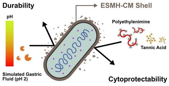
The one-step approach is especially beneficial in the field of single-cell nanoencapsulation (SCNE) [15,16,17,18,19,20], where cytoprotective nanometric-shells are constructed on individual living cells, leading to the creation of cell-in-shell nanobiohybrids. Its procedural simplicity also would be seamlessly coupled with the existing industrial processes, for instance, for fabrication of probiotic capsules, as well as enhancing biocompatibility with living cells, probiotics in this study. As a related work, tea polyphenols and poly(N-vinylpyrrolidone) (PVPON) were added simultaneously to an aqueous cell suspension for cytocompatible formation of cell-in-shell structures with a polymeric complex of tea polyphenols and PVPON [21], as an alternative to the LbL-based SCNE with TA and PVPON [22]. It is envisaged that the identification of other nature-derived materials, structurally different from polyphenolics, would further upscale the usability and applicability of one-step film construction in the practical applications as well as in the research domain of single-cell surface engineering including SCNE [23]. In this work, we constructed a cell-in-shell structure of probiotic Lactobacillus acidophilus by one-step, cytocompatible formation of the cytoprotective shells comprising nature-derived eggshell membrane hydrolysates (ESMHs) and coffee melanoidins (CMs) (Figure 1).
ESMHs, hydrolyzed ESMs, have recently been suggested as nature-derived biomaterials in the LbL-based formation of thin films [24]. In consideration of high content of proteins (80–85%) in ESMs, including collagens (types I, V, and X) and glycosaminoglycans, ESMHs could serve as extracellular matrix-mimetic materials in the manipulation of living cells. The LbL-SCNE methods with ESMH-TA [25] and ESMH-CM pairs [26] have previously been demonstrated to show the great cytocompatibility of ESMHs. Utilization of ESMHs as biomaterials also would contribute to the waste upcycling in the aspect of circular economy; eggshells, comprising about 11% of egg weight, have been discarded as food waste [27,28]. In the aspect of waste upcycling, spent coffee grounds also have been explored as a sustainable solution in various fields, such as photothermal materials in sterilization, fertilizers and soil conditioners in the agricultural industry, raw materials for biodiesel and biochar production, and sorbents in water management [29,30,31,32]. CMs, a product of the Maillard reaction during coffee-roasting process, exhibit antioxidant activity as well as containing biologically active molecules [33]. Aside from potential use as nutraceuticals [34], CMs, extracted from spent coffee ground, form thin films with Fe3+, which has been utilized in SCNE [35]. The combined use of ESMHs and CMs as next-generation biomaterials would widen the material scope in biomedical engineering and related fields, assisted by the development of simple, one-step methods for forming ultrathin films and shells.

2. Materials and Methods

2.1. One-Step Formation of ESMH-CM Films and Shells on Abiotic Substrates

The ESMHs and CMs were prepared according to our previous reports [26,35]. The stock solution of ESMHs or CMs was made to the final concentration of 2 mg/mL in a sodium chloride (NaCl) solution (50 mM). Prior to use, gold substrates were cleaned with ethanol and acetone. The cleaned gold substrates were immersed in a 1:1 mixture of the ESMH and CM stock solutions (500 μL each), stirred at 120 rpm for 3 h, washed with deionized (DI) water, and dried under a stream of argon gas. The same protocol was employed for other flat substrates (silver, aluminum, copper, nickel, tin, titanium, silicon, stainless steel, poly(acrylic acid), polycarbonate, polyethylene, polyurethane, and polytetrafluoroethylene). Calcium carbonate (CaCO3) particles were prepared by mixing 4 mL of an aqueous poly(sodium 4-styrenesulfonate) solution (PSS, MW: ca. 70,000, 2 mg/mL), 48 µL of an aqueous sodium carbonate solution (Na2CO3, 1 M), and 96 µL of an aqueous calcium chloride solution (CaCl2, 1 M) under vigorous stirring for 40 s, incubating for 7 min at room temperature, and calcinating at 450 °C for 2 h. ESMH-CM shells were formed on the resulting CaCO3 particles with a 1:1 mixture of the ESMH and CM stock solutions (500 µL each).

2.2. Single-Cell Nanoencapsulation (SCNE) and Characterizations

A single colony of Saccharomyces cerevisiae, picked from the YPD agar plate, was cultured for 30 h in a YPD broth medium at 33 °C. After washing with DI water, S. cerevisiae were immersed for 3 h in a 1:1 mixture of the ESMH and CM stock solutions (500 μL each) and washed with DI water three times. The same SCNE protocol was employed for L. acidophilus and Levilactobacillus brevis, after culturing for 24 h in an MRS broth medium at 33 °C. For viability assay of S. cerevisiae, 5 μL of the stock solution of fluorescein diacetate (FDA, 10 mg/mL in acetone) and 2 μL of an aqueous solution of propidium iodide (PI, 1 mg/mL) were added to a S. cerevisiae suspension (1 mL), and the mixture was incubated for 15 min at 33 °C. SYTO 9 was used instead of FDA for the viability assay of L. acidophilus and L. brevis. Both 2 μL of the SYTO 9 stock solution (3.34 mM in DMSO) and 2 μL of the PI stock solution (20 mM in DMSO) were added to a cell suspension (1 mL). The mixture was then incubated for 20 min at 33 °C. To form the ESMH-CM[Fe3+] shell, ESMH-CM-encapsulated cells (denoted as cell@ESMH-CM) were immersed in an aqueous solution of FeCl3 (10 mM) for 30 min. The t 2.0 OD 600 values were calculated based on the results of cell culture in the MRS broth medium. In short, 1 mL of an aqueous cell suspension (L. acidophilus, L. acidophilus@ESMH-CM, or L. acidophilus@ESMH-CM[Fe3+], OD 600 = 0.15) was added to 150 mL of the MRS broth medium (final OD 600 = 0.001) and incubated at 33 °C. The 100 μL of the culture mixture was picked at the predetermined time, and the cell density was measured at 600 nm with a microplate reader. Linear fitting of ln OD 600 , from −4.0 to +1.0, with incubation time (in hour) gave t 2.0 OD 600 , the time for ln OD 600 of −2.0. For cytoprotection studies, cells were suspended in 1 mL of an aqueous polyethylenimine solution (PEI, 0.5, 1, 10, or 50 mg/mL) for 30 min or 1 mL of an aqueous TA solution (1, 5, 10, 25, or 50 mg/mL) for 1 h. The simulated gastric fluid (SGF) was prepared by dissolving NaCl (0.2% (w/v)) and pepsin (3 mg/mL) in DI water, followed by pH adjustment to 2 with 1 M HCl. The cells were suspended in the SGF solution and incubated for 2 h at 37 °C for cytoprotection studies against the SGF.

3. Results and Discussion

3.1. One-Step Formation of ESMH-CM Films and Shells on Abiotic Substrates

Prior to SCNE, we investigated the feasibility of one-step film formation with ESMH-CM complex and optimized the reaction conditions, with a gold substrate as a model. The concentrations of ESMHs and CMs were set to be 1 mg/mL each. The ellipsometric thickness measurement, after 3 h of reaction, indicated that a film was formed with 6.2 nm of thickness. It was also found that the addition of NaCl to the coating mixture increased film thickness, and 50 mM of NaCl was chosen as an optimized concentration in this study. The film thickness significantly increased to 10.3 nm with 50 mM of NaCl as an additive after 3 h of reaction (166% increase). Detailed thickness analysis showed that the film growth stopped after about 3 h without NaCl (thickness: 7.1 nm), but in stark contrast, the ESMH-CM film grew continuously at least up to 24 h (thickness: 14.0 nm) (Figure 2a). As a control, we used only a single component (ESMHs or CMs) in the film formation. The thickness analysis showed that 1.6-nm-thick and 0.8-nm-thick films were formed with ESMHs and CMs, respectively, under the same conditions, confirming the significance of pre-association of ESMHs and CMs in solution (Figure S1). On the other hand, it was observed that the pre-mixed ESMH-CM pair also could be utilized in the LbL-type film formation: film thickness increased in a linear fashion, with 2.8 nm per 10 min of incubation, making 28 nm thick films after 10 deposition cycles (Figure S2).
The films formed after 3 h of reaction were characterized by Fourier transform infrared (FT-IR) spectroscopy, X-ray photoelectron spectroscopy (XPS), field-emission scanning electron microscopy (FE-SEM), and atomic force microscopy (AFM). The signature bands for ESMHs and CMs at 1666 (amide-I stretching) and 1550 cm−1 (amide-II stretching), in addition to the v(C−H) band at 2960 cm−1 and v(O−H) band at 3297 cm−1, in the FT-IR spectrum indicated the successful formation of ESMH-CM films (Figure 2b), further supported by the XPS analysis showing C 1s and N 1s peaks (Figure S3a). The C 1s XPS peak was deconvoluted into three peaks at binding energies of 283.6 (C−C and C−H), 284.8 (C−O and C−N), and 287.0 eV (C=O and C=N), and the one for the N 1s peak was further deconvoluted into two peaks at 398.4 (C−NH) and 399.1 eV (O=C−N), additionally providing evidence for the presence of carbohydrates and peptides in the film (Figure S3b,c) [35,36]. The FE-SEM and AFM analysis showed that the ESMH-CM films were composed of nanoparticulates, clearly distinct from the bare gold surface (Figure S3d,e). The water-contact angle measurements also indicated the successful formation of ESMH-CM films: the contact angle was changed to 28.2° from 76.6°, after film formation.
We also examined whether our one-step method for ESMH-CM-film formation was universal, applicable to interface engineering of various different substrates, including silver, aluminum, copper, nickel, tin, titanium, silicon, stainless steel, poly(acrylic acid), polycarbonate, polyethylene, polyurethane, and polytetrafluoroethylene (PTFE), in addition to gold. The water-contact angle measurements clearly showed that the one-step method was material-independent (Figure 2c). The water-contact angles of all the substrates tested were changed to be below 60° after film formation, regardless of their intact angles: for example, the contact angle of PTFE was changed to 52.8° from 115.7°. In addition to the flat substrates, our one-step method was employed for the construction of core-shell structures in particle engineering, exemplified with CaCO3 and amine-terminated silica (SiO2) particles. Changes in the zeta (ζ) potential indicated the formation of ESMH-CM shells on the particles: −15.7 eV from +4.1 eV for CaCO3 particles (diameter: 2–4 μm) and −35.5 eV from +44.3 eV for SiO2 particles (diameter: 3.92 μm) (Figure S4a). It is of note that the shell formation occurred regardless of the surface charge of particles. The formation of ESMH-CM shells was visualized with a rhodamine-linked ESMH (ESMH_TAMRA, λemission: 575 nm) [25,26] by confocal laser-scanning microscopy (CLSM) (Figure S4b). The ability to form shells on individual particles under biocompatible synthetic conditions suggested the potential of our system in the SCNE of living cells.

3.2. One-Step SCNE of S. cerevisiae

After confirming the one-step formation of films and cells on abiotic substrates with ESMHs and CMs, the protocol was applied to the SCNE with S. cerevisiae as a model. S. cerevisiae was chosen for investigation of cytocompatibility of our method and cytoprotectability of the formed ESMH-CM shell, because of the availability of numerous reference reports on the SCNE of S. cerevisiae [37,38].
S. cerevisiae was incubated for 3 h in a 50 mM NaCl solution of ESMHs (1 mg/mL) and CMs (1 mg/mL), leading to the construction of S.cerevisiae@ESMH-CM. The cell viability, after SCNE, was analyzed with FDA (λemission: 521 nm; for live cells) and PI (λemission: 636 nm; for dead cells). FDA is a membrane-permeable, fluorogenic viability-probe that measures both enzymatic activity and membrane integrity, and PI is a membrane-impermeable, nucleic-acid-intercalating agent that is commonly used to detect dead cells. The CLSM images showed that most S.cerevisiae@ESMH-CM cells were viable (Figure 3a), and the quantitative analysis showed 98.7% of %viability (calculated by dividing the viability of S.cerevisiae@ESMH-CM (97.3 ± 0.9%) by the viability of intact, bare S. cerevisiae as a reference (98.6 ± 1.0%)), indicating no noticeable harm to the cells. That is, the viability assay confirmed that the one-step shell formation with ESMHs and CMs was extremely cytocompatible. The ESMH-CM shells on S. cerevisiae were visualized with use of ESMH_TAMRA by CLSM, which showed green/red core/shell structures of FDA-stained S. cerevisiae (Figure 3b).
Cytoprotectability of the ESMH-CM shells on S. cerevisiae was tested and demonstrated with PEI (branched, MW: 25,000). The viability of bare S. cerevisiae decreased significantly with the PEI concentration, after 30 min of incubation in a PEI solution (pH 7, in DI water) (Figure 3c): for instance, the viability was calculated to be 18.1 ± 2.1% in the case of 1 mg/mL, and no bare S. cerevisiae survived the PEI concentration of 10 mg/mL. In striking contrast, the viability of S.cerevisiae@ESMH-CM was 75.2 ± 5.1% (ca. 5-fold increase in viability) for 1 mg/mL of PEI, and 27.6 ± 8.8% of S.cerevisiae@ESMH-CM was viable in the case of 10 mg/mL of PEI. In addition, the enhanced viability against various concentrations of TA was also observed for S.cerevisiae@ESMH-CM (Figure 3d). The SCNE results with S. cerevisiae arguably confirmed that our one-step ESMH-CM method created cytoprotective shells in the cytocompatible manner, which is the primary requirement of the first-generation cell-in-shell nanobiohybrids or artificial spores [19,20]. It was also noticeable that the ESMH-CM shell of only ca. 10 nm in thickness had such cytoprotective power. The cytoprotectability and durability of the shells could be enhanced further by forming ESMH-CM shells for a longer time than 3 h and/or repeating the shell-forming process. For instance, the thickness of ESMH-CM films on gold increased to 67.5 nm after 10 cycles of the film deposition ([ESMH] = [CM] = 1 mg/mL; 3 h of reaction) (Figure S5).

3.3. One-Step SCNE of Probiotic L. acidophilus and L. brevis

The ESMH-CM shells were formed on L. acidophilus, producing L. acidophilus@ESMH-CM. L. acidophilus is a gram-positive, microaerophilic probiotic bacterium, naturally present in the gastrointestinal (GI) tract, vagina, and others. It is one of the major probiotic species in commercialized products, such as yogurt and probiotic capsules, along with L. bulgaricus, Streptococcus thermophilus, and Bifidobacterium bifidum.
Innumerable encapsulation methods for L. acidophilus (and also other probiotics) have been attempted and reported to enhance the survival during food/nutraceutical processing as well as against harsh conditions in the stomach and GI tract [39,40,41]. In addition to the long-pursued microencapsulation approach [42,43], recent research efforts have intensively been devoted to the formation of nanometric shells on probiotics in SCNE [44,45,46]. Notable examples include the recent utilization of TA-Fe3+ nanoshells [47,48,49] for cytoprotective SCNE of anaerobic Bacteroides thetaiotaomicron [50] and L. casei [51] for potential development of microbial biotherapeutics, in addition to the autonomous nanoencapsulations of L. rhamnosus with polydopamine [52] and engineered S. thermophilus with hyaluronic acid [53]. Nanoshells of metal–organic frameworks and silica nanoparticles also have been used for potential cytoprotection of L. acidophilus, B. infantis, and B. breve [54,55]. Other endeavors in this direction involve the LbL construction of polyelectrolyte multilayers (PEMs), exemplified by the PEM shells on L. acidophilus [56], L. rhamnosus [57], L. pentosus [58], L. plantarum [59], and Bacillus coagulans [60].
L. acidophilus@ESMH-CM was constructed by simply incubating L. acidophilus in a 50-mM NaCl solution of ESMHs (1 mg/mL) and CMs (1 mg/mL) for 3 h. The viability of L. acidophilus@ESMH-CM, after SCNE, was investigated with SYTO 9 (λemission: 503 nm) and PI. SYTO 9 is a nucleic-acid stain for bacteria [61,62], and the combination of SYTO 9 and PI has widely been used for bacterial-viability assays [63]. The assay showed 98.9% of %viability for L. acidophilus@ESMH-CM (viability: showing 93.8 ± 1.4%) with pristine L. acidophilus as a reference (viability: 94.8 ± 2.3%) (Figure 4a). In addition to the construction of L. acidophilus@ESMH-CM, inspired by our previous report on Fe3+-mediated shell augmentation [24,28], we formed Fe3+-fortified L. acidophilus@ESMH-CM, denoted as L. acidophilus@ESMH-CM[Fe3+], by incubating L. acidophilus@ESMH-CM for 30 min in an aqueous solution of FeCl3 (10 mM). No decrease in viability was observed after Fe3+ fortification (viability: 93.7 ± 1.3%). It was also verified that the Fe3+-mediated shell augmentation prolonged the lag phase of L. acidophilus, implying that the Fe3+ fortification would be another chemical tool for manipulation and control of cellular activities and metabolism (Figure 4b). Quantitatively, the t 2.0 OD 600 values [64] were calculated to be 11.8, 12.2, and 19.9 h for pristine L. acidophilus, L. acidophilus@ESMH-CM, and L. acidophilus@ESMH-CM[Fe3+], respectively.
Considering the importance of sustained survival after passage through the stomach in the development and formulation of probiotic capsules, viabilities of bare L. acidophilus, L. acidophilus@ESMH-CM, and L. acidophilus@ESMH-CM[Fe3+] were measured and compared after incubation in SGF (pH 2) (Figure 4c). After 1 h of incubation, the viability of bare L. acidophilus was calculated to be 69.5 ± 1.7%, in comparison with 78.3 ± 1.5% for L. acidophilus@ESMH-CM and 92.1 ± 1.3% for L. acidophilus@ESMH-CM[Fe3+], signifying the Fe3+-fortified L. acidophilus@ESMH-CM would be protected effectively during the passage though the stomach. The cytoprotection of L. acidophilus@ESMH-CM[Fe3+] was much more discernable for 2 h of incubation. The viability decreased significantly to 29.6 ± 2.8% and 30.0 ± 5.8% for bare L. acidophilus and L. acidophilus@ESMH-CM, respectively (not significant between the two values, based on Student’s t-test). In stark contrast, 78.5 ± 1.3% of %viability was observed for L. acidophilus@ESMH-CM[Fe3+]. The results clearly confirmed the cytoprotection capability of ESMH-CM and ESMH-CM[Fe3+] shells, suggesting great potential in the construction of probiotic capsules and/or microbial biotherapeutics [65]. It is yet to mention that the cytoprotection degree of ESMH-CM-based shells was species-dependent in the absolute sense. For example, L. brevis, a species in vaginal microbiota [66], was observed to be more labile than L. acidophilus in SGF. Neither bare L. brevis nor L. brevis@ESMH-CM survived after 1 h of incubation, whereas 42.7 ± 6.0% of L. brevis@ESMH-CM[Fe3+] were viable even after 2 h of incubation in SGF (Figure 4d). Although the value (ca. 43 %) was less than the %viability for L. acidophilus (ca. 79%), the ESMH-CM[Fe3+] showed reasonable protection of L. brevis against the attack of SGF.
It could be thought that the observed enhancement in cell viability against SGF was attributed to the stability of ESMH-CM-based shells under acidic conditions, and a model study was carried out. The ESMH-CM and ESMH-CM[Fe3+] films on gold were incubated for 2 h at various pH values (1, 2, 3, 4, 5, 6, and 7) as well as in SGF, and % decrease in film thickness (%ΔTh) was calculated after ellipsometric-thickness measurements (Figure 4e). The ESMH-CM film was observed to be stable at pH 3 and 4, but not at pH 2 and below. Accordingly, about 24% of the ESMH-CM film remained after 2 h of SGF treatment. In contrast, the Fe3+ fortification made the film much more durable at all the pHs tested, as well as in SGF. For example, 84% of the ESMH-CM[Fe3+] film was maintained after 2 h of SGF treatment, in a good agreement with the viability studies above. Of interest, the ESMH-CM[Fe3+] film decreased to about 74% in thickness at pH 7, implying that the nanoencapsulated probiotics could inhabit the gut epithelium after protected passage of the stomach.

4. Conclusions

In summary, we developed a simple but scalable method for constructing ultrathin films and shells, based on nature-derived biomaterials—eggshell membrane hydrolysates (ESMHs) and coffee melanoidins (CMs). The process developed proved extremely biocompatible with living microbial cells, applied seamlessly to probiotic bacteria, Lactobacillus acidophilus (in gut microbiota) and Levilactobacillus brevis (in vaginal microbiota). The ESMH-CM shells protected the probiotic bacteria in the simulated gastric fluid, suggesting potential in the probiotics nanoencapsulation. The cytoprotectability was further augmented by the Fe3+-mediated cross-linking of ESMH-CM shells. Considering that multivalent metal cations are present in body fluids, such as Fe3+, Ca2+, Mg2+, Zn2+, and Cu2+, the shell augmentation also could occur autonomously in the body, which is our next research thrust. In addition, our formulation might be combined with mineral supplements for shell augmentation during oral administration. Furthermore, in the aspect of biomaterials, ESMHs and CMs would add to the set of nature-derived biocompatible materials for fabrication of nanobiohybrid structures, with a characteristic of waste upcycling.

Supplementary Materials

The following supporting information can be downloaded at: https://www.mdpi.com/article/10.3390/polym15051104/s1, Figure S1: Graph of film thickness after 3 h of incubation (ESMHs, CMs, and ESMH-CM). Figure S2: Graph of film thickness vs. number of depositions. Figure S3: XPS spectrum, and FE-SEM and AFM images of ESMH-CM films. Figure S4: Characterizations of particle@ESMH-CM. Figure S5: Graph of film thickness vs. the number of depositions (3 h of incubation).

Author Contributions

Conceptualization, B.J.K. and I.S.C.; methodology, S.Y.H., B.J.K., N.K. and I.S.C.; validation, S.Y.H., D.T.N., N.K., E.K.K. and I.S.C.; investigation, S.Y.H., B.J.K., J.H.P. and I.S.C.; writing, S.Y.H., B.J.K. and I.S.C.; visualization, S.Y.H. and I.S.C.; supervision, B.J.K. and I.S.C.; project administration, I.S.C.; funding acquisition, B.J.K. and I.S.C. All authors have read and agreed to the published version of the manuscript.

Funding

This work supported by the Basic Science Research Program through the National Research Foundation of Korea (NRF) funded by the Ministry of Science, ICT & Future Planning (MSIP) (2021R1A3A3002527 and 2021R1F1A1063108). Part of the work was supported by Hansol RootOne, Inc.

Institutional Review Board Statement

Not applicable.

Data Availability Statement

Not applicable.

Conflicts of Interest

The authors declare no conflict of interest.

Abbreviations

ACpoly(acrylic acid)
AFMatomic force microscopy
CaCl2calcium chloride
CaCO3calcium carbonate
CLSMconfocal laser-scanning microscopy
CMcoffee melanoidin
DMSOdimethylsulfoxide
ESMHeggshell membrane hydrolysate
FDAfluorescein diacetate
FE-SEMfield-emission scanning electron microscopy
FT-IRFourier-transform infrared
GIgastrointestinal
LbLlayer-by-layer
NaClsodium chloride
Na2CO3sodium carbonate
PCpolycarbonate
PEpolyethylene
PEIpolyethylenimine
PIpropidium iodide
PSSpoly(sodium 4-styrenesulfonate)
PTFEpolytetrafluoroethylene
PUpolyurethane
PVPONpoly(N-vinylpyrrolidone)
SCNEsingle-cell nanoencapsulation
SGFsimulated gastric fluid
SSstainless steel
SiO2Silica
TAtannic acid
TAMRAcarboxytetramethylrhodamine
YPDyeast-extract-peptone-dextrose
XPSX-ray photoelectron spectroscopy

References

  1. Oliveira, M.B.; Hatami, J.; Mano, J.F. Coating strategies using layer-by-layer deposition for cell encapsulation. Chem. Asian J. 2016, 11, 1753–1764. [Google Scholar] [CrossRef] [Green Version]
  2. Fakhrullin, R.F.; Lvov, Y.M. “Face-lifting” and “make-up” for microorganisms: Layer-by-layer polyelectrolyte nanocoating. ACS Nano 2012, 6, 4557–4564. [Google Scholar] [CrossRef]
  3. Liu, T.; Wang, Y.; Zhung, W.; Li, B.; Mequanint, K.; Luo, G.; Xing, M. Biomedical applications of layer-by-layer self-assembly for cell encapsulation: Current status and future perspective. Adv. Healthcare Mater. 2019, 8, 1800939. [Google Scholar] [CrossRef] [Green Version]
  4. Richardson, J.J.; Cui, J.; Björnmalm, M.; Braunger, J.A.; Ejima, H.; Caruso, F. Innovation in layer-by-layer assembly. Chem. Rev. 2016, 116, 14828–14867. [Google Scholar] [CrossRef] [Green Version]
  5. Yun, G.; Kang, D.G.; Rheem, H.B.; Lee, H.; Han, S.Y.; Park, J.; Cho, W.K.; Han, S.M.; Choi, I.S. Reversed anionic Hofmeister effect in metal-phenolic-based film formation. Langmuir 2020, 36, 15552–15557. [Google Scholar] [CrossRef]
  6. Yun, G.; Youn, W.; Lee, H.; Han, S.Y.; Oliveira, M.B.; Cho, H.; Caruso, F.; Mano, J.F.; Choi, I.S. Dynamic electrophoretic assembly of metal-phenolic films: Accelerated formation and cytocompatible detachment. Chem. Mater. 2020, 32, 7746–7753. [Google Scholar] [CrossRef]
  7. Yun, G.; Richardson, J.J.; Capelli, M.; Hu, Y.; Besford, Q.A.; Weiss, A.C.G.; Lee, H.; Choi, I.S.; Gibson, B.C.; Reineck, P.; et al. The biomolecular corona in 2D and reverse: Patterning metal-phenolic networks on proteins, lipids, nucleic acids, polysaccharides, and fingerprints. Adv. Funct. Mater. 2020, 30, 1905805. [Google Scholar] [CrossRef]
  8. Yun, G.; Richardson, J.J.; Biviano, M.; Caruso, F. Tuning the mechanical behavior of metal-phenolic networks through building block composition. ACS Appl. Mater. Interfaces 2019, 11, 6404–6410. [Google Scholar] [CrossRef]
  9. Yun, G.; Besford, Q.A.; Johnston, S.T.; Richardson, J.J.; Pan, S.; Biviano, M.; Caruso, F. Self-assembly of nano- to macroscopic metal-phenolic materials. Chem. Mater. 2018, 30, 5750–5758. [Google Scholar] [CrossRef]
  10. Lee, H.; Kim, W.I.; Youn, W.; Park, T.; Lee, S.; Kim, T.-S.; Mano, J.F.; Choi, I.S. Iron gall ink revisited: In situ oxidation of Fe(II)-tannin complex for fluidic-interface engineering. Adv. Mater. 2018, 30, 1805091. [Google Scholar] [CrossRef]
  11. Han, S.Y.; Kang, E.K.; Choi, I.S. Iron gall ink revisited: A surfactant-free emulsion technology for black hair-dyeing formulation. Cosmetics 2021, 8, 9. [Google Scholar] [CrossRef]
  12. Han, S.Y.; Hong, S.-P.; Kang, E.K.; Kim, B.J.; Lee, H.; Kim, W.I.; Choi, I.S. Iron gall ink revisited: Natural formulation for black hair-dyeing. Cosmetics 2019, 6, 23. [Google Scholar] [CrossRef] [Green Version]
  13. Lee, H.; Park, J.; Han, S.Y.; Han, S.; Youn, W.; Choi, H.; Yun, G.; Choi, I.S. Ascorbic acid-mediated reductive disassembly of Fe3+-tannic acid shells in degradable single-cell nanoencapsulation. Chem. Commun. 2020, 56, 13748–13751. [Google Scholar] [CrossRef]
  14. Lee, H.; Nguyen, D.T.; Kim, N.; Han, S.Y.; Hong, Y.J.; Yun, G.; Kim, B.J.; Choi, I.S. Enzyme-mediated kinetic control of Fe3+-tannic acid complexation for interface engineering. ACS Appl. Mater. Interfaces 2021, 13, 52385–52394. [Google Scholar] [CrossRef]
  15. Youn, W.; Kim, J.Y.; Park, J.; Kim, N.; Choi, H.; Cho, H.; Choi, I.S. Single-cell nanoencapsulation: From passive to active shells. Adv. Mater. 2020, 32, 1907001. [Google Scholar] [CrossRef]
  16. Kim, B.J.; Cho, H.; Park, J.H.; Mano, J.F.; Choi, I.S. Strategic advances in formation of cell-in-shell structures: From syntheses to applications. Adv. Mater. 2018, 30, 1706063. [Google Scholar] [CrossRef]
  17. Park, J.H.; Hong, D.; Lee, J.; Choi, I.S. Cell-in-shell hybrids: Chemical nanoencapsulation of individual cells. Acc. Chem. Res. 2016, 49, 792–800. [Google Scholar] [CrossRef]
  18. Park, J.H.; Yang, S.H.; Lee, J.; Ko, E.H.; Hong, D.; Choi, I.S. Nanocoating of single cells: From maintenance of cell viability to manipulation of cellular activities. Adv. Mater. 2014, 26, 2001–2010. [Google Scholar] [CrossRef]
  19. Hong, D.; Park, M.; Yang, S.H.; Lee, J.; Kim, Y.-G.; Choi, I.S. Artificial spores: Cytoprotective nanoencapsulation of living cells. Trends Biotechnol. 2013, 31, 442–447. [Google Scholar] [CrossRef]
  20. Yang, S.H.; Hong, D.; Lee, J.; Ko, E.H.; Choi, I.S. Artificial spores: Cytocompatible encapsulation of individual living cells within thin, though artificial shells. Small 2013, 9, 178–186. [Google Scholar] [CrossRef]
  21. Chen, W.; Yang, Z.; Fu, X.; Du, L.; Tian, Y.; Wang, J.; Cai, W.; Guo, P.; Wu, C. Synthesis of a removable cytoprotective exoskeleton by tea polyphenol complexes for living cell encapsulation. ACS Biomater. Sci. Eng. 2021, 7, 764–771. [Google Scholar] [CrossRef]
  22. Kozlovskaya, V.; Harbaugh, S.; Drachuk, I.; Shchepelina, O.; Kelley-Loughnane, N.; Stone, M.; Tsukruk, V.V. Hydrogen-bonded LbL shells for living cell surface engineering. Soft Matter 2011, 7, 2364–2372. [Google Scholar] [CrossRef]
  23. Fakhrullin, R.F.; Choi, I.S.; Lvov, Y.M. (Eds.) Cell Surface Engineering: Fabrication of Functional Nanoshells; Royal Society of Chemistry: London, UK, 2014. [Google Scholar]
  24. Kim, S.; Youn, W.; Choi, I.S.; Park, J.H. Thickness-tunable eggshell membrane hydrolysate nanocoating with enhanced cytocompatibility and neurite outgrowth. Langmuir 2019, 35, 12562–12568. [Google Scholar] [CrossRef]
  25. Han, S.Y.; Lee, H.; Nguyen, D.T.; Yun, G.; Kim, S.; Park, J.H.; Choi, I.S. Single-cell nanoencapsulation of Saccharomyces cerevisiae by cytocompatible layer-by-layer assembly of eggshell membrane hydrolysate and tannic acid. Adv. NanoBiomed Res. 2021, 1, 2000037. [Google Scholar] [CrossRef]
  26. Han, S.Y.; Yun, G.; Nguyen, D.T.; Kang, E.K.; Lee, H.; Kim, S.; Kim, B.J.; Park, J.H.; Choi, I.S. Hydrogen bonding-based layer-by-layer assembly of nature-derived eggshell membrane hydrolysates and coffee melanoidins in single-cell nanoencapsulation. ChemNanoMat 2022, 8, e202100535. [Google Scholar] [CrossRef]
  27. Tsai, W.T.; Yang, J.M.; Lai, C.W.; Cheng, Y.H.; Lin, C.C.; Yeh, C.W. Characterization and adsorption properties of eggshells and eggshell membrane. Bioresour. Technol. 2006, 97, 488–493. [Google Scholar] [CrossRef]
  28. Baláž, M. Eggshell membrane biomaterials as a platform for applications in material science. Acta Biomater. 2014, 10, 3827–3843. [Google Scholar] [CrossRef]
  29. Campos-Vega, R.; Loarca-Piña, G.; Vergara-Castañeda, H.A. Spent coffee grounds: A review on current research and future prospects. Trends Food Sci. Technol. 2015, 45, 24–36. [Google Scholar] [CrossRef]
  30. Chen, X.E.; Mangindaan, D.; Chien, H.-W. Green sustainable photothermal materials by spent coffee grounds. J. Taiwan Inst. Chem. Eng. 2022, 137, 104259. [Google Scholar] [CrossRef]
  31. Chien, H.-W.; Chen, X.-E. Spent coffee grounds as potential green photothermal materials for biofilm elimination. J. Environ. Chem. Eng. 2022, 10, 107131. [Google Scholar] [CrossRef]
  32. Chrysargyris, A.; Antoniou, O.; Xylia, P.; Petropoulos, S.; Tzortzakis, N. The use of spent coffee grounds in growing media for the production of Brassica seedlings in nurseries. Environ. Sci. Pollut. Res. 2021, 28, 24279–24290. [Google Scholar] [CrossRef]
  33. Moreira, A.S.P.; Nunes, F.M.; Domingues, M.R.; Coimbra, M.A. Coffee melanoidins: Structures, mechanisms of formation and potential health impacts. Food Funct. 2012, 3, 903–915. [Google Scholar] [CrossRef]
  34. Iriondo-Dehond, A.; Casas, A.R.; Castillo, M.D. Interest of coffee melanoidins as sustainable healthier food ingredients. Front. Nutr. 2021, 8, 730343. [Google Scholar] [CrossRef]
  35. Kim, J.Y.; Kim, S.; Han, S.; Han, S.Y.; Passos, C.P.; Seo, J.; Lee, H.; Kang, E.K.; Mano, J.F.; Coimbra, M.A.; et al. Coffee melanoidin-based multipurpose film formation: Application to single-cell nanoencapsulation. ChemNanoMat 2020, 6, 379–385. [Google Scholar] [CrossRef]
  36. Ray, P.G.; Pal, P.; Srivas, P.K.; Basak, P.; Roy, S.; Dhara, S. Surface modification of eggshell membrane with electrospun chitosan/polycaprolactone nanofibers for enhanced dermal wound healing. ACS Appl. Bio Mater. 2018, 1, 985–998. [Google Scholar]
  37. Drachuk, I.; Gupta, M.K.; Tsukruk, V.V. Biomimetic coatings to control cellular function through cell surface engineering. Adv. Funct. Mater. 2013, 23, 4437–4453. [Google Scholar] [CrossRef]
  38. Chong, L.S.H.; Zhang, J.; Bhat, K.S.; Yong, D.; Song, J. Bioinspired cell-in-shell systems in biomedical engineering and beyond: Comparative overview and prospects. Biomaterials 2021, 266, 120473. [Google Scholar] [CrossRef]
  39. Altamirano-Ríos, A.V.; Guadarrama-Lezama, A.Y.; Arroyo-Maya, I.J.; Hernández-Álvarez, A.-J.; Orozco-Villafuerte, J. Effect of encapsulation methods and materials on the survival and viability of Lactobacillus acidophilus: A review. Int. J. Food Sci. Technol. 2022, 57, 4027–4040. [Google Scholar] [CrossRef]
  40. Gu, Q.; Yin, Y.; Yan, X.; Liu, X.; Liu, F.; McClements, D.J. Encapsulation of multiple probiotics, synbiotics, or nutrabiotics for improved health effects: A review. Adv. Colloid Interface Sci. 2022, 309, 102781. [Google Scholar] [CrossRef]
  41. Arepally, D.; Reddy, R.S.; Goswami, T.K.; Coorey, R. A review on probiotic microencapsulation and recent advances of their application in bakery products. Food Bioprocess Technol. 2022, 15, 1677–1699. [Google Scholar] [CrossRef]
  42. Yao, M.; Xie, J.; Du, H.; McClements, D.J.; Xiao, H.; Li, L. Progress in microencapsulation of probiotics: A review. Compr. Rev. Food Sci. Food Saf. 2020, 19, 857–874. [Google Scholar] [CrossRef] [Green Version]
  43. Sbehat, M.; Mauriello, G.; Altamimi, M. Microencapsulation of probiotics for food functionalization: An update on literature reviews. Microorganisms 2022, 10, 1948. [Google Scholar] [CrossRef]
  44. Centurion, F.; Basit, A.W.; Liu, J.; Gaisford, S.; Rahim, M.A.; Kalantar-Zadeh, K. Nanoencapsulation for probiotic delivery. ACS Nano 2021, 15, 18653–18660. [Google Scholar] [CrossRef]
  45. Razavi, S.; Janfaza, S.; Tasnim, N.; Gibson, D.L.; Hoorfar, M. Nanomaterial-based encapsulation for controlled gastrointestinal delivery of viable probiotic bacteria. Nanoscale Adv. 2021, 3, 2699–2709. [Google Scholar] [CrossRef]
  46. Xu, C.; Ban, Q.; Wang, W.; Hou, J.; Jiang, Z. Novel nano-encapsulated probiotic agents: Encapsulate materials, delivery, and encapsulation systems. J. Control. Release 2022, 349, 184–205. [Google Scholar] [CrossRef]
  47. Park, J.H.; Kim, K.; Lee, J.; Choi, J.Y.; Hong, D.; Yang, S.H.; Caruso, F.; Lee, Y.; Choi, I.S. A cytoprotective and degradable metal-polyphenol nanoshell for single-cell encapsulation. Angew. Chem. Int. Ed. 2014, 53, 12420–12425. [Google Scholar] [CrossRef]
  48. Lee, J.; Cho, H.; Choi, J.; Kim, D.; Hong, D.; Park, J.H.; Yang, S.H.; Choi, I.S. Chemical sporulation and germination: Cytoprotective nanocoating of individual mammalian cells with a degradable tannic acid-FeIII complex. Nanoscale 2015, 7, 18918–18922. [Google Scholar] [CrossRef] [Green Version]
  49. Kim, B.J.; Han, S.; Lee, K.-B.; Choi, I.S. Biphasic supramolecular self-assembly of ferric ions and tannic acid across interfaces for nanofilm formation. Adv. Mater. 2017, 29, 1700784. [Google Scholar] [CrossRef]
  50. Fan, G.; Wasuwanich, P.; Rodriguez-Otero, M.R.; Furst, A.L. Protection of anaerobic microbes from processing stressors using metal-phenolic networks. J. Am. Chem. Soc. 2022, 144, 2438–2443. [Google Scholar] [CrossRef]
  51. Pan, J.; Gong, G.; Wang, Q.; Shang, J.; He, Y.; Catania, C.; Birnbaum, D.; Li, Y.; Jia, Z.; Zhang, Y.; et al. A single-cell nanocoating of probiotics for enhanced amelioration of antibiotic-associated diarrhea. Nat. Commun. 2022, 13, 2117. [Google Scholar] [CrossRef]
  52. Centurion, F.; Merhebi, S.; Baharfar, M.; Abbasi, R.; Zhang, C.; Mousavi, M.; Xie, W.; Yang, J.; Cao, Z.; Allioux, F.-M.; et al. Cell-mediated biointerfacial phenolic assembly for probiotic nano encapsulation. Adv. Funct. Mater. 2022, 32, 2200775. [Google Scholar] [CrossRef]
  53. Ma, D.-X.; Zhou, Y.; Wu, L.-D.; Li, Z.-Y.; Jiang, W.-J.; Huang, S.-L.; Guo, X.-P.; Sheng, J.-Z.; Wang, F.-S. Enhanced stability and function of probiotic Streptococcus thermophilus with self-encapsulation by increasing the biosynthesis of hyaluronan. ACS Appl. Mater. Interfaces 2022, 14, 42963–42975. [Google Scholar] [CrossRef]
  54. Wei, H.; Yang, X.-Y.; Geng, W.; van der Mei, H.C.; Busscher, H.J. Interfacial interactions between protective, surface-engineered shells and encapsulated bacteria with different cell surface composition. Nanoscale 2021, 13, 7220–7233. [Google Scholar] [CrossRef]
  55. Yuan, L.; Wei, H.; Yang, X.-Y.; Geng, W.; Peterson, B.W.; van der Mei, H.C.; Busscher, H.J. Escherichia coli colonization of intestinal epithelial layers in vitro in the presence of encapsulated Bifidobacterium breve for its protection against gastrointestinal fluids and antibiotics. ACS Appl. Mater. Interfaces 2021, 13, 15973–15982. [Google Scholar] [CrossRef]
  56. Priya, A.J.; Vijayalakshmi, S.P.; Raichur, A.M. Enhanced survival of probiotic Lactobacillus acidophilus by encapsulation with nanostructured polyelectrolyte layers through layer-by-layer approach. J. Agric. Food Chem. 2011, 59, 11838–11845. [Google Scholar] [CrossRef]
  57. Sbehat, M.; Altamimi, M.; Sabbah, M.; Mauriello, G. Layer-by-layer coating of single-cell Lacticaseibacillus rhamnosus to increase viability under simulated gastrointestinal conditions and use in film formation. Front. Microbiol. 2022, 13, 838416. [Google Scholar] [CrossRef]
  58. Wang, M.; Yang, J.; Li, M.; Wang, Y.; Wu, H.; Xiong, L.; Sun, Q. Enhanced viability of layer-by-layer encapsulated Lactobacillus pentosus using chitosan and sodium phytate. Food Chem. 2019, 285, 260–265. [Google Scholar] [CrossRef]
  59. Li, S.; Fan, L.; Li, S.; Sun, X.; Di, Q.; Zhang, H.; Li, B.; Liu, X. Validation of layer-by-layer coating as a procedure to enhance Lactobacillus plantarum survival during in vitro digestion, storage, and fermentation. J. Agric. Food Chem. 2023, 71, 1701–1712. [Google Scholar] [CrossRef]
  60. Anselmo, A.C.; McHugh, K.J.; Webster, J.; Langer, R.; Jaklenec, A. Layer-by-layer encapsulation of probiotics for delivery to the microbiome. Adv. Mater. 2016, 28, 9486–9490. [Google Scholar] [CrossRef] [Green Version]
  61. Ou, F.; McGoverin, C.; Swift, S.; Vanholsbeeck, F. Rapid and cost-effective evaluation of bacterial viability using fluorescence spectroscopy. Anal. Bioanal. Chem. 2019, 411, 3653–3663. [Google Scholar] [CrossRef] [Green Version]
  62. Buysschaert, B.; Byloos, B.; Leys, N.; Houdt, R.V.; Boon, N. Reevaluating multicolor flow cytometry to assess microbial viability. Appl. Microbiol. Biotechnol. 2016, 100, 9037–9051. [Google Scholar] [CrossRef]
  63. Deng, Y.; Wang, L.; Chen, Y.; Long, Y. Optimization of staining with SYTO 9/propidium iodide: Interplay, kinetics and impact on Brevibacillus brevis. BioTechniques 2020, 69, 88–98. [Google Scholar] [CrossRef]
  64. Lee, J.; Yang, S.H.; Hong, S.-P.; Hong, D.; Lee, H.; Lee, H.-Y.; Kim, Y.-G.; Choi, I.S. Chemical control of yeast cell division by cross-linked shells of catechol-grafted polyelectrolyte multilayers. Macromol. Rapid Commun. 2013, 34, 1351–1356. [Google Scholar] [CrossRef]
  65. Ghani, R.; Mullish, B.H.; Roberts, L.A.; Davies, F.J.; Marchesi, J.R. The potential utility of fecal (or intestinal) microbiota transplantation in controlling infectious diseases. Gut Microbes 2022, 14, e2038856. [Google Scholar] [CrossRef]
  66. Cribby, S.; Taylor, M.; Reid, G. Vaginal microbiota and the use of probiotics. Interdiscip. Perspect. Infect. Dis. 2008, 2008, 256490. [Google Scholar] [CrossRef] [Green Version]
Figure 1. Schematic for formation of ESMH-CM shells on individual Lactobacillus acidophilus.
Polymers 15 01104 g001
Figure 2. (a) Thickness of ESMH-CM films: (red) 50-mM NaCl and (gray) no NaCl. (b) FT-IR spectrum of the ESMH-CM film on a gold substrate. (c) Static water contact angles (white) before and (gray) after ESMH-CM-film formation. Au: gold; Ag: silver; Al: aluminum; Cu: copper; Ni: nickel; Sn: tin; Ti: titanium; Si: silicon; SS: stainless steel; AC: poly(acrylic acid); PC: polycarbonate; PE: polyethylene; PU: polyurethane; PTFE: polytetrafluoroethylene.
Polymers 15 01104 g002
Figure 3. (a) Viability: CLSM images of S. cerevisiae before and after formation of ESMH-CM shells. Green: live; red: dead. Scale bar: 20 μm. (b) CLSM image of FDA-treated S.cerevisiae@ESMH_TAMRA-CM. Scale bar: 20 μm. (c,d) Cytoprotection against (c) PEI and (d) TA: (white) bare S. cerevisiae and (gray) S.cerevisiae@ESMH-CM. Data are expressed as mean ± standard deviation. Statistical significance was analyzed by Student’s t-test. *** p < 0.001; nd: not detected; ns: not significant.
Polymers 15 01104 g003
Figure 4. (a) Viability of L. acidophilus: CLSM images of (left) bare L. acidophilus, (middle) L. acidophilus@ESMH-CM, and (right) L. acidophilus@ESMH-CM[Fe3+]. Scale bar: 20 μm. (b) Linear-fitted plots from −4.0 to +1.0 of ln(OD600) of (open black square) L. acidophilus, (brown circle) L. acidophilus@ESMH-CM, and (gray triangle) L. acidophilus@ESMH-CM[Fe3+] (c) Cytoprotection of L. acidophilus against SGF: (white) bare L. acidophilus, (brown) L. acidophilus@ESMH-CM, and (gray) L. acidophilus@ESMH-CM[Fe3+]. Data are expressed as mean ± standard deviation. Statistical significance was analyzed by Student’s t-test. *** p < 0.001; ns: not significant. (d) Cytoprotection of L. brevis against SGF: (white) bare L. brevis, (brown) L. brevis@ESMH-CM, and (gray) L. brevis@ESMH-CM[Fe3+]. nd: not detected. (e) Film degradation after 2 h of incubation at various pHs and in SGF: (white) ESMH-CM and (gray) ESMH-CM[Fe3+] films. %ΔTh: percent film thickness with initial film thickness as a reference. Data are expressed as mean ± standard deviation.
Polymers 15 01104 g004
Disclaimer/Publisher’s Note: The statements, opinions and data contained in all publications are solely those of the individual author(s) and contributor(s) and not of MDPI and/or the editor(s). MDPI and/or the editor(s) disclaim responsibility for any injury to people or property resulting from any ideas, methods, instructions or products referred to in the content.