Review

Towards the Future of Polymeric Hybrids of Two-Dimensional Black Phosphorus or Phosphorene: From Energy to Biological Applications

Department of Industrial Chemistry and CECS Research Institute, Pukyong National University, Busan 48513, Republic of Korea
*
Author to whom correspondence should be addressed.
Polymers 2023, 15(4), 947; https://doi.org/10.3390/polym15040947
Submission received: 18 January 2023 / Revised: 6 February 2023 / Accepted: 9 February 2023 / Published: 14 February 2023
(This article belongs to the Special Issue Advances in Multifunctional Polymer-Based Nanocomposites)

Abstract

:
With the advent of a new 2D nanomaterial, namely, black phosphorus (BP) or phosphorene, the scientific community is now dedicated to focusing on and exploring this 2D material offering elusive properties such as a higher carrier mobility, biocompatibility, thickness-dependent band gap, and optoelectronic characteristics that can be harnessed for multiple applications, e.g., nanofillers, energy storage devices, field effect transistors, in water disinfection, and in biomedical sciences. The hexagonal ring of phosphorus atoms in phosphorene is twisted slightly, unlike how it is in graphene. Its unique characteristics, such as a high carrier mobility, anisotropic nature, and biocompatibility, have attracted much attention and generated further scientific curiosity. However, despite these interesting features, the phosphorene or BP poses challenges and causes frustrations when it comes to its stability under ambient conditions and processability, and thus in order to overcome these hurdles, it must be conjugated or linked with the suitable and functional organic counter macromolecule in such a way that its properties are not compromised while providing a protection from air/water that can otherwise degrade it to oxides and acid. The resulting composites/hybrid system of phosphorene and a macromolecule, e.g., a polymer, can outperform and be exploited for the aforementioned applications. These assemblies of a polymer and phosphorene have the potential for shifting the paradigm from exhaustively used graphene to new commercialized products offering multiple applications.

1. Introduction

In the family of low dimensional materials, particularly inorganic ones, two-dimensional (2D) black phosphorus or BP, an allotrope of phosphorus, has gained tremendous attention due to its elusive properties [1,2,3,4,5,6,7,8,9,10,11,12]. The few-layered form of BP is commonly known as phosphorene [13,14]. Being a competitor of graphene, it has distinct features such as a thickness-dependent band-gap (0.3 to 2 eV), high carrier mobility (up to ~1000 cm2/V·s) at room temperature, anisotropic nature (mechanical, thermal, optical, and electrical), and biocompatibility. The applications of BP nanosheets are mainly being explored in photoelectronic devices, biomedicine, catalysis, and energy storage [15,16,17,18,19,20]. In 2D BP nanosheets or phosphorene, the phosphorus atoms are hexagonally packed, leading to the formation of a puckered ring, as observed in its crystal structure. Initially, BP was prepared from its precursor white phosphorus under a high pressure and temperature (1.2 GPa and 200 °C, respectively) [21,22,23]. Since then, several other methods have emerged to obtain the bulk BP as well its nanosheets with a certain thickness [24,25,26,27,28,29,30]. Currently, the nanosheets of BP can be afforded via mechanical and solvent mediated exfoliation [24,25,26,27,28,29,30]. However, there are still some challenges to synthesize the BP nanosheets with an improved quality and in a larger quantity for commercial applications. Nevertheless, the overall properties of phosphorene can be optimized accordingly by reducing its thickness during the synthesis. One of the greatest challenges that the researchers working with phosphorene are facing is its instability, due to which the industrial usage of BP nanosheets remains still a milestone to be achieved [31,32,33,34,35,36,37,38]. When exposed to the air or ambient conditions, the BP nanosheets can degrade by reacting with oxygen and produce the oxide of phosphorus, thereby lowering the performance of a fabricated device [34,35,36,37,38]. In order to protect the BP nanosheets from the oxidative degradation and to maintain its tailored properties, the efforts to couple or sheath it with the organic materials, particularly polymers, have been ongoing for a considerable amount of time [39,40,41,42].
Polymers or macromolecules with the active functionality and controllable characteristics have become an essential commodity in our daily life [43,44,45,46,47,48,49,50,51,52,53,54,55,56,57,58,59,60,61,62,63]. These large molecules with guided attributes and a complementary chemical component can provide phosphorene nanosheets with a protective environment and also contribute further towards the performance of BP nanosheets by increasing the photocatalytic activity, in case of a water split process, or biocompatibility for drug delivery or sensing applications [64,65,66,67,68,69,70,71]. Further research with regard to 2D BP nanosheets and suitable polymers for a commercial purpose is still necessary so that phosphorene-based devices can be fabricated and used on a large scale in energy production and biomedical applications. Therefore, with a focus on related research and development efforts, this review mainly aims at the hybrid materials systems composed of BP nanosheets and active polymers in order to discuss the current on-going attempts to exploit BP nanosheets, in combination with polymers, for multiple applications. Scheme 1 depicts the BP nanosheets, quantum dots, their hybrids with the counterpart polymers, together with the current applications of such hybrid material systems [72,73]. Another emphasis of this review is directed on the synergetic function of various polymers, such as conducting or being peptide-based in combination with the BP nanosheets or phosphorene (Scheme 1). This review shows further perspectives for developing the hybrid system of BP nanosheets and polymers, offering a high performance in a device. An overview of various polymers with the active functional groups attached or adsorbed to the surface of BP nanosheets or quantum dots is also shown in Scheme 1 for a better understanding.

2. BP Nanosheets or Phosphorene Synthesis and Characterization

As mentioned earlier, bulk black phosphorus (BP) can be prepared from white phosphorus under a high pressure of about 1.2 GPa and a temperature of 200 °C [21,22,23]. The few-layered BP nanosheets that are the focus of interest and are usually synthesized from its bulk BP precursor. In 2014, the scotch tape-based mechanical exfoliation of bulk BP crystal (bottom-down synthesis) was carried out to obtain the flakes of BP [74]. These few-layer phosphorene or BP flakes were fabricated on a silicon wafer surface for FETs (field effect transistors). Usually, the exfoliation of bulk BP can be complex, owing to the stronger interlayer interactions (~151 meV per P atom) and electron lone pairs on phosphorus atoms. Thus, liquid phase exfoliation (bottom-up synthesis) is sought to delaminate the bulk BP, yielding the few-layer nanosheets in a reasonable quantity or large scale [75]. Various organic solvents, such as N-methyl-2-pyrrolidone (NMP), in [76,77,78], N-vinyl pyrrolidone (NVP), in [79], dimethyl formamide (DMF), in [80,81], dimethyl sulfoxide (DMSO), and in [82,83,84,85,86,87,88], isopropyl alcohol (IPA), have been used for preparing 2D BP nanosheets. Figure 1 shows the exfoliation of BP under sonication into different shapes and sizes. In a typical liquid phase process, the exfoliation of BP is conducted in the presence of a specific solvent under ultra-sonication, which can lead to the formation of 2–5 layered phosphorene depending on the solvent and time taken for ultra-sonication.
Table 1 summarizes the solvent exfoliation methods for preparing BP nanosheets in different solvents and dispersants. The nature of the solvent and frequency of sonication cn directly impact the size of the BP nanosheets, which could be very crucial for their certain applications. In a separate method, both NaOH and NMP were used for exfoliating the BP to produce the phosphorene with an improved dispersion and stability in water [89,90,91]. It was observed that the high power of ultra-sonication can greatly influence the exfoliation of BP by destroying the weak van der Waals interaction between the sheets of bulk BP, thus allowing the evolution of thin layers of BP. Apart from the nanosheets, the BP can be transformed into quantum dots (QDs) by following a similar liquid phase exfoliation procedure and optimizing the sonication power and time [92,93]. These BPQDs could be as small as 2.6 nm with a thickness of 1.5 nm. To improve the scale and stability of BP nanosheets via exfoliation, third-phase dispersants such as surfactants, polymers, and ionic liquids have also been employed and reported already in the literature [94,95]. Following the bottom-up synthesis, a facile, scalable, and low-cost approach for producing BP nanosheets in a large quantity has been described. In this approach, the one-pot wet-chemical synthesis of BP nanosheets in gram-scale quantities was conducted by using white phosphorus and ethylenediamine as a solvent [96]. Based on the results, it was concluded that BP nanosheets can be manufactured via a facile solvothermal process using white phosphorus at a low temperature. For the morphology of 2D BP nanosheets, in Raman spectra, three distinct peaks at 360.2, 437.5, and 464.6 cm−1 (Figure 2) are seen and ascribed to the characteristic Ag1, B2g, and Ag2 vibrational modes of BP. In some cases, when BP nanosheets are partially oxidized, a peak at about 400 cm−1 could be observed for the P–O species.
The surface morphology and crystal structure of 2D BP nanosheets are usually investigated under AFM and TEM. Under transmission electron microscopy, a two-dimensional structure of the nanosheets can be observed, in which the lattice and its interlayer spacing can be estimated (Figure 2). As explained, the interlayer or d spacing of about 0.507 nm corresponds to the (020) plane of the orthorhombic morphology of BP (Figure 3). The thickness and complementary structure of BP nanosheets can be also studied by AFM, which can further provide information regarding the thickness and number of layers in BP nanosheets. Usually, for the thickness of 1–15 nm, about 2–28 atomic layers of BP are stacked together, forming the two-dimensional structure.
The thermogravimetric analysis of BP nanosheets or phosphorene has suggested their stability up to 350 °C [97]. At ambient conditions, when exposed to the atmosphere or water, BP nanosheets can form oxides (PxOy) and phosphoric acid. Such modifications can lead to the perturbation in molecular orbitals and thus alter the electronic properties. In X-ray photoelectron spectroscopy analysis, the appearance of two peaks at 129.5 and 130.4 eV corresponding to the single spin-orbit P 2P3/2 and doublet P 2P1/2 have indicated the crystalline nature of BP nanosheets (Figure 3a) [98,99,100]. However, the sub-bands for the oxidized species, such as P2O4 or P2O5, pointed out the presence of defects that may have originated during the exfoliation. Nevertheless, for some cases, the content of a PxOy has been reported to be as high as ~15%, which can be reduced or diminished by stabilizing the BP nanosheets by solvent molecules or other dispersants.
Figure 3. XPS, UV-VIS, PL, and X-ray diffractions of BP nanosheets. (a) High-resolution XPS spectra of P2p for the holey phosphorene; XRD pattern of the holey phosphorene, (b) The UV–Vis absorption spectra of holey phosphorene obtained by electrochemical assistance and phosphorene obtained by ultrasonic exfoliation, (c) X-ray diffractions of BP nanosheets, and (d) PL of BP nanosheets. Copyright 2018, Elsevier Inc. Reproduced with permission from Electrochemistry Communications [98], Copyright 2018, Elsevier Inc. Amsterdam, The Netherlands.
Polymers 15 00947 g003
The X-ray experiments have shown that BP or phosphorene have arrays of phosphorus atoms hexagonally packed in its layer (Figure 3). However, the hexagonal ring of phosphorus atoms is slightly puckered. Discussing the electronic properties of phosphorene, as stated earlier, the band-gap for BP nanosheets is directly related with the thickness and can vary depending on the number of the layers. In Figure 3b–d, the optical properties of phosphorene are displayed. In the literature, the band gap of 0.3 to 2 eV is reported for the bulk and monolayer, respectively [101,102]. In UV–Vis spectrum of BP nanosheets, typically a broad absorption band in the visible and NIR region, demonstrates that BP nanosheets can harness solar energy and produce the sufficient amount of electric conductivity necessary for their applications in photocatalysis, field effect transistors, and biomedical applications such as phototherapy (Figure 3b–d).
As proposed earlier, phosphorene as a competitor of graphene, can provide better modulated electronic properties and suitability for integrating in a biological system. In order to support this view, a comparison between the properties of phosphorene and graphene is displayed in Table 2.
As phosphorene can react with O2 and H2O, it can, therefore, degrade in a biological system, whereas graphene with a higher content of the aromatic carbonaceous fraction does not degrade easily and thus is more toxic towards living cells. The optical absorption ability of phosphorene is also superior due to the absence of impurities; thus, it can be a promising material for an FET device compared to graphene.

3. Nanocomposites or Hybrids of Polymers and BP Nanosheets

For processing, stabilizing, and modulating the properties of 2D BP nanosheets, it is highly imperative to combine an appropriate polymer with them either by a covalent linkage or simply by the adsorption process. A schematic diagram to explain the functionalization of nanosheets with polymer chains via a covalent linkage or adsorption is shown in Figure 4. To overview the synthesis, a dispersion of as-obtained BP nanosheets from the exfoliation method discussed previously is taken to react the surface with an active initiator molecule. The surface of the nanosheets becomes decorated with the initiator molecules, through which a desired monomer is then polymerized in a suspension to afford the BP nanosheets–polymer.
Alternatively, both the polymer and BP nanosheets are mixed in a solvent, in which polymer chains with active side groups are adsorbed on the surface of nanosheets via non-covalent interactions, resulting in the composite of a BP nanosheet–polymer which can be used for processing in device fabrication (Figure 4). Such hybrids of BP nanosheets and polymers can be easily fabricated on a device for commercial purpose or real-world applications. Polymers with unique features and ’active’ functional groups are used for decorating or anchoring the 2D surface of BP nanosheets. In a number of reports, the chemically different polymer chains have also been linked to the surface of nanosheets via a covalent bond. In these examples, different types of polymers, for instance, conjugated polyacetylene, and polyglycerol chains were linked to the surface of BP nanosheets by following diazonium salt chemistry and ring-opening polymerization, respectively [103,104,105]. The further covalent functionalization of BP nanosheets with small organic molecules, such as porphyrins, has been also described in the literature. As the focus of this review is directed on the composites of polymers and BP, herein, several successful attempts using different polymers with ionic moieties or peptide building blocks in combination with BP nanosheets are described, including their applications. The following criteria can be applied for selecting a suitable polymer counterpart that can be coupled with the BP surface for achieving the maximum efficiency: (1) it must bear the appropriate functional group to interact and break down the interlayer interactions within the layered nanostructured BP, (2) the polymer must not degenerate the structure and properties of the individual components and must rather act synergistically with the BP nanosheets or nanodots, (3) depending on the application, the polymer must enhance the processability of the relevant composite for its fabrication in a device, and (4) for biological applications, the polymer must be biocompatible and be responsive to certain external stimuli or a signal.

3.1. Hybrids of Polymers and BP Nanosheets as Flame Retardants

The ecofriendly and inexpensive fire retardants, such as phosphorus-, nitrogen-, and silicon-containing materials, are sought as an alternative to the harmful and high-cost halogenated ones. In this regard, phosphorus nitrogen-based intumescent flame retardants (IFRs) are potential flame retardants for PU, benefiting from their halogen-free, low-toxicity, low-cost, and impressive fire hazard control [106]. The mechanism of flame retardancy undergoes a number of processes in which physical and chemical changes take place. For an overview, initially, it occurs with a combustion (endothermic) of the material when the temperature reaches its ignition point. During this step, the flame-retardant material is able to absorb a fraction of heat (endothermic reaction) released by the burning material, thereby reducing the surface temperature of the burning material. As a result, the fire and generation of smoke are prevented. Usually, the combustion proceeds with the formation of free radicals. Therefore, in order to decrease the flame density and burning rate of the combustible materials, the later must react with these free radicals, which in turn can extinguish the combustion process. Furthermore, a shielding layer is developed by the combustible material to minimize the exposure of the surface to the air so that the combustion gas does not escape, and fire can be stopped. Owing to the capability of black phosphorus to react with these free radicals, it is also the fire-retardant material of choice. It can promote the formation of char and capture the free radicals during the combustion. The layered structure of BP also acts as a physical barrier to oxygen and heat, allowing for the combustion process to slow down.
Recently, in an attempt to create the self-assemblies of BP nanosheets and a thermoplastic polymer, e.g., polyurethane (PU), the ionic liquid molecule 1-allyl-3-methylimidazolium chloride was employed to cover the surface of the nanosheets, which in the next steps were linked with PU chains (Figure 5) [107,108,109,110].
The vinyl group in ionic liquid was polymerized via a free radical process, resulting in the polymerized coating of ionic liquid on the 2D surface of nanosheets. These functionalized nanosheets were added to the matrix of PU for enhancing its flame retardant and mechanical property, as contributed from the layered structure and high P content and mechanical robustness of the nanosheets. As observed in TGA analysis, the fraction of ionic liquid on the surface of the nanosheets was only 6.2 wt%. However, the crystal structure of BP nanosheets was found to be stable even after functionalization with the ionic liquid, as confirmed by the XRD peaks appearing at 16.6, 27.3, 34.4, 35.1, and 52.3° (2θ) corresponding to (0 2 0), (0 2 1), (0 4 0), (1 1 1), and (0 6 0) planes of orthorhombic BP crystal. In the Raman spectra of the self-assemblies of BP nanosheets with ionic liquid, the typical peaks for the nanosheets were shifted to a lower wavenumber, showing a red-shift phenomenon. Obviously, the structural properties correlations in these composite were affected significantly, particularly with regard to their applications as flame retardants. Further analytical results from Raman and digital photos of the char residue (Figure 6a–c) have revealed that the degree of graphitization can be decreased from 0.35 to 0.29 or 0.31 for pure thermoplastic PU and composite, respectively (Figure 6d–f) [107,108].
The degree of graphitization was estimated from the intensity ratio of the peaks for disorder and order structures in the hexagonal graphitic layers, corresponding to the D band (~1360 cm−1) and G band (~1605 cm−1) in the Raman spectra, respectively [107].
In another example, the hybrids or composites of BP nanosheets with polydopamine and polyvinyl alcohol were developed for enhancing the flame retardant and mechanical properties of polyvinyl alcohol. In this strategy, shown in Figure 7, polydopamine or PDA, a bio-polymer with a superior structural stability and adhesive properties, was adsorbed on the surface of BP nanosheets under weakly alkaline conditions by using its building block protonated dopamine to produce PDA-encapsulated BP (BP-PDA) [111].
BP-PDA was added to the matrix of polyvinyl alcohol (PVA) to evaluate the flame-retardant and mechanical properties of its films. The microscale combustion calorimeter (MCC) of PVA/BP-PDA nanocomposite films has demonstrated a reduction in the heat release rate (HRR) and total heat release (THR), as the BP-PDA loading increased from 0.5 to 5 wt% (Figure 8) [111].
It was further concluded from the above results that the BP-PDA extends fire stability to PVA as compared to pure BP. Due to the interfacial interactions between PDA and PVA, the fire-retardant capacity of the nanocomposites increased significantly [112,113,114].
In Table 3, the properties of graphene and polymers-based fire retardants are depicted in order to compare their performance with BP polymers-based fire retardants. For BP/IL/TPU composites, the higher heat release rate and residual char fraction indicated their better performance.

3.2. Hybrids of Polymers and BP Nanosheets as Supercapacitors

Although the 2D BP nanosheets are already being used as the material of choice for supercapacitors and other energy storage devices due to their light weight, outstanding power density, superior long-term stability, and safety [116,117,118]. However, in order to modulate and improve their energy densities over a longer period of time, conducting polymers, such as semiconducting ones, with redox-active properties have been customized together with BP nanosheets for producing a hybrid nanomaterial with a high supercapacitive performance. As per one such example, the BP nanosheets or phosphorene were functionalized with polypyrrole by following the ball mill method to attach urea molecules in the first step and the polymerization of carboxylated pyrrole onto FP nanosheets using a FeCl3 initiator in a successive step (Figure 9) [119]. The investigations of their supercapacitive properties by electrochemical impedance spectroscopy (EIS) have indicated that the functionalized BP nanosheets with polypyrrole acted almost as an ideal capacitor, as evidenced by the vertical slope in the low-frequency region (Figure 10). The cyclic voltammetry (CV) curves and specific capacitances of the FPPY hybrid nanomaterials have also suggested an enhancement in the specific capacitance to 176.0 F g−1 when the PY/FP ratio was changed (Figure 10a).
These functionalized BP nanosheets with polypyrrole have shown a specific capacitance four times higher (411.5 F g−1) as compared to that of the pristine PPY, whose capacitance was 106 F g−1 (Figure 10b).
The capacitance retention or cycle stability has also increased by a factor of >2 (56.5%), demonstrating their high performance as supercapacitors (Figure 10d) [114]. In a separate development with regard to the supercapacitors, the authors have claimed to obtain a free-standing film of BP nanosheets and polypyrrole (Figure 11). In this unique method, a free-standing film was prepared via a facile one-step electrochemical polymerization method [120]. Herein, the as-prepared BP nanosheets were allowed to be captured by the polypyrrole chains produced during the electrochemical polymerization of pyrrole monomer dispersed into an electrolyte (Figure 11a). These hybrids were then deposited onto a surface of ITO (indium tin oxide) to fabricate a film which, in a subsequent step, was peeled off after a continued electrochemical polymerization process. A large area (5 × 5 cm2) film of the PPy/BP film was found to be robust with an excellent mechanical strength and flexibility [120].
The electrochemical properties of a free-standing film studied by CV have revealed a low resistance and good capacitive behavior, with an excellent reversibility in the charge/discharge process (Figure 12). Moreover, the free-standing super capacitor films also retained their flexibility under different bending angles (0°–180°) even after fabrication in a device (Figure 12c). Further observations from the CV curves of the device indicated no obvious degradation, and the corresponding capacitance was almost unchanged, exhibiting a superior mechanical flexibility and excellent structural integrity.
After 10,000 charging/discharging cycles (Figure 12e), the cycling stability of this PPy/BP-based SC was found to be without any decay, pointing towards the compact configuration of the film (Figure 12d), showing exceptional gravimetric (based on the total mass of active material) and volumetric (combined volume) capacitance, achieving high values of 452.8 F g–1 and 7.7 F cm–3 at a current density of 0.5 A g–1, respectively [119]. In Table 4, the properties of supercapacitors made up of the nanocomposites of BP/polymers and graphene/polymers are shown in order to compare the efficiency of two different systems.
The capacitance for a free-standing film of BP nanosheets and polypyrrole was found to be 452.8 F g–1, whereas for graphene and polypyrrole it was 417 F g−1, indicating the superior efficiency.

3.3. Hybrids of Polymers and BP Nanosheets as Ionic Batteries

BP nanosheets have attracted further attention for their usage in Li and Na ionic batteries due to their high theoretical capacity and low working potential [123,124,125]. The sufficient interlayer space in BP nanosheets allows a feasible intercalation and diffusion of metal ions, prompting the scientific community to exploit BP nanosheets for green energy production using ionic batteries. In this regard, a new type of Li-ionic conducting composites of BP nanosheets have been proposed in which a ternary polymer electrolyte containing poly(ethylene oxide) (PEO)/glycol dimethyl ether (TEGDME)/1-ethyl-2,3-dimethylimidazolium bis(trifluoromethylsulfonyl)imide (EMIM-TFSI) and Li-TFSI as lithium salt was combined with the passivated BP nanosheets to establish a correlation between the composition of BP nanosheets and ionic conductivity (Figure 13) [126].
The structural integrity and stability of BP nanosheets were maintained after passivation using the above method. BP nanosheets of over ≈200 nm in lateral size and thickness of 1–8 nm were formed, as evidenced by the TEM images (Figure 13a). The residual Au/Sn alloy and Si contamination were eliminated after exfoliation (Figure 13b,c). The binding energies in the XPS spectra at 529.7 and 529.4 eV were associated with the signals for O 1 s in BP crystal and passivated BP respectively (Figure 13c). The composition distribution of elements such as C, F, O, S, and P was done by EDS mapping. The polymer backbone was mainly responsible for C atoms, whereas the uniform distribution of the 1-ethyl-3-methylimidazolium bis(trifluoromethylsulfonyl)imide (EMIM-TFSI) and Li-TFSI salts was originally pointed towards the fluorine (F) and sulfur (S) [125]. The uniform distribution of phosphorus and oxygen linked to the passivated BP nanosheets additives and electrolyte components, as shown in the density map, explained that the synthesis of the BP nanocomposite electrolyte was homogenous [126].
The temperature-dependent ionic conductivity and other electrochemical properties, such as the electrochemical stability window (ESW) and electrical conductivity (EC), were evaluated for above the BP nanocomposite electrolyte (Figure 14). The highest ionic conductivity, about 2.4 × 10⁻³ S cm−1 for CPE-0.5P, was observed and related with the facilitated intercalation and diffusion of Li+ in BP nanosheets and the polymer network (Figure 14a) [121]. A current change at 5.5 V (vs. Li/Li+) suggested their oxidative electrochemical stability in a wide range of voltage. To prove whether the conductivity is mainly originated from the Li+ upon the addition of BP nanosheets, the corresponding EC plots with a polarization voltage of 1 V have shown a considerably low electrical conductivity (EC) (Figure 14b). From these tests, it was confirmed that the ≈106 times lower electrical conductivity of these composites assisted ionic transport in a safe network without any short circuit [126].

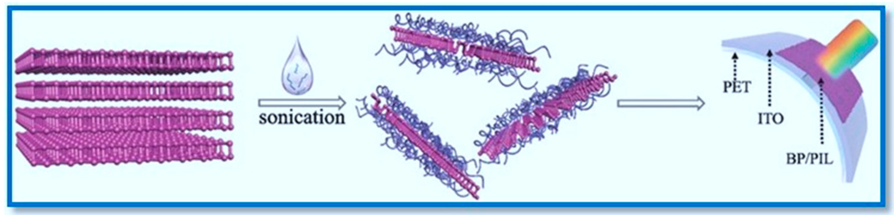
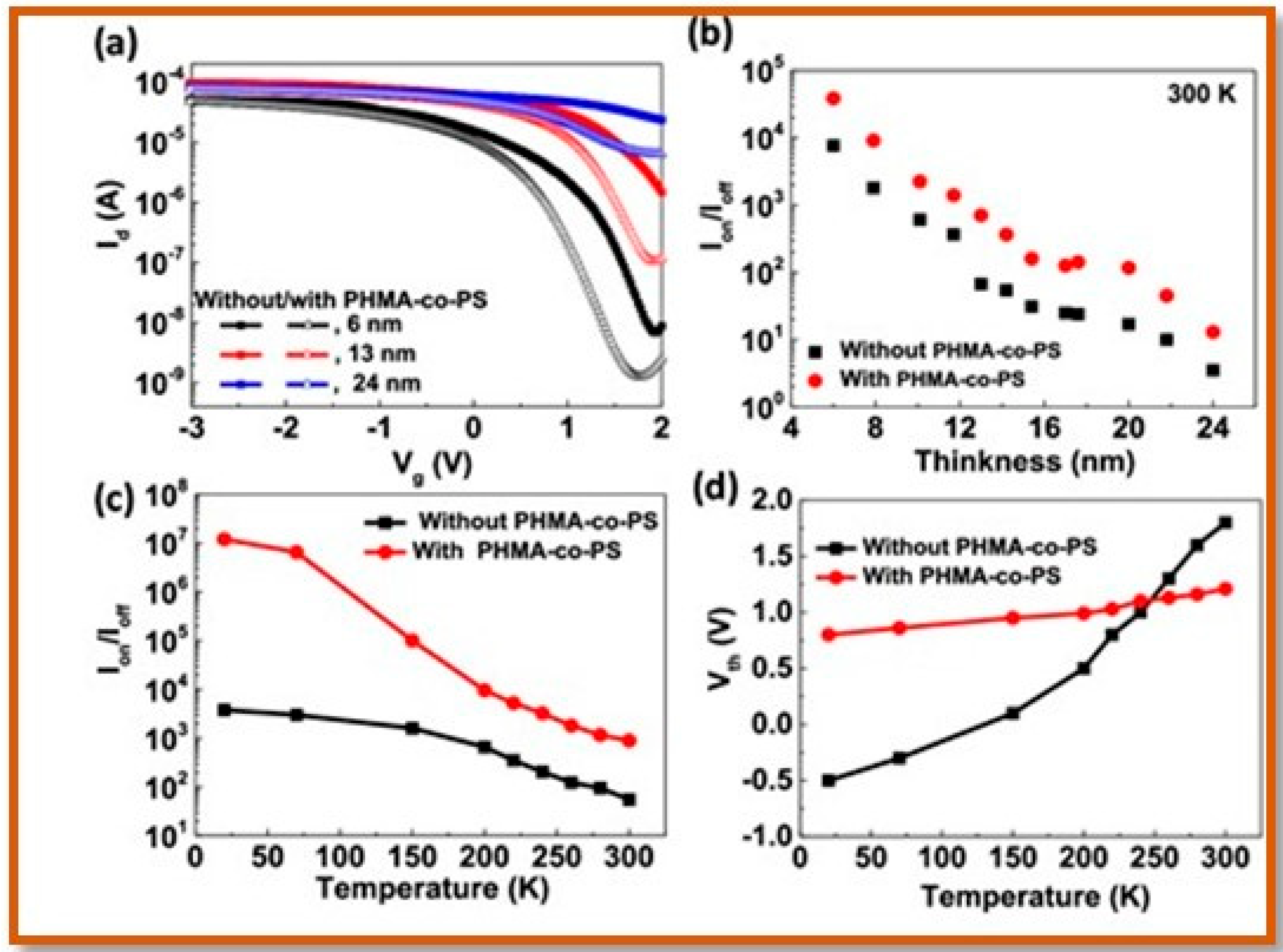
3.4. Hybrids of Polymers and BP Nanosheets in Optoelectronics

The semiconducting properties of BP nanosheets in the combination of suitable (non)conducting polymers have been also exploited for applications in optoelectronics, such as random-access memories, light-emitting diodes (LEDs), and laser technology [127,128,129,130,131,132]. Obviously, for further improving the combined performance of such systems, the BP nanosheets must be decorated or linked with the well-designed conducting polymer chains that can not only protect the BP nanosheets from the oxidative degradation, but also do not allow any compromise with the properties of either the BP nanosheets and their own. The few reports describing such optoelectronic active nanocomposites of BP/conducting polymers, along with their performance, are highlighted here. A study focusing on flexible photodetectors has proposed a novel method for modifying the BP nanosheets with diluted polymer ionic liquids.
Figure 13. Preparation of the nanocomposite polymer electrolyte with passivated BP nanosheets additive. (a) TEM micrograph of the BP nanosheets, (b) survey XPS spectra, and (c) high-resolution XPS spectra of the P 2p and O 1 s signals of the pristine BP crystal and passivated BP nanosheets. (d) Overall synthesis procedure. Inset: photograph of the developed electrolytes. From left to right: CPE-0P (no additive) and nanocomposite polymer electrolytes with 0.05, 0.1, 0.5, 1, and 2 wt% of passivated BP nanosheets. (e) SEM image of the 0.5 wt% with the corresponding EDS mapping. Copyright 2020 WILEY-VCH Verlag GmbH & Co. KGaA, Weinheim, Reproduced with permission from Adv. Funct. Mater. [128], Copyright 2020 WILEY-VCH Verlag GmbH & Co. KGaA, Weinheim, Germany.
Polymers 15 00947 g013
Figure 14. Electrochemical evaluation of the developed electrolytes. (a) Ionic conductivity as a function of temperature for CPE–0P, CPE–0.1P, and CPE–0.5P. The inset graph shows the Nyquist plots corresponding samples at 25 °C. (b) Linear sweep voltammetry showing electrochemical stability window. (c) Direct current polarization tests to measure the electronic conductivity of the developed electrolytes. Copyright 2020 WILEY-VCH Verlag GmbH & Co. KGaA, Weinheim, Reproduced with permission from Adv. Funct. Mater. [128], Copyright 2020 WILEY-VCH Verlag GmbH & Co. KGaA, Weinheim, Germany.
Polymers 15 00947 g014
In this noncovalent functionalization method, highly charged polymer ionic liquids (PIL), such as P ([VPIm]Br), P ([VPIm]PF6), and P ([VPIm]TFSI), were used to passivate the BP nanosheets in the liquid phase (Figure 15) [128]. The unique feature of this approach included the exfoliation of BP nanosheets in the presence of diluted polymer ionic liquids instead of ionic liquid alone. Here, using the polymer ionic liquids for exfoliation has shown a clear advantage over the stability and aggregation of BP nanosheets.
A comparative study on the effect of the exfoliation and absorption intensity of the suspension of BP nanosheets in diluted polymer ionic liquids and two imidazole small molecule ILs, [EmIm]BF4 and [BmIm]BF4, has suggested that the exfoliation of BP was improved significantly (Figure 16). Depending on the high intensity and the darkest brown color of the suspension, it was concluded that the BP/P ([VPIm]TFSI) and DMF produced a higher yield of BP nanosheets wrapped with the relevant polymer ionic liquid (Figure 16b). From the Raman spectra and XPS binding energies data, it was clear that the BP nanosheets were protected very well with the polymer ionic liquid chains, thereby excluding any oxidative degradation. For fabricating the photodetector devices, a concentrated solution (≈0.64 mg mL−1) of BP/P ([VPIm]TFSI) nanosheets was deposited on the surface of indium tin oxides/polyethylene-terephthalate (ITO/PET) substrate by the drop casting method in order to obtain a thin film with the thickness of ≈5 µm, a width of ≈0.9 cm, and a length of ≈0.6 cm. The photocurrent response of the device under the bias voltages and with the light source power intensity of around 20 mW cm−2 was recorded [127]. An increment in the photocurrent density from ≈5 nA cm−2 under 0 V bias to 51 nA cm−2 under 3 V was observed, highlighting the presence of electron–hole in the BP/P ([VPIm]TFSI) composites (Figure 17a). The consistency in the responsibility of the BP/P ([VPIm]TFSI)-based flexible photodetector device was found to be 4.6 µA W−1, indicating no severe effect on the electrical performance of BP nanoflakes, and offering these materials as potential candidates for flexible optoelectronics [131].
Furthermore, when an external bending stress was applied to the device, its photocurrents with the corresponding bending curvatures did not change under dark and light condition, proving its outstanding electrical stability (Figure 17b). The further photostability of the device was supported from the investigation of the time-dependent photocurrent density, which was improved up to ≈50 nA cm−2 when exposed to an ambient environment for 120 h (Figure 17d) [131].
In another work, the authors have reported using poly(2-hydroxyethyl methacrylate)-co-poly(styrene) (PHMA-co-PS) for passivating and encapsulating BP nanosheets and achieving greatly improved electrical transport properties at an electric high field for the BP-based transistor (Figure 18) [132].
Here, a block copolymer based on the poly(2-hydroxyethyl methacrylate) (PHMA) and styrene monomer was chosen for a passivation purpose, owing to its good thermal stability and hydrophobicity (Figure 18a) [132]. Well-defined copolymer PHMA-co-PS was synthesized by following reversible addition fragmentation chain transfer polymerization. The resulting polymer with a low polydispersity (PDI = 1.26) possessed a high contact angle (θ = 75°) and thermal stability up to 347 °C (Figure 18c). As shown in the Scheme above (Figure 18), the polymer was used to encapsulate the layers of BP nanosheets and Al2O3 that were previously implemented on the surface of the atomic layer deposited (ALD), with HfSiO acting as the gate dielectric (Figure 18). The channel length of the gate dielectric was 0.16 μm, as estimated from the scanning electron microscopy image of a BP device. The electrical properties of the BP nanosheets in the FETs device were also evaluated before and after their encapsulation with the PHMA-co-PS layer. The IdVg curves of BP FETs with three different thickness at Vd = −0.05 V for the 0.16 μm channel length have shown an improvement in on/off current ratios for the corresponding thickness (Figure 19a). Further experiments have suggested that the current on/off ratio increased by a factor of 4–10 after the BP nanosheets were coated with the polymer (Figure 19b). The temperature-dependent on/off current ratio of the device with PHMA-co-PS encapsulation was also found to be larger compared to the bare BP nanosheets [132].
The threshold voltage (Vth) of BP FETs as a function of temperature has shown no deviation and remained almost 0.8–1.2 V. after encapsulation, while for bare BP nanosheets, the Vth decreased, providing insights regarding the charged impurities such as H2O and O2 on the BP surface that were eliminated by the passivation; hence, the Vth was stabilized (Figure 19c,d). Clearly, BP nanosheets encapsulated with the polymer layer have demonstrated a better stability with almost unchanged electrical properties that can be harnessed in FET device applications in ambient conditions [132].

3.5. Hybrids of Polymers and BP Nanosheets as Drug Delivery Platform

As compared to other 2D nanomaterials such as graphene, transition metal dichalcogenides (molybdenum disulfide), and Mxene, the phosphorene or BP nanosheets can offer a superior biocompatibility and a great biodegradability in the physiological environment owing to its reactivity with the water and oxygen; therefore, its applications can be extended to the biomedical field e.g., phototherapy, drug delivery, biosensing, theranostics, and bone regeneration [133,134,135,136,137,138]. Some of the examples describing the strategies and applications of BP nanosheets with conjugated polymers in biomedical sciences are summarized here. In one such case, the surface of the BP nanosheets were fabricated with the poly-L-lysine (PLL) or poly(ethylene glycol) (PEG) linker together with the two different fragments of peptides, namely RGD and KRK (Figure 20), for using the assembly against breast cancer [139].
In this two-step process, first, the covalent bioconjugation of the peptide with the poly-L-lysine (PLL) or poly(ethylene glycol) (PEG) was carried out, followed by the non-covalent immobilization of PLL-peptide or PEG-peptide to the surface of BP nanosheets (Figure 20). Finally, the effect of peptides RGD and KRK in combination with functionalized BP nanosheets as potential drugs in breast cancer therapy was investigated. The analysis of functionalized BP nanosheets with both the polymers was done by means of Raman and SEM to examine the morphology [139].
As shown in the Figure 21A, the several vibrational bands in the range of 3600–3300 cm−1, 3100–2200 cm−1, 1750–1550 cm−1, 1550–1300 cm−1, and 1300–900 cm−1 appeared for the FLBP-PEG-peptide assembly. A characteristic narrow and intense band at 1112 cm−1 was associated with the C–O–C group in FLBP-PEG, which was not observed for FLBP-PLL-peptide conjugates (Figure 21A). The presence of amino groups was confirmed with the presence of a corresponding band at 1300–900 cm−1. The further morphological analysis before/after functionalization under Scanning electron microscopy has revealed that the non-ionic linker PEG assisted in the interconnection of adjacent layers, whereas the positively charged PLL induced the formation of dispersion throughout the sample (Figure 21(Bb–e)). Nevertheless, the above tests proved the formation of bioconjugates successfully. To target the breast, e.g., breast cancer cell lines, MCF-7, MDA-MB-231, and HB2 cells, these conjugates of BP nanosheets and PEG/PLL were scrutinized for their cytotoxicity (Figure 22).
The treatment of breast cancerous cells with the different concentrations of these bioconjugates has indicated a loss in viability among the MCF-7 and MDA-MB-231 infected cells at 4 μg ml−1 [135]. Before functionalization, BP nanosheets exhibited toxicity for HB2 (normal mammary cells) at a concentration of (20 μg mL−1) (Figure 22a). However, after modification, BP nanosheets were found to be less harmful towards the normal mammary cells; HB2 with ~70% cell viability at 20 μg mL−1 (Figure 22b). The modified BP nanosheets are more toxic to the triple-negative breast cancer cell culture MDA-MB-231 (viability < 34%) compared to the luminal breast cancer cell culture MCF7 (viability < 46%). From these results, it can be concluded that the decoration of the BP nanosheets surface definitely extends their biocompatibility with regard to their biomedical applications and the possibilities of using these bioconjugates as drug delivery system against cancer [139].

3.6. Hybrids of Polymers and BP Nanosheets for Tissue Engineering

Following up with the biomedical usage of BP nanosheets, a research group has combined polymers-based hydrogels with BP nanosheets to improve the mineralization of CaP crystal nanoparticles for bone-tissue regeneration. In this combinatorial approach, double-network (DN) hydrogels based on three distinct polymers were generated via photo polymerization (Figure 23) [140].
As shown in the Figure 23(Aa), the biocompatible synthetic polymers such as [poly(2-hydroxyethylacrylate) (PHEA), poly(N,N-dimethyl acrylamide) (PDMA), or polyacrylamide (PAM)], were cross-linked in combination of one of three methacrylate-decorated natural polymers to form the nanoengineered network with a high mechanical strength and tunable toughness. In these networks, BP nanosheets were introduced, adding multiple functions to the exceptional mineralized matrix’s formation ability and excellent bioactivity to the hydrogels. Covalent bonds alone or together with hydrogen bonds in a network weaken the strength of DN hydrogels, whereas the presence of covalent bonds and the high density of polymer chain entanglements offered a high strength to DN hydrogels (Figure 23(Ab)). The successive addition of BP nanosheets into the DN hydrogels resulted in NE hydrogels that induced the formation of the CaP matrix for bone regenerations (Figure 23(Ab)). The networks with outstanding mechanical properties were able to withstand high levels of compression in longitudinal and transverse directions even at an extremely high strain. However, upon the removal of the compression force, the initial cylindrical shapes were regained by both the DN and NE hydrogels. These tough DN and NE hydrogels were also resistant towards and further deformations induced by bending (Figure 23(Bb), upper right), knotting (Figure 23(Bc), upper right), and crossover/knotting stretching (Figure 23(Bd)) without any rupture, indicating a high degree of strength, stiffness, and toughness.
The ability of these DN gels to regenerate the bone was evaluated in vivo in the rat calvarial defect model. Under micro-computed tomography (micro-CT) imaging and from the quantification of the bone volume/tissue volume ratio (BV/TV) (Figure 23C(a,b)), including the studies of bone mineral density (Figure 23(Cc)), it was suggested that these BP nanosheet-encapsulated NE hydrogels (PAM/ChiMA/BP and PAM/AlgMA/BP) significantly improved bone regeneration. As the mineralization occurred within NE hydrogels, the amount of newly formed bone also increased simultaneously [140,141].

3.7. Hybrids of Polymers and BP Nanosheets for Bioimaging and Photothermal Cancer Therapy

It is very well known that cancer is responsible for millions of deaths per year across the globe. Until today, considerable efforts have been ongoing to develop an effective therapy against this terminal disease. Currently, two unique therapies, namely photothermal therapy (PTT) and photodynamic therapy (PDT), are gaining a tremendous amount of attention as they can offer a good efficiency without any major injury. In PTT, an active material system absorbs near-infrared (NIR) light and turns it into heat under light irradiation, leading to the elimination of cancer cells, whereas in PDT, the reactive oxygen species (1O2) are generated during the photosensitizing process in order to kill the infected cells. In PDT, the efficiency of the system depends upon the concentration of 1O2 species which are reduced under a hypoxia environment in tumors. Therefore, by combining the PTT with PDT, this issue can be resolved as the heat in the PTT process can enhance the blood flow and thereby increase the oxygen supply for an increased 1O2 production. One such successful attempt to combine PTT and PDT is summarized here, in which BPQDs (black phosphorus quantum dots) smaller than 10 nm were functionalized with water-soluble PEG for enhancing their biocompatibility, excellent NIR photothermal performance, and 1O2 generation capability (Figure 24) [141].
First, the BPQDs with a smaller size were obtained by liquid exfoliation methods (Figure 24a). These as-obtained BPQDs were decorated with the water-soluble PEG-NH2 via electrostatic bonding. To this, a photosensitizer known as rhodamine B (RdB) was conjugated to obtain the RdB/PEG-BPQDs that were analyzed by UV–Vis and Raman spectrometry to confirm the functionalization (Figure 24b,c). These RdB/PEG-BPQDs were evaluated for their anti-cancerous capabilities in 4T1 tumor-bearing Balb/c mice under in vivo conditions [141].
The real-time temperature changes in tumors were monitored under the infrared thermal camera (Figure 25a). The tumors injected with the PEGylated BPQDs (groups V and VI) have shown a considerable increment in temperature within 2 min (ΔT ≈ 30 °C) under NIR irradiation, as compared to the group III that was treated only with a laser. These observations indicated towards the excellent photothermal efficiency of PEGylated BPQDs [141].
Other studies involving the biodegradable BPQDs functionalized with poly (lactic-co-glycolic acid) reported an enhanced photothermal stability for cancer therapy (Figure 26) [142].
In this study, BPQDs as small as 3 nm were loaded into poly (lactic-co-glycolic acid) (PLGA) and processed by an oil-in-water emulsion solvent evaporation method to produce ∼100 nm BPQDs/PLGA nanospheres (NSs). Owing to the biodegradability and biocompatibility of PLGA, encapsulated BPQDs increased the blood circulation and effective accumulation in tumors for an effective cancer therapy. The biodegradability tests of control BPQDs and BPQDs/PLGA NSs in the phosphate-buffered saline (PBS; pH 7.4) have revealed that after functionalization, BPQDs retained their stability and performance during the initial 24 h (Figure 26a,b) in comparison to bare BPQDs. After 8 weeks, the absorption and photothermal behavior of the BPQDs/PLGA NSs started to diminish because of the degradation of PLGA under similar conditions. According to the measurements done for residual weight over the time period, the BPQDs/PLGA NSs displayed a negative trend during first week; however, further rapid weight loss occurred after 8 weeks (Figure 26c). The degradation behavior was also observed visually under SEM and TEM. During the first week, the NSs retained their shape and morphology, however, after 8 weeks, they appeared to degrade, resulting in a total disruption in their morphology (Figure 26d, SEM and TEM images). An illustration of the degradation is shown in Figure 26, in which the PLGA shell degrades under acidic conditions via desertification, leading to the evolution of water and carbon dioxide. Such degradation induces the disintegration of NSs, exposing the BPQDs to air and water directly, which further results in the formation of byproducts such as nontoxic phosphate and phosphonate [142].
The in vivo cytotoxicity investigations of these BPQDs/PLGA NSs were also conducted first on the healthy female Balb/c mice. Compared to the control group, the standard hematology markers, e.g., white blood cells, red blood cells, hemoglobin, mean corpuscular volume, mean corpuscular hemoglobin, mean corpuscular hemoglobin concentration, platelets, and hematocrit, were found to be normal, with no major difference pointing towards the absence of an obvious infection and inflammation in the treated mice (Figure 27a). For the blood analyses, the parameters, including alanine transaminase, aspartate transaminase, total protein, globulin, total bilirubin, blood urea nitrogen, creatinine, and albumin, appeared to be normal as well (Figure 27b). These observations have indicated that NSs treatment does not affect the chemical nature of blood in mice [136]. The histological changes in the tissue obtained from the liver, spleen, kidney, heart, and lung were also investigated. The corresponding tissue stained with eosin have shown no signal for any organ damage during the period of treatment (Figure 27c). These results have suggested that BPQDs/PLGA NSs are biocompatible and have no toxicity towards healthy cells [142].
In Table 5, the therapeutic properties of the nanocomposites of BP polymers and graphene polymers obtained for a different test or model with their relevant mode are summarized. The nanocomposites of PEGylated BPQDs and PLGA–BPQDs were evaluated against the breast cancerous cells and were found to be effective and highly efficient during photothermal and photodynamic therapy.

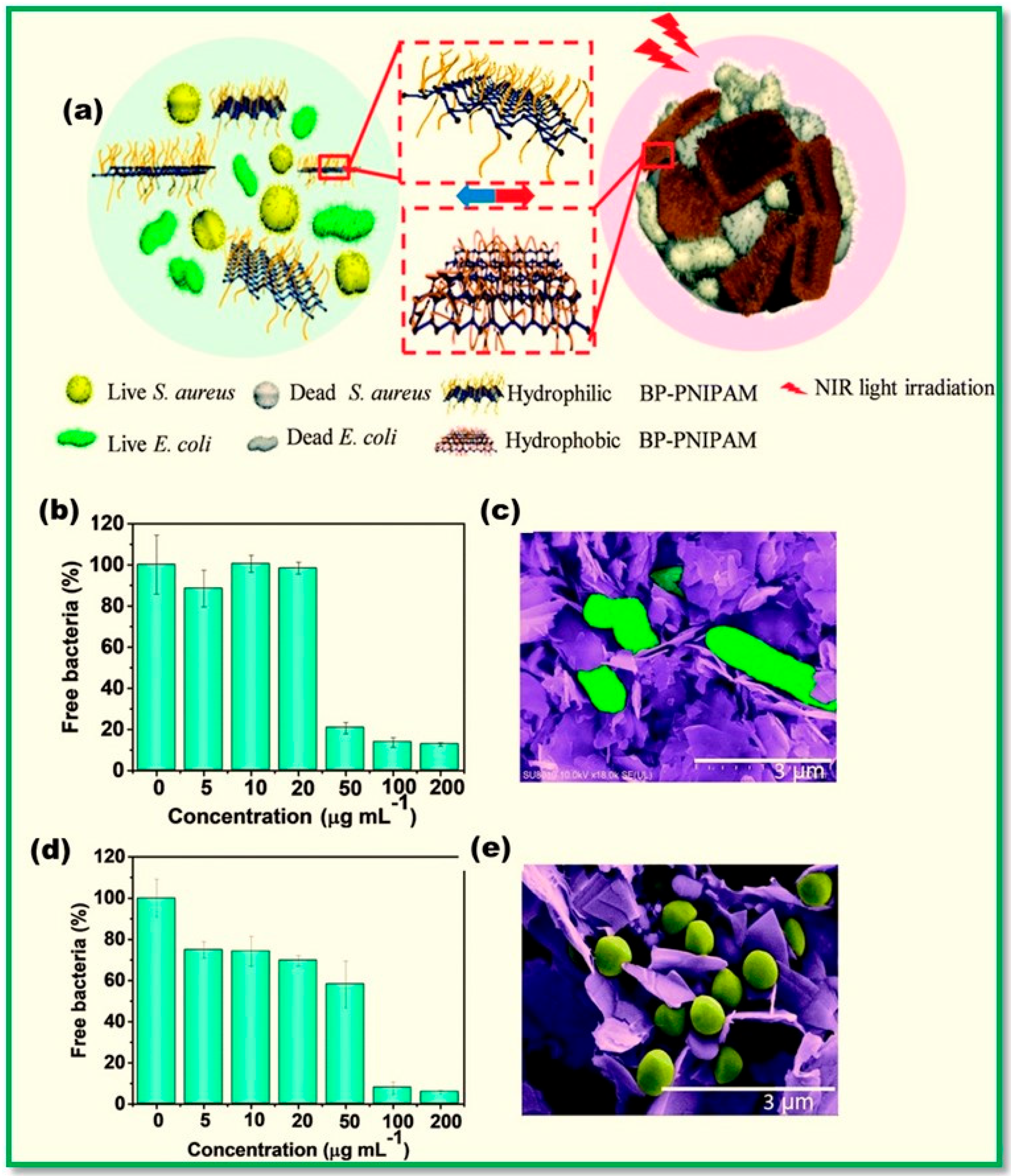
3.8. Hybrids of Polymers and BP Nanosheets for Bacteria Capture and Elimination

Pollutants or contaminants pollute drinking water; due to which, some parts of the world are facing drinking water scarcity. The byproducts produced during the disinfection process via the currently used purification methods can be environmental hazards. In a very recent report, a group has made efforts to design a rather environmentally friendly disinfection method by using a thermo-responsive poly(N–isopropyl acrylamide)–functionalized black phosphorus (named BP–PNIPAM) to capture and eliminate bacterial cells under the stimuli of near-infrared irradiation. An overview of this thermoresponsive composite, followed by its capacity for capturing E. coli (A and B) and S. aureus, is shown in Figure 28 [145]. Here, the thermo-responsive PNIPAM brushes were linked on to the surface of BP nanosheets by following in situ atom transfer radical polymerization (ATRP). Upon irradiation, these brushes can undergo a hydrophilic-to-hydrophobic transition in order to capture the bacterial cells in water (Figure 28a).
The ability of these nanocomposites to capture the bacterial strains such as E. coli and S. aureus was examined via the colony counting method. A concentration of BP–PNIPAM, for instance higher than 50 μg mL−1, was found to be sufficient for capturing the 80% colony of E. coli. However, for S. aureus, the capturing efficiency was only 25.16%. The bacterial capture efficiency of BP–PNIPAM composites further increased to 94.25% at 200 μg mL−1 (Figure 28b,d).
As per the proposed mechanism for the bacterial capture, in first step, the hydrophobic PNIPAM binds to the surface of bacterial cells via non-specific interactions, and in turn, the bacterial cells become wrapped into the BP–PNIPAM aggregation as triggered by the irradiation. Scanning electron microscope images have demonstrated that the BP–PNIPAM aggregations were able to capture both Gram-negative E. coli and Gram-positive S. aureus successfully (Figure 28c,e). Further tests under near-infrared (NIR) irradiation have indicated that these BP–PNIPAM aggregations could eradicate both E. coli and S. aureus for the BP–PNIPAM concentrations of 100 and 200 μg mL−1 and thus could be a promising candidate for a safe application in water disinfection [145].

4. Conclusions

Since the elusive properties of black phosphorus or phosphorene came to light, the researchers working on 2D materials have shifted their focus to exploring this fascinating material, its modulation, and optimizing its properties. There are few methods in which the different shape and size of phosphorene can be obtained with the precise control over its electronic characteristic, as explained in the article. However, for the processing and fabrication of different nanostructures of phosphorene, such as nanosheets or nanoribbons, it has to be coupled with a polymer in order to prevent its degradation without compromising its features for the applications. Various attempts to combine the phosphorene nanosheets or quantum dots with a suitable and active polymer chain have been carried out successfully. Active polymers (ionic) conducting and non-conducting, such as poly(vinyl alcohol), poly(ethylene glycol), poly(imidazole), poly(styrene sulfonic acid), poly(pyrrole), poly(dopamine), and polypeptides, have been linked to the surface of phosphorene nanosheets via a covalent linkage or adsorption process. In these composites, the oxidation or degradation of nanosheets was prevented and the solubility of phosphorene in common organic solvents was increased for a better processability. Some of these hybrid materials or composites appeared as hydrogels and have provided a superior biocompatibility to the phosphorene nanosheets. After their thorough chemical analysis, these composites have been fabricated and employed in a wide range of applications that include fire retardants, capacitors, ionic batteries, field effect transistors, drug delivery, and cancer therapy. The efficiency of capacitors and batteries was found to be higher. For cancer therapy, the cell toxicity experiments have proven that some of the biocompatible composites of phosphorene nanosheets and a relevant polymer can effectively be used for treating the tumor in mice. Some of the BP polymers nanocomposites summarized here can also be applied for the water disinfection process, in which the bacterial cells are captured and eradicated under irradiation. Obviously, the progress made so far with regard to the BP polymers nanocomposites has caused a greater impact, thereby reducing the burden on other similar 2D nanomaterials, such as graphene, for multiple applications. The future of BP polymer nanocomposites appears to be bright; we will witness their usage in space technology as well.
However, despite significant progress with the composites of phosphorene and polymers, there is still an improvement with regard to their robustness and performance. The high-scale production of phosphorene nanoobjects with a controlled shape and structure is highly demanded. Second, multiple polymers, with a superior performance and the ability to synergize with BP nanoobjects, are to be developed. Last but not the least, the fabrication of these hybrid composites for a commercialization purpose or for real-life applications would need technological advancements which, as of yet, have not been developed.

Author Contributions

Conceptualization, A.K.; methodology, A.K.; software, A.K.; validation, A.K. and D.W.C.; formal analysis, A.K.; investigation, A.K.; resources, D.W.C.; data curation, A.K.; writing—original draft preparation, A.K.; writing—review and editing, D.W.C.; visualization, A.K.; supervision, A.K.; project administration, A.K. All authors have read and agreed to the published version of the manuscript.

Funding

This work was supported by the National Research Foundation of Korea (NRF-2021R1A2C1003755 and 2022R1A6A1A03051158) and the BB21 plus funded by Busan Metropolitan City and Institute for Talent and Lifelong Education (BIT).

Institutional Review Board Statement

Not applicable.

Data Availability Statement

Not applicable.

Acknowledgments

The authors thank National Research Foundation of Republic of Korea and Pukyong National University, Busan, South Korea for generous support.

Conflicts of Interest

The authors declare no conflict of interest.

References

  1. Novoselov, K.S.; Geim, A.K.; Morozov, S.V.; Jiang, D.; Zhang, Y.; Dubonos, S.V.; Grigorieva, I.V.; Firsov, A.A. Electric field effect in atomically thin carbon films. Science 2004, 306, 666. [Google Scholar] [CrossRef] [PubMed]
  2. Chen, Y.; Fan, Z.; Zhang, Z.; Niu, W.; Li, C.; Yang, N.; Chen, B.; Zhang, H. Two–dimensional metal nanomaterials: Synthesis, properties, and applications. Chem. Rev. 2018, 118, 6409–6455. [Google Scholar] [CrossRef]
  3. Li, X.; Cai, W.; An, J.; Kim, S.; Nah, J.; Yang, D.; Piner, R.; Velamakanni, A.; Jung, I.; Tutuc, E.; et al. Large area synthesis of high quality and uniform graphene films on copper foils. Science 2009, 324, 1312–1314. [Google Scholar] [CrossRef] [PubMed]
  4. Duong, D.L.; Han, G.H.; Lee, S.M.; Gunes, F.; Kim, E.S.; Kim, S.T.; Kim, H.; Ta, Q.H.; So, K.P.; Yoon, S.J.; et al. Probing graphene boundaries with optical microscopy. Nature 2012, 490, 235. [Google Scholar] [CrossRef] [PubMed]
  5. Geim, A.K.; Novoselov, K.S. The rise of graphene. In Nanoscience and Technology: A Collection of Reviews from Nature Journals; World Scientific: Singapore, 2010; pp. 11–19. [Google Scholar]
  6. Lee, C.; Wei, X.; Kysar, J.W.; Hone, J. Measurement of the elastic properties and intrinsic strength of monolayer graphene. Science 2008, 321, 385–388. [Google Scholar] [CrossRef] [PubMed]
  7. Lee, G.-H.; Cooper, R.C.; An, S.J.; Lee, S.; Van Der Zande, A.; Petrone, N.; Hammerberg, A.G.; Lee, C.; Crawford, B.; Oliver, W.; et al. High strength chemical vapor deposited graphene and grain boundaries. Science 2013, 340, 1073–1076. [Google Scholar] [CrossRef]
  8. Wang, G.; Dai, Z.; Wang, Y.; Tan, P.; Liu, L.; Xu, Z.; Wei, Y.; Huang, R.; Zhang, Z. Measuring interlayer shear stress in bilayer graphene. Phys. Rev. Lett. 2017, 119, 036101. [Google Scholar] [CrossRef]
  9. Lloyd, D.; Liu, X.; Christopher, J.W.; Cantley, L.; Wadehra, A.; Kim, B.L.; Goldberg, B.B.; Swan, A.K.; Bunch, J.S. Band gap engineering with with ultra large biaxial strains in suspended monolayer MoS2. Nano Lett. 2016, 16, 5836–5841. [Google Scholar] [CrossRef]
  10. Zhang, P.; Ma, L.; Fan, F.; Zeng, Z.; Peng, C.; Loya, P.E.; Liu, Z.; Gong, Y.; Zhang, J.; Zhang, X.; et al. Fracture toughness of graphene. Nat. Commun. 2014, 5, 3782. [Google Scholar] [CrossRef]
  11. Li, P.; Jiang, C.; Xu, S.; Zhuang, Y.; Gao, L.; Hu, A.; Wang, H.; Lu, Y. In situ nanomechanical characterization of multilayer MoS2 membranes: From intraplanar to interplanar fracture. Nanoscale 2017, 9, 9119–9128. [Google Scholar] [CrossRef]
  12. Cao, Y.; Fatemi, V.; Fang, S.; Watanabe, K.; Taniguchi, T.; Kaxiras, E.; Jarillo-Herrero, P. Unconventional superconductivity in magic angle graphene superlattices. Nature 2018, 556, 43. [Google Scholar] [CrossRef]
  13. Watts, M.C.; Picco, L.; Russell-Pavier, F.S.; Cullen, P.L.; Miller, T.S.; Bartuś, S.P.; Payton, O.D.; Skipper, N.T.; Tileli, V.; Howard, C.A. Production of phosphorene nanoribbons. Nature 2019, 568, 216. [Google Scholar] [CrossRef]
  14. Liu, Z.; Sun, Y.; Cao, H.; Xie, D.; Li, W.; Wang, J.; Cheetham, A.K. Unzipping of black phosphorus to form zigzag phosphorene nanobelts. Nat. Commun. 2020, 11, 3917. [Google Scholar] [CrossRef] [PubMed]
  15. Saito, Y.; Iizuka, T.; Koretsune, T.; Arita, R.; Shimizu, S.; Iwasa, Y. Gate tuned thermoelectric power in black phosphorus. Nano Lett. 2016, 16, 4819. [Google Scholar] [CrossRef]
  16. Smith, B.; Vermeersch, B.; Carrete, J.; Ou, E.; Kim, J.; Mingo, N.; Akinwande, D.; Shi, L. Temperature and thickness dependences of the anisotropic in plane thermal conductivity of black phosphorus. Adv. Mater. 2017, 29, 1603756. [Google Scholar] [CrossRef]
  17. Liu, H.; Liu, J.; Jing, R.; You, C. Anisotropic thermal conductivity in direction specific black phosphorus nanoflakes. MRS Commun. 2019, 9, 1311. [Google Scholar] [CrossRef]
  18. Qin, G.; Hu, M. Thermal transport in phosphorene. Small 2018, 14, 1702465. [Google Scholar] [CrossRef]
  19. Selamneni, V.; Sahatiya, A.B.S.P. Highly air stabilized phosphorus on disposable paper substrate as a tunneling effect based highly sensitive piezoresistive strain sensor. Med. Devices Sens. 2020, 3, e10099. [Google Scholar] [CrossRef]
  20. Nourbakhsh, Z.; Asgari, R. Phosphorene as nanoelectromechanical material. Phys. Rev. B 2018, 98, 125427. [Google Scholar] [CrossRef]
  21. Bridgman, P.W. Two new modifications of phosphorus. J. Am. Chem. Soc. 1914, 36, 1344–1363. [Google Scholar] [CrossRef] [Green Version]
  22. Shirotani, I. Growth of large single crystals of black phosphorus at high pressures and temperatures, and its electric properties. Mol. Cryst. Liq. Cryst. 1982, 86, 203–211. [Google Scholar] [CrossRef]
  23. Maruyama, Y.; Suzuki, S.; Kobayashi, K.; Tanuma, S. Synthesis and some properties of black phosphorus single crystals. Physical 1981, 105, 99–102. [Google Scholar] [CrossRef]
  24. Gusmão, R.; Sofer, Z.; Pumera, M. Black phosphorus rediscovered: From bulk material to monolayers. Angew. Chem. Int. Ed. 2017, 56, 8052–8072. [Google Scholar] [CrossRef]
  25. Dhanabalan, S.C.; Ponraj, J.S.; Guo, Z.; Li, S.; Bao, Q.; Zhang, H. Emerging trends in phosphorene fabrication towards next generation devices. Adv. Sci. 2017, 4, 1600305. [Google Scholar] [CrossRef]
  26. Lin, S.; Liu, S.; Yang, Z.; Li, Y.; Ng, T.W.; Xu, Z.; Bao, Q.; Hao, J.; Lee, C.-S.; Surya, C.; et al. Solution processable ultrathin black phosphorus and effective electron layer on organic photovoltaics. Adv. Funct. Mater. 2016, 26, 864–871. [Google Scholar] [CrossRef]
  27. Batmunkh, M.; Shrestha, A.; Bat-Erdene, M.; Nine, M.J.; Shearer, C.J.; Gibson, C.T.; Slattery, A.D.; Tawfik, S.A.; Ford, M.J.; Dai, S.; et al. Electrocatalytic activity of a 2D phosphorene based heteroelectrocatalyst for photoelectrochemical cells. Angew. Chem. Int. Ed. 2018, 57, 2644–2647. [Google Scholar] [CrossRef]
  28. Fu, N.; Huang, C.; Lin, P.; Zhu, M.; Li, T.; Ye, M.; Lin, S.; Zhang, G.; Du, J.; Liu, C.; et al. Black phosphorus quantum dots as dual functional electron selective materials for efficient plastic perovskite solar cells. J. Mater. Chem. A 2018, 6, 8886–8894. [Google Scholar] [CrossRef]
  29. Yang, Y.; Gao, J.; Zhang, Z.; Xiao, S.; Xie, H.-H.; Sun, Z.-B.; Wang, J.-H.; Zhou, C.-H.; Wang, Y.-W.; Guo, X.-Y.; et al. Black phosphorus based photocathodes in wideband bifacial dye sensitized solar cells. Adv. Mater. 2016, 28, 8937–8944. [Google Scholar] [CrossRef]
  30. Batmunkh, M.; Bat-Erdene, M.; Shapter, J.G. Black phosphorus: Synthesis and application for solar cells. Adv. Energy Mater. 2018, 8, 1701832. [Google Scholar] [CrossRef]
  31. Castellanos-Gomez, A.; Vicarelli, L.; Prada, E.; Island, J.O.; Narasimha-Acharya, K.L.; Blanter, S.I.; Groenendijk, D.J.; Buscema, M.; Steele, G.A.; Alvarez, J.V. Isolation and characterization of few layer black phosphorus. 2D Mater. 2014, 1, 025001. [Google Scholar] [CrossRef]
  32. Island, J.O.; Steele, G.A.; van der Zant, H.S.; Castellanos-Gomez, A. Environmental instability of few layer black phosphorus. 2D Mater. 2015, 2, 011002. [Google Scholar] [CrossRef]
  33. Favron, A.; Gaufrès, E.; Fossard, F.; Phaneuf-L’Heureux, A.-L.; Tang, N.Y.-W.; Lévesque, P.L.; Loiseau, A.; Leonelli, R.; Francoeur, S.; Martel, R. Photooxidation and quantum confinement effects in exfoliated black phosphorus. Nat. Mater. 2015, 14, 826. [Google Scholar] [CrossRef]
  34. Ahmed, T.; Balendhran, S.; Karim, M.N.; Mayes, E.L.; Field, M.R.; Ramanathan, R.; Singh, M.; Bansal, V.; Sriram, S.; Bhaskaran, M.; et al. Degradation of black phosphorus is contingent on UV-blue light exposure. NPJ 2D Mater. Appl. 2017, 1, 18. [Google Scholar] [CrossRef]
  35. Gamage, S.; Li, Z.; Yakovlev, V.S.; Lewis, C.; Wang, H.; Cronin, S.B.; Abate, Y. Nanoscopy of black phosphorus degradation. Adv. Mater. Interfaces 2016, 3, 1600121. [Google Scholar] [CrossRef]
  36. Wood, J.D.; Wells, S.A.; Jariwala, D.; Chen, K.-S.; Cho, E.; Sangwan, V.K.; Liu, X.; Lauhon, L.J.; Marks, T.J.; Hersam, M.C. Effective passivation of exfoliated black phosphorus transistors against ambient degradation. Nano Lett. 2014, 14, 6964–6970. [Google Scholar] [CrossRef]
  37. Huang, Y.; Qiao, J.; He, K.; Bliznakov, S.; Sutter, E.; Chen, X.; Luo, D.; Meng, F.; Su, D.; Decker, J.; et al. Interaction of black phosphorus with oxygen and water. Chem. Mater. 2016, 28, 8330–8339. [Google Scholar] [CrossRef]
  38. Edmonds, M.T.; Tadich, A.; Carvalho, A.; Ziletti, A.; O’Donnell, K.M.; Koenig, S.P.; Coker, D.F.; Özyilmaz, B.; Neto, A.H.C.; Fuhrer, M.S. Creating a stable oxide at the surface of black phosphorus. ACS Appl. Mater. Interfaces 2015, 7, 14557–14562. [Google Scholar] [CrossRef]
  39. Passaglia, E.; Cicogna, F.; Lorenzetti, G.; Legnaioli, S.; Caporali, M.; Serrano-Ruiz, M.; Ienco, A.; Peruzzini, M. Novel polystyrene based nanocomposites by phosphorene dispersion. RSC Adv. 2016, 6, 53777. [Google Scholar] [CrossRef]
  40. Fonsaca, J.E.S.; Domingues, S.H.; Orth, E.S.; Zarbin, A.J.G. Air stable black phosphorus in polyaniline based nanocomposite. Sci. Rep. 2017, 7, 10165. [Google Scholar] [CrossRef]
  41. Kumar, A. Controlled nanostructures and simultaneous passivation of black phosphorus (phosphorene) with Nafion. J. Mater. Res. 2020, 35, 141–152. [Google Scholar] [CrossRef]
  42. Kumar, A. Simultaneous passivation and encapsulation of black phosphorus nanosheets (phosphorene) by optically active polypeptide micelles for biosensors. ACS Appl. Nano Mater. 2019, 2, 2397–2404. [Google Scholar] [CrossRef]
  43. Häußler, M.; Lam, J.W.Y.; Qin, A.; Tse, K.K.C.; Li, M.K.S.; Liu, J.; Jim, C.K.W.; Gao, P.; Tang, B.Z. Metallized hyperbranched polydiyne: A photonic material with a large refractive index tenability and a spin-coatable catalyst for facile fabrication of carbon nanotubes. Chem. Commun. 2007, 25, 2584–2586. [Google Scholar] [CrossRef]
  44. Liu, J.; Zhong, Y.; Lam, J.W.Y.; Lu, P.; Hong, Y.; Yu, Y.; Yue, Y.; Faisal, M.; Sung, H.H.Y.; Williams, I.D.; et al. Hyperbranched conjugated polysiloles: Synthesis, structure, aggregation-enhanced emission, multicolor fluorescent photo patterning, and superamplified detection of explosives. Macromolecules 2010, 43, 4921–4936. [Google Scholar] [CrossRef]
  45. Jim, C.K.W.; Qin, A.; Lam, J.W.Y.; Häußler, M.; Liu, J.; Yuen, M.M.F.; Kim, J.K.; Ng, K.M.; Tang, B.Z. Facile polycyclotrimerization of “simple” arylene bipropiolates: A metal free, regioselective route to functional hyperbranched polymer with high optical transparency, tunable refractive index, low chromatic aberration, and photoresponsive patternability. Macromolecules 2009, 42, 4099–4109. [Google Scholar] [CrossRef]
  46. Sekitani, T.; Nakajima, H.; Maeda, H.; Fukushima, T.; Aida, T.; Hata, K.; Someya, T. Stretchable active-matrix organic light-emitting diode display using printable elastic conductors. Nat. Mater. 2009, 8, 494. [Google Scholar] [CrossRef] [PubMed]
  47. Yao, Y.; Dong, H.; Hu, W. Charge transport in organic and polymeric semiconductors for flexible and stretchable devices. Adv. Mater. 2015, 28, 4213. [Google Scholar] [CrossRef]
  48. Rogers, J.A.; Someya, T.; Huang, Y. Materials and mechanics for stretchable electronics. Science 2010, 327, 1603. [Google Scholar] [CrossRef] [PubMed]
  49. Gustafsson, G.; Ingan, O.; Stafstrom, S.; Osterholm, H.; Laakso, J. Stretch-oriented poly(3-alkylthiophenes). Synth. Met. 1991, 41, 593. [Google Scholar] [CrossRef]
  50. Forsyth, M.; Sun, J.Z.; MacFarlane, D.R. Novel polymer-in-salt electrolytes based on polyacrylonitrile (PAN) lithium triflate salt mixtures. Solid State Ionics 1998, 112, 161. [Google Scholar]
  51. Allcock, H.R.; Oconnor, S.J.M.; Olmeijer, D.L.; Napierala, M.E.; Cameron, C.G. Polyphosphazenes bearing branched and linear oligoethyleneoxy side groups as solid solvents for ionic conduction. Macromolecules 1996, 29, 7544. [Google Scholar] [CrossRef]
  52. Voit, B.I. Dendritic polymers: From aesthetic macromolecules to commercially interesting materials. Acta Polym. 1995, 46, 87. [Google Scholar] [CrossRef]
  53. Buffeteau, T.; Natansohn, A.; Rochon, P.; Pezolet, M. Study of cooperative side group motions in amorphous polymers by time dependent infrared spectroscopy. Macromolecules 1996, 29, 8783. [Google Scholar] [CrossRef]
  54. Fukuda, T.; Kim, J.Y.; Barada, D.; Yase, K. Photoinduced cooperative molecular reorientation on azobenzene side-chain-type copolymers. J. Photobiol. Chem. 2006, 183, 273. [Google Scholar] [CrossRef]
  55. Hickner, M.A.; Ghassemi, H.; Kim, Y.S.; Einsla, B.R.; McGrath, J.E. Alternative polymer systems for proton exchange membranes (PEMs). Chem. Rev. 2004, 104, 4587. [Google Scholar] [CrossRef]
  56. Peckham, T.J.; Holdcroft, S. Structure–morphology–property relationships of non-perfluorinated proton-conducting membranes. Adv. Mater. 2010, 22, 4667. [Google Scholar] [CrossRef]
  57. Miyatake, K.; Chikashige, Y.; Higuchi, E.; Watanabe, M. Tuned polymer electrolyte membranes based on aromatic polyethers for fuel cell applications. J. Am. Chem. Soc. 2007, 129, 3879. [Google Scholar] [CrossRef]
  58. Badami, A.S.; Roy, A.; Lee, H.-S.; McGrath, J.E. Morphological investigations of disulfonated poly(arylene ether sulfone)-b-naphthalene dianhydride-based polyimide multiblock copolymers as potential high temperature proton exchange membranes. J. Membr. Sci. 2009, 328, 156. [Google Scholar] [CrossRef]
  59. Capelot, M.; Montarnal, D.; Tournilhac, F.; Leibler, L. Metal-catalyzed transesterification for healing and assembling of thermosets. J. Am. Chem. Soc. 2012, 134, 7664. [Google Scholar] [CrossRef]
  60. Cordier, P.; Tournilhac, F.; Soulié-Ziakovic, C.; Leibler, L. Self-healing and thermoreversible rubber from supramolecular assembly. Nature 2008, 451, 977–980. [Google Scholar] [CrossRef]
  61. Das, A.; Sallat, A.; Böhme, F.; Suckow, M.; Basu, D.; Wießner, S. Ionic modification turns commercial rubber into a self-healing material. ACS Appl. Mater. Interfaces 2015, 7, 20623. [Google Scholar] [CrossRef]
  62. Markova, D.; Kumar, A.; Klapper, M.; Müllen, K. Phosphonic acid containing homo–, AB and BAB block copolymers via ATRP designed for fuel cell applications. Polymer 2009, 50, 3411. [Google Scholar] [CrossRef]
  63. Kumar, A.; Markova, D.; Klapper, M.; Müllen, K. Proton conducting poly(phenylene oxide)–poly(vinyl benzyl phosphonic acid) block copolymers via atom transfer radical polymerization. Macromol. Chem. Phys. 2012, 213, 489. [Google Scholar] [CrossRef]
  64. Ge, Y.; Lyu, Z.; Marcos-Hernández, M.; Villagran, D. Free base porphyrin polymer for bifunctional electrochemical water splitting. Chem. Sci. 2022, 13, 8597. [Google Scholar] [CrossRef]
  65. Zhu, Y.; Zhou, W.; Zhong, Y.; Bu, Y.; Chen, X.; Zhong, Q.; Liu, M.; Shao, Z. A perovskite nanorod as bifunctional electrocatalyst for overall water splitting. Adv. Energy Mater. 2017, 7, 1602122. [Google Scholar] [CrossRef]
  66. Wurster, B.; Grumelli, D.; Hötger, D.; Gutzler, R.; Kern, K. Driving the oxygen evolution reaction by nonlinear cooperativity in bimetallic coordination catalysts. J. Am. Chem. Soc. 2016, 138, 3623–3626. [Google Scholar] [CrossRef] [PubMed]
  67. Barraza-Lopez, S.; Kaloni, T.P. Water splits to degrade two dimensional group IV monochalcogenides in nanoseconds. ACS Cent. Sci. 2018, 4, 1436–1446. [Google Scholar] [CrossRef] [PubMed]
  68. Liang, W.; Luo, X. Theoretical studies of MoS2 and phosphorene drug delivery for anti-tuberculosis drugs. J. Phys. Chem. C 2020, 24, 8279–8287. [Google Scholar] [CrossRef]
  69. Liu, W.; Dong, A.; Wang, B.; Zhang, H. Current advances in black phosphorus based drug delivery systems for cancer therapy. Adv. Sci. 2021, 8, 2003033. [Google Scholar] [CrossRef]
  70. Kou, L.; Chen, C.; Smith, S. Phosphorene: Fabrication, Properties, and Applications. J. Phys. Chem. Lett. 2015, 6, 2794–2805. [Google Scholar] [CrossRef]
  71. Hu, T.; Dong, J. Geometric and Electronic Structures of Mono- and Di-vacancies in Phosphorene. Nanotechnology 2015, 26, 065705. [Google Scholar] [CrossRef]
  72. Valappil, M.O.; Alwarappan, S.; Pillai, V.K. Phosphorene quantum dots: Synthesis, properties and catalytic applications. Nanoscale 2022, 14, 1037–1053. [Google Scholar] [CrossRef]
  73. De Alwis, W.U.G.; Weerawardene, K.D.M.; Ellington, T.L.; Shuford, K.L. Electronic Structure Modification of Rectangular Phosphorene Quantum Dots Via Edge Passivation. J. Phys. Chem. C 2021, 125, 5029–5036. [Google Scholar] [CrossRef]
  74. Liu, H.; Neal, A.T.; Zhu, Z.; Luo, Z.; Xu, X.; Tomanek, D.; Ye, P.D. Phosphorene: An unexplored 2D semiconductor with a high mobility. ACS Nano 2014, 8, 4033–4041. [Google Scholar] [CrossRef]
  75. Baboukani, A.R.; Khakpour, I.; Drozd, V. Liquid based exfoliation of black phosphorus into phosphorene and its application for energy storage devices. Small Struct. 2021, 2, 2000148. [Google Scholar] [CrossRef]
  76. Ren, X.; Zhou, J.; Qi, X.; Liu, Y.; Huang, Z.; Li, Z.; Ge, Y.; Dhanabalan, S.C.; Ponraj, J.S.; Wang, S. Few layer black phosphorus nanosheets as electrocatalyst for highly efficient oxygen evolution reaction. Adv. Energy Mater. 2017, 7, 1700396. [Google Scholar] [CrossRef]
  77. Lin, S.; Lai, W.K.; Li, Y.; Lu, W.; Bai, G.; Lu, S.P. Liquid-phase exfoliation of violet phosphorus for electronic applications. SmartMat 2021, 2, 226. [Google Scholar] [CrossRef]
  78. Brent, J.R.; Savjani, N.; Lewis, E.A.; Haigh, S.J.; Lewis, D.J.; O’Brien, P. Production of few layer phosphorene by liquid exfoliation of black phosphorus. Chem. Commun. 2014, 50, 13338. [Google Scholar] [CrossRef] [PubMed]
  79. Late, D.J. Liquid exfoliation of black phosphorus nanosheets and its application as humidity sensor. Microporous Mesoporous Mater. 2016, 225, 494. [Google Scholar] [CrossRef]
  80. Batmunkh, M.; Bat Erdene, M.; Shapter, J.G. Phosphorene and phosphorene based materials– prospects and future applications. Adv. Mater. 2016, 28, 8586. [Google Scholar] [CrossRef] [PubMed]
  81. Yasaei, P.; Kumar, B.; Foroozan, T.; Wang, C.; Asadi, M.; Tuschel, D.; Indacochea, J.E.; Klie, R.F.; Salehi-Khojin, A. High quality black phosphorus atomic layers by liquid phase exfoliation. Adv. Mater. 2015, 27, 1887. [Google Scholar] [CrossRef]
  82. Yasaei, P.; Behranginia, A.; Foroozan, T.; Asadi, M.; Kim, K.; Khalili-Araghi, F.; Salehi-Khojin, A. Stable and selective humidity sensing using stacked black phosphorus flakes. ACS Nano 2015, 9, 9898. [Google Scholar] [CrossRef] [PubMed]
  83. Serrano-Ruiz, M.; Caporali, M.; Ienco, A.; Piazza, V.; Heun, S.; Peruzzini, M. The role of water in the preparation and stabilization of high quality phosphorene flakes. Adv. Mater. Interfaces 2016, 3, 1500441. [Google Scholar] [CrossRef] [PubMed]
  84. Woomer, A.H.; Farnsworth, T.W.; Hu, J.; Wells, R.A.; Donley, C.L.; Warren, S.C. Phosphorene: Synthesis, scale up, and quantitative optical spectroscopy. ACS Nano 2015, 9, 8869. [Google Scholar] [CrossRef]
  85. Hao, C.; Wen, F.; Xiang, J.; Yuan, S.; Yang, B.; Li, L.; Wang, W.; Zeng, Z.; Wang, L.; Liu, Z.; et al. Liquid exfoliated black phosphorus nanosheet thin films for flexible resistive random access memory applications. Adv. Funct. Mater. 2016, 26, 2016–2024. [Google Scholar] [CrossRef]
  86. Zhang, B.; Lou, F.; Zhao, R.; He, J.; Li, J.; Su, X.; Ning, J.; Yang, K. Exfoliated layers of black phosphorus as saturable absorber for ultrafast solid state laser. Opt. Lett. 2015, 40, 3691. [Google Scholar] [CrossRef]
  87. Lin, S.; Li, Y.; Lu, W.; Chui, Y.S.; Rogée, L.; Bao, Q.; Lau, S.P. In situ observation of the thermal stability of black phosphorus. 2D Mater. 2017, 4, 025001. [Google Scholar] [CrossRef]
  88. Wang, R.; Yan, X.; Ge, B.; Zhou, J.; Wang, M.; Zhang, L.; Jiao, T. Facile preparation of self-assembled black phosphorus dye composite films for chemical gas sensors and surface enhanced Raman scattering performances. ACS Sustain. Chem. Eng. 2020, 8, 4521. [Google Scholar] [CrossRef]
  89. Liu, W.; Zhang, Y.; Zhang, Y.; Dong, A. Black phosphorus nanosheets counteract bacteria without causing antibiotic resistance. Chemistry 2020, 26, 2478. [Google Scholar] [CrossRef] [PubMed]
  90. Wan, S.; Zhang, B.; Li, S.; He, B.; Pu, Y. Combination of PEG–decorated black phosphorus nanosheets and immunoadjuvant for photoimmunotherapy of melanoma. J. Mater. Chem. B 2020, 8, 2805. [Google Scholar] [CrossRef]
  91. Yi, J.; Chen, X.; Weng, Q.; Zhou, Y.; Han, Z.; Chen, J.; Li, C. A simple electrochemical pH sensor based on black phosphorus nanosheets. Electrochem. Commun. 2020, 118, 106796. [Google Scholar] [CrossRef]
  92. Du, J.; Zhang, M.; Guo, Z.; Chen, J.; Xhu, X.; Hu, G.; Peng, P.; Zheng, Z.; Zhang, H. Phosphorene quantum dot saturable absorbers for ultrafast fiber lasers. Sci. Rep. 2017, 7, 42357. [Google Scholar] [CrossRef] [Green Version]
  93. Thakur, T.; Szafiran, B. Wigner molecules in phosphorene quantum dots. Phys. Rev. B 2022, 106, 205304. [Google Scholar] [CrossRef]
  94. Zeng, Y.; Guo, Z. Synthesis and stabilization of black phosphorus and phosphorene: Recent progress and prospectives. iScience 2021, 24, 103116. [Google Scholar] [CrossRef] [PubMed]
  95. Chaban, V.V.; Fileti, E.E.; Prezhdo, O.V. Imidazolium ionic liquid mediates black phosphorus exfoliation while preventing phosphorene decomposition. ACS Nano 2017, 11, 6459–6466. [Google Scholar] [CrossRef] [PubMed]
  96. Tian, B.; Tian, B.; Smith, B.; Liu, Y. Facile bottom up synthesis of partially oxidized black phosphorus nanosheets as metal free catalyst for hydrogen evolution. Proc. Natl. Acad. Sci. USA 2018, 115, 4345–4350. [Google Scholar] [CrossRef] [PubMed]
  97. Tiouitchi, G.; Raji, M.; Mounkachi, O.; AitAli, M.; Mahmoud, A.; Boschini, F.; Essabir, H.; Bouhfid, R.; Qaiss, A. Black phosphorus based polyvinylidene fluoride nanocomposites: Synthesis, processing and characterization. Compos. Part B Eng. 2019, 175, 107165. [Google Scholar] [CrossRef]
  98. Liu, H.; Liana, H.; Zhanga, Q.; Yanga, Y.; Mei, Y. The preparation of holey phosphorene by electrochemical assistance. Electrochem. Commun. 2019, 98, 124. [Google Scholar] [CrossRef]
  99. Jain, R.; Singh, Y.; Cho, S.Y.; Sasikala, S.P.; Koo, S.H.; Narayan, R.; Jung, H.T.; Jung, Y.; Kim, S.O. Ambient stabilization of few layer phosphorene via noncovalent functionalization with surfactants: Systematic 2D NMR characterization in aqueous dispersion. Chem. Mater. 2019, 31, 2786. [Google Scholar] [CrossRef]
  100. Kang, J.; Wells, S.A.; Wood, J.D.; Hersam, M.C. Stable aqueous dispersions of optically and electronically active phosphorene. Proc. Natl. Acad. Sci. USA 2016, 113, 11688. [Google Scholar] [CrossRef] [PubMed]
  101. Batmunkh, M.; Vimalanathan, K.; Wu, C.; Bati, A.S.R.; Yu, L.; Tawfik, S.A.; Ford, M.J.; Macdonald, T.J.; Raston, C.L.; Priya, S.; et al. Efficient production of phosphorene nanosheets via sheer stress mediated exfoliation for low temperature perovskite solar cells. Small Methods 2019, 3, 1800521. [Google Scholar] [CrossRef]
  102. Zhang, J.; Cao, Y.; Chen, C.; Wang, Q.; Shao, Y. Preparation of black phosphorus nanosheets and research in nonlinear optical response characteristics. Phys. Status Solidi B 2022, 259, 2100543. [Google Scholar] [CrossRef]
  103. Tian, X.; Gu, J.; Liu, B.; Zhang, B.; Song, S.; Fan, F.; Chen, Y. Covalent Functionalization of Black Phosphorus with Conjugated Polymer for Information Storage. Angew. Chem. Int. Ed. 2018, 57, 4543–4548. [Google Scholar]
  104. Bawadkji, O.; Cherri, M.; Schäfer, A.; Herziger, S.; Nickl, P.; Achazi, K.; Donskyi, I.S.; Adeli, M.; Haag, R. One pot covalent functionalization of black phosphorus by anionic ring opening polumerization. Adv. Mat. Interfaces 2022, 9, 2201245. [Google Scholar] [CrossRef]
  105. Thurakkal, S.; Zhang, X. Covalent functionalization of two dimensional black phosphorus nanosheets with porphyrins and their photophysical characterization. Mater. Chem. Front. 2021, 5, 2824–2831. [Google Scholar] [CrossRef]
  106. Hou, Y.; Xu, Z.; An, R.; Zheng, H.; Hu, W.; Zhou, K. Recent progress in black phosphorus nanosheets for improving the fire safety of polymer nanocomposites. Compos. Part B Eng. 2022, 249, 110404. [Google Scholar] [CrossRef]
  107. Cai, W.; Hu, Y.; Pan, Y.; Zhou, X.; Chu, F.; Han, L.; Mu, X.; Zhuang, Z.; Wang, X.; Xing, W. Self-assembly followed by radical polymerization of ionic liquid for interfacial engineering of black phosphorus nanosheets: Enhancing flame retardancy, toxic gas suppression, and mechanical performance of polyurethane. J. Colloid Interface Sci. 2020, 561, 32. [Google Scholar] [CrossRef] [PubMed]
  108. Cai, W.; Mu, X.; Li, Z.; Hu, W.; Hu, Y. Poly(dimethyl siloxane) grafted black phosphorus nanosheets as fillers to enhance moisture–resistance and flame retardancy of thermoplastic polyurethane. Mater. Chem. Phys. 2022, 286, 126189. [Google Scholar] [CrossRef]
  109. Du, X.; Qui, J.; Deng, S.; Du, Z.; Cheng, X.; Wang, H. Flame retardant and form stable phase change composites based on black phosphorus nanosheets/cellulose nanofiber aerogels with extremely high energy storage density and superior solar–thermal conversion efficiency. J. Mater. Chem. A 2020, 8, 14126–14134. [Google Scholar] [CrossRef]
  110. Qu, Z.; Wu, K.; Jiao, E.; Chen, W.; Hu, Z.; Xu, C.; Shi, J.; Wang, S.; Tan, Z. Surface functionalization of few layer black phosphorene and its flame retardancy in epoxy resin. Chem. Eng. J. 2020, 382, 122991. [Google Scholar] [CrossRef]
  111. Qiu, S.; Zhou, Y.; Ren, X.; Zou, B.; Guo, W.; Song, L.; Hu, Y. Construction Construction of hierarchical functionalized black phosphorus with polydopamine: A novel strategy for enhancing flame retardancy and mechanical properties of polyvinyl alcohol. Chem. Eng. J. 2020, 402, 126212. [Google Scholar] [CrossRef]
  112. Shi, Y.; Yu, B.; Duan, L.; Gui, Z.; Wang, B.; Hu, Y.; Yuen, R.K. Graphitic carbon nitride/phosphorus-rich aluminum phosphinates hybrids as smoke suppressants and flame retardants for polystyrene. J. Hazard. Mater. 2017, 332, 87–96. [Google Scholar] [CrossRef] [PubMed]
  113. Qiu, S.; Zhou, Y.; Zhou, X.; Zhang, T.; Wang, C.; Yuen, R.; Hu, W.; Hu, Y. Air-Stable Polyphosphazene-Functionalized Few-Layer Black Phosphorene for Flame Retardancy of Epoxy Resins. Small 2019, 15, 1805175. [Google Scholar] [CrossRef]
  114. Zou, B.; Qiu, S.; Ren, X.; Zhou, Y.; Zhou, F.; Xu, Z.; Zhao, Z.; Song, L.; Hu, Y.; Gong, X. Combination of black phosphorus nanosheets and MCNTs via phosphorus- carbon bonds for reducing the flammability of air stable epoxy resing nanocomposites. J. Hazard. Mater. 2020, 383, 121069. [Google Scholar] [CrossRef] [PubMed]
  115. Yu, B.; Wang, X.; Qian, X.; Xing, W.; Yang, H.; Ma, L.; Lin, Y.; Jiang, S.; Song, L.; Hu, Y.; et al. Functionalized graphene oxide/phosphoramide oligomer hybrids flame retardant prepared via in situ polymerization for improving the fire safety of polypropylene. RSC Adv. 2014, 4, 31782–31794. [Google Scholar] [CrossRef]
  116. Yang, B.; Hao, C.; Wen, F.; Wang, B.; Mu, C.; Xiang, J.; Li, L.; Xu, B.; Zhao, Z.; Liu, Z.; et al. Flexible black phosphorus nanoflake/carbon nanotubes composite paper for high performance all solid state supercapacitors. ACS Appl. Mater. Interfaces 2017, 9, 44478–44484. [Google Scholar] [CrossRef] [PubMed]
  117. Wu, X.; Xu, Y.; Hu, Y.; Wu, G.; Cheng, H.; Yu, Q.; Zhang, K.; Chen, W.; Chen, S. Microfluidic spinning construction of black phosphorus–hybrid microfibers for non-woven fabrics toward a high energy density flexible supercapacitors. Nat. Commun. 2018, 9, 4573. [Google Scholar] [CrossRef]
  118. Sun, J.; Zheng, G.; Lee, H.W.; Liu, N.; Wang, H.; Yao, H.; Yang, W.; Cui, Y. Formation of stable phosphorus-carbon bond for enhanced performance in black phosphorus nanoparticle-graphite composite battery anodes. Nano Lett. 2014, 14, 4573–4580. [Google Scholar] [CrossRef]
  119. Kim, Y.K.; Shin, K.Y. Functionalized phosphorene/polypyrrole hybrid nanomaterial by covalent bonding and its supercapacitor application. J. Ind. Eng. Chem. 2021, 94, 122. [Google Scholar] [CrossRef]
  120. Luo, S.; Zhao, J.; Zou, J.; He, Z.; Xu, C.; Liu, F.; Huang, Y.; Dong, L.; Wang, L.; Zhang, H. Self-standing polypyrrole/black phosphorus laminated film: Promising electrode for flexible supercapacitor with enhanced capacitance and cyclic stability. ACS Appl. Mater. Interfaces 2018, 10, 3538. [Google Scholar] [CrossRef]
  121. Zhang, S.; Li, Y.; Pan, N. Graphene based supercapacitor fabricated by vacuum filtration deposition. J. Power Sources 2012, 206, 476–482. [Google Scholar] [CrossRef]
  122. Bose, S.; Kim, N.; Kuila, T.; Lau, K.; Lee, J.H. Electrochemical performance of a graphene-polypyrrole nanocomposite as a supercapacitor electrode. Nanotechnology 2011, 22, 295202. [Google Scholar] [CrossRef]
  123. Jin, H.; Zhang, T.; Chuang, C.; Lu, Y.R.; Chan, T.S.; Du, Z.; Ji, H.; Wan, L.J. Synergy of black phosphorus-graphite-polyaniline based ternary composites for stable high reversible Na-Ion battery anodes. ACS Appl. Mater. Interfaces 2019, 11, 16656–16661. [Google Scholar] [CrossRef]
  124. Kulish, V.V.; Malyi, O.I.; Persson, C.; Wu, P. Phosphorene as an anode material for Na-ion batteries: A first principles study. Phys. Chem. Chem. Phys. 2015, 17, 13921–13928. [Google Scholar] [CrossRef] [PubMed]
  125. Sun, J.; Lee, H.-W.; Pasta, M.; Yuan, H.; Zheng, G.; Sun, Y.; Li, Y.; Cui, Y. A phosphorene-graphene hybrid material as a high capacity anode for sodium ion batteries. Nat. Nanotechnol. 2015, 10, 980–985. [Google Scholar] [CrossRef] [PubMed]
  126. Hultgren, R.; Gingrich, N.; Warren, B. The atomic distribution in red and black phosphorus and the crystal structure of black phosphorus. J. Chem. Phys. 1935, 3, 351–355. [Google Scholar] [CrossRef]
  127. Zhang, Y.; Sun, W.; Luo, Z.-Z.; Zheng, Y.; Yu, Z.; Zhang, D.; Yang, J.; Tan, H.T.; Zhu, J.; Wang, X.; et al. Functionalized few layer black phosphorus with super wettability towards enhanced reaction kinetics for rechargeable batteries. Nano Energy 2017, 40, 576–586. [Google Scholar] [CrossRef]
  128. Rojaee, R.; Cavallo, S.; Mogurampelly, S.; Wheatle, B.K.; Yurkiv, V.; Deivanayagam, R.; Foroozan, T.; Rasul, M.G.; Sharifi-Asl, S.; Phakatkar, A.H.; et al. Highly-Cyclable Room-Temperature Phosphorene Polymer Electrolyte Composites for Li Metal Batteries. Adv. Funct. Mater. 2020, 30, 1910749. [Google Scholar] [CrossRef]
  129. Gu, M.; Zhang, B.; Liu, B.; Che, Q.; Zhao, Z.; Chen, Y. Solution processable black phosphorus nanosheets covalently modified with polyacrylonitrile for nonvolatile resistive random access memory. J. Mater. Chem. C 2020, 8, 1231–1238. [Google Scholar] [CrossRef]
  130. Wu, D.; Cui, X.; El-Khouly, M.E.; Gu, M.; Zhang, B.; Chen, Y. Covalent Functionalization of Black Phosphorus Nanosheets with Photochromic Polymer for Transient Optoelectronic Memory Devices. Adv. Electron. Mater. 2022, 9, 2200925. [Google Scholar] [CrossRef]
  131. Wang, J.; Rousseau, A.; Yang, M.; Low, T.; Francoeur, S.; Kéna-Cohen, S. Mid-infrared polarized emission from black phosphorus light emitting diodes. Nano Lett. 2020, 20, 3651–3655. [Google Scholar] [CrossRef]
  132. Geng, B.; Shen, W.; Li, P.; Fang, F.; Quin, H.; Li, X.K.; Pan, D.; Shen, L. Carbon dot passivated black phosphorus nanosheet hybrids for synergistic cancer therapy in the NIR-II window. ACS Appl. Mater. Interfaces 2019, 11, 44949–44960. [Google Scholar] [CrossRef]
  133. Hu, C.X.; Xiao, Q.; Ren, Y.Y.; Zhao, M.; Dun, G.H.; Wu, H.R.; Li, X.Y.; Yang, Q.Q.; Sun, B.; Peng, Y.; et al. Polymeric ionic stabilized black phosphorus for environmental robust flexile optoelectronics. Adv. Funct. Mater. 2018, 28, 1805311. [Google Scholar] [CrossRef]
  134. Li, X.; Wu, J.; Ye, Y.; Li, S.; Li, T.; Xiong, X.; Xu, X.; Gao, T.; Xie, X.; Wu, Y. Performance and reliability improvement under high current densities in black phosphorus transistors by interface engineering. ACS Appl. Mater. Interfaces 2019, 11, 1587–1594. [Google Scholar] [CrossRef]
  135. Kang, Y.; Li, Z.; Lu, F.; Su, Z.; Ji, X.; Zhang, S. Synthesis of red/black phosphorus based composite nanosheets with a Z-scheme heterostructure for high performance cancer phototherapy. Nanoscale 2022, 14, 766–779. [Google Scholar] [CrossRef]
  136. Hai, L.; Zhang, A.; Wu, X.; Cheng, H.; He, D.; Wang, T.; He, X.; Wang, K. Liposome stabilized black phosphorus for photothermal drug delivery and oxygen self-enriched photodynamic therapy. ACS Appl. Nano Mater. 2020, 3, 563–575. [Google Scholar] [CrossRef]
  137. Chen, Y.; Ren, R.; Pu, H.; Chang, J.; Mao, S.; Chen, J. Field effect transistor biosensors with two dimensional black phosphorus nanosheets. J. Biosens. Bioelectron. 2017, 89, 505. [Google Scholar] [CrossRef] [PubMed]
  138. Qian, X.; Gu, Z.; Chen, Y. Two dimensional black phosphorus nanosheets for theranostic nanomedicine. Mater. Horiz. 2017, 4, 800–816. [Google Scholar] [CrossRef]
  139. Biedulska, M.; Jakóbczyk, P.; Sosnowska, M.; Dec, B.; Muchlińska, A.; Zaczek, A.J.; Nidzworski, D.; Bogdanowicz, R. Cytocompatibility of stabilized black phosphorus nanosheets tailored by directly conjugated polymeric micelles for human breast cancer therapy. Sci. Rep. 2021, 11, 9304. [Google Scholar] [CrossRef] [PubMed]
  140. Wang, Z.; Zhao, J.; Tang, W.; Hu, L.; Chen, X.; Su, Y.; Zou, C.; Wang, J.; Lu, W.W.; Zhen, W.; et al. Multifunctional nanoengineered hydrogels consisting of black phosphorus nanosheets upregulate bone formation. Small 2019, 15, 1901560. [Google Scholar] [CrossRef]
  141. Li, Y.; Liu, Z.; Hou, Y.; Yang, G.; Fei, X.; Zhao, H.; Guo, Y.; Su, C.; Wang, Z.; Zhong, H.; et al. Multifunctional nanoplatform based on black phosphorus quantum dots for bioimaging and photodynamic/photothermal synergistic cancer therapy. ACS Appl. Mater. Interfaces 2017, 9, 25098–25106. [Google Scholar] [CrossRef]
  142. Shao, J.; Xie, H.; Huang, H.; Li, Z.; Sun, Z.; Xu, Y.; Xiao, Q.; YU, X.F.; Zhao, Y.; Zhang, H.; et al. Biodegradable black phosphorus based nanospheres for in vivo photothermal cancer therapy. Nat. Commun. 2016, 7, 12967. [Google Scholar] [CrossRef] [PubMed]
  143. Tian, B.; Wang, C.; Zhang, S.; Feng, L.; Liu, Z. Photothermally enhanced photodynamic therapy delivered by nanographene oxide. ACS Nano 2011, 5, 7000–7009. [Google Scholar] [CrossRef] [PubMed]
  144. Sahu, A.; Choi, W.; Lee, J.H.; Tae, G. Graphene oxide mediated delivery of methylene blue for combined photodynamic and photothermal therapy. Biomaterials 2013, 34, 6239–6248. [Google Scholar] [CrossRef] [PubMed]
  145. Deng, S.; Wang, D.; Xiong, Z.; Zhang, S.; Li, D.; Zeng, J.; Zhang, X.; Zhao, Q. Thermo-responsive polymer black phosphorus nanocomposites for NIR-triggered bacterial capture and elimination. Environ. Sci. Nano 2022, 9, 1330–1340. [Google Scholar] [CrossRef]
Scheme 1. Hybrid nanomaterials of BP (black phosphorus or phosphorene) nanosheets or quantum dots and polymers. Bulk black phosphorus (BP) transforms into either nanosheets or quantum dots after exfoliation and mechanical sonication, which, in the next step, are converted to their polymer hybrids by following covalent functionalization or the adsorption process. The resulting hybrid system can be used for many sought applications in energy and biomedical fields, including the water disinfection process.
Polymers 15 00947 sch001
Figure 1. (a) Synthesis of nanosheets and quantum dots of black phosphorus or phosphorene via solvent exfoliation in the presence of a polar/nonpolar solvent and a suitable polymer. (b) Exfoliation in the presence of a solvent and polymer. During exfoliation, the molecules of solvent and polymer can penetrate the layers and thus assist in unzipping the multilayer of BP under stress caused by the sonication. The type of solvent, sonication, and time can significantly determine the shape and size of resulted nanosheets. At the bottom, a chemical structure of single phosphorene nanosheet is shown. Copyright 2020, the Materials Research Society, reproduced with permission from the Journal of Materials Research [41]. Copyright 2020, the Materials Research Society.
Polymers 15 00947 g001
Figure 2. (a) Raman spectrum of BP nanosheets with (b) typical vibrational bands and, (c) surface morphology as evaluated by AFM studies, and (d) TEM image; (e) TEM image with high resolution (Scale 5 nm). On top and right-hand side, the chemical structure of monolayer of phosphorene nanosheet is depicted. Copyright 2020, the Materials Research Society Reproduced with permission from ACS Appl. Nano Mater. [42]. Copyright 2019, American Chemical Society, Washington, DC, USA.
Polymers 15 00947 g002
Figure 4. Synthesis of hybrids of polymers and BP nanosheets or quantum dots via solvent exfoliation in the presence of a polar/nonpolar solvent and a suitable polymer. Additionally, the active polymer chains can be linked covalently on the surface of the nanosheets via polymerization initiated with the active small molecules that are decorated in first step at the surface of the BP nanosheets.
Polymers 15 00947 g004
Figure 5. (a) A schematic illustration of (ionic liquid) IL-functionalized BP nanosheets with polyurethane (PU) matrix for flame retardants. (b) Different ration of TPU and IL are introduced and tested for stress and total production of CO during combustion. Copyright 2019, Elsevier Inc. Reproduced with permission from Journal of Colloid and Interface Science [107], Copyright 2019, Elsevier Inc. Amsterdam, The Netherlands.
Polymers 15 00947 g005
Figure 6. Photos of the chars and Raman spectra of (a,d) pure TPU, (b,e) TPU/IL–BP–2.0, and (c,f) TPU/pure–BP–2.0. Copyright 2019, Elsevier Inc. Amsterdam, The Netherlands. Reproduced with permission from Journal of Colloid and Interface Science [107], Copyright 2019, Elsevier Inc. Amsterdam, The Netherlands.
Polymers 15 00947 g006
Figure 7. Schematic diagram for synthesis of BP-PDA hybrid and fabrication of PVA/BP-PDA nanocomposites. Copyright 2020, Elsevier Inc. Reproduced with permission from Chemical Engineering Journal [111], Copyright 2020, Elsevier Inc. Amsterdam, The Netherlands.
Polymers 15 00947 g007
Figure 8. Combustion calorimeter (MCC) results of pure PVA and its nanocomposites: (a) heat release rate (HRR) and (b) total heat release (THR) versus temperature curves. Copyright 2020, Elsevier Inc. Reproduced with permission from Chemical Engineering Journal [111], Copyright 2020, Elsevier Inc. Amsterdam, The Netherlands.
Polymers 15 00947 g008
Figure 9. Schematic of the fabrication of FPPY hybrid nanomaterials via the polymerization of carboxylated PY on to FP nanosheets using a FeCl3 initiator. The FP was prepared via mechanical milling of phosphorene with urea. Copyright 2020, Elsevier Inc. Reproduced with permission from Journal of Industrial and Engineering Chemistry [119], Copyright 2020 the Korean Society of Industrial and Engineering Chemistry. Published by Elsevier B.V.
Polymers 15 00947 g009
Figure 10. (a) CV curves for the series of FPPY hybrid nanomaterials, obtained at a scan rate of 2 mVs–1 and (b) a comparison of the specific capacitance of the FPPY as a function of the PY/FP ratio. (c) Nyquist plot and (d) cycle stability of PPY– and FPPY_5.0-based supercapacitors. The inset in (c) presents a magnified Nyquist plot. The capacitance retentions were characterized at a scan rate of 25 mV s–1. Copyright 2020, Elsevier Inc. Reproduced with permission from Journal of Industrial and Engineering Chemistry [119], Copyright 2020 the Korean Society of Industrial and Engineering Chemistry. Published by Elsevier B.V.
Polymers 15 00947 g010
Figure 11. (a) The design and manufacturing process flow of self-standing PPy/black phosphorus (BP) laminated film. BP nanosheets were dispersed into electrolyte at the very beginning, and subsequently captured by PPy under electrodeposition process. Afterward, this hybrid film can be readily peeled off from substrate. (b) The demo of peeling off process of laminated PPy/BP film from PET substrate. (c) Photograph of a large-area self-standing PPy/BP laminated film with a size of 5 cm × 5 cm, which is robust enough and can be folded into a paper plane. Reprinted (adapted) with permission from {ACS Applied Materials and Interfaces [120], Copyright 2018 American Chemical Society.
Polymers 15 00947 g011
Figure 12. (a) CVs and (b) GCDs of as-prepared flexible solid-state device obtained at different scan rates and current densities. (c) Performance stability of flexible SC under different bending angles. (d) Capacitance stability of flexible SC device under bending test, and (e) flexible SC cycling stability of 10,000 charging/discharging cycles at 3 A g−1. (f) Digital photographs of flexible device and its thickness (~400 μm). Reprinted (adapted) with permission from ACS Applied Materials and Interfaces [120], Copyright 2018 American Chemical Society.
Polymers 15 00947 g012
Figure 15. Schematic illustration of the exfoliation of BP assisted by diluted polymer ionic liquid (PIL) solution. The PIL-modified few-layer BP nanosheets were then used in the active layer of the flexible photodetector. Copyright 2018, WILEY-VCH Verlag GmbH & Co. KGaA, Weinheim, Reproduced with permission from Adv. Funct. Mater. [133], Copyright 2018, WILEY-VCH Verlag GmbH & Co. KGaA.
Polymers 15 00947 g015
Figure 16. (a) Structure details of the employed PILs and ILs. (b) Photographs of BP suspensions exfoliated with different solvents (the supernatant after a centrifugation for 10 min at 3000 rpm). (c) Comparison of absorption intensity (wavelength at 600 nm) of BP solutions exfoliated with different solvents. (a,b) 1 to 9 stand for water + P([VPIm]Br) (≈3.49 mg mL−1), DMF + P([VPIm]TFSI) (≈7.61 mg mL−1), DMF + P([VPIm]PF6) (≈13.01 mg mL−1), water, DMF, [EmIm]BF4, [BmIm]BF4, water + [EmIm]BF4 (v/v = 5/1) and water + [BmIm]BF4 (v/v = 5/1), respectively. Copyright 2018, WILEY-VCH Verlag GmbH & Co. KGaA, Weinheim, Reproduced with permission from Adv. Funct. Mater. [133], Copyright 2018, WILEY-VCH Verlag GmbH & Co. KGaA.
Polymers 15 00947 g016
Figure 17. (a) Photocurrent density under different potential of BP/P([VPIm]TFSI)-based photodetectors. Inset graph is the enlarged view of the photocurrent response for rise and decay times. (b) The flexibility of BP/P([VPIm]TFSI) nanosheets-based photodetectors under a bias voltage of 3 V. Photocurrent density of the photodetector when bent it at different curvatures. The insets photographs indicated the corresponding bending states of the device. (c) Performances of BP/P([VPIm]TFSI) based photodetectors. I–V characteristics of the photodetector in the dark and under irradiation with a Xe lamp (20 mW cm−2). (d) Stability test of the photodetector (1, 72, and 120 h). Copyright 2018, WILEY–VCH Verlag GmbH & Co. KGaA, Weinheim, Reproduced with permission from Adv. Funct. Mater. [133], Copyright 2018, WILEY-VCH Verlag GmbH & Co. KGaA.
Polymers 15 00947 g017
Figure 18. Schematic of the few-layer BP devices before (a) and after Al2O3 (b) and PHMA–co–PS (c) encapsulation. S: source. D: drain. (d) An SEM image of a BP back-gate transistor with a 0.16 μm channel length. (e) NMR spectra of polymer, (f) DSC, and (g) TGA curves of PHMA–co–PS. (h) Contact angle for silica wafer (left side) and silicon wafer coated with PHMA–co–PS (right side). Reprinted (adapted) with permission from ACS Appl. Mater. Interfaces [134]. Copyright 2019 American Chemical Society.
Polymers 15 00947 g018
Figure 19. (a) Transfer characteristics of the 0.16 μm BP transistors with three different thicknesses before and after PHMA-co-PS encapsulation with Vd = −0.05 V at 300 K. (b) Comparison of the on/off ratios for encapsulated and non-encapsulated BP FETs versus different BP flake thicknesses. Comparison of (c) on/off ratio and (d) Vth for 13 nm BP devices before and after PHMA-co-PS encapsulation from 300 to 20 K. Reprinted (adapted) with permission from ACS Appl. Mater. Interfaces [134]. Copyright 2019 American Chemical Society.
Polymers 15 00947 g019
Figure 20. Scheme processes to yield FLBP-linker-peptide conjugates [partially drawn with QuantumATK Q-2019.12, Scientific Reports [139], open access article distributed under the terms of the Creative Commons CC BY license, which permits unrestricted use, distribution, and reproduction in any medium, provided the original work is properly cited.
Polymers 15 00947 g020
Figure 21. (A) Fourier-transform infrared (FTIR) spectra for few-layered black phosphorus (FLBP) and its conjugates: FLBP–PLL–RGD, FLBP–PLL–KRK, FLBP–PEG–RGD, and FLBP–PEG–KRK. (B) Scanning electron microscopy before and after functionalization of FLBP: (a) FLBP, (b) FLBP–PEGKRK, (c) FLBP–PEG–RGD, (d) FLBP–PLL–KRK, and (e) FLBP–PLL–RGD. Scientific Reports [139], open access article distributed under the terms of the Creative Commons CC BY license, which permits unrestricted use, distribution, and reproduction in any medium, provided the original work is properly cited.
Polymers 15 00947 g021
Figure 22. Cell viability [%] of three different cell lines HB2, MCF-7, and MDA-MB-231 after 72 h incubation with various concentrations (0.8, 4, 20 μg mL−1) of (a) PEG, KRK, RGD, PLL, and FLBP and (b) FLBP-PEG-RGD, FLBP-PEG-KRK, FLBP-PLL-RGD, and FLBP-PLL-KRK determined by MTT assay. Data are normalized to control (cells incubated without any additives [0 μg mL−1]) and presented as means ± SD (n = 3; Student’s t-test; * p < 0.05, ** p < 0.01, *** p < 0.001). Scientific Reports, [139], open access article distributed under the terms of the Creative Commons CC BY license, which permits unrestricted use, distribution, and reproduction in any medium, provided the original work is properly cited.
Polymers 15 00947 g022
Figure 23. (A) (a) Schematic illustration of combinatorial screening of double network (DN) hydrogels with tunable strength and subsequently construction of bioactive nanoengineered (NE) hydrogels composed of black phosphorus (BP) nanosheets. The combinatorial screening DN hydrogels were prepared using one of three established biocompatible synthetic macromolecules to form the first ductile network (poly(2-hydroxyethylacrylate) (PHEA), poly(N,N-dimethyl acrylamide) (PDMA), or polyacrylamide (PAM) and one of three methacrylate-decorated natural polymers originated from ECM molecules as the second brittle network chitosan methacrylate (ChiMA), alginate methacrylate (AlgMA), or gelatin methacrylate (GelMA). (b) As a result, the networks composed of covalent bonds (only) or combined with hydrogen bonds resulted in weak strength of DN hydrogels, while the networks made of covalent bonds combined with high density of polymer entanglements formed high strength DN hydrogels. The BP nanosheets were further introduced into the various DN hydrogels to obtain NE hydrogels that induced CaP matrix formation; (B) the ultrahigh strength and tough covalently cross-linked DN gels (PAM/AlgMA) and NE hydrogels (PAM/AlgMA/BP hydrogels) exhibited extraordinary mechanical toughness and could withstand high levels of deformation by (a) compression, (b) bending, (c) knotting, and (d) crossover knotted stretching, (C) (a) representative micro–CT images of regeneration in calvarial bone defects within DN and NE gels. The circular defect areas were around 4.5 mm in diameter, and the defect areas implanted with PAM scaffolds and untreated defects served as control groups. Quantitative analysis of (b) bone volume/tissue volume (BV/TV) and (c) average bone mineral density (BMD) of newly regenerated bone. (d) Whole photoimages of histologic images of new bone tissues and remaining hydrogels stained with hematoxylin and eosin (HE) for calvarial bone defects 4 weeks post-surgery. (e) High-resolution images depicting HE staining of a portion of newly regenerated bone tissues after treatment with the NE gels (PAM/ChiMA/BP, PAM/ChiMA/BP-M, PAM/AlgMA/BP, and PAM/AlgMA/BP-M). Copyright 2019, WILEY-VCH Verlag GmbH & Co. KGaA, Weinheim, Reproduced with permission from Small [140], Copyright 2019, WILEY-VCH Verlag GmbH & Co. KGaA, Weinheim.
Polymers 15 00947 g023
Figure 24. Schematic illustration of PEGylated BPQDs and the biomedical applications; (a) UV–vis–NIR absorbance spectra of PEGylated BPQDs at different concentrations. Inset: photos of PEGylated BPQDs solutions.(b) UV–Vis spectra of BPQDs with different concentration of BP. (c) Raman spectra of BPQDs and bulk BP crystal. Reprinted (adapted) with permission from ACS Appl. Mater. Interfaces [141]. Copyright 2017 American Chemical Society.
Polymers 15 00947 g024
Figure 25. (a) Infrared thermal images of 4T1 tumor-bearing mice after 808 nm laser irradiation. (b) Tumor growth curves in different groups subjected with different treatments. Data are presented as mean ± SD. (c) Photographs of tumors collected from different groups of mice at the end of treatments (16 day). (d) Average weights of tumors collected from different groups of mice. (e) Representative micrographs of H&E-stained tumor tissue slices obtained from different groups of mice. (d) group I, control; group II, BPQDs; group III, 625 nm light + 808 nm laser; group IV, 625 nm light + BPQDs; group V, 808 nm laser + BPQDs; group VI, 625 nm light + 808 nm laser + BPQDs. scale bar, 200 μm). Reprinted (adapted) with permission from ACS Appl. Mater. Interfaces [141]. Copyright 2017 American Chemical Society.
Polymers 15 00947 g025
Figure 26. (a) Absorbance spectra of the BPQDs/PLGA NSs (internal BPQDs concentration is 10 p.p.m.) dispersed in PBS for 0 h, 24 h, and 8 weeks with the inset showing the corresponding photographs. (b) Photothermal heating curves of the BPQDs/PLGA NSs dispersed in PBS for 0 h, 24 h, and 8 weeks and irradiated with the 808 nm laser (1 W cm−2) for 10 min. (c) Residual weight of the BPQDs/PLGA NSs after degradation in PBS as a function of time (n = 5; * p < 0.05, ** p < 0.01, *** p < 0.001; ANOVA). (d) SEM images (scale bars, 500 nm) of the BPQDs/PLGA NSs after degradation in PBS for 1, 4, and 8 weeks together with the corresponding TEM image (scale bar, 200 nm) of the NSs after degradation for 8 weeks. (e) Schematic representation of the degradation process of the BPQDs/PLGA NSs in the physiological environment. Copyright 2016, open access article distributed under the terms of the Creative Commons CC BY license, which permits unrestricted use, distribution, and reproduction in any medium, provided the original work is properly cited, J. Shao, H. Xie, H. Huang, Z. Li, Z. Sun, Y. Xu, Q. Xiao, X. F. YU, Y. Zhao, H. Zhang, H. Wang, P. K. Chu, Nature Communications [142].
Polymers 15 00947 g026
Figure 27. (a) Hematological data of the mice intravenously injected with the BPQDs/PLGA NSs at 1, 7, and 28 days post–injection. The terms are as follows: white blood cells, red blood cells, hemoglobin, mean corpuscular volume, mean corpuscular hemoglobin, mean corpuscular hemoglobin concentration, platelets, and hematocrit. (b) Blood biochemical analysis of the NSs–treated mice at 1, 7, and 28 days post–injection. The results show the mean and s.d. of aminotransferase, aminotransferase, total protein, globulin, total bilirubin, blood urea nitrogen, creatinine, and albumin (AL B). (c) Histological data (hematoxylin and eosin-stained images) obtained from the liver, spleen, kidney, heart, and lung of the NSs-treated mice at 1, 7, and 28 days post–injection (scale bars, 100 μm for all panels). Copyright 2016, open access article distributed under the terms of the Creative Commons CC BY license, which permits unrestricted use, distribution, and reproduction in any medium, provided the original work is properly cited, J. Shao, H. Xie, H. Huang, Z. Li, Z. Sun, Y. Xu, Q. Xiao, X. F. YU, Y. Zhao, H. Zhang, H. Wang, P. K. Chu, Nature Communications [142].
Polymers 15 00947 g027
Figure 28. (a) The process of NIR-triggered bacterial capture and elimination by BP–PNIPAM; (b) and (c) the capacity of BP–PNIPAM for capturing E. coli; (d) and (e) the capacity of BP–PNIPAM for capturing S. aureus. The numbers of free E. coli (b) and S. aureus (d) in the supernatant under treatment with different concentrations of BP–PNIPAM; the control experiment was performed by adding sterile water and leaving to stand for 5 min; SEM images of E. coli (c) and S. aureus (e) treated with BP–PNIPAM at 100 μg mL−1. All the treatments were performed under 5 min NIR (808 nm, 2 W cm−2) irradiation. The scale bar is 3 μm.
Polymers 15 00947 g028
Table 1. Synthesis of BP nanosheets via liquid exfoliation and their properties as required for certain applications.
Solvent Sonication (Power/Frequency)TimeDimension (Size/Thickness)ApplicationsReferences
NMP 30–820 W/20–40 kHz1–24 h6 nm–μm range/3.5–5 nmField effect transistors, optical devices, ionic batteries, sensors, flame retardant.[76,77,78]
DMSO 130–300 W/19–37 kHz10–20 h4.5–1200 nm/2–26 nmField effect transistors, sensor, therapeutic application.[82,83]
IPA 300–650 W/20 kHz3–24 h50 nm–μm range/0.26–22 nmSolid-state laser, field effect transistors, flame retardant.[84,85,86,87,88]
DMF 130–500 W/40 kHz6–15 h190–200 nm/1–8 nmField effect transistors, Li-metal batteries.[80,81]
NMP/NaOH 300–400 W/25–40 kHz4–12 h100–670 nm/2–12 nmSupercapacitors, antibacterial application, therapeutic application, sensors.[89,90,91]
DI Water 90–950 W/–1.5–10 h200 nm–μm range/2–9.4 nmCatalytic devices, Li-ion batteries, supercapacitors.[94,95]
DMF/ionic liquids −/50 kHz6 h−/1.6–4.9 nmOptoelectronic devices.[94,95]
Table 2. A comparison between the properties of phosphorene and graphene.
PhosphoreneGraphene
Bandgap0.3–2 eV0
Effective mass0.146 me (1.246)~0
Carrier mobility~1000 cm2 V1 s−1200,000 cm2 V−1 s−1
On/off ratio103–105~5.5–44
Young’s modulus44 (166) GPa1 TPa
Poisson’s ratio0.4 (0.93)0.186
Optical absorptionHigherLesser
Reactive to O2 and H2O×
CytotoxicityAlmost noneToxic
Degradability×
Table 3. A comparative fire retardants properties of few nanocomposites of polymers with BP and graphene.
Composite
Material
BP Content [wt%]PHRRTHRResidual Char [wt%]Ref.
BP/IL/TPU1.5700.0 W m−270.4 MJ m−26.3[107,108,109,110]
BP/PVA/PDA5.0216.1 W g−134.2 kJ g−15.4[111]
PP-grafted maleic anhydride-GO20 (graphene oxide)397 kW m−273.9 MJ m−26.55[115]
Table 4. Comparative properties of few composites of BP polymers and graphene polymers for supercapacitors.
Composite
Material
PotentialCapacitanceCapacity RetentionReference
BP nanosheets–polypyrrole−0.2–0.8 V411.5 F g−156% (500 cycles)[119]
Free-standing film of BP nanosheets–polypyrrole−0.1–0.7 V452.8 F g–198% (10,000 cycles)[120]
Graphene–PVDF−0.1–0.8 V152 F g−195% (2000 cycles)[121]
Graphene–PPy−0.1–0.8 V417 F g−190% (500 cycles)[122]
Table 5. A comparative photo-thermal/dynamic therapeutic properties of composites of BP polymers and graphene polymers.
Composite
Material
Size (nm)LaserModelType or ModeReference
PEGylated BPQDs2.5 ± 0.7 nm808 nm, (2 W/cm2, 2 min)4T1 breast cancerEffective for bioimaging, photo-thermal and dynamic therapy[139]
PLGA-BPQDs102.8 ± 35.7 nm808 nm, (1 W/cm2, 10 min)MCF7 breast cancerHighly efficient, photo-thermal therapy[140]
PEGylated GO50 nm660 nm, (0.1 W/cm2, 10 min)Human nasopharyngeal epidermal carcinoma KB cellPhotodynamic treatment[143]
Pluronic-coated GO38.4 ± 3.1 nm808 nm (2 W/cm2, 3 min)Cervical cancer cell line HeLaCombined PDT-PTT effect[144]
Disclaimer/Publisher’s Note: The statements, opinions and data contained in all publications are solely those of the individual author(s) and contributor(s) and not of MDPI and/or the editor(s). MDPI and/or the editor(s) disclaim responsibility for any injury to people or property resulting from any ideas, methods, instructions or products referred to in the content.

