

Multiple Self-Healing Effects of Water-Absorbing Microcapsules in Cementitious Materials
 ,
,
Abstract
:1. Introduction
2. Materials and Methods
2.1. Materials
2.2. Specimens
2.2.1. Preparation of Microcapsules
2.2.2. Preparation of Paste Specimens
2.2.3. Preparation of Mortar Specimens
2.3. Testing Methods
2.3.1. Optical Microscopy (OM)
2.3.2. Scanning Electron Microscopy (SEM)
2.3.3. Three-Dimensional X-Ray Computed Tomography (3D-XCT)
2.3.4. Mechanical Testing
2.3.5. Water Permeability Testing
3. Results and Discussion
3.1. Characterization of the Microcapsule and Its Morphology in Cement Matrix
3.2. Multiple Self-Healing Mechanisms of Water-Absorbing Microcapsules
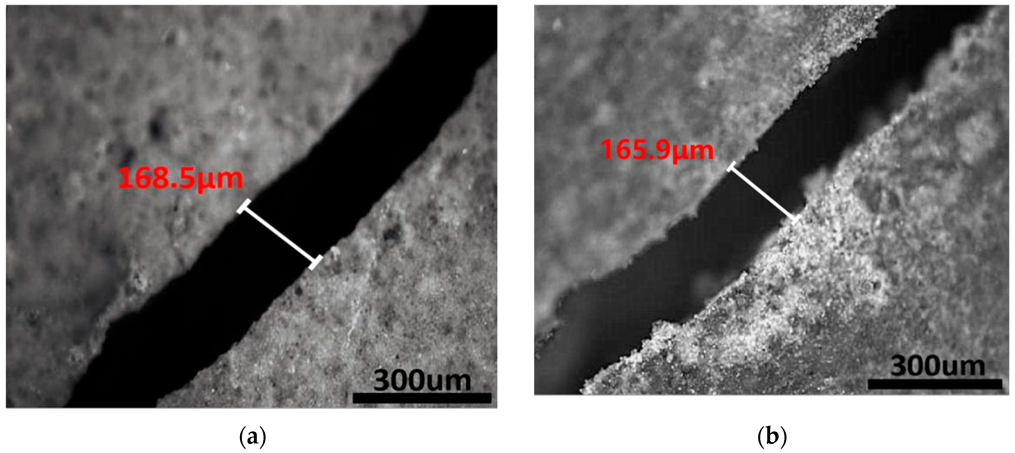
3.2.1. Microcapsules Core Healing Agent Bonded Cracks in a Dry Environment
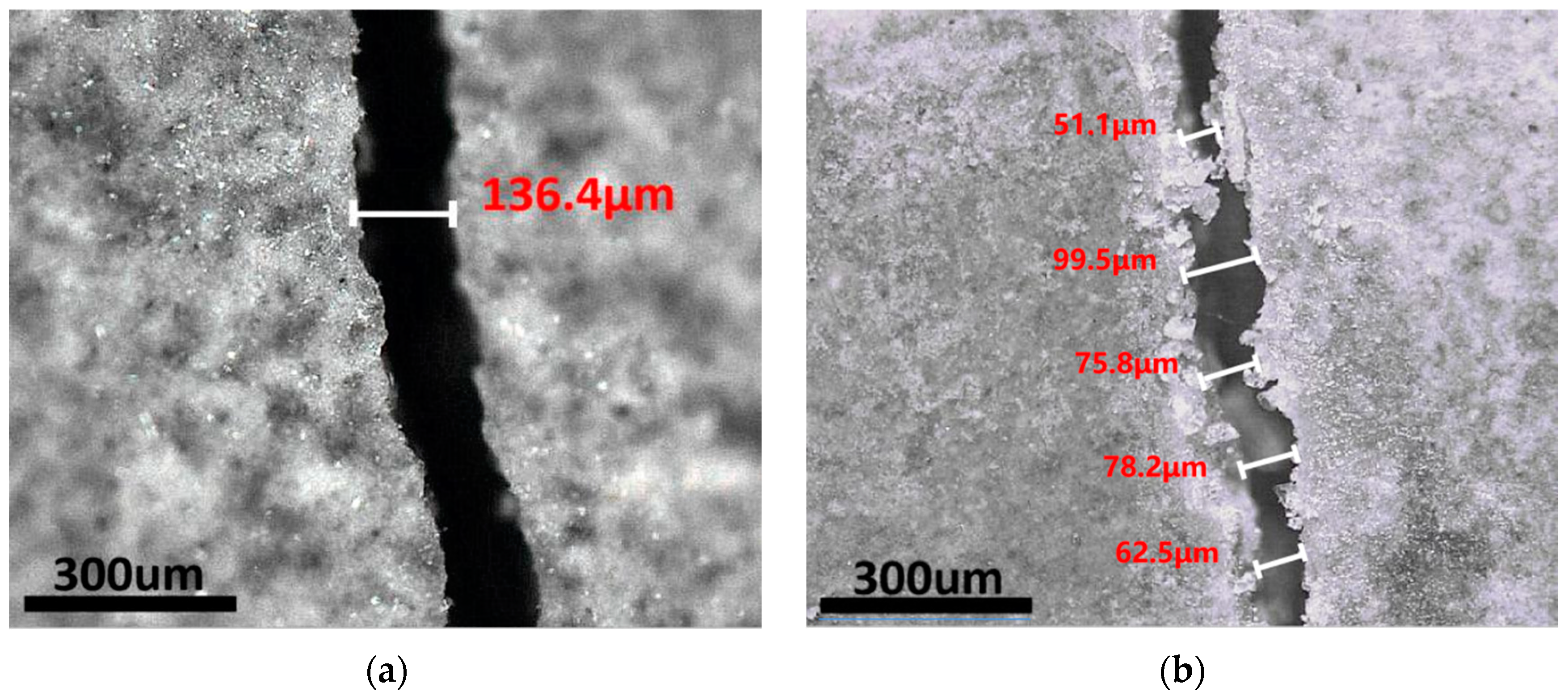
3.2.2. Microcapsule Swelling to Seal Cracks in a Water Environment
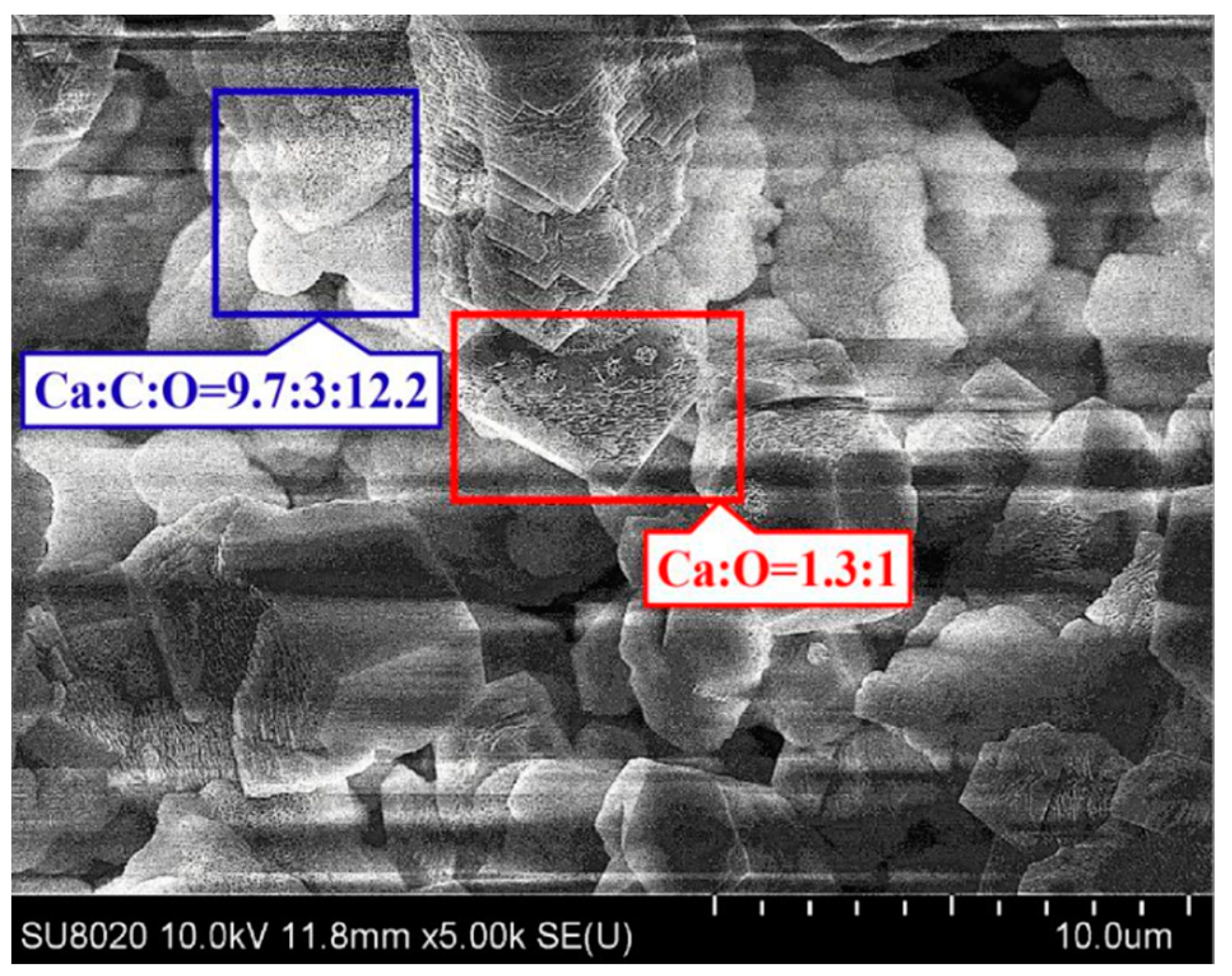
3.2.3. Microcapsules Promoting Autogenous Healing in Dry-Wet Cycles Environment
3.3. Compressive Strength Healing Effect
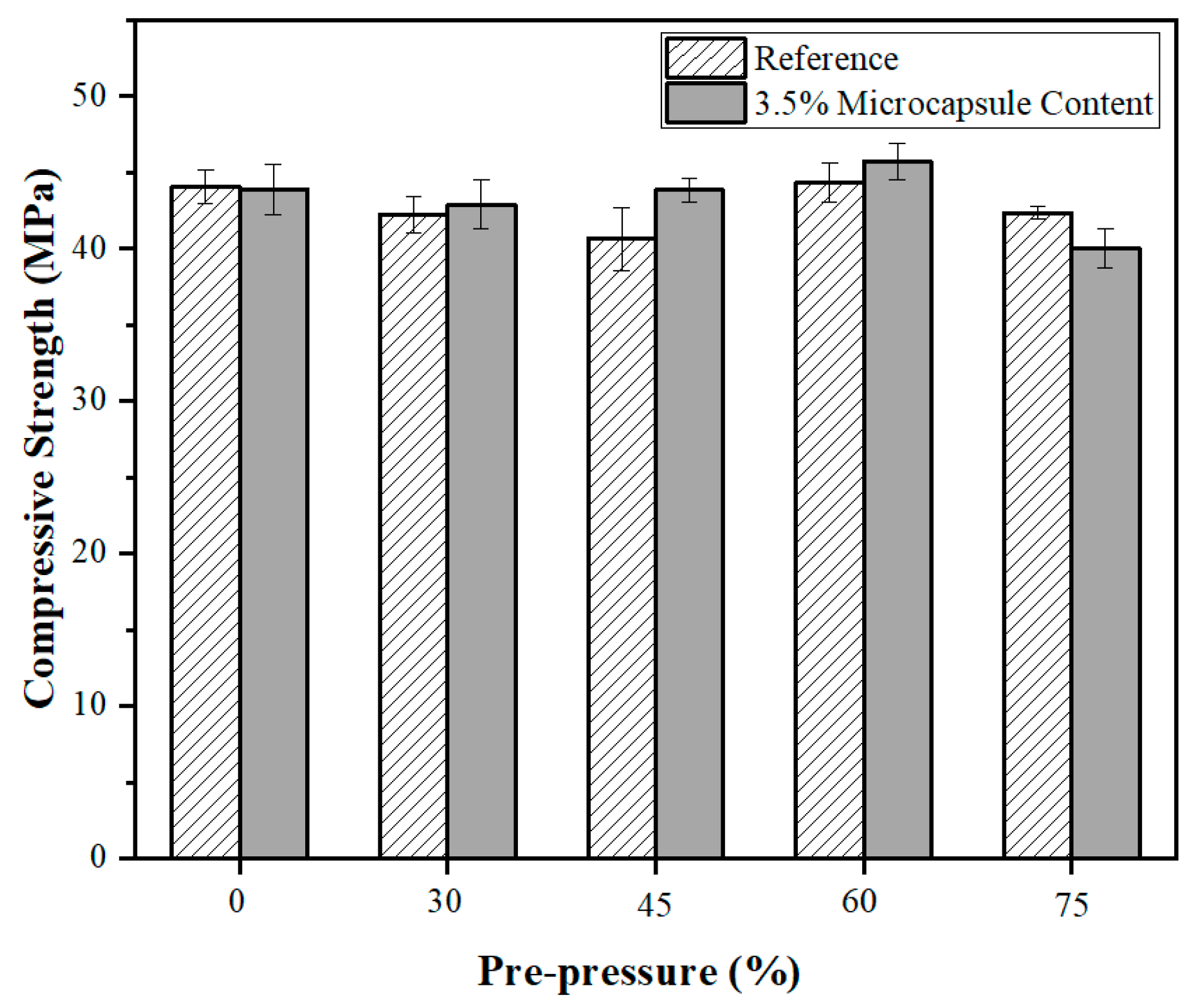
3.3.1. The Influence of Loading Pressure on the Compressive Strength Healing Effect
3.3.2. The Influence of Microcapsule Content on Compressive Strength Healing Effect
3.3.3. Self-Healing Effect in Drying Curing
3.3.4. Self-Healing Effect in Dry–Wet Cycles Curing
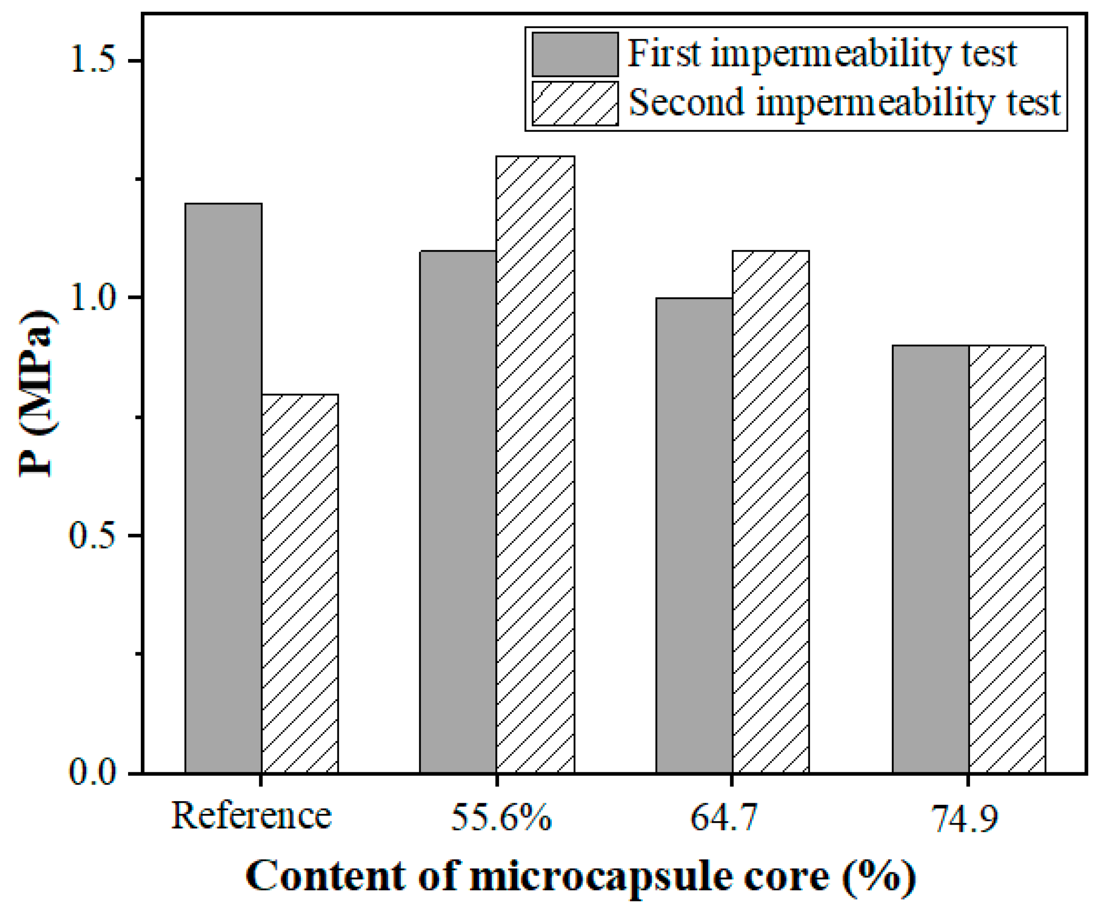
3.4. Water Permeability Healing Effect
4. Conclusions
Author Contributions
Funding
Institutional Review Board Statement
Conflicts of Interest
Appendix A

| Drug Name | Application | Pure Degree | Manufacturer |
|---|---|---|---|
| Sodium alginate | Preparation of Microcapsules | 99.9% | Shanghai McLean Biochemical Technology Co., Ltd. (Shanghai, China) |
| Epoxy resin (E-51) | Preparation of Microcapsules | 99.8% | Sinopec Baling Petrochemical Co., Ltd. (Hunan, China) |
| Cyclooxygen dilutions | Preparation of Microcapsules | - | Shanghai Aotun Huagong Technology Co., Ltd. (Shanghai, China) |
| Sodium dodecyl phenyl sulfonate | Preparation of Microcapsules | AR | Tianjin Forchen Chemical Reagent Factory (Tianjin, China) |
| Anhydrous calcium chloride (CaCl2) | Preparation of Microcapsules | AR | Beijing Tongguang Fine Chemical Co., Ltd. (Beijing, China) |
| Absolute ethyl alcohol | Washing of Microcapsules | AR | Beijing Tongguang Fine Chemical Co., Ltd. (Beijing, China) |
| Portland cement | Preparation of Mortarand Paste | P∙I 42.5 | Qufu Zhonglian Cement Co., Ltd. (Shandong, China) |
| ISO standard sand | Preparation of Mortar | — | Xiamen Europe Standard Sand Co., Ltd. (Xiamen, China) |
| Tetraethylenepentamine | Preparation of Mortar | AR | Shanghai McLean Biochemical Technology Co., Ltd. (Shanghai, China) |
References
- Singh, M.; Saini, B.; Chalak, H.D. Performance and com- position analysis of engineered cementitious composite (ecc) a review. J. Build. Eng. 2019, 26, 100851. [Google Scholar] [CrossRef]
- Yildirim, G.; Lachemi, M.; Keskin, O.; Kasap, S.; Bahadir, S. A review of intrinsic self-healing capability of engineered cementi- tious composites: Recovery of transport and mechanical properties. Constr. Build. Mater. 2015, 101, 10–21. [Google Scholar] [CrossRef]
- Yunovich, M.; Thompson, N.G. Corrosion of highway bridges: Economic impact and control methodologies. Concr. Int. 2003, 25, 52–57. [Google Scholar]
- Fernandez, C.A.; Correa, M.; Nguyen, M.T.; Rod, K.A.; Glezakou, V.A. Progress and challenges in self-healing cementitious materials. J. Mater. Sci. 2020, 56, 201–230. [Google Scholar] [CrossRef]
- Al-Tabbaa, A.; Litina, C.; Giannaros, P.; Kanellopoulos, A.; Souza, L. First UK field application and performance of microcapsule-based self-healing concrete. Constr. Build. Mater. 2019, 208, 669–685. [Google Scholar] [CrossRef]
- Patrick Jason, F. Polymers with autonomous life-cycle control. Nature 2016, 540, 363–370. [Google Scholar] [CrossRef] [PubMed]
- Sidiq, A.; Gravina, R.; Giustozzi, F. Is concrete healing really efficient? A review. Constr. Build. Mater. 2019, 205, 257–273. [Google Scholar] [CrossRef]
- Edvardsen, C. Water Permeability and Autogenous Healing of Cracks in Concrete. ACI 1999, 96, 448–454. [Google Scholar] [CrossRef]
- Tomczak, K.; Jakubowski, J. The effects of age, cement content, and healing time on the self-healing ability of high-strength concrete. Constr. Build. Mater. 2018, 187, 149–159. [Google Scholar] [CrossRef]
- Yıldırım, G.; Khiavi, A.H.; Yeşilmen, S.; Şahmaran, M. Self-healing performance of aged cementitious composites. Cem. Concr. Compos. 2018, 87, 172–186. [Google Scholar] [CrossRef]
- Qureshi, T.; Kanellopoulos, A.; Al-Tabbaa, A. Autogenous self-healing of cement with expansive minerals-II: Impact of age and the role of optimised expansive minerals in healing performance. Constr. Build. Mater. 2019, 194, 264–275. [Google Scholar] [CrossRef]
- Xue, C.H.; Li, W.G.; Luo, Z.Y.; Wang, K.J.; Castel, A. Effect of chloride ingress on self-healing recovery of smart cementitious composite incorporating crystalline admixture and MgO expansive agent. Cem. Concr. Res. 2021, 139, 106252. [Google Scholar] [CrossRef]
- Nasim, M. Autonomous healing in concrete by crystalline admixture: A review. Mater. Today Proc. 2020, 5, 638–644. [Google Scholar] [CrossRef]
- Meng, D.; Dewangan, U.K.; Deo Shirish, V. Mechanical behaviour of a polyvinyl alcohol fibre reinforced engineered cementitious composite (PVA-ECC) using local ingredients. Constr. Build. Mater. 2017, 141, 259–270. [Google Scholar] [CrossRef]
- Liu, H.Z.; Zhang, Q.; Gu, C.S.; Su, H.Z.; Li, V. Influence of microcrack self-healing behavior on the permeability of Engineered Cementitious Composites. Cem. Concr. Compos. 2017, 82, 14–22. [Google Scholar] [CrossRef]
- He, J.L.; Shi, X. Developing an abiotic capsule-based self-healing system for cementitious materials: The state of knowledge. Constr. Build. Mater. 2017, 156, 1096–1113. [Google Scholar] [CrossRef]
- Jakubovskis, R.; Jankut, A.; Urbonaviius, J.; Gribniak, V. Analysis of mechanical performance and durability of self-healing biological concrete. Constr. Build. Mater. 2020, 260, 1–15. [Google Scholar] [CrossRef]
- Yatish Reddy, P.V.; Ramesh, B.; Prem Kumar, L. Influence of bacteria in self healing of concrete—A review. Mater. Today Proc. 2020, 33, 4212–4218. [Google Scholar] [CrossRef]
- Khaliq, W.; Ehsan, M.B. Crack healing in concrete using various bio influenced self-healing techniques. Constr. Build. Mater. 2016, 102, 349–357. [Google Scholar] [CrossRef]
- White, S.R. Autonomic healing of polymer composites. Nature 2001, 409, 6822. [Google Scholar] [CrossRef]
- Kanellopoulos, A.; Qureshi, T.; Al-tabbaa, A. Glass encapsulated minerals for self-healing in cement based composites. Constr. Build. Mater. 2015, 98, 780–791. [Google Scholar] [CrossRef]
- Beglarigale, A.; Seki, Y.; Demir, N.Y.; Yazıcı, H. Sodium silicate/polyurethane microcapsules used for self-healing in cementitious materials: Monomer optimization, characterization, and fracture behavior. Constr. Build. Mater. 2018, 162, 57–64. [Google Scholar] [CrossRef]
- Chindasiriphan, P.; Yokota, H.; Pimpakan, P. Effect of fly ash and superabsorbent polymer on concrete self-healing ability. Constr. Build. Mater. 2020, 233, 116975. [Google Scholar] [CrossRef]
- Mignon, A. Crack Mitigation in Concrete: Superabsorbent Polymers as Key to Success? Materials 2017, 10, 237. [Google Scholar] [CrossRef] [PubMed] [Green Version]
- Lee, H.X.D.; Snoeck, D.; Dubruel, P.; Van Vlierberghe, S.; De Belie, N. Self-sealing of cracks in concrete using superabsorbent polymers. Cem. Concr. Res. 2016, 79, 194–208. [Google Scholar] [CrossRef] [Green Version]
- Chen, W.H.; Lin, B.X.; Feng, K.; Cui, S.S.; Zhang, D. Effect of shape memory alloy fiber content and preloading level on the self-healing properties of smart cementitious composite (SMA-ECC). Constr. Build. Mater. 2022, 314, 127797. [Google Scholar] [CrossRef]
- Chen, W.H.; Feng, K.; Wang, Y.; Lin, Y.W.; Qian, H. Evaluation of self-healing performance of a smart composite material(SMA-ECC). Constr. Build. Mater. 2021, 290, 123216. [Google Scholar] [CrossRef]
- Ali, M.A.E.M.; Nehdi, M.L. Innovative crack-healing hybrid fiber reinforced engineered cementitious composite. Constr. Build. Mater. 2021, 290, 123216. [Google Scholar] [CrossRef]
- Selvarajoo, T.; Davies, R.E.; Freeman, B.L.; Jefferson, A.D. Mechanical response of a vascular self-healing cementitious material system under varying loading conditions. Constr. Build. Mater. 2020, 254, 119245. [Google Scholar] [CrossRef]
- Huang, H.L.; Ye, G.; Shui, Z.H. Feasibility of self-healing in cementitious materials-By using capsules or a vascular system? Constr. Build. Mater. 2014, 63, 108–118. [Google Scholar] [CrossRef]
- Wang, R.Y.; Yu, J.Y.; Gu, S.J.; He, P.; Han, X.B.; Liu, Q.T. Investigation of migration and self-healing ability of ion chelator in cement-based materials by a novel method. Constr. Build. Mater. 2020, 262, 120917. [Google Scholar] [CrossRef]
- Li, Y.; Yu, J.Y.; Cao, Z.L.; Du, W.; Zhang, Y.C.; Zou, Y. Preparation and characterization of nano-Fe3O4/paraffin encapsulated isocyanate microcapsule by electromagnetic controlled rupture for self-healing cementitious materials. Constr. Build. Mater. 2020, 265, 120703. [Google Scholar] [CrossRef]
- Wang, X.G.; Yin, H.; Chen, Z.; Xia, L. Epoxy resin/ethyl cellulose microcapsules prepared by solvent evaporation for repairing microcracks: Particle properties and slow-release performance. Mater. Today Commun. 2020, 22, 100854. [Google Scholar] [CrossRef]
- Lv, L.Y.; Guo, P.Y.; Xing, F.; Han, N.X. Trigger efficiency enhancement of polymeric microcapsules for self-healing cementitious materials. Constr. Build. Mater. 2020, 235, 117443. [Google Scholar] [CrossRef]
- Wang, X.G.; Zhu, J.L.; Zou, F.B.; Zhou, N.G.; Li, Y.G.; Lei, W.Y. Ca-Al LDH hybrid self-healing microcapsules for corrosion protection. Chem. Eng. J. 2022, 447, 137125. [Google Scholar] [CrossRef]
- Han, T.L.; Wang, X.F.; Li, D.W.; Li, D.F.; Xing, F.; Han, N.X. Impermeability characteristics of cementitious materials with self-healing based on epoxy/urea-formaldehyde microcapsules using an immersion test. Constr. Build. Mater. 2020, 259, 119782. [Google Scholar] [CrossRef]
- Han, T.L.; Wang, X.F.; Li, D.W.; Li, D.F.; Xing, F.; Ren, J.; Han, N.X. Stress-strain behaviour and pore structure of microcapsule-based self-healing cementitious composite under triaxial tests. Constr. Build. Mater. 2020, 241, 118009. [Google Scholar] [CrossRef]
- Zhou, G.; Jiang, W.J.; Li, S.L.; Liu, R.L.; Zhang, Q.T.; Qi, G.S.; He, Z.L. Preparation and performance analysis of dopamine hydrochloride functionalized E-51@MPF/SiO2 double-wall microcapsules for microcracks self-healing in cement-based materials. Constr. Build. Mater. 2022, 325, 126622. [Google Scholar] [CrossRef]
- Li, W.T.; Jiang, Z.W.; Yang, Z.H.; Zhao, N.; Yuan, W.Z.; Costa-Rodrigues, J. Self-healing efficiency of cementitious materials containing microcapsules filled with healing adhesive: Mechanical restoration and healing process monitored by water absorption. PLoS ONE 2013, 8, 81616. [Google Scholar] [CrossRef] [Green Version]
- Tittelboom, K.V.; De Belie, N.; Van Loo, D.; Jacobs, P. Self-healing efficiency of cementitious materials containing tubular capsules filled with healing agent. Cem. Concr. Compos. 2011, 33, 497–505. [Google Scholar] [CrossRef]
- Feng, J.H.; Dong, H.; Wang, R.X.; Su, Y.L. A novel capsule by poly (ethylene glycol) granulation for self-healing concrete. Cem. Concr. Res. 2020, 133, 106053. [Google Scholar] [CrossRef]
- Fang, G.H.; Liu, Y.Q.; Qin, S.F.; Ding, W.J.; Zhang, J.C.; Hong, S.X.; Xing, F.; Dong, B.Q. Visualized tracing of crack self-healing features in cement/microcapsule system with X-ray microcomputed tomography. Constr. Build. Mater. 2018, 179, 336–347. [Google Scholar] [CrossRef]
- Beglarigale, A.; Eyice, D.; Seki, Y.; Yalçınkaya, Ç.; Çopuroğlu, O.; Yazıcı, H. Sodium silicate/polyurethane microcapsules synthesized for enhancing self-healing ability of cementitious materials: Optimization of stirring speeds and evaluation of self-healing efficiency. J. Build. Eng. 2021, 39, 102297. [Google Scholar] [CrossRef]
- Song, C.W.; Choi, Y.C.; Choi, S. Effect of internal curing by superabsorbent polymers—Internal relative humidity and autogenous shrinkage of alkali-activated slag mortars. Constr. Build. Mater. 2016, 123, 198–206. [Google Scholar] [CrossRef]
- Hu, M.M.; Guo, J.T.; Du, J.B.; Liu, Z.X.; Li, P.P.; Ren, X.K.; Feng, Y.K. Development of Ca 2+ -based, ion-responsive superabsorbent hydrogel for cement applications: Self-healing and compressive strength. J. Colloid Interface Sci. 2018, 538, 397–403. [Google Scholar] [CrossRef]
- Gwon, S.; Ahn, E.; Shin, M. Self-healing of modified sulfur composites with calcium sulfoaluminate cement and superabsorbent polymer. Compos. Part B 2019, 162, 469–483. [Google Scholar] [CrossRef]
- Wang, C.Y.; Bu, Y.H.; Guo, S.L.; Lu, Y.; Sun, B.J.; Shen, Z.H. Self-healing cement composite: Amine- and ammonium-based pH-sensitive superabsorbent polymers. Cem. Concr. Compos. 2018, 96, 154–162. [Google Scholar] [CrossRef]
- Snoeck, D.; Dewanckele, J.; Cnudde, V.; De Belie, N. X-ray computed microtomography to study autogenous healing of cementitious materials promoted by superabsorbent polymers. Cem. Concr. Compos. 2016, 65, 83–93. [Google Scholar] [CrossRef]
- Hong, G.; Song, C.; Choi, S. Autogenous Healing of Early-Age Cracks in Cementitious Materials by Superabsorbent Polymers. Materials 2020, 13, 690. [Google Scholar] [CrossRef] [Green Version]
- Mao, Q.J.; Wu, W.W.; Liang, P.; Wang, Z.M.; Cui, S.P. Self-healing effect of calcium alginate/epoxy microcapsules in cementitious materials. Mater. Rev. 2018, 32, 4016–4021. [Google Scholar] [CrossRef]
- Mao, Q.J.; Chen, J.Y.; Qi, W.J.; Liu, H.; Wang, Z.M.; Cui, S.P. Improving Self-Healing and Shrinkage Reduction of Cementitious Materials Using Water-Absorbing Polymer Microcapsules. Materials 2022, 15, 847. [Google Scholar] [CrossRef]
- Wang, X.G.; Zhou, Z.; Zhao, H.; Zhang, C.Y.; Li, Y.J. Capillary Transport Mechanism of Epoxy Resin Repairing Micro-cracks in Cement-Based Materials. J. Build. Mater. 2021, 24, 1200–1207. [Google Scholar] [CrossRef]
- Wang, X.F.; Zhang, M.; Xing, F.; Han, N.X. Effect of a Healing Agent on the Curing Reaction Kinetics and Its Mechanism in a Self-Healing System. Appl. Sci. 2018, 8, 2241. [Google Scholar] [CrossRef] [Green Version]
- Schiessl, P.; Brauer, N. Influence of Autogenous Healing of Cracks on Corossion of Reinforcement. In Proceedings of the 1996 7th International Conference on Durability of Building Materials and Components, Stockholm, Sweden, 19–23 May 1996; pp. 542–552. [Google Scholar]
- Jacobsen, S.; Marchand, J.; Boisvert, L. Effect of cracking and healing on chloride transport in OPC concrete. Cem. Concr. Res. 1996, 26, 869–881. [Google Scholar] [CrossRef]
- Snoeck, D.; Van Tittelboom, K.; Steuperaert, S. Self-Healing Cementitious Materials by the Combination of Microfibres and Superabsorbent Polymers. J. Intell. Mater. Syst. Struct. 2014, 25, 13–24. [Google Scholar] [CrossRef] [Green Version]
- Han, N.X.; Xing, F. A Comprehensive Review of the Study and Development of Microcapsule Based Self-Resilience Systems for Concrete Structures at Shenzhen University. Materials 2017, 10, 2. [Google Scholar] [CrossRef]
- Du, W.; Liu, Q.T.; Lin, R.S.; Su, X. Preparation and Characterization of Microcrystalline Wax/Epoxy Resin Microcapsules for Self-Healing of Cementitious Materials. Materials 2021, 14, 1725. [Google Scholar] [CrossRef]
- Snoeck, D.; De Belie, N. Repeated Autogenous Healing in Strain-Hardening Cementitious Composites by Using Superabsorbent Polymers. J. Mater. Civ. Eng. 2016, 28, 04015086. [Google Scholar] [CrossRef]


| Shell–Core Ratio | Core Content | Water Absorbing Rate | |
|---|---|---|---|
| A | 1:5.5 | 55.57% | 39.86% |
| B | 1:7.5 | 64.12% | 30.12% |
| C | 1:9.0 | 74.35% | 24.01% |
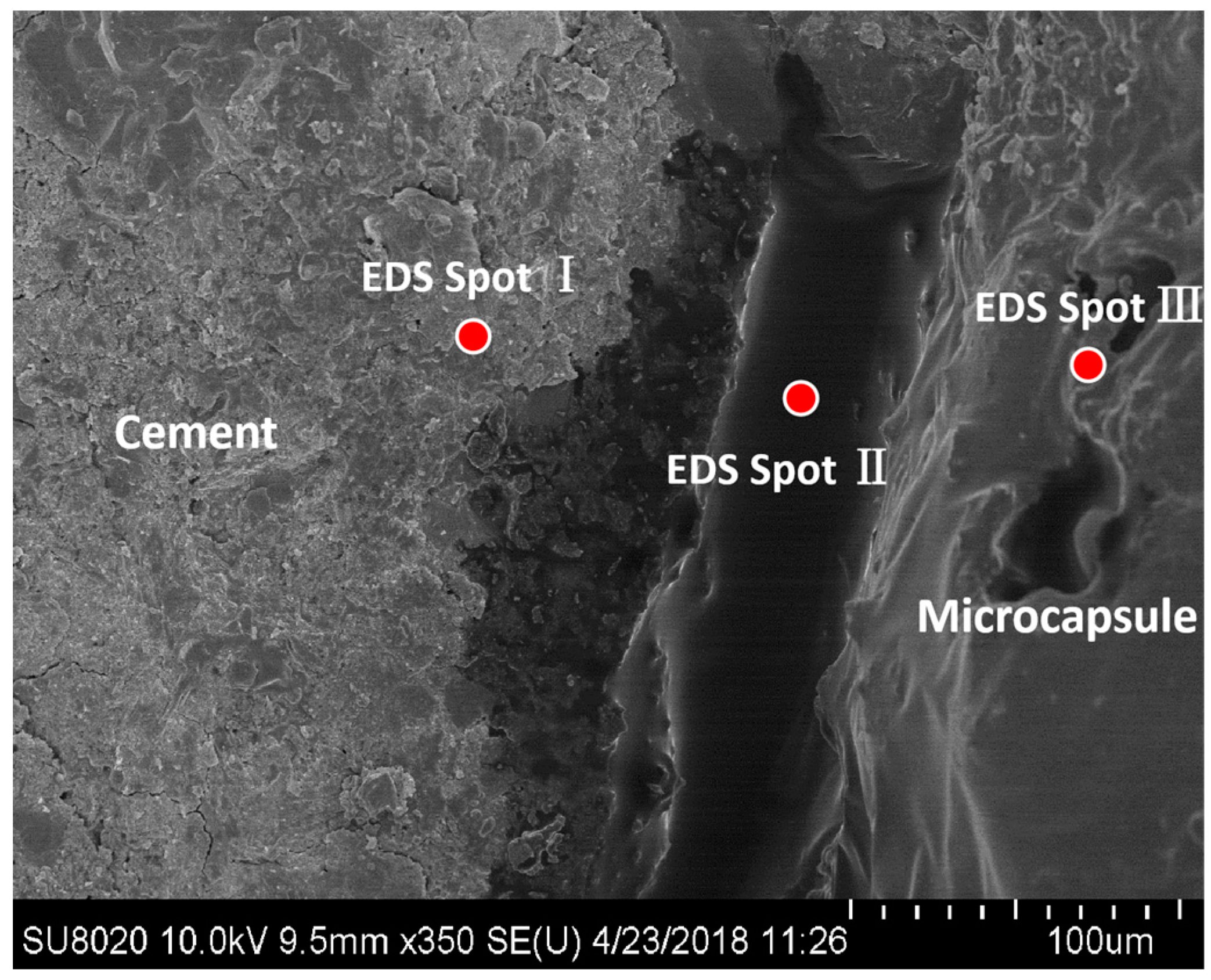
| Point | Element Composition | ||||||
|---|---|---|---|---|---|---|---|
| C | O | Ca | Si | Al | Cl | N | |
| I | 3.25 | 31.98 | 42.15 | 11.73 | 2.05 | 0.00 | 0.05 |
| II | 59.90 | 24.15 | 9.04 | 0.00 | 0.00 | 2.20 | 5.11 |
| III | 29.12 | 19.69 | 28.08 | 0.00 | 0.00 | 22.58 | 0.01 |
Disclaimer/Publisher’s Note: The statements, opinions and data contained in all publications are solely those of the individual author(s) and contributor(s) and not of MDPI and/or the editor(s). MDPI and/or the editor(s) disclaim responsibility for any injury to people or property resulting from any ideas, methods, instructions or products referred to in the content. |
© 2023 by the authors. Licensee MDPI, Basel, Switzerland. This article is an open access article distributed under the terms and conditions of the Creative Commons Attribution (CC BY) license (https://creativecommons.org/licenses/by/4.0/).
Share and Cite
Mao, Q.; Chen, J.; Wu, W.; Li, R.; Shi, S.; Wang, Z.; Cui, S. Multiple Self-Healing Effects of Water-Absorbing Microcapsules in Cementitious Materials. Polymers 2023, 15, 428. https://doi.org/10.3390/polym15020428
Mao Q, Chen J, Wu W, Li R, Shi S, Wang Z, Cui S. Multiple Self-Healing Effects of Water-Absorbing Microcapsules in Cementitious Materials. Polymers. 2023; 15(2):428. https://doi.org/10.3390/polym15020428
Chicago/Turabian StyleMao, Qianjin, Jiayi Chen, Wenwen Wu, Runfeng Li, Shuqing Shi, Ziming Wang, and Suping Cui. 2023. "Multiple Self-Healing Effects of Water-Absorbing Microcapsules in Cementitious Materials" Polymers 15, no. 2: 428. https://doi.org/10.3390/polym15020428