

Peripheral Nerve Regeneration at 1 Year: Biodegradable Polybutylene Succinate Artificial Scaffold vs. Conventional Epineurial Sutures

 , , , , and
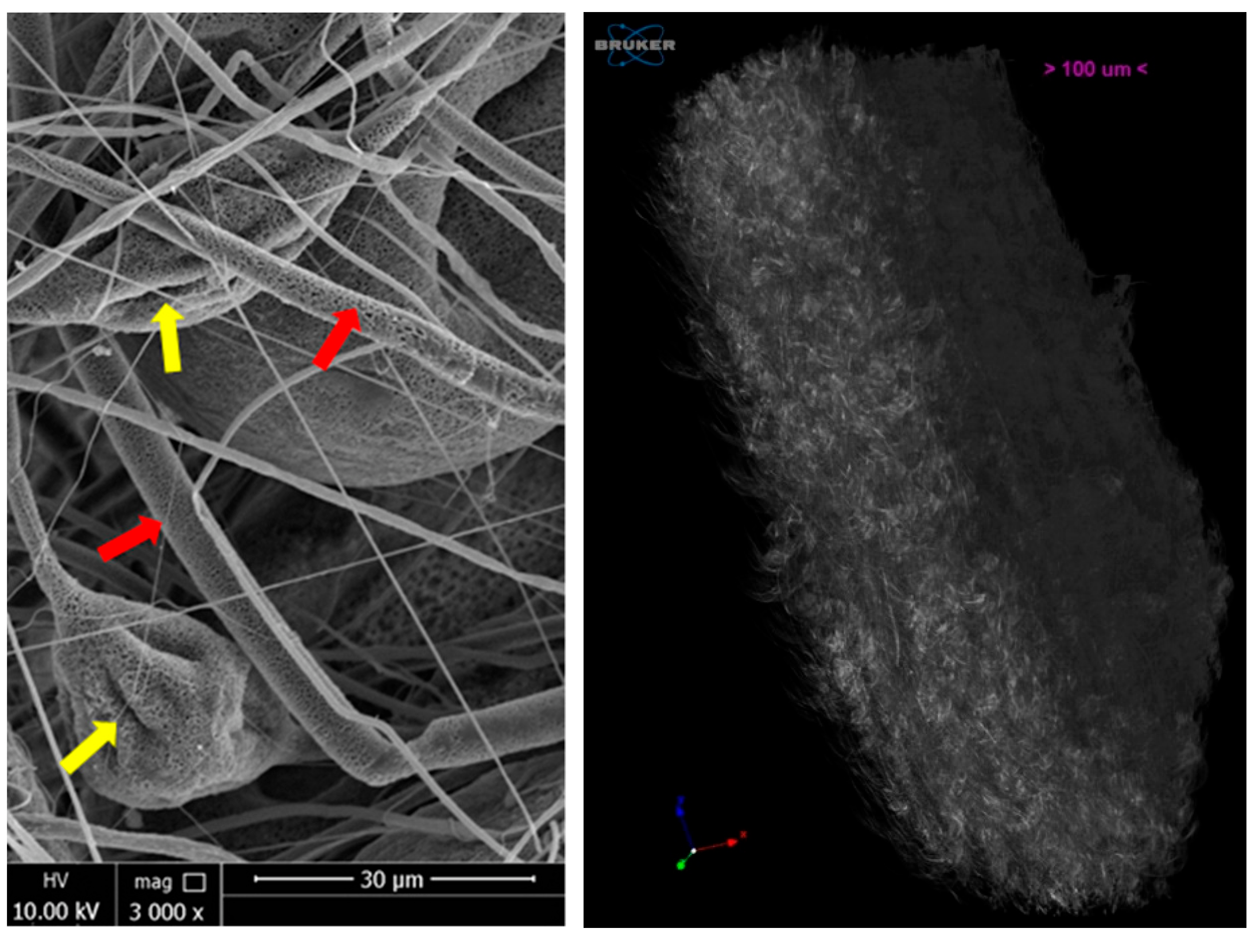
Abstract
:1. Introduction
2. Materials and Methods
2.1. Poly-Butylene Succinate (PBS) Scaffolds Fabrication by Electrospinning Technique
2.2. PBS Scaffolds Characterization by Scanning Electron Microscopy (SEM) and Microcomputed Tomography (μCT)
2.3. Animals
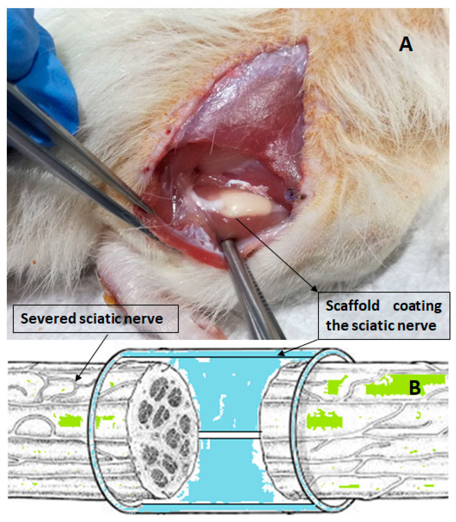
2.4. Surgical Procedure and Scaffold Implantation
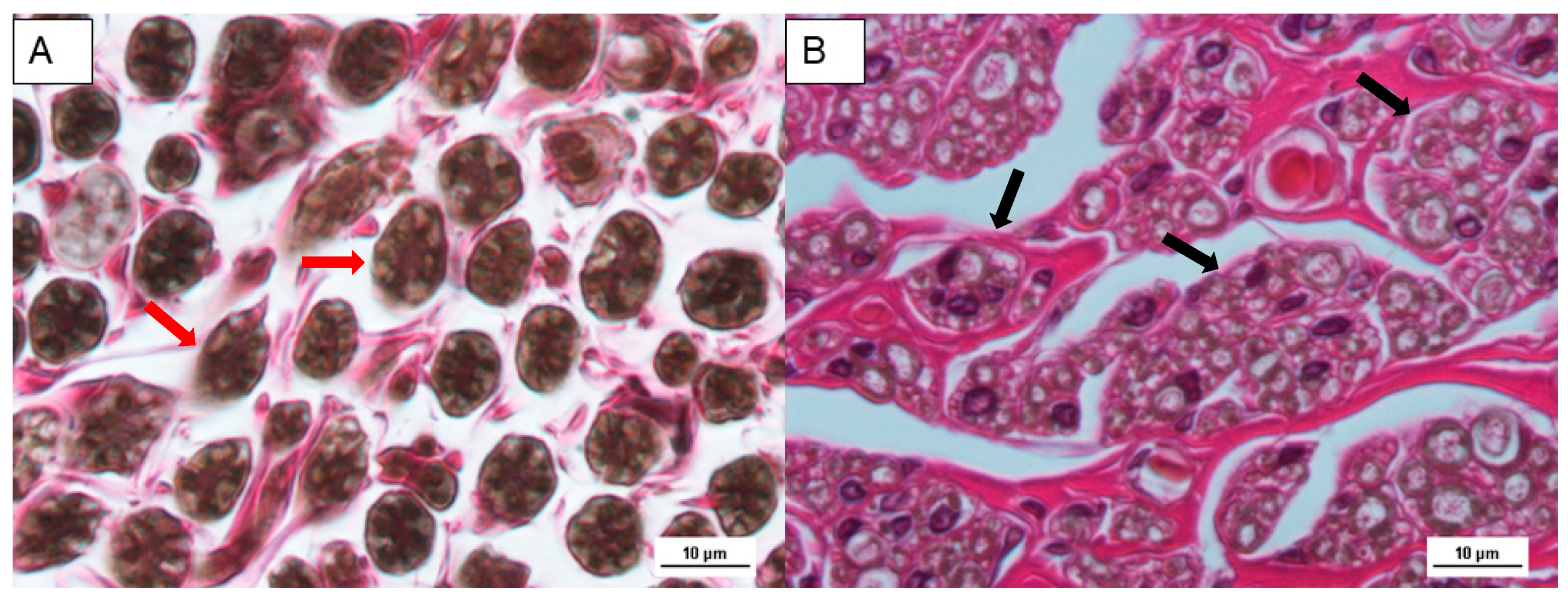
2.5. Histological Analysis
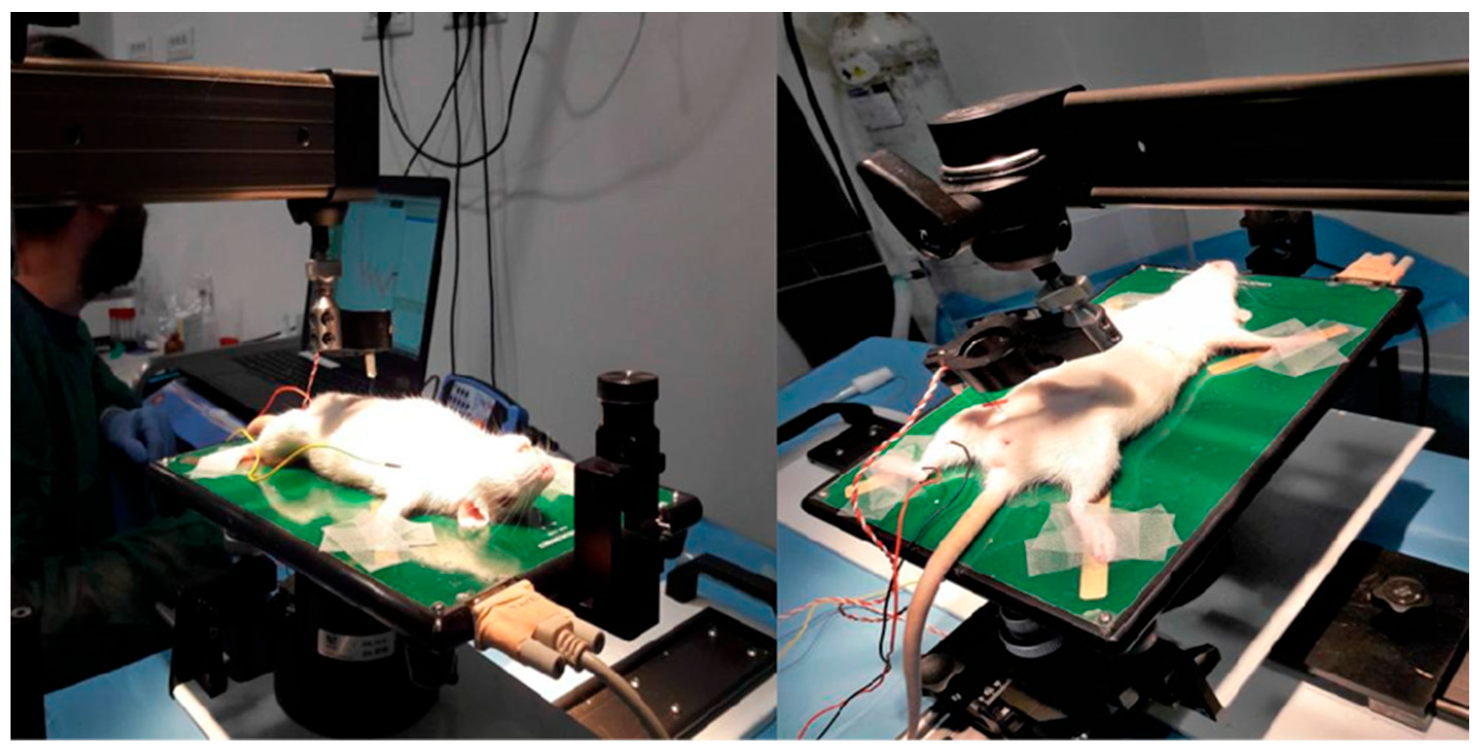
2.6. Compound Muscle Action Potential (CMAP) Response Evaluation
2.7. Statistical Analysis
3. Results and Discussion
3.1. Fabrication and Characterization of PBS Scaffolds
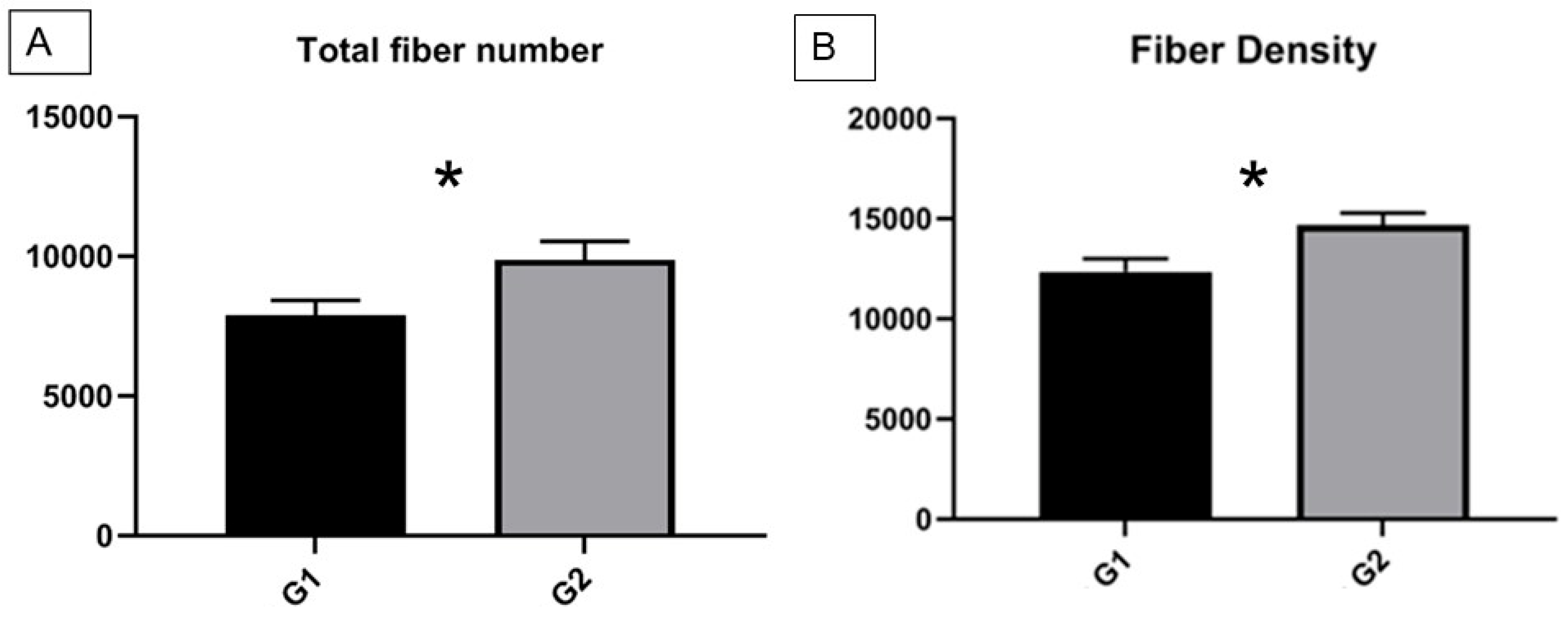
3.2. Evaluation of Nerve Regeneration by Histological Analysis: Counting of Regenerated Fibers
3.3. Electrophysiological Evaluation
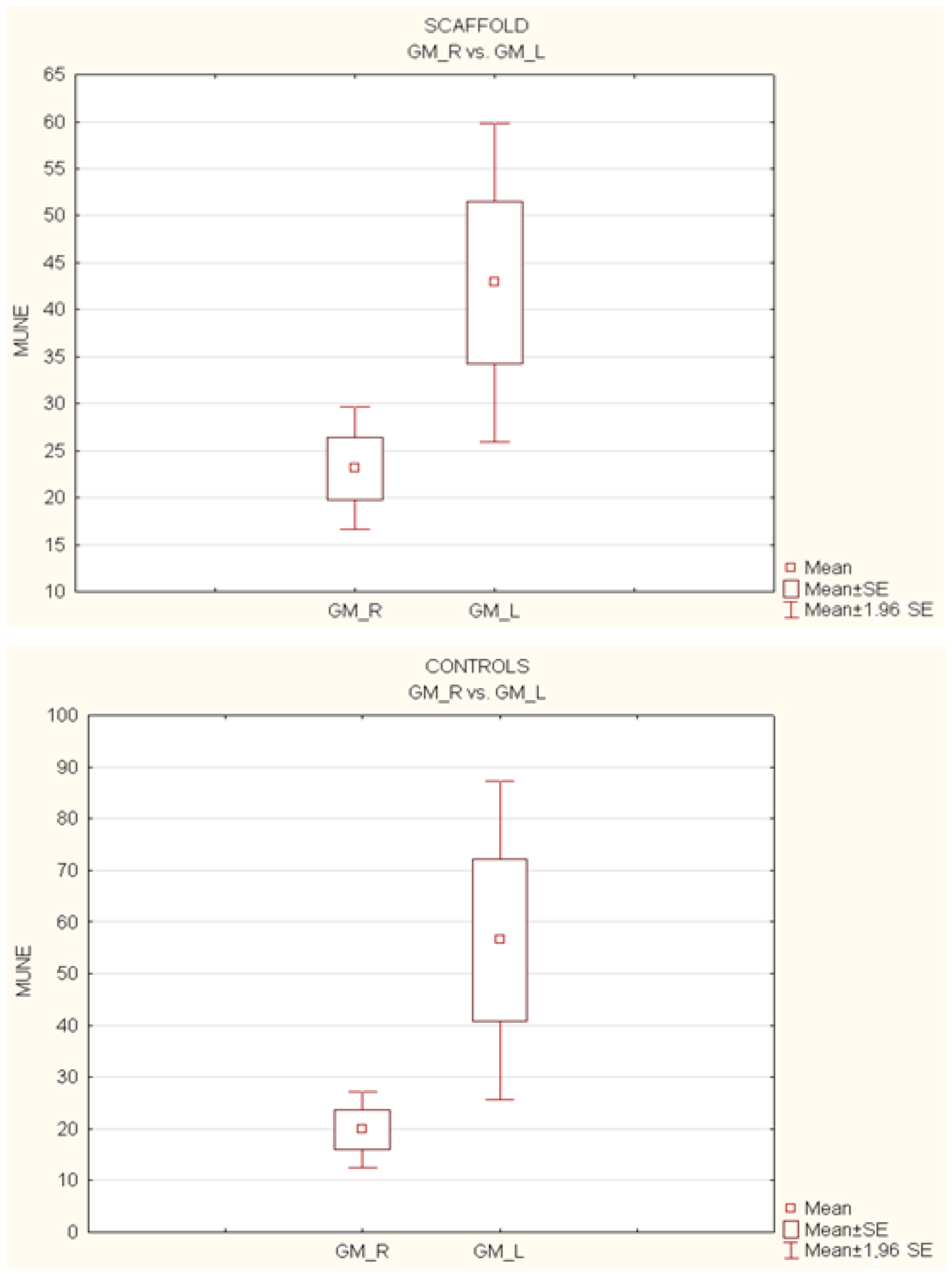
- Gastrocnemius muscle (MG-R vs. MG-L):
- -
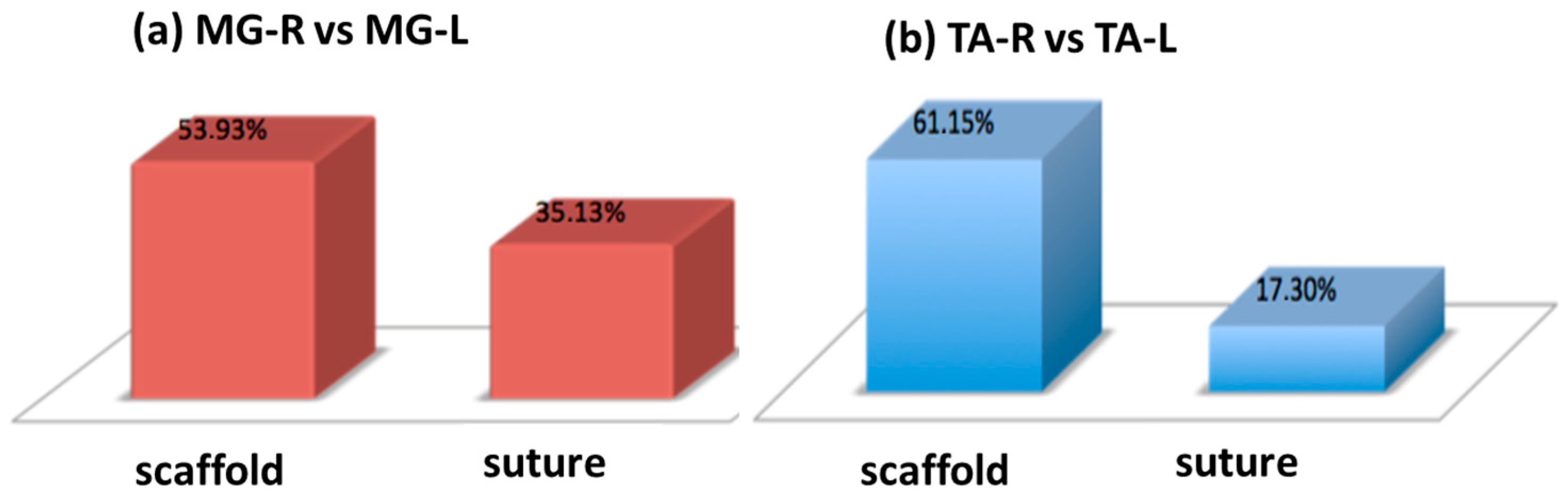
- In the study group (scaffold), the gastrocnemius muscle recovered 53.93% of its muscular function.
- -
- In the control group (suture), the gastrocnemius muscle recovered 35.13% of its muscular function (see Figure 8a).
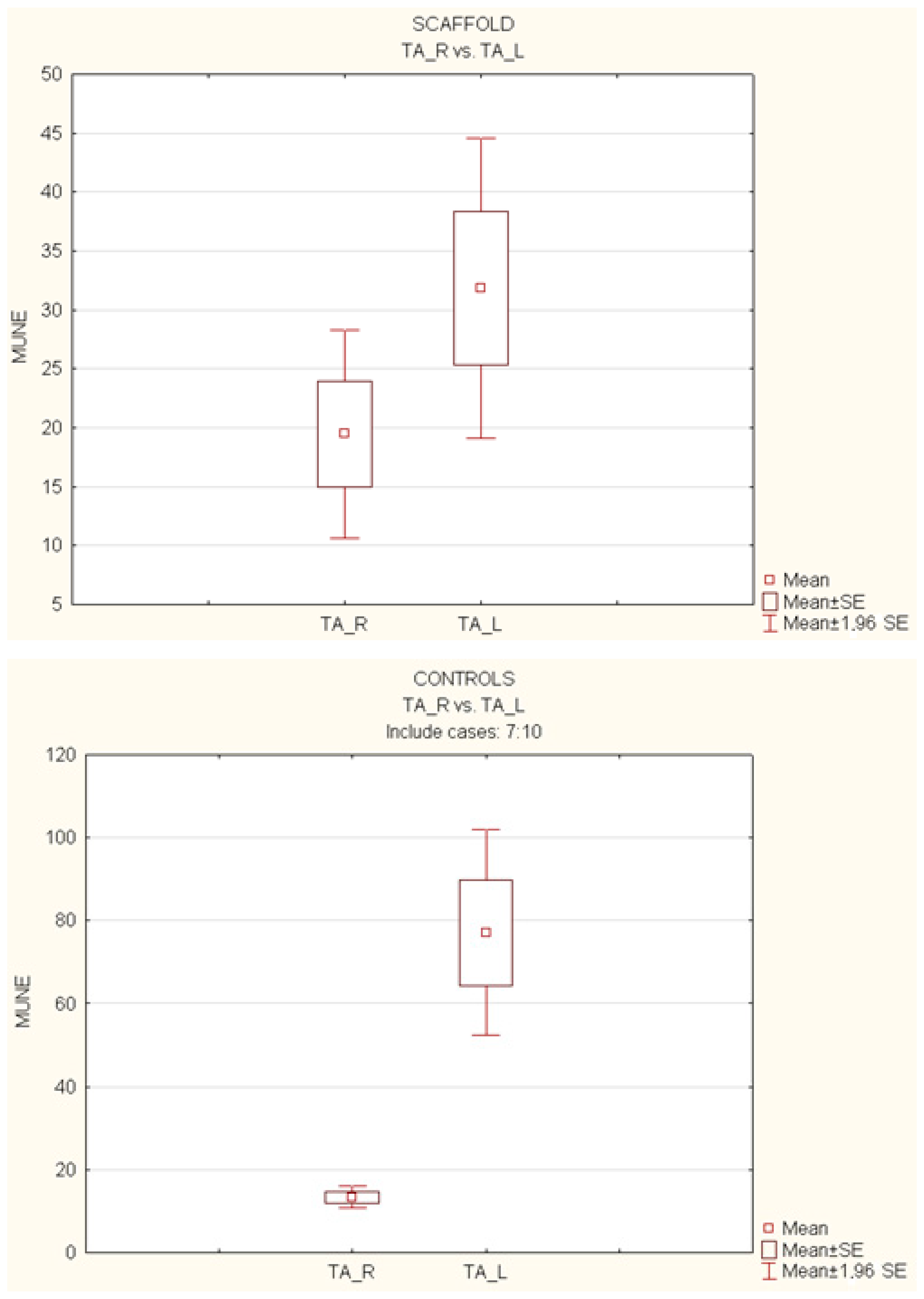
- Tibialis anterior muscle (TA-R vs. TA-L):
- -
- In the study group (scaffold), the tibialis anterior muscle showed a recovery of 61.15% of its muscular function.
- -
- In the control group (suture), the tibialis anterior muscle recovered 17.30% of its muscular function (see Figure 8b).
4. Conclusions
Author Contributions
Funding
Institutional Review Board Statement
Data Availability Statement
Acknowledgments
Conflicts of Interest
References
- Lopes, B.; Sousa, P.; Alvites, R.; Branquinho, M.; Sousa, A.C.; Mendonça, C.; Atayde, L.M.; Luís, A.L.; Varejão, A.S.P.; Maurício, A.C. Peripheral Nerve Injury Treatments and Advances: One Health Perspective. Int. J. Mol. Sci. 2022, 23, 918. [Google Scholar] [CrossRef] [PubMed]
- Wang, M.L.; Rivlin, M.; Graham, J.G.; Beredjiklian, P.K. Peripheral nerve injury, scarring, and recovery. Connect. Tissue Res. 2019, 60, 3–9. [Google Scholar] [CrossRef] [PubMed]
- Dong, R.; Liu, Y.; Yang, Y.; Wang, H.; Xu, Y.; Zhang, Z. MSC-Derived Exosomes-Based Therapy for Peripheral Nerve Injury: A Novel Therapeutic Strategy. BioMed Res. Int. 2019, 2019, 6458237. [Google Scholar] [CrossRef] [PubMed] [Green Version]
- Slavin, B.R.; Sarhane, K.A.; von Guionneau, N.; Hanwright, P.J.; Qiu, C.; Mao, H.-Q.; Höke, A.; Tuffaha, S.H. Insulin-Like Growth Factor-1: A Promising Therapeutic Target for Peripheral Nerve Injury. Front. Bioeng. Biotechnol. 2021, 9, 695850. [Google Scholar] [CrossRef] [PubMed]
- Alvites, R.; Caseiro, A.R.; Pedrosa, S.S.; Branquinho, M.V.; Ronchi, G.; Geuna, S.; Varejão, A.S.; Maurício, A.C. Peripheral nerve injury and axonotmesis: State of the art and recent advances. Cogent Med. 2018, 5, 1466404. [Google Scholar] [CrossRef]
- Kornfeld, T.; Vogt, P.M.; Radtke, C. Nerve grafting for peripheral nerve injuries with extended defect sizes. Wien. Med. Wochenschr. 2019, 169, 240–251. [Google Scholar] [CrossRef] [Green Version]
- Rayner, M.L.D.; Grillo, A.; Williams, G.; Tawfik, E.; Zhang, T.; Volitaki, C.; Craig, D.Q.; Healy, J.; Phillips, J. Controlled local release of PPARγ agonists from biomaterials to treat peripheral nerve injury. J. Neural Eng. 2020, 17, 046030. [Google Scholar] [CrossRef]
- Rayner, M.L.; Day, A.G.; Bhangra, K.S.; Sinden, J.; Phillips, J.B. Engineered neural tissue made using clinical-grade human neural stem cells supports regeneration in a long gap peripheral nerve injury model. Acta Biomater. 2021, 135, 203–213. [Google Scholar] [CrossRef]
- Vijayavenkataraman, S. Nerve guide conduits for peripheral nerve injury repair: A review on design, materials and fabrication methods. Acta Biomater. 2020, 106, 54–69. [Google Scholar] [CrossRef]
- Niu, Y.; Galluzzi, M. A biodegradable block polyurethane nerve-guidance scaffold enhancing rapid vascularization and promoting reconstruction of transected sciatic nerve in Sprague-Dawley rats. J. Mater. Chem. B 2020, 8, 11063–11073. [Google Scholar] [CrossRef]
- Kasmaie, F.M.; Zamani, F.; Sayad-Fathi, S.; Zaminy, A. Promotion of nerve regeneration by biodegradable nanofibrous scaffold following sciatic nerve transection in rats. Prog. Biomater. 2021, 10, 53–64. [Google Scholar] [CrossRef] [PubMed]
- Niu, Y.; Stadler, F.J.; Fu, M. Biomimetic electrospun tubular PLLA/gelatin nanofiber scaffold promoting regeneration of sciatic nerve transection in SD rat. Mater. Sci. Eng. C 2021, 121, 111858. [Google Scholar] [CrossRef]
- Zhang, M.; Li, C.; Zhou, L.-P.; Pi, W.; Zhang, P.-X. Polymer scaffolds for biomedical applications in peripheral nerve reconstruction. Molecules 2021, 26, 2712. [Google Scholar] [CrossRef] [PubMed]
- Kong, Y.; Xu, J.; Han, Q.; Zheng, T.; Wu, L.; Li, G.; Yang, Y. Electrospinning porcine decellularized nerve matrix scaffold for peripheral nerve regeneration. Int. J. Biol. Macromol. 2022, 209, 1867–1881. [Google Scholar] [CrossRef] [PubMed]
- Raza, C.; Riaz, H.A.; Anjum, R.; Shakeel, N.U.A. Repair strategies for injured peripheral nerve: Review. Life Sci. 2020, 243, 117308. [Google Scholar] [CrossRef]
- Cicero, L.; Licciardi, M.; Cirincione, R.; Puleio, R.; Giammona, G.; Giglia, G.; Sardo, P.; Vigni, G.E.; Cioffi, A.; Sanfilippo, A.; et al. Polybutylene succinate artificial scaffold for peripheral nerve regeneration. J. Biomed. Mater. Res. B Appl. Biomater. 2022, 110, 125–134. [Google Scholar] [CrossRef]
- Miceli, G.C.; Palumbo, F.S.; Bonomo, F.P.; Zingales, M.; Licciardi, M. Polybutylene Succinate Processing and Evaluation as a Micro Fibrous Graft for Tissue Engineering Applications. Polymers 2022, 14, 4486. [Google Scholar] [CrossRef]
- Vigni, G.E.; Cassata, G.; Caldarella, G.; Cirincione, R.; Licciardi, M.; Miceli, G.C.; Puleio, R.; D’itri, L.; Coco, R.L.; Camarda, L.; et al. Improved Bone Regeneration Using Biodegradable Polybutylene Succinate Artificial Scaffold in a Rabbit Model. J. Funct. Biomater. 2023, 14, 22. [Google Scholar] [CrossRef]
- Di Scipio, F.; Raimondo, S.; Tos, P.; Geuna, S. A simple protocol for paraffin-embedded myelin sheath staining with osmium tetroxide for light microscope observation. Microsc. Res. Tech. 2008, 71, 497–502. [Google Scholar] [CrossRef]
- Prodanov, D.; Feirabend, H.K. Morphometric analysis of the fiber populations of the rat sciatic nerve, its spinal roots, and its major branches. J. Comp. Neurol. 2007, 503, 85–100. [Google Scholar] [CrossRef]
- Soundarya, S.P.; Menon, A.H.; Chandran, S.V.; Selvamurugan, N. Bone tissue engineering: Scaffold preparation using chitosan and other biomaterials with different design and fabrication techniques. Int. J. Biol. Macromol. 2018, 119, 1228–1239. [Google Scholar] [CrossRef] [PubMed]
- Xu, K.; Li, X.-K.; Zhang, H.-Y.; Ye, L.-X.; An, N.-C.; Huang, P.; Li, D.-H.; Zheng, Z.-L.; Ji, H.; Li, H.; et al. Exogenous platelet-derived growth factor improves neurovascular unit recovery after spinal cord injury. Neural Regen. Res. 2021, 16, 765–771. [Google Scholar] [CrossRef] [PubMed]
- Gu, X.; Ding, F.; Yang, Y.; Liu, J. Construction of tissue engineered nerve grafts and their application in peripheral nerve regeneration. Prog. Neurobiol. 2011, 93, 204–230. [Google Scholar] [CrossRef] [PubMed]


Disclaimer/Publisher’s Note: The statements, opinions and data contained in all publications are solely those of the individual author(s) and contributor(s) and not of MDPI and/or the editor(s). MDPI and/or the editor(s) disclaim responsibility for any injury to people or property resulting from any ideas, methods, instructions or products referred to in the content. |
© 2023 by the authors. Licensee MDPI, Basel, Switzerland. This article is an open access article distributed under the terms and conditions of the Creative Commons Attribution (CC BY) license (https://creativecommons.org/licenses/by/4.0/).
Share and Cite
Cicero, L.; Puleio, R.; Cassata, G.; Cirincione, R.; Camarda, L.; Caracappa, D.; D’Itri, L.; Licciardi, M.; Vigni, G.E. Peripheral Nerve Regeneration at 1 Year: Biodegradable Polybutylene Succinate Artificial Scaffold vs. Conventional Epineurial Sutures. Polymers 2023, 15, 3398. https://doi.org/10.3390/polym15163398
Cicero L, Puleio R, Cassata G, Cirincione R, Camarda L, Caracappa D, D’Itri L, Licciardi M, Vigni GE. Peripheral Nerve Regeneration at 1 Year: Biodegradable Polybutylene Succinate Artificial Scaffold vs. Conventional Epineurial Sutures. Polymers. 2023; 15(16):3398. https://doi.org/10.3390/polym15163398
Chicago/Turabian StyleCicero, Luca, Roberto Puleio, Giovanni Cassata, Roberta Cirincione, Lawrence Camarda, Dario Caracappa, Lorenzo D’Itri, Mariano Licciardi, and Giulio Edoardo Vigni. 2023. "Peripheral Nerve Regeneration at 1 Year: Biodegradable Polybutylene Succinate Artificial Scaffold vs. Conventional Epineurial Sutures" Polymers 15, no. 16: 3398. https://doi.org/10.3390/polym15163398