Article

Ultrastructural Evidence of Synapse Preservation and Axonal Regeneration Following Spinal Root Repair with Fibrin Biopolymer and Therapy with Dimethyl Fumarate

by
Paula Regina Gelinski Kempe
1,
Mateus Vidigal de Castro
1,
Victor Campos Khuriyeh
1,
Benedito Barraviera
2,
Rui Seabra Ferreira, Jr.
2 and
Alexandre Leite Rodrigues de Oliveira
1,*
1
Laboratory of Nerve Regeneration, Department of Structural and Functional Biology, Institute of Biology, University of Campinas, Campinas 13083-862, SP, Brazil
2
Center for the Study of Venoms and Venomous Animals (CEVAP), São Paulo State University (UNESP), Botucatu 18610-307, SP, Brazil
*
Author to whom correspondence should be addressed.
Polymers 2023, 15(15), 3171; https://doi.org/10.3390/polym15153171
Submission received: 28 June 2023 / Revised: 16 July 2023 / Accepted: 19 July 2023 / Published: 26 July 2023
(This article belongs to the Special Issue Advanced Polymers for Medical Applications)

Abstract

:
Spinal cord injury causes critical loss in motor and sensory function. Ventral root avulsion is an experimental model in which there is the tearing of the ventral (motor) roots from the surface of the spinal cord, resulting in several morphological changes, including motoneuron degeneration and local spinal cord circuitry rearrangements. Therefore, our goal was to test the combination of surgical repair of lesioned roots with a fibrin biopolymer and the pharmacological treatment with dimethyl fumarate, an immunomodulatory drug. Thus, adult female Lewis rats were subjected to unilateral ventral root avulsion of L4–L6 roots followed by repair with fibrin biopolymer and daily treatment with dimethyl fumarate (15 mg/Kg; gavage) for 4 weeks, the survival time post-surgery being 12 weeks; n = 5/group/technique. Treatments were evaluated by immunofluorescence and transmission electron microscopy, morphometry of the sciatic nerve, and motor function recovery. Our results indicate that the combination between fibrin biopolymer and dimethyl fumarate is neuroprotective since most of the synapses apposed to alfa motoneurons were preserved in clusters. Also, nerve sprouting occurred, and the restoration of the ‘g’ ratio and large axon diameter was achieved with the combined treatment. Such parameters were combined with up to 50% of gait recovery, observed by the walking track test. Altogether, our results indicate that combining root restoration with fibrin biopolymer and dimethyl fumarate administration can enhance motoneuron survival and regeneration after proximal lesions.

Graphical Abstract

1. Introduction

Brachial and lumbar plexuses injuries occur in high-energy trauma, such as car accidents, falls from heights, and extreme sports. Due to the great amplitude of movement in the shoulder joint, the cervical and upper thoracic roots are more susceptible to traction forces and rupture. In turn, spinal motoneurons present in the ventral horn are axotomized significantly close to the cell body, resulting in retrograde degeneration, gliosis, and inflammation of the gray matter. Attempts to restore the avulsed roots started about fifty years ago [1], and, although some positive outcomes have been obtained by Carlsted and colleagues [2,3], the investigation of the cellular and molecular aspects of the injury remain key elements for the development of more effective therapeutic approaches. For that, the establishment of animal models of root avulsion allowed the advance in understanding the several reactions to proximal axotomy as well as aspects of the regeneration process [4,5,6,7,8,9,10,11]. Thus, ventral root avulsion (VRA) in rats has become an animal model widely used since it can recapitulate both acute and chronic phases following injury [3,12,13,14,15,16,17,18,19,20,21].
In the acute phase following VRA, motoneurons display stereotyped morphological changes due to disconnection between the cell body and its target. Such interruption of axonal continuity results in metabolic and electrophysiological changes in the cell soma, well-known as chromatolysis [22]. Together with that, robust modifications in the spinal circuitry take place. A hallmark is the retraction of pre-synaptic inputs to alpha motoneurons, with a preferential loss in excitatory glutamatergic terminals, which originate mostly from the dorsal root ganglia conveying proprioceptive information [11,23,24,25]. Nevertheless, the remaining inhibitory boutons from interneurons [26,27] seem to contribute to a firing resting state of the axotomized motoneurons so that the metabolism is focused on cell repair and regrowth.
In the long term, such changes determine whether the injured neurons survive or degenerate, which has been interpreted as a window of opportunity for the application of neuroprotective therapies. In turn, the use of neurotrophic molecules, such as GDNF and BDNF, can increase the survival rate of avulsed motoneurons [28]. However, antioxidant and anti-inflammatory drugs show advantages regarding systemic administration over longer periods without strong side effects. In this line of research, we have shown the positive effects of dimethyl fumarate daily administration over four weeks after ventral root avulsion and reimplantation. Neuroprotection, downregulation of glial reaction, and motor recovery were obtained up to 12 weeks survival time, reinforcing the concept that acute pharmacological treatment is crucial for long-term recovery [29]. Herein, we show ultrastructural evidence that DMF therapy preserves inputs to alpha motoneurons, which in turn correlates with the regrowth and myelination of large axons of the sciatic nerve, and motor recovery over time. As a result, functional recovery was achieved, indicating a possible translation to the clinic.

2. Materials and Methods

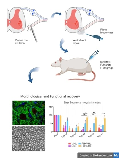
2.1. Experimental Animals and Groups

This study was approved by the Institutional Committee for Ethics in Animal Experimentation (Committee for Ethics in Animal Use—Institute of Biology—CEUA/IB/UNICAMP, proc. Nr. 4500-1, Campinas, SP, Brazil). All experiments were performed by the guidelines of the Brazilian College for Animal Experimentation. Forty adult 8-week-old female Lewis rats (LEW/HsdUnib) were housed with free access to food and water in a 12 h light/dark cycle. The animals were subjected to unilateral ventral root avulsion of the lumbar L4, L5, and L6 roots. Twelve weeks after injury, the animals were euthanized; the lumbar spinal cords and sciatic nerves were processed for light and transmission electron microscopy.
The animals were divided into 4 groups: (1) ventral root avulsion without reimplantation and vehicle treatment (VHL); (2) ventral root avulsion without reimplantation and treatment with dimethyl fumarate—15 mg/kg (DMF); (3) ventral root avulsion followed by root repair with fibrin biopolymer and vehicle treatment (FB+VHL); and (4) ventral root avulsion followed by root repair with fibrin biopolymer and treatment with DMF—15 mg/kg (FB+DMF; n = 10 for all groups). The contralateral (CTL) sciatic nerve and spinal cord side were used as the control.
DMF (242926-100 g, Sigma-Aldrich, Waltham, MA, USA) was diluted in 0.08% methylcellulose solution and administered daily for 4 weeks after injury at a dose of 15 mg/kg by gavage [29]; methylcellulose solution was administered as the vehicle. Both treatments started 30 min after the surgical procedures. Fibrin biopolymer derived from the venom of Crotalus durissus terrificus (Cdt), combined with buffalo fibrinogen, was applied at the injury site as follows: 3 μL of fibrinogen-rich cryoprecipitate derived from buffalo blood, 2 μL of calcium chloride, and 1 μL of a thrombin-like enzyme-enriched fraction extracted from Cdt venom [30,31,32,33,34,35]. FB components and application formulas are listed in its patent (BR1020140114327) and were kindly provided by the Center for the Study of Venoms and Venomous Animals (CEVAP, Brazil).

2.2. VRA and Lesioned Motor Root Reimplantation

Animals were induced to anesthesia with a combination of xylazine hydrochloride (Anasedan®, 10 mg/kg, Sespo Indústria e Comércio, Paulínia, SP, Brazil) and ketamine hydrochloride (Dopalen®, 50 mg/kg, Sespo Indústria e Comércio, Paulínia, SP, Brazil) at a ratio of 1:1.5, applied intraperitoneally. After loss in reflexes, the animals were kept in deep anesthesia with isoflurane (2%). A lubricating ophthalmic gel (Liposic, 2 mg/g; 0.2% w/w) was applied to the eyes, and the animals were kept on a heated pad during the surgical procedure.
To assess the spinal cord, the rat was put in a prone position, the back was shaved, and an incision was made parallel to the vertebral column from the lower thoracic region to the upper lumbar segment. The musculature was released for visualization of the thoracic and lumbar vertebrae. A laminectomy of approximately two vertebrae was performed (T13-L1) so that the lumbar intumescence was exposed. The dissection of the dura mater and the denticulate ligament was performed. After exposing the spinal cord, the ventral roots associated with lumbar segments were exposed. Avulsion was performed unilaterally on the ventral lumbar roots referring to L4, L5, and L6 spinal segments with the aid of fine forceps. The animals in the reimplanted groups had the ventral lumbar roots repositioned to their original sites before avulsion immediately after the injury and reimplanted using FB. The stability of the root fixation was observed to evaluate the success of the repair. After the surgical procedures, the spinal cord roots were repositioned to their original position, and the musculature, fascia, and skin were sutured in layers.
Surgical recovery was performed in a postsurgical environment with heating for 4 h. Tramadol hydrochloride (Germed Farmacêutica Ltda, Hortolândia, SP, Brazil) was administered at a dose of 0.5 mg/kg by gavage every 24 h until the third postoperative day. If any signs of pain (prostration, ruffled fur, decreased food consumption) and discomfort were detected, the administration of tramadol hydrochloride was extended appropriately.

2.3. Specimen Preparation

Twelve weeks post-lesion, animals were euthanized with a lethal dose of a combination of xylazine hydrochloride (Anasedan®, 30 mg/kg, Sespo Indústria e Comércio, Paulínia, SP, Brazil) and ketamine hydrochloride (Dopalen®, 150 mg/kg, Sespo Indústria e Comércio, Paulínia, SP, Brazil) at a ratio of 1:1.5 applied intraperitoneally, and the vascular system was transcardially perfused.
For immunofluorescence analysis, after rinsing with buffered saline (PB), animals were perfused with 4% formaldehyde in PB 0.1 M (pH 7.4). The lumbar intumescence and a 1 cm mid-thigh segment of the sciatic nerve (ipsi and contralateral) were collected and postfixed for 24 h at 4 °C in the same fixative. They were then kept in a solution of 10%, 20%, and 30% sucrose for 24 h each at 4 °C. Samples were embedded in Tissue-Tek (Miles Inc., Oro Valley, AZ, USA) and frozen at −35 to −40 °C. Twelve micrometer-thick cross-sections of the spinal cord and longitudinal sections of the sciatic nerves were obtained in a cryostat (Micron HM25) and subsequently stored at −20 °C until use.
For morphometry and transmission electron microscopy, phosphate buffer 0.1 M (pH 7.4) saline was perfused through the left ventricle, followed by Karnovski fixative solution (glutaraldehyde 2% + paraformaldehyde 1%) in the same buffer (pH 7.4). The lumbar spinal cord and a 1 cm mid-thigh segment of the sciatic nerve (ipsi and contralateral) were collected and stored for 7 days in the same fixative solution at 4 °C. The specimens were then trimmed into smaller segments and separated between contra and ipsilateral, osmicated (3 h for nerve segments and 4 h for spinal cord segments), dehydrated through an ethanol series, and embedded in Durcupan ACM (Fluka, Steinheim, Switzerland).

2.4. Immunofluorescence

The slides containing the spinal cord and sciatic nerve cross-sections were initially acclimatized, washed with phosphate buffer (0.01 M CP) for 3 × 5 min, and incubated with blocking solution (bovine serum albumin—3% BSA + PB 0.1 M) for 45 min. Then, the slides were incubated for 3 h with anti-synaptophysin, anti-neurofilament, and anti-NeuN antibodies (Table 1) in an incubation solution (BSA 1% + Triton 0.2% + PB 0.1 M) and kept in a humid chamber at room temperature. They were then washed with PB 0.01 M (3 × 5 min), and the respective secondary antibodies were added (Alexa 488 or 594—1:400) and diluted in incubation solution for 45 min. Slides were washed once more in PB 0.01 M (3 × 5 min) and mounted in glycerol + DAPI (1:1000)/PB 0.01 M (3:1).
Immunostained specimens were observed and photographed under a fluorescence microscope (Leica DM 5500B, Wetzlar, Germany) coupled to a high-sensitivity camera (Leica DFC-345FX) at 20× or 40× magnification, depending on the analysis; three representative images were obtained for each animal on each side (injured/ipsilateral and uninjured/contralateral) of lamina IX of Rexed at the spinal cord ventral column and the sciatic nerve.
For quantification, the integrated density of pixels was measured, which represents the intensity of immunostaining, using ImageJ software (version 1.51 h). For all images at 20× magnification, the entire image was used for analysis, which is indicative of the microenvironment; for the anti-synaptophysin marker, the analysis was performed around the Neu-N-positive neurons at 40× magnification to assess the synaptic preservation of each motoneuron. The results are expressed as the ratio of the ipsilateral/contralateral side for each animal.

2.5. Sciatic Nerve Regeneration

Resin blocks containing sciatic nerve segments were trimmed, and semithin sections (500 nm) were obtained in an ultramicrotome (Leica Ultracut UCT Ultramicrotome) and stained with Sudan black for examination under light microscopy (Leica DM 5500B). Each nerve was photographed at 10× magnification to obtain the total area using ImageJ software (version 1.51 h). Then, we obtained 6 images per animal at 100× magnification, sampling at least 15% of each nerve cross-section. These images were used for counting and measuring myelin fibers (Adobe Photoshop CS4 extended, Adobe Systems Incorporated, San Jose, CA, USA). For that, the following parameters were used: external diameter—myelin fiber diameter (FD), internal diameter—axon diameter (AD), myelin sheath thickness (MST), and ‘g’ ratio (GR), which is the ratio of axon diameter divided by the fiber diameter and indicates the degree of myelination (normal-, hypo-, or hyper-myelination).

2.6. Synaptic Analysis of the Ultrathin Sections

The resin blocks containing spinal cord segments were trimmed, and semithin sections (500 nm) were obtained in an ultramicrotome (Leica Ultracut UCT Ultramicrotome) and stained with toluidine blue for examination under light microscopy (Leica DM 5500B) to identify the presence of motor neurons at lamina IX of Rexed. Then, ultrathin sections (70 nm) from the L4–L6 segments were collected on formvar-coated single-slot copper grids, contrasted with uranyl acetate 4% and lead citrate, and examined under a Tecnai G2 Spirit BioTwin (FEI, Eindhoven, The Netherlands) transmission electron microscope operating at 80 KV. This procedure was performed for the ipsilateral side of all groups; the contralateral side used as the control was obtained from the VHL group.
Neurons with large cell bodies in the nuclear plane were identified as α-motoneurons by the presence of at least one C-type (cholinergic) nerve terminal. The neuronal membrane of these cells was then sequentially digitized at a magnification of 11,000× using a video camera (Eagle, FEI, Eindhoven) connected to a computerized system. The images were then mounted together in Corel Draw software (version 19), and the total perimeter of the neurons was measured using ImageJ software (version 1.51 h).
The synaptic terminals apposing the motoneuron membrane were identified and classified as inhibitory (F-type—with flattened synaptic vesicles), excitatory (S-type—with spherical synaptic vesicles), and cholinergic (C-type), identified by a subsynaptic cistern with continuity with the rough endoplasmic reticulum [36,37]. After classification, they were counted and measured using the ImageJ program (version 1.51 h) to quantify synaptic coverage and proportion of buttons/100 μm of membrane; the distance between consecutive nerve buttons was also measured (GAPs). In total, two neurons per animal in the CTL, VHL, and DMF groups (n = 10 neurons for each group) and four neurons per animal in the FB+VHL and FB+DMF groups (n = 20 neurons for each group) were examined.

2.7. Motor Function Recovery Assessment

To evaluate the recovery of motor function, the Catwalk System (CatWalk XT 5.0, Noldus, Leesburg, VA, USA) was used [29,38]. The CatWalk system allows the detailed evaluation of gait recovery by using illuminated footprints and provides data such as size, pressure, width, and length of paw prints. One baseline motor measurement (before injury) was obtained as normal values for each animal. After the injury, assessments were performed every 18 days for 12 weeks. In each evaluation, 4 sessions were recorded per animal. An average was calculated for each baseline value and each of the days analyzed per animal and then for each experimental group ± standard error of the mean (SEM).

2.8. Statistical Analysis

From the numerical results from all the analyses, the mean and standard deviation were calculated for each group, and Prism 6 software was used. Immunofluorescence, nerve area, and myelin fiber count data were evaluated by the analysis of variance method—one-way ANOVA followed by Tukey’s post-test. Myelinated fiber morphometry data were analyzed using frequency distribution. The functional analysis data (walking track test) were evaluated by the analysis of variance method—two-way ANOVA and Mann–Whitney. Significance levels were assumed when * p < 0.05, ** p < 0.01, *** p < 0.001, and **** p < 0.0001 for all analyses.

3. Results

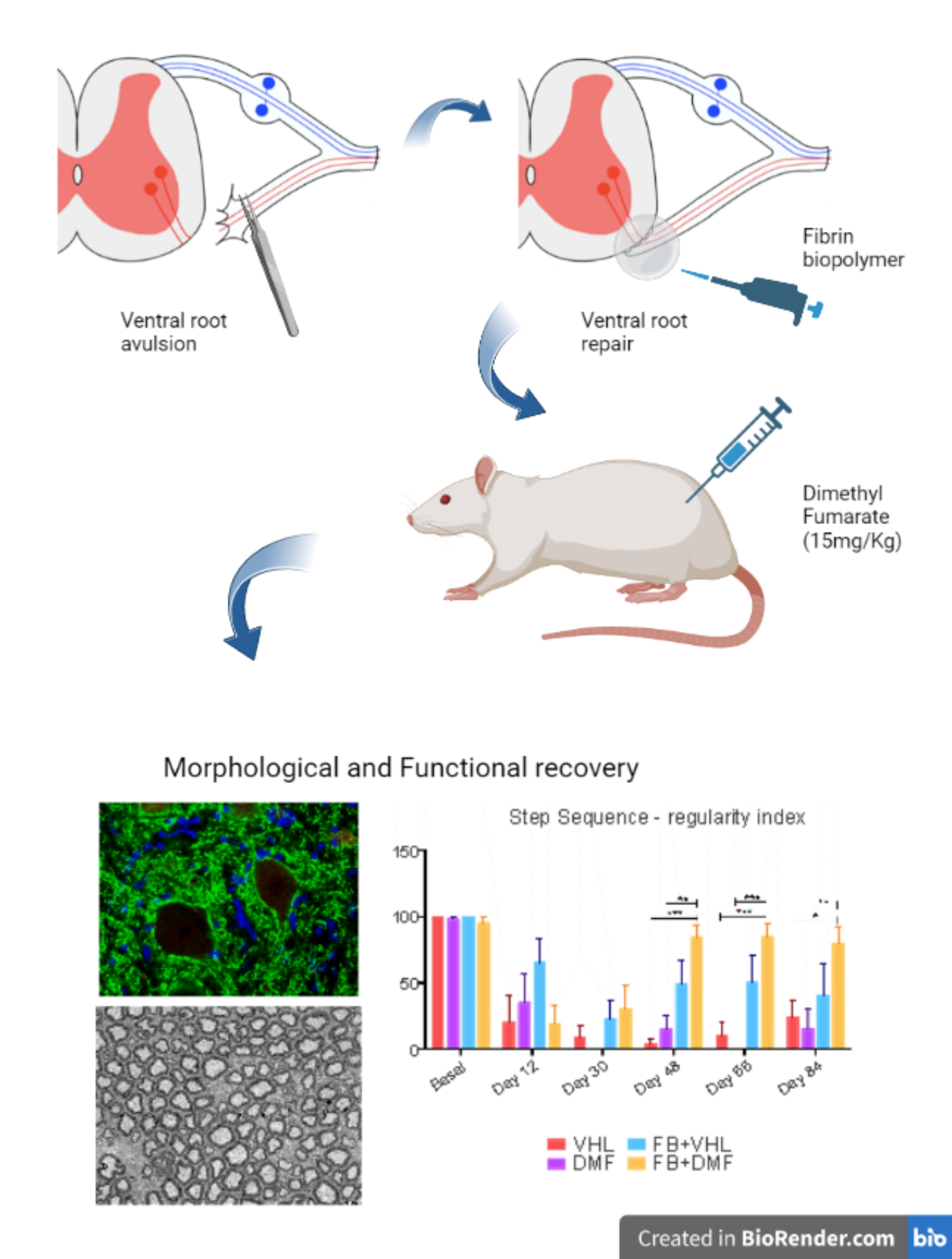
3.1. Synapse Preservation after Ventral Root Avulsion and DMF Treatment

To evaluate the overall synaptic preservation 12 weeks after avulsion/repair of the motor nerve roots, an anti-synaptophysin antibody was used. Photomicrographs were taken in the region of Rexed’s lamina IX at 20× magnification to analyze the microenvironment and 40× magnification to analyze the surface of the motoneurons. Immunostaining revealed baseline reactivity for the contralateral side of all groups for both magnifications.
We observed lower synaptic density (40% decrease) after injury, indicating an intense retraction of terminals in apposition to axotomized motoneurons both in the neuropile and on the cell surface. DMF treatment provided synaptic preservation in the neuropile (63%) but not at the motoneuron surface level (54%) since coverage analysis showed no difference with the VHL group. However, reimplantation of lesioned roots significantly improved preservation (70%) both in the microenvironment and on the motoneuron surface (Figure 1, Table 2).

3.2. Ultrastructural Analysis of Alpha Motoneurons

Ultrastructural analysis of the alpha motoneurons, present in the CTL group, revealed the expected perimeter of the neuronal membrane (140 μm on average), combined robust synaptic coverage (63%), and a significant proportion of synaptic buttons per 100 μm of membrane (approximately 40 buttons in apposition to the soma membrane). In the groups without repair of the lesioned root, we observed changes in the ultrastructure of the neuronal coverage of alpha motoneurons, where the presynaptic terminals were partially or completely retracted. We observed many glial projections, identified as astrocytes due to their low electron density, also in contact with the neuronal membrane, which filled the space between presynaptic terminals and the postsynaptic membrane. After injury, there was a reduction in neuronal perimeter in the VHL group (105 μm). For all groups that received any of the interventions, we observed that the perimeter was close to the value observed in the contralateral group, being 135 μm for the DMF group, 148 μm for the FB+VHL group, and 141 μm for the FB+DMF group. For synaptic coverage, there was an intense loss after axotomy, only 43% remaining in the VHL group and 51% in the DMF group when compared to the CTL group (63%). However, both groups that had their roots reimplanted, FB+VHL and FB+DMF, showed preservation of synaptic coverage, which was greater than 60%. A similar result was observed about the number of buttons apposed to every 100 μm of the neuronal membrane. After injury, the VHL group displayed an average of 23 buttons, and the DMF group showed about 30 buttons. Both groups submitted to root reimplantation preserved more than 35 buttons, which was closer to the values of the CTL group (40 buttons on average) (Figure 2, Table 3).
Furthermore, we obtained the synaptic coverage values and the number of buttons/100 μm for each type of terminal (F, S, and C). After injury, we observed a reduction in the synaptic coverage of F-type inhibitory terminals in all groups analyzed, except in the FB+VHL group. Interestingly, this group was similar to the CTL group. Regarding the S-type excitatory terminals (glutamatergic), we observed a reduction in synaptic coverage only in the VHL group when compared to all other groups. C-type cholinergic terminals showed no differences among groups. For the number of normalized buttons per 100 μm of neuronal membrane, we observed a reduction in synaptic coverage of F-type terminals after injury in the VHL and the FB+DMF groups. Both groups that received the therapies separately showed no differences from the CTL group. For S-type terminals, we observed a reduction only in the VHL group when compared to all other groups. Again, C-type cholinergic terminals showed no differences among groups (Figure 2, Table 4).

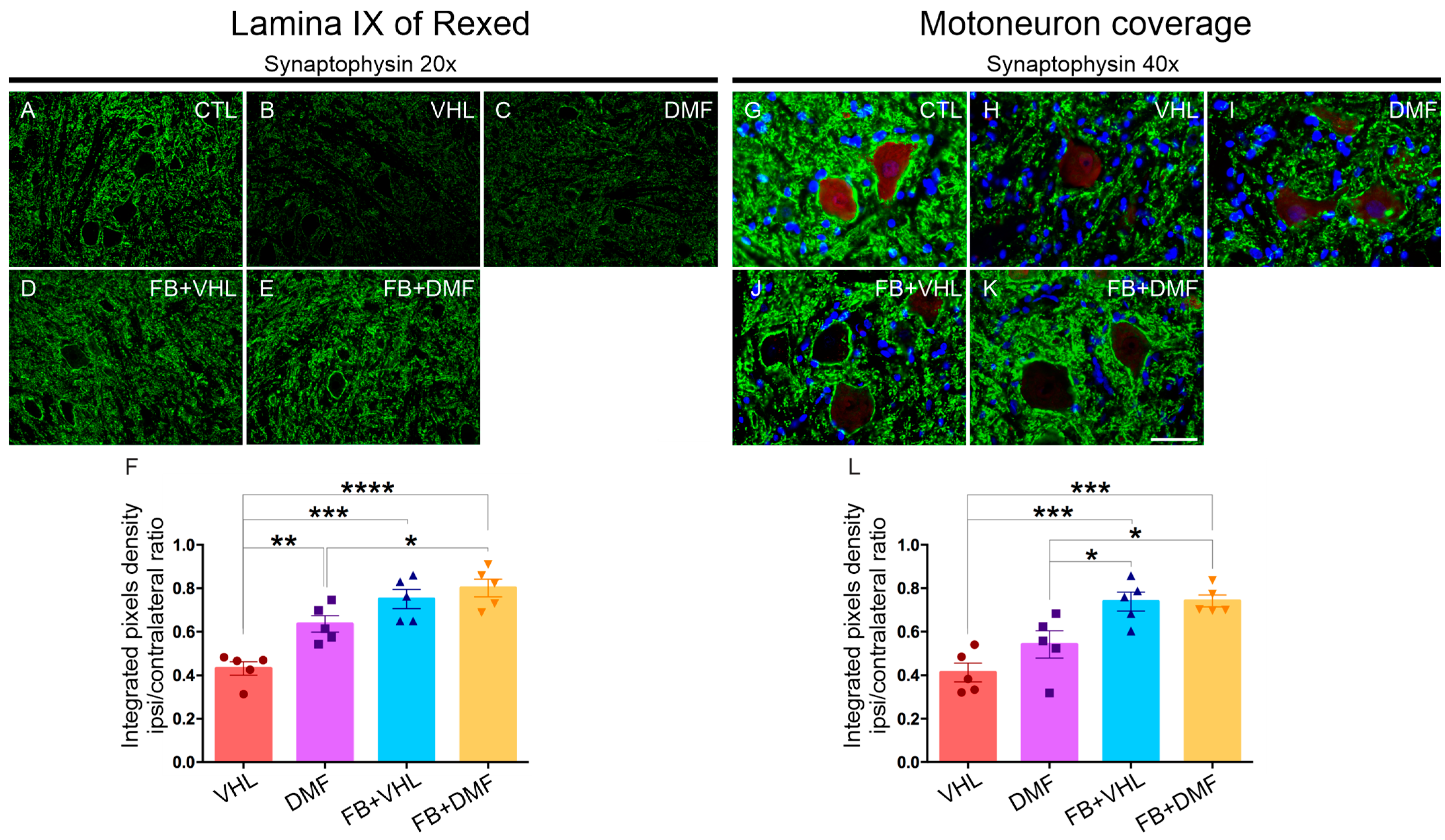
3.3. Nerve Terminals Distribution

The gaps between the terminals were measured, and their frequency distribution in 0.5 μm intervals was evaluated. These intervals were identified as filled with astroglial projections due to the low electron density. In normal neurons (CTL group), the terminals were grouped, with a large number of smaller intervals between the inputs, the largest being 4.5 μm. However, in the VHL group, a reduction in smaller intervals and an increase in the frequency of longer gaps between terminals were observed, with the longest gaps exceeding 15.0 μm. The DMF group presented intervals that increased to 8.5 μm. The FB groups, with lesioned root reimplanted, presented intervals very close to the CTL group (Figure 3).
After the injury (VHL group), a reduction in the number of GAPs was observed, with approximately 24 GAPs, and, for the DMF group, approximately 33 GAPs. However, we observed a greater number of GAPs in the CTL group (42 GAPs) and in the groups that were reimplanted, with 42 GAPs for the FB+VHL group and 38 GAPs for the FB+DMF group. These values indicate a better distribution of clusters of synaptic buttons after root reimplantation and DMF therapy (Figure 3).

3.4. Neurons with Incomplete Regeneration

Throughout the present study, we observed that many motoneurons in the FB+DMF group, which received both therapies, showed very similar characteristics to those of the VHL group. Thus, the regeneration of these neurons was incomplete or insufficient, possibly blocked by the glial scar. Synaptic coverage in motoneurons in the CTL group was 63%, while, in the VHL group, it was 43%. For neurons that showed insufficient regeneration, synaptic coverage was about 30%. Furthermore, the normalization of synaptic buttons per 100 μm of the neuronal membrane was also greatly reduced. For the CTL group, we observed an average of 40 buttons; for the VHL group, we observed 23 buttons; and, for neurons with insufficient regeneration, we observed only 17 buttons per 100 μm of the neuronal membrane. These results align with the distribution and size of gaps (GAPs) between synaptic terminals, where we observed that these neurons presented a distribution similar to that of the VHL group and were significantly different from the CTL group. There was a great decrease in the frequency of small intervals and an increase in the number of larger intervals, with several exceeding 15.0 μm (Figure 4, Table 5).

3.5. Nerve Preservation

After 12 weeks of injury, the non-reimplanted groups showed a significant reduction in the immunostaining of the fibers by the neurofilament, which labels intermediate filaments present in the axon cytoskeleton, when compared to the reimplanted and contralateral groups. In parallel with this analysis, we observed that the number of myelinated axons was reduced in both non-reimplanted groups and the FB+VHL group. In contrast, in the FB+DMF group, which received both therapies, there was a recovery in the number of fibers, which approached the values obtained in the CTL group, demonstrating axon sprouting and regrowth towards the target muscles. However, we did not observe a reduction in the area of the sciatic nerves in any of the experimental groups, indicating that the loss in axons in the endoneurial environment was compensated with extracellular matrix synthesis, mostly collagen fibers (Figure 5, Table 6).

3.6. Sciatic Nerve Histomorphometry

3.6.1. Myelinated Axon Diameter (FD)

The nerves in the CTL group presented myelin fibers with diameters ranging from 2 μm to 14 μm, distributed in a bimodal manner, with peaks at 4 and 7 μm. The VHL group has a distribution of fibers with smaller dimensions, from 1 to 14 μm, with a predominance of fibers with 4, 7, and 8 μm. The DMF group showed a distribution from 2 to 15 μm, with a predominance of fibers with 7 μm and a reduction in fibers within the 4 μm range. In both SF groups, a distribution of fibers between 2 and 15 μm was observed, distributed in a bimodal way as in the CTL group, with a predominance of fibers within 4 and 7 μm. Thus, the combination of therapeutic approaches was able to preserve the distribution of large and small fibers closer to normal, demonstrating a regenerative effect (Figure 6A–E, Table 7).

3.6.2. Axon Diameter (AD)

We observed that the frequency distribution of axonal diameter among all avulsed groups was similar to each other and differed from that in the CTL group. This similarity is due to the relative increase in the frequency of smaller-diameter fibers, confirming the loss in larger-caliber fibers, compatible with motor axons. The nerves in the CTL group had myelinated axons with minimum diameters ranging from 2 to 12 μm. For the VHL group, myelin axons varied between 1 and 12 μm and between 2 and 11 μm for the groups where there was some therapeutic intervention (Figure 6F–J, Table 7).

3.6.3. Myelin Sheath Thickness (MST)

We observed that the MST frequency distribution among all avulsed groups was similar to each other and differed from that of the control group. The shift in the histogram pattern to the left is highly suggestive, indicating loss in large-caliber fibers, compatible with axons of avulsed motoneurons. Thus, the avulsion-alone group showed an increase in fibers of 0.4, 0.6, and 0.8 μm to almost 20% for each, while, in the other groups, this percentage was lower. Furthermore, all groups that received any of the therapeutic strategies had a higher percentage of fibers within the 1.4 μm (approximately 7%) range compared to the lesion-only group (less than 5%), indicating the presence of larger and more preserved fibers (Figure 6K–O, Table 7).

3.6.4. ’g’ Ratio

The ‘g’ ratio determines whether the myelin sheath thickness is adequate for the axonal diameter so that normal myelinated axons are expected to display 0.7 to 0.8 ratio (in the graph, such an interval is expressed in the column of 0.8). Therefore, the distribution at 0.9 indicates thinly myelinated fibers, possibly with reduced speed in action potential transmission. Interestingly, an increased frequency of fibers in the 0.9 range in the VHL group, but not in the groups that received any of the therapeutic approaches, indicates that regeneration occurred in those groups (Figure 6P–U, Table 7).

3.7. Motor Functional Recovery

Recovery of motor function is a result of neuronal survival and regeneration of axons to the target muscle. As demonstrated before by our group Kempe et al. 2020 [29], we observed that considerable early improvements around day 48 were obtained in the FB+DMF group, while other groups showed poor or delayed recovery regarding nerve functionality, depicted by the peroneal function index. The same positive results were observed regarding the strength (max contact max intensity) and motor coordination (step sequence and base of support hind paws) in which animals almost reached basal values (Figure 7).

4. Discussion

Motor root avulsion is characterized by root pullout from the spinal cord surface, causing disconnection of spinal motoneurons from muscle fibers, resulting in neuronal loss, degeneration of nerve fibers, and paralysis of the target skeletal muscle [14,39,40,41,42]. This experimental model is of relevance since it allows the study of structural changes that occur in the spinal cord microenvironment, such as neuronal degeneration and neurotransmission circuitry modifications, as well as changes in the affected peripheral nerve. After injury, extensive synaptic plasticity occurs in the cell body and proximal dendrites of the motoneuron in response to ventral root avulsion, with retraction, reduction, or elimination of presynaptic terminals apposed to injured neurons [3,9,11,13,43,44,45,46,47,48]. These events may reduce synaptic transmission to low levels or even block it in the acute post-injury period [26,27,49], probably also affecting nearby spinal segments that have not been directly affected by the primary lesion [42,50]. In turn, such an imbalance in synaptic connections impairs voluntary movements and can cause neuropathic pain and/or hyperalgesia. If injury repair and reinnervation do not occur, synaptic transmission can be abolished totally [26,49] and irreversibly [43].
Our results are in line with the literature, both regarding the lesion-only group without any intervention, where synapse loss was noted, as well as in the group where there was reimplantation using FB, restoring the CNS/PNS anatomical continuity [14,41]. Treatment with DMF without reimplantation seems to have a minor effect on the stabilization of spinal circuits, although significant preservation concerning excitatory circuits was previously observed by immunohistochemistry [29]. Therefore, it is clear that the early repair of the lesioned roots is crucial for both motoneuron survival and the preservation of intrinsic spinal circuitry. Further, the early repair positive outcome can be enhanced with the combination of a neuroprotective drug such as DMF. Herein, we demonstrate marked preservation of the spinal circuits by synaptophysin immunolabeling, which was confirmed by the ultrastructural evaluation. This result is strongly related to the reduction in inflammation, possibly due to the reduction in microglial and astroglial activation [29], since these cells have a role in the retraction and elimination of synaptic terminals after injury [51,52,53,54]. This may, in turn, aid in the recovery of motor coordination and function once regenerating motor axons reach the target muscle.
Regarding the ultrastructural organization of synaptic terminals that support the neuronal membrane, we observed that reimplantation alone or associated with DMF seems to provide greater preservation of these terminals and decrease the space between the remaining terminals, possibly preserving the clustering formation. This is in line with a previous work of our group, indicating that the combination of DMF and root reimplantation preserves immunoreactivity to pre-synaptic markers of excitatory and inhibitory inputs present in the motor nucleus [29].
The present work further investigated the synaptic preservation by using transmission electron microscopy, thus providing the typing of the synaptic terminals that remained in apposition to the axotomized alpha motoneuron membrane. Thus, we observed that there is intense elimination of excitatory terminals after injury. This effect is believed to decrease neuronal transmission activity and prevent excitotoxicity caused by glutamate, which can activate apoptotic and necrotic pathways [23,55]. However, all therapies used, alone or in combination, showed preservation of such inputs, demonstrating that they possibly contribute to a pro-regenerative state of the neurons. In fact, Parodi and colleagues [56] pointed out that, in mice with experimental autoimmune encephalomyelitis (EAE), a model for multiple sclerosis, the neuroprotective effect of DMF occurs directly on presynaptic terminals through immunomodulation of glutamate release and indirectly, post-synaptic, through the modulation of microglial function. Kempe and colleagues [29] observed an intense loss in inhibitory inputs through immunostaining with anti-GAD65 after avulsion, and significant preservation of such inputs after reimplantation and DMF treatment. In line with that, the present data based on the typing of the synaptic buttons indicated that they remained close to the values obtained for the uninjured neurons. Thus, the reimplantation by itself or in combination with DMF seems to allow the axotomized neurons to remain wired to the motor nucleus in a similar fashion to the uninjured counterpart.
After a peripheral nerve injury, several events are triggered in order to allow repair and reconnection to the target. Thus, the distal stump develops Wallerian degeneration, with degeneration of the myelin sheath and injured axons. In the case of ventral root avulsion, axons initially regenerate within the CNS and cross the glial scar formed at the edge of the spinal cord, which plays a fundamental role in the failure of central axons to regenerate efficiently as it acts as a physical barrier to their passage. In a second moment, the axons must regenerate towards the reinnervation of the target, through the PNS. Macrophages and Schwann cells assist in these steps, clearing debris and providing interleukins, neurotrophic factors [57,58,59,60], and an extracellular matrix [61], which are fundamental to the regenerative process. Thus, lesioned root repair with FB and pharmacological treatment with DMF has the potential for inducing the regeneration of nerve fibers due to their neuroprotective and immunomodulatory properties. In particular, through the control of gliosis [29], reduction or decrease in the glial scar formation can be achieved, combined with stimulation of Schwann cells in the nerve, allowing enhanced regeneration of myelinated fibers.
The present results show a marked decrease in the number of large, myelinated axons after ventral root avulsion. When therapeutic interventions were used, there was a restoration of a number of fibers, indicating the occurrence of regeneration of motor axons. Thus, due to the ability of DMF to act on Schwann cells [56,62,63], we suggest that the treatment leads to protection and substantial regenerative response. The FB, in turn, by re-establishing the CNS/PNS connection, allows a similar positive outcome [14,64]. Therefore, when both therapies were combined, more robust results in protection and regeneration were observed.
Of note, it has already been shown that the administration of neurotrophic factors after spinal cord injury in rodents and primates [65,66] promotes axonal regeneration, inducing increased expression of neurofilaments necessary for the elongation of the regrowing axons [67]. Thus, our results demonstrated greater expression of neurofilament after the association of therapies when compared to that observed in animals that did not receive treatment. This is probably related to the fact that DMF treatment induces the production of neurotrophic factors, such as BDNF, GDNF, and NT3 [68]. The present morphometric results show that the frequency distribution pattern of fibers changes between uninjured and injured nerves, demonstrated by the large increase in thin fibers with a ‘g’ ratio of 0.9, which is in part the result of the interactions between Schwann cells and growing axons [69,70,71]. All groups that received any of the therapies presented improved “g” ratio frequency distribution, close to the value found in nerves that were not injured. Together with the “g” ratio, the myelin sheath thickness (MST) is a parameter closely related with Schwann cell activity. Our results indicate that the association between early repair with FB and DMF accelerates the regrowth and myelination of large-diameter fibers, with their distribution being closer to the uninjured nerve. Regarding the diameter of the axons, fibers with smaller dimensions after injury were observed in all experimental groups when compared to the uninjured side, which is compatible with what is described in the literature [72]. Regarding large fibers, since the ventral root avulsion does not affect sensitive axons, the presence of large axons is expected even in the avulsion-alone group. Overall, the frequency distribution of such fibers showed improvements after reimplantation and DMF therapy.
Although DMF alone induces nerve fiber regeneration, the long-lasting effects observed herein were very robust. DMF increases the expression of the enzyme HO-1 (heme oxygenase), which has cytoprotective and anti-inflammatory effects, participating in the anti-oxidative pathway [63,73,74,75], which may support the long-term regenerative response. Szepanowski and colleagues [76], using a model of sciatic nerve crush, related the increase in the NRF2 factor with the increase in the HO-1 enzyme and glutathione. These results are in line with what was observed in the current work, where greater recovery of fibers, combined with a close-to-normal ‘g’ ratio, was achieved.
The functional improvement suggests that the repaired nerve fibers were successful in crossing the glial scar, which functions as a physical barrier at the interface between the CNS and the PNS. Furthermore, peripheral regeneration depends on the long-term survival of the injured neuron and the entry of regenerating axons into the endoneurial tubes, which provide guided axon growth [77]. Thus, given that both DMF and FB have neuroprotective and immunomodulatory potential, as previously observed by Kempe and colleagues [29], the enhanced motor recovery observed herein demonstrates that newly formed motor axons reach target muscles and restore, to a certain degree, the motor coordination.
DMF is capable of providing neurotrophic factors, such as BDNF, GDNF, and NT3 [68,78], these neurotrophic molecules being directly related to axonal elongation after peripheral nerve injury and during development [79] and also having been associated with motor recovery after cerebral ischemia [80]. Also, Szepanowski and colleagues [76] related the improvement in the grip strength test with the re-establishment of the ‘g’ ratio, which indicates nerve fiber functionality, as observed in our work. Furthermore, these authors observed an increase in the expression of antioxidant enzymes and the NRF2 factor, showing a decrease in malondialdehyde, a marker for lipid stress [68,76]. In addition, the control or reduction in inflammatory responses provides a pro-regenerative environment that allows improvements in functional performance [29,81,82].

5. Conclusions

Pharmacological treatment with DMF associated with lesioned root reimplantation showed neuroprotective and anti-inflammatory capacity. Thus, we obtained a preservation of spinal circuits combined with a close-to-normal clustering of inputs in direct contact with alpha motoneurons, which may have contributed to the motor recovery over time. Therefore, the combined therapy of DMF with root replantation with fibrin biopolymer may be further studied, aiming at future therapeutic application.

Author Contributions

P.R.G.K. designed and performed the experiments, interpreted the results, and wrote the original draft. M.V.d.C. performed transmission electron microscopy and interpreted the results. V.C.K. performed morphometry analysis. B.B. and R.S.F.J. provided the heterologous fibrin sealant and revised the original draft. A.L.R.d.O. designed the experiments, interpreted the results, wrote the original draft, and obtained financial support. All authors have read and agreed to the published version of the manuscript.

Funding

This work was supported by the São Paulo Research Foundation—FAPESP (grant numbers 2016/25478-9, 2018/25845-7, 2018/05006-0, and 2021/11936-3 - Center for Translational Sciences and Biopharmaceuticals Development). A.L.R.d.O. is a CNPq 1C fellow researcher (303050/2021-7); R.S.F.J. is a CNPq PQ1C fellow researcher (303224/2018-5); B.B. is a CNPq PQ-2 fellow researcher (306339/2020-0).

Institutional Review Board Statement

Not applicable.

Data Availability Statement

The datasets generated during the current study are available from the corresponding author upon reasonable request.

Conflicts of Interest

The authors declare no conflict of interest.

References

  1. Bonney, G.; Birch, R.; Jamieson, A.M.; Eames, R.A. Experience with vascularized nerve grafts. Clin. Plast. Surg. 1984, 11, 137–142. [Google Scholar] [CrossRef] [PubMed]
  2. Carlstedt, T.P. Spinal nerve root injuries in brachial plexus lesions: Basic science and clinical application of new surgical strategies. A review. Microsurgery 1995, 16, 13–16. [Google Scholar] [CrossRef] [PubMed]
  3. Carlstedt, T. Experimental studies on surgical treatment of avulsed spinal nerve roots in brachial plexus injury. J. Hand Surg. Br. 1991, 16, 477–482. [Google Scholar] [CrossRef]
  4. Carlstedt, T.; Cullheim, S.; Risling, M.; Ulfhake, B. Nerve fibre regeneration across the PNS-CNS interface at the root-spinal cord junction. Brain Res. Bull. 1989, 22, 93–102. [Google Scholar] [CrossRef]
  5. Carlstedt, T.; James, N.; Risling, M. Surgical reconstruction of spinal cord circuit provides functional return in humans. Neural Regen. Res. 2017, 12, 1960–1963. [Google Scholar] [CrossRef] [PubMed]
  6. Carlstedt, T.; Linda, H.; Cullheim, S.; Risling, M. Reinnervation of hind limb muscles after ventral root avulsion and implantation in the lumbar spinal cord of the adult rat. Acta Physiol. Scand. 1986, 128, 645–646. [Google Scholar] [CrossRef]
  7. Carlstedt, T.; Risling, M.; Linda, H.; Cullheim, S.; Ulfhake, B.; Sjogren, A.M. Regeneration after spinal nerve root injury. Restor. Neurol. Neurosci. 1990, 1, 289–295. [Google Scholar] [CrossRef]
  8. Cullheim, S.; Carlstedt, T.; Hallin, R.; Linda, H.; Risling, M. Motor neurons regenerate axons after CNS-injury. Surgery in cases of rotavulsion restore motor functions. Lakartidningen 1996, 93, 4537–4541. [Google Scholar]
  9. Cullheim, S.; Carlstedt, T.; Linda, H.; Risling, M.; Ulfhake, B. Motoneurons reinnervate skeletal muscle after ventral root implantation into the spinal cord of the cat. Neuroscience 1989, 29, 725–733. [Google Scholar] [CrossRef]
  10. Cullheim, S.; Risling, M. Observations on the morphology and the axon conduction velocity of axotomized and regenerating sciatic motoneurons in the kitten. Exp. Brain Res. 1982, 45, 428–432. [Google Scholar] [CrossRef]
  11. Cullheim, S.; Wallquist, W.; Hammarberg, H.; Linda, H.; Piehl, F.; Carlstedt, T.; Risling, M. Properties of motoneurons underlying their regenerative capacity after axon lesions in the ventral funiculus or at the surface of the spinal cord. Brain Res. Brain Res. Rev. 2002, 40, 309–316. [Google Scholar] [CrossRef] [PubMed]
  12. Koliatsos, V.E.; Price, W.L.; Pardo, C.A.; Price, D.L. Ventral root avulsion: An experimental model of death of adult motor neurons. J. Comp. Neurol. 1994, 342, 35–44. [Google Scholar] [CrossRef]
  13. Cullheim, S.; Carlstedt, T.; Risling, M. Axon regeneration of spinal motoneurons following a lesion at the cord-ventral root interface. Spinal Cord. 1999, 37, 811–819. [Google Scholar] [CrossRef] [Green Version]
  14. Vidigal de Castro, M.; Barbizan, R.; Seabra Ferreira, R., Jr.; Barraviera, B.; Leite Rodrigues de Oliveira, A. Direct Spinal Ventral Root Repair following Avulsion: Effectiveness of a New Heterologous Fibrin Sealant on Motoneuron Survival and Regeneration. Neural Plast. 2016, 2016, 2932784. [Google Scholar] [CrossRef] [Green Version]
  15. Chan, S.Y.; Kuo, C.W.; Liao, T.T.; Peng, C.W.; Hsieh, T.H.; Chang, M.Y. Time-course gait pattern analysis in a rat model of foot drop induced by ventral root avulsion injury. Front. Hum. Neurosci. 2022, 16, 972316. [Google Scholar] [CrossRef]
  16. Huang, Y.; Zhang, X.; Huang, Q.; Dou, Y.; Qu, C.; Xu, Q.; Yuan, Q.; Xian, Y.F.; Lin, Z.X. Quercetin enhances survival and axonal regeneration of motoneurons after spinal root avulsion and reimplantation: Experiments in a rat model of brachial plexus avulsion. Inflamm. Regen. 2022, 42, 56. [Google Scholar] [CrossRef]
  17. Torok, D.G.; Fekecs, Z.; Pajer, K.; Pinter, S.; Nogradi, A. The use of a detailed video-based locomotor pattern analysis system to assess the functional reinnervation of denervated hind limb muscles. J. Neurosci. Methods 2022, 365, 109398. [Google Scholar] [CrossRef]
  18. Zhong, K.; Huang, Y.; Zilundu, P.L.M.; Wang, Y.; Zhou, Y.; Yu, G.; Fu, R.; Chung, S.K.; Tang, Y.; Cheng, X.; et al. Motor neuron survival is associated with reduced neuroinflammation and increased autophagy after brachial plexus avulsion injury in aldose reductase-deficient mice. J. Neuroinflamm. 2022, 19, 271. [Google Scholar] [CrossRef]
  19. Eggers, R.; de Winter, F.; Tannemaat, M.R.; Malessy, M.J.A.; Verhaagen, J. GDNF Gene Therapy to Repair the Injured Peripheral Nerve. Front. Bioeng. Biotechnol. 2020, 8, 583184. [Google Scholar] [CrossRef] [PubMed]
  20. Eggers, R.; de Winter, F.; Smit, L.; Luimens, M.; Muir, E.M.; Bradbury, E.J.; Tannemaat, M.R.; Verhaagen, J. Combining timed GDNF and ChABC gene therapy to promote long-distance regeneration following ventral root avulsion and repair. FASEB J. Off. Publ. Fed. Am. Soc. Exp. Biol. 2020, 34, 10605–10622. [Google Scholar] [CrossRef] [PubMed]
  21. Gunther, M.; Skold, M.K. Temporal gene expression changes after acute and delayed ventral root avulsion-reimplantation. Restor. Neurol. Neurosci. 2020, 38, 23–40. [Google Scholar] [CrossRef] [PubMed]
  22. Kreutzberg, G.W. Acute Neural Reaction to Injury; Springer: Berlin/Heidelberg, Germany, 1982; pp. 57–69. [Google Scholar]
  23. Linda, H.; Shupliakov, O.; Ornung, G.; Ottersen, O.P.; Storm-Mathisen, J.; Risling, M.; Cullheim, S. Ultrastructural evidence for a preferential elimination of glutamate-immunoreactive synaptic terminals from spinal motoneurons after intramedullary axotomy. J. Comp. Neurol. 2000, 425, 10–23. [Google Scholar] [CrossRef] [PubMed]
  24. Oliveira, A.L.; Thams, S.; Lidman, O.; Piehl, F.; Hokfelt, T.; Karre, K.; Linda, H.; Cullheim, S. A role for MHC class I molecules in synaptic plasticity and regeneration of neurons after axotomy. Proc. Natl. Acad. Sci. USA 2004, 101, 17843–17848. [Google Scholar] [CrossRef] [PubMed]
  25. Carlstedt, T.; Cullheim, S. Spinal cord motoneuron maintenance, injury and repair. Prog. Brain Res. 2000, 127, 501–514. [Google Scholar] [PubMed]
  26. Takata, M.; Nagahama, T. Synaptic Efficacy of Inhibitory Synapses in Hypoglossal Motoneurons after Transection of the Hypoglossal Nerves. Neuroscience 1983, 10, 23–29. [Google Scholar] [CrossRef]
  27. Delgado-Garcia, J.M.; Del Pozo, F.; Spencer, R.F.; Baker, R. Behavior of neurons in the abducens nucleus of the alert cat--III. Axotomized motoneurons. Neuroscience 1988, 24, 143–160. [Google Scholar] [CrossRef]
  28. Rodrigues Hell, R.C.; Silva Costa, M.M.; Goes, A.M.; Oliveira, A.L. Local injection of BDNF producing mesenchymal stem cells increases neuronal survival and synaptic stability following ventral root avulsion. Neurobiol. Dis. 2009, 33, 290–300. [Google Scholar] [CrossRef]
  29. Kempe, P.R.G.; Chiarotto, G.B.; Barraviera, B.; Ferreira, R.S., Jr.; de Oliveira, A.L.R. Neuroprotection and immunomodulation by dimethyl fumarate and a heterologous fibrin biopolymer after ventral root avulsion and reimplantation. J. Venom. Anim. Toxins Incl. Trop. Dis. 2020, 26, e20190093. [Google Scholar] [CrossRef]
  30. Thomazini-Santos, I.A.; Giannini, M.J.S.M.; Toscano, E.; Machado, P.E.A.; Lima, C.R.G.; Barraviera, B. The evaluation of clotting time in bovine thrombin, reptilase®, and thrombin-like fraction of Crotalus durissus terrificus venom using bovine, equine, ovine bubaline and human crioprecipitates. J. Venom. Anim. Toxins 1998, 4, 16. [Google Scholar] [CrossRef]
  31. Barros, L.C.; Soares, A.M.; Costa, F.L.; Rodrigues, V.M.; Fuly, A.L.; Giglio, J.R.; Gallacci, M.; Thomazini-Santos, I.A.; Barraviera, S.R.C.S.; Barraviera, B.; et al. Biochemical and biological evaluation of gyroxin isolated from Crotalus durissus terrificus venom. J. Venom. Anim. Toxins Incl. Trop. Dis. 2011, 17, 23–33. [Google Scholar] [CrossRef] [Green Version]
  32. Gasparotto, V.P.; Landim-Alvarenga, F.C.; Oliveira, A.L.; Simoes, G.F.; Lima-Neto, J.F.; Barraviera, B.; Ferreira, R.S., Jr. A new fibrin sealant as a three-dimensional scaffold candidate for mesenchymal stem cells. Stem Cell Res. Ther. 2014, 5, 78. [Google Scholar] [CrossRef] [PubMed] [Green Version]
  33. Abbade, L.P.F.; Barraviera, S.; Silvares, M.R.C.; Lima, A.; Haddad, G.R.; Gatti, M.A.N.; Medolago, N.B.; Rigotto Carneiro, M.T.; Dos Santos, L.D.; Ferreira, R.S., Jr.; et al. Treatment of Chronic Venous Ulcers With Heterologous Fibrin Sealant: A Phase I/II Clinical Trial. Front. Immunol. 2021, 12, 627541. [Google Scholar] [CrossRef] [PubMed]
  34. Ferreira Junior, R.S.; de Barros, L.C.; Abbade, L.P.F.; Barraviera, S.R.C.S.; Silvares, M.R.C.; de Pontes, L.G.; Dos Santos, L.D.; Barraviera, B. Heterologous fibrin sealant derived from snake venom: From bench to bedside—An overview. J. Venom. Anim. Toxins Incl. Trop. Dis. 2017, 23, 21. [Google Scholar] [CrossRef] [PubMed] [Green Version]
  35. Barros, L.C.; Ferreira, R.S., Jr.; Barraviera, S.R.; Stolf, H.O.; Thomazini-Santos, I.A.; Mendes-Giannini, M.J.; Toscano, E.; Barraviera, B. A new fibrin sealant from Crotalus durissus terrificus venom: Applications in medicine. J. Toxicol. Environ. Health. Part. B Crit. Rev. 2009, 12, 553–571. [Google Scholar] [CrossRef]
  36. Conradi, S. Observations on the ultrastructure of the axon hillock and initial axon segment of lumbosacral motoneurons in the cat. Acta Physiol. Scandinavica. Suppl. 1969, 332, 65–84. [Google Scholar]
  37. Conradi, S.; Kellerth, J.O.; Berthold, C.H. Electron microscopic studies of serially sectioned cat spinal alpha-motoneurons. II. A method for the description of architecture and synaptology of the cell body and proximal dendritic segments. J. Comp. Neurol. 1979, 184, 741–754. [Google Scholar] [CrossRef]
  38. Bain, J.R.; Mackinnon, S.E.; Hunter, D.A. Functional evaluation of complete sciatic, peroneal, and posterior tibial nerve lesions in the rat. Plast. Reconstr. Surg. 1989, 83, 129–138. [Google Scholar] [CrossRef]
  39. Barbizan, R.; Oliveira, A.L. Impact of acute inflammation on spinal motoneuron synaptic plasticity following ventral root avulsion. J. Neuroinflamm. 2010, 7, 29. [Google Scholar] [CrossRef] [Green Version]
  40. Barbizan, R.; Castro, M.V.; Rodrigues, A.C.; Barraviera, B.; Ferreira, R.S.; Oliveira, A.L. Motor recovery and synaptic preservation after ventral root avulsion and repair with a fibrin sealant derived from snake venom. PLoS ONE 2013, 8, e63260. [Google Scholar] [CrossRef] [Green Version]
  41. Barbizan, R.; Castro, M.V.; Ferreira, R.S., Jr.; Barraviera, B.; Oliveira, A.L. Long-term spinal ventral root reimplantation, but not bone marrow mononuclear cell treatment, positively influences ultrastructural synapse recovery and motor axonal regrowth. Int. J. Mol. Sci. 2014, 15, 19535–19551. [Google Scholar] [CrossRef] [Green Version]
  42. Kachramanoglou, C.; Li, D.; Andrews, P.; East, C.; Carlstedt, T.; Raisman, G.; Choi, D. Novel strategies in brachial plexus repair after traumatic avulsion. Br. J. Neurosurg. 2011, 25, 16–27. [Google Scholar] [CrossRef]
  43. Brannstrom, T.; Kellerth, J.O. Changes in synaptology of adult cat spinal alpha-motoneurons after axotomy. Exp. Brain Res. 1998, 118, 1–13. [Google Scholar] [PubMed]
  44. Hoang, T.X.; Nieto, J.H.; Dobkin, B.H.; Tillakaratne, N.J.; Havton, L.A. Acute implantation of an avulsed lumbosacral ventral root into the rat conus medullaris promotes neuroprotection and graft reinnervation by autonomic and motor neurons. Neuroscience 2006, 138, 1149–1160. [Google Scholar] [CrossRef]
  45. Hammarberg, H.; Lidman, O.; Lundberg, C.; Eltayeb, S.Y.; Gielen, A.W.; Muhallab, S.; Svenningsson, A.; Linda, H.; van Der Meide, P.H.; Cullheim, S.; et al. Neuroprotection by encephalomyelitis: Rescue of mechanically injured neurons and neurotrophin production by CNS-infiltrating T and natural killer cells. J. Neurosci. Off. J. Soc. Neurosci. 2000, 20, 5283–5291. [Google Scholar] [CrossRef] [PubMed] [Green Version]
  46. Carlstedt, T. Functional recovery after ventral root avulsion and implantation in the spinal cord. Clin. Neurol. Neurosurg. 1993, 95, S109–S111. [Google Scholar] [CrossRef] [PubMed]
  47. Carlstedt, T. Root repair review: Basic science background and clinical outcome. Restor. Neurol. Neurosci. 2008, 26, 225–241. [Google Scholar]
  48. Carlstedt, T. Nerve root replantation. Neurosurg. Clin. N. Am. 2009, 20, 39–50. [Google Scholar] [CrossRef]
  49. Purves, D. Functional and structural changes in mammalian sympathetic neurones following interruption of their axons. J. Physiol. 1975, 252, 429–463. [Google Scholar] [CrossRef]
  50. Kachramanoglou, C.; De Vita, E.; Thomas, D.L.; Wheeler-Kingshott, C.A.; Balteau, E.; Carlstedt, T.; Choi, D.; Thompson, A.J.; Ciccarelli, O. Metabolic changes in the spinal cord after brachial plexus root re-implantation. Neurorehabilit. Neural Repair. 2013, 27, 118–124. [Google Scholar] [CrossRef] [Green Version]
  51. Oliet, S.H.; Piet, R.; Poulain, D.A. Control of glutamate clearance and synaptic efficacy by glial coverage of neurons. Science 2001, 292, 923–926. [Google Scholar] [CrossRef]
  52. Wang, D.D.; Bordey, A. The astrocyte odyssey. Prog. Neurobiol. 2008, 86, 342–367. [Google Scholar] [CrossRef]
  53. Tom, V.J.; Steinmetz, M.P.; Miller, J.H.; Doller, C.M.; Silver, J. Studies on the development and behavior of the dystrophic growth cone, the hallmark of regeneration failure, in an in vitro model of the glial scar and after spinal cord injury. J. Neurosci. Off. J. Soc. Neurosci. 2004, 24, 6531–6539. [Google Scholar] [CrossRef] [Green Version]
  54. Aldskogius, H.; Liu, L.; Svensson, M. Glial responses to synaptic damage and plasticity. J. Neurosci. Res. 1999, 58, 33–41. [Google Scholar] [CrossRef]
  55. Lindå, H.; Risling, M.; Cullheim, S. ‘Dendraxons’ in regenerating motoneurons in the cat: Do dendrites generate new axons after central axotomy? Brain Res. 1985, 358, 329–333. [Google Scholar] [CrossRef]
  56. Parodi, B.; Rossi, S.; Morando, S.; Cordano, C.; Bragoni, A.; Motta, C.; Usai, C.; Wipke, B.T.; Scannevin, R.H.; Mancardi, G.L.; et al. Fumarates modulate microglia activation through a novel HCAR2 signaling pathway and rescue synaptic dysregulation in inflamed CNS. Acta Neuropathol. 2015, 130, 279–295. [Google Scholar] [CrossRef] [PubMed] [Green Version]
  57. Taniuchi, M.; Clark, H.B.; Johnson, E.M., Jr. Induction of nerve growth factor receptor in Schwann cells after axotomy. Proc. Natl. Acad. Sci. USA 1986, 83, 4094–4098. [Google Scholar] [CrossRef] [PubMed]
  58. Taniuchi, M.; Clark, H.B.; Schweitzer, J.B.; Johnson, E.M., Jr. Expression of nerve growth factor receptors by Schwann cells of axotomized peripheral nerves: Ultrastructural location, suppression by axonal contact, and binding properties. J. Neurosci. Off. J. Soc. Neurosci. 1988, 8, 664–681. [Google Scholar] [CrossRef] [Green Version]
  59. Lewin, S.L.; Utley, D.S.; Cheng, E.T.; Verity, A.N.; Terris, D.J. Simultaneous treatment with BDNF and CNTF after peripheral nerve transection and repair enhances rate of functional recovery compared with BDNF treatment alone. Laryngoscope 1997, 107, 992–999. [Google Scholar] [CrossRef]
  60. Zochodne, D.W.; Cheng, C. Neurotrophins and other growth factors in the regenerative milieu of proximal nerve stump tips. J. Anat. 2000, 196 Pt 2, 279–283. [Google Scholar] [CrossRef]
  61. Tona, A.; Perides, G.; Rahemtulla, F.; Dahl, D. Extracellular matrix in regenerating rat sciatic nerve: A comparative study on the localization of laminin, hyaluronic acid, and chondroitin sulfate proteoglycans, including versican. J. Histochem. Cytochem. Off. J. Histochem. Soc. 1993, 41, 593–599. [Google Scholar] [CrossRef] [Green Version]
  62. Al-Jaderi, Z.; Maghazachi, A.A. Utilization of Dimethyl Fumarate and Related Molecules for Treatment of Multiple Sclerosis, Cancer, and Other Diseases. Front. Immunol. 2016, 7, 278. [Google Scholar] [CrossRef] [PubMed] [Green Version]
  63. Albrecht, P.; Bouchachia, I.; Goebels, N.; Henke, N.; Hofstetter, H.H.; Issberner, A.; Kovacs, Z.; Lewerenz, J.; Lisak, D.; Maher, P.; et al. Effects of dimethyl fumarate on neuroprotection and immunomodulation. J. Neuroinflamm. 2012, 9, 163. [Google Scholar] [CrossRef] [PubMed] [Green Version]
  64. Barbizan, R.; Castro, M.V.; Barraviera, B.; Ferreira, R.S., Jr.; Oliveira, A.L. Influence of Delivery Method on Neuroprotection by Bone Marrow Mononuclear Cell Therapy following Ventral Root Reimplantation with Fibrin Sealant. PLoS ONE 2014, 9, e105712. [Google Scholar] [CrossRef]
  65. Kishino, A.; Ishige, Y.; Tatsuno, T.; Nakayama, C.; Noguchi, H. BDNF prevents and reverses adult rat motor neuron degeneration and induces axonal outgrowth. Exp. Neurol. 1997, 144, 273–286. [Google Scholar] [CrossRef]
  66. Brock, J.H.; Rosenzweig, E.S.; Blesch, A.; Moseanko, R.; Havton, L.A.; Edgerton, V.R.; Tuszynski, M.H. Local and remote growth factor effects after primate spinal cord injury. J. Neurosci. Off. J. Soc. Neurosci. 2010, 30, 9728–9737. [Google Scholar] [CrossRef] [Green Version]
  67. Hoffman, P.N.; Griffin, J.W.; Price, D.L. Control of axonal caliber by neurofilament transport. J. Cell Biol. 1984, 99, 705–714. [Google Scholar] [CrossRef]
  68. Cordaro, M.; Casili, G.; Paterniti, I.; Cuzzocrea, S.; Esposito, E. Fumaric Acid Esters Attenuate Secondary Degeneration after Spinal Cord Injury. J. Neurotrauma 2017, 34, 3027–3040. [Google Scholar] [CrossRef]
  69. Ikeda, M.; Oka, Y. The relationship between nerve conduction velocity and fiber morphology during peripheral nerve regeneration. Brain Behav. 2012, 2, 382–390. [Google Scholar] [CrossRef]
  70. Waxman, S.G. Determinants of conduction velocity in myelinated nerve fibers. Muscle Nerve 1980, 3, 141–150. [Google Scholar] [CrossRef]
  71. Waxman, S.G.; Foster, R.E. Development of the axon membrane during differentiation of myelinated fibres in spinal nerve roots. Proc. R. Soc. London. Ser. B Contain. Pap. A Biol. Character. R. Soc. 1980, 209, 441–446. [Google Scholar]
  72. Schmalbruch, H. Fiber composition of the rat sciatic nerve. Anat. Rec. 1986, 215, 71–81. [Google Scholar] [CrossRef] [PubMed]
  73. Han, R.; Xiao, J.; Zhai, H.; Hao, J. Dimethyl fumarate attenuates experimental autoimmune neuritis through the nuclear factor erythroid-derived 2-related factor 2/hemoxygenase-1 pathway by altering the balance of M1/M2 macrophages. J. Neuroinflamm. 2016, 13, 97. [Google Scholar] [CrossRef] [PubMed] [Green Version]
  74. Lin, S.X.; Lisi, L.; Dello Russo, C.; Polak, P.E.; Sharp, A.; Weinberg, G.; Kalinin, S.; Feinstein, D.L. The anti-inflammatory effects of dimethyl fumarate in astrocytes involve glutathione and haem oxygenase-1. ASN Neuro 2011, 3, AN20100033. [Google Scholar] [CrossRef] [PubMed] [Green Version]
  75. Linker, R.A.; Lee, D.H.; Ryan, S.; van Dam, A.M.; Conrad, R.; Bista, P.; Zeng, W.; Hronowsky, X.; Buko, A.; Chollate, S.; et al. Fumaric acid esters exert neuroprotective effects in neuroinflammation via activation of the Nrf2 antioxidant pathway. Brain A J. Neurol. 2011, 134, 678–692. [Google Scholar] [CrossRef] [Green Version]
  76. Szepanowski, F.; Donaldson, D.M.; Hartung, H.P.; Mausberg, A.K.; Kleinschnitz, C.; Kieseier, B.C.; Stettner, M. Dimethyl fumarate accelerates peripheral nerve regeneration via activation of the anti-inflammatory and cytoprotective Nrf2/HO-1 signaling pathway. Acta Neuropathol. 2017, 133, 489–491. [Google Scholar] [CrossRef]
  77. Fraher, J.P. The transitional zone and CNS regeneration. J. Anat. 1999, 194 Pt 2, 161–182. [Google Scholar] [CrossRef]
  78. Jing, X.; Shi, H.; Zhang, C.; Ren, M.; Han, M.; Wei, X.; Zhang, X.; Lou, H. Dimethyl fumarate attenuates 6-OHDA-induced neurotoxicity in SH-SY5Y cells and in animal model of Parkinson’s disease by enhancing Nrf2 activity. Neuroscience 2015, 286, 131–140. [Google Scholar] [CrossRef]
  79. Mendell, L.M.; Munson, J.B.; Arvanian, V.L. Neurotrophins and synaptic plasticity in the mammalian spinal cord. J. Physiol. 2001, 533, 91–97. [Google Scholar] [CrossRef]
  80. Zhang, Z.H.; Wang, R.Z.; Wang, R.Z.; Li, G.L.; Wei, J.J.; Li, Z.J.; Feng, M.; Kang, J.; Du, W.C.; Ma, W.B.; et al. Transplantation of neural stem cells modified by human neurotrophin-3 promotes functional recovery after transient focal cerebral ischemia in rats. Neurosci. Lett. 2008, 444, 227–230. [Google Scholar] [CrossRef]
  81. Paterniti, I.; Impellizzeri, D.; Crupi, R.; Morabito, R.; Campolo, M.; Esposito, E.; Cuzzocrea, S. Molecular evidence for the involvement of PPAR-delta and PPAR-gamma in anti-inflammatory and neuroprotective activities of palmitoylethanolamide after spinal cord trauma. J. Neuroinflamm. 2013, 10, 20. [Google Scholar] [CrossRef] [Green Version]
  82. Carvalho, N.Z.M.; Chiarotto, G.B.; Bernardes, D.; Kempe, P.R.G.; Oliveira, A.L.R. Neuroprotection by dimethyl fumarate following ventral root crush in C57BL/6J mice. Brain Res. Bull. 2020, 164, 184–197. [Google Scholar] [CrossRef] [PubMed]
Figure 1. Immunostaining analysis of the neuropile and motoneuron coverage with anti-synaptophysin at lamina IX of Rexed 12 weeks postinjury. (AE) Neuropile microenvironment analysis of all groups; groups that received reimplantation or DMF showed significant preservation of labeling intensity when compared to the group treated with vehicle alone. (F) Quantification (ipsi/contralateral ratio) of labeling, 12 weeks after ventral root avulsion. 20× magnification; scale bar = 100 μm. (GK) Motoneuron coverage analysis in all groups showing root reimplantation is effective in preserving the spinal circuitry in apposition to motoneurons. (L) Synaptophysin motoneuron coverage quantification (ipsi/contralateral ratio), 12 weeks after motor root avulsion. 40× magnification; scale bar = 50 μm. N = 5. Mean values per group ± standard error of the mean. * p < 0.05, ** p < 0.01, *** p < 0.001 and **** p < 0.0001.
Polymers 15 03171 g001
Figure 2. Analysis of the alpha motoneuron coverage, 12 weeks after injury. (A) Ultrastructural reconstruction of the membrane surface of an alpha-motoneuron present in the lumbar intumescence. Scale = 5 μm. (B) Normal pre-synaptic bouton apposition to the motoneuron membrane surface. (C) Synaptic loss following ventral root avulsion. (D,E) Representative examples of partially retracted boutons following motoneuron axotomy. (F) Neuronal perimeter. (G) Total synaptic coverage. (H) Number of buttons/100 μm of neuronal membrane. (I) Total synaptic coverage of Type F, S, and C buttons. (J) Types F, S, and C number of buttons/100 μm of neuronal membrane. N = 5. Mean values per group ± standard error of the mean. Scale bar = 500 nm. * p < 0.05, ** p < 0.01, *** p < 0.001 and **** p < 0.0001.
Polymers 15 03171 g002
Figure 3. Consecutive space distribution (GAPs) between synaptic terminals along the neuronal membrane. Distribution values vary by 0.5 μm. (A) Normal distribution of intervals between nerve terminals—CTL group. (BE) Distribution of intervals between nerve terminals in all experimental groups. (F) Number of intervals (GAPs) for each group. Mean values per group ± standard error of the mean. * p < 0.05, ** p < 0.01 and **** p < 0.0001.
Polymers 15 03171 g003
Figure 4. Incomplete or insufficient motoneuron regeneration. Analysis of surface ultrastructure and synapses of alpha-motoneurons on lamina IX of Rexed, 12 weeks after injury, regarding (A) total synaptic coverage, (B) number of buttons every 100 μm of neuronal membrane, (C) total number of intervals and (DF) distribution of spaces (GAPs) between consecutive synaptic terminals along the neuronal membrane. Distribution values vary by 0.5 μm. Mean values per group ± standard error of the mean in (AC). *** p < 0.001 and **** p < 0.0001.
Polymers 15 03171 g004
Figure 5. Nerve preservation analysis 12 weeks after injury. (AE) Immunostaining analysis of the mid-thigh portion of the sciatic nerve stained with anti-neurofilament; groups that received some therapeutic approach showed a significant increase in labeling compared to the VHL group. 20× magnification; scale bar = 100 μm. (FJ) Myelin axon counts in cross-sections of the sciatic nerve stained with Sudan black; groups that received some therapeutic approach showed a significant increase in myelinated axon counts compared to the VHL group. 100× magnification; scale bar = 10 μm. (KO) Total nerve area in cross-sections of the sciatic nerve stained with Sudan black; no difference was observed between groups. 10× magnification; scale bar = 50 μm. (P) Neurofilament quantification (ipsi/contralateral ratio). (Q) Myelinated axon count quantification (ipsi/contralateral ratio). (R) Total nerve area quantification (ipsi/contralateral ratio). Mean values per group ± standard error of the mean. N = 5. * p < 0.05, ** p < 0.01, *** p < 0.001 and **** p < 0.0001.
Polymers 15 03171 g005
Figure 6. Frequency distribution histogram 12 weeks after injury, referring to (AE) myelinated fiber diameter distribution, (FJ) axon diameter, (KO) myelin sheath thickness, (PT) ‘g’ ratio, and (U) highlighted differences in the percentage of fibers with a ‘g’ ratio of 0.7 to 0.9 in each group. N = 5. * p < 0.05, ** p < 0.01 and *** p < 0.001.
Polymers 15 03171 g006
Figure 7. Motor function recovery 12 weeks after injury. Significant motor function improvement was observed from day 48 on all parameters evaluated when lesioned root was reimplanted and animals were orally treated with DMF. (A) Functional index of the peroneal nerve. (B) Maximum contact maximum intensity, demonstrating the strength with which the particles touch the walkway. (C) Step sequence-regularity index, showing improvement in the coordination of the paws in the FB groups. (D) The mean base of support of the hind paws, where both FB groups showed restoration of the distance between the paws. N = 5. * p < 0.05, ** p < 0.01, *** p < 0.001 and **** p < 0.0001.
Polymers 15 03171 g007
Table 1. Antibodies used for the immunofluorescence assay. Details regarding the supplier, host, product catalog number, and concentration used are provided.
AntibodySupplierHost Product Cat No.Concentration
SynaptophysinSynaptic Systems (Göttingen, Germany)rabbitNBP2-251701:1000
NeurofilamentMillipore (Burlington, MA, USA)rabbitAB19891:2000
NeuNMilliporemouseMAB3771:1500
Alexa 488Jackson (West Grove, PA, USA)rabbit711-545-1521:400
Alexa 594Jacksonmouse715-585-1501:400
Table 2. Overall synapse preservation in the neuropile (20× magnification) and in close apposition to spinal motoneurons (40× magnification). The integrated density of pixels, ratio ipsi/contralateral.
GroupsMicroenvironmentMotoneuron Coverage
VHL0.432 ± 0.0310.412 ± 0.043
DMF0.635 ± 0.0370.541 ± 0.061
FB+VHL0.750 ± 0.0440.738 ± 0.043
FB+DMF0.801 ± 0.0400.741 ± 0.027
Table 3. Ultrastructural quantification of alpha-motoneuron regarding neuronal perimeter, total synapse coverage, and number of buttons/100 μm of neuronal membrane.
GroupsNeuronal Perimeter (μm)Total Synapse
Coverage (%)
Number of Buttons/100 μm of Neuronal Membrane
CTL140.0 ± 7.263.03 ± 1.8240.36 ± 2.05
VHL105.0 ± 9.743.06 ± 4.3823.22 ± 2.37
DMF135.1 ± 5.251.7 ± 3.7230.80 ± 2.75
FB+VHL148.9 ± 3.164.87 ± 1.5136.20 ± 1.26
FB+DMF141.5 ± 4.263.32 ± 2.2435.64 ± 1.32
Table 4. Ultrastructural quantification of alpha-motoneuron regarding type F, S, and C total synapse coverage and type F, S, and C number of buttons/100 μm of neuronal membrane.
GroupsTotal Synaptic Coverage (%)Number of Boutons/100 μm
FSCFSC
CTL51.98 ± 1.957.10 ± 0.893.94 ± 0.6635.55 ± 2.213.72 ± 0.461.45 ± 0.15
VHL36.08 ± 4.201.63 ± 0.865.34 ± 1.2123.80 ± 3.140.80 ± 0.241.50 ± 0.26
DMF40.13 ± 2.826.33 ± 1.615.22 ± 1.3026.30 ± 2.082.55 ± 0.661.40 ± 0.22
FB+VHL54.14 ± 1.664.54 ± 0.796.17 ± 0.7332.40 ± 1.272.20 ± 0.431.60 ± 0.16
FB+DMF43.35 ± 3.425.12 ± 0.875.14 ± 0.6826.45 ± 2.062.60 ± 0.381.35 ± 0.13
Table 5. Quantitative values of incomplete or insufficient regeneration of alpha motoneurons, regarding total synaptic coverage, number of buttons/100 μm of neuronal membrane, and the total number of intervals.
GroupsTotal Synaptic
Coverage (%)
Number of Buttons/100 μm of Neuronal MembraneNumber of Intervals
CTL63.03 ± 1.8240.36 ± 2.0544.73 ± 2.05
VHL43.06 ± 4.3823.22 ± 2.3724.30 ± 3.40
FB+DMF30.96 ± 3.3117.83 ± 2.3820.17 ± 1.90
Table 6. Quantitative values regarding neurofilament immunostaining, myelinated axon counts, and total nerve area.
GroupsNeurofilament
Immunostaining
Myelinated Axon CountingTotal Nerve Area (μm2)
CTL23,105 ± 5331064 ± 84257,097 ± 15,843
VHL15,526 ± 642756 ± 36286,198 ± 29,407
DMF17,021 ± 1254876 ± 62264,682 ± 10,953
FB+VHL19,361 ± 362893 ± 35284,019 ± 13,227
FB+DMF22,052 ± 6421041 ± 46239,858 ± 18,166
Table 7. Sciatic nerve histomorphometry. Mean values of fiber diameter (FD), axon diameter (AD), myelin sheath thickness (MST), and ‘g’ ratio in the different experimental groups, 12 weeks after injury. Values are presented as the mean ± standard error of the mean.
GroupsFD (μm)AD (μm)MST (μm)‘g’ Ratio
CTL6.193 ± 0.0334.658 ± 0.0260.767 ± 0.0040.750 ± 0.0005
VHL6.418 ± 0.0434.974 ± 0.0360.721 ± 0.0040.768 ± 0.0008
DMF 156.237 ± 0.0354.670 ± 0.0280.783 ± 0.0040.746 ± 0.0007
FB+VHL6.199 ± 0.0384.673 ± 0.0300.762 ± 0.0040.749 ± 0.0007
FB+DMF5.997 ± 0.0334.462 ± 0.0260.767 ± 0.0040.739 ± 0.0006
Disclaimer/Publisher’s Note: The statements, opinions and data contained in all publications are solely those of the individual author(s) and contributor(s) and not of MDPI and/or the editor(s). MDPI and/or the editor(s) disclaim responsibility for any injury to people or property resulting from any ideas, methods, instructions or products referred to in the content.

