
In Vitro and In Vivo Evaluation of a Polycaprolactone (PCL)/Polylactic-Co-Glycolic Acid (PLGA) (80:20) Scaffold for Improved Treatment of Chondral (Cartilage) Injuries
 , , , , and
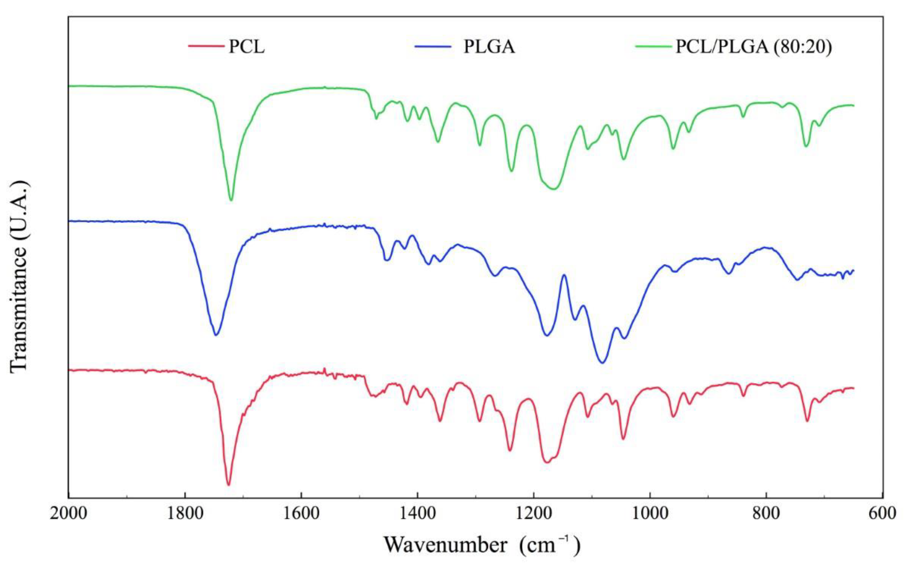
Abstract
:1. Introduction
2. Materials and Methods
2.1. Polymer Blends Preparation by Chloroform and Ethanol
2.2. Scaffold Manufacture
2.3. Scaffold Characterization
2.3.1. Fourier Transform Infrared Spectroscopy (FTIR)
2.3.2. Contact Angle Measurement
2.3.3. Differential Scanning Calorimetry (DSC) and Thermogravimetric Analysis (TGA)
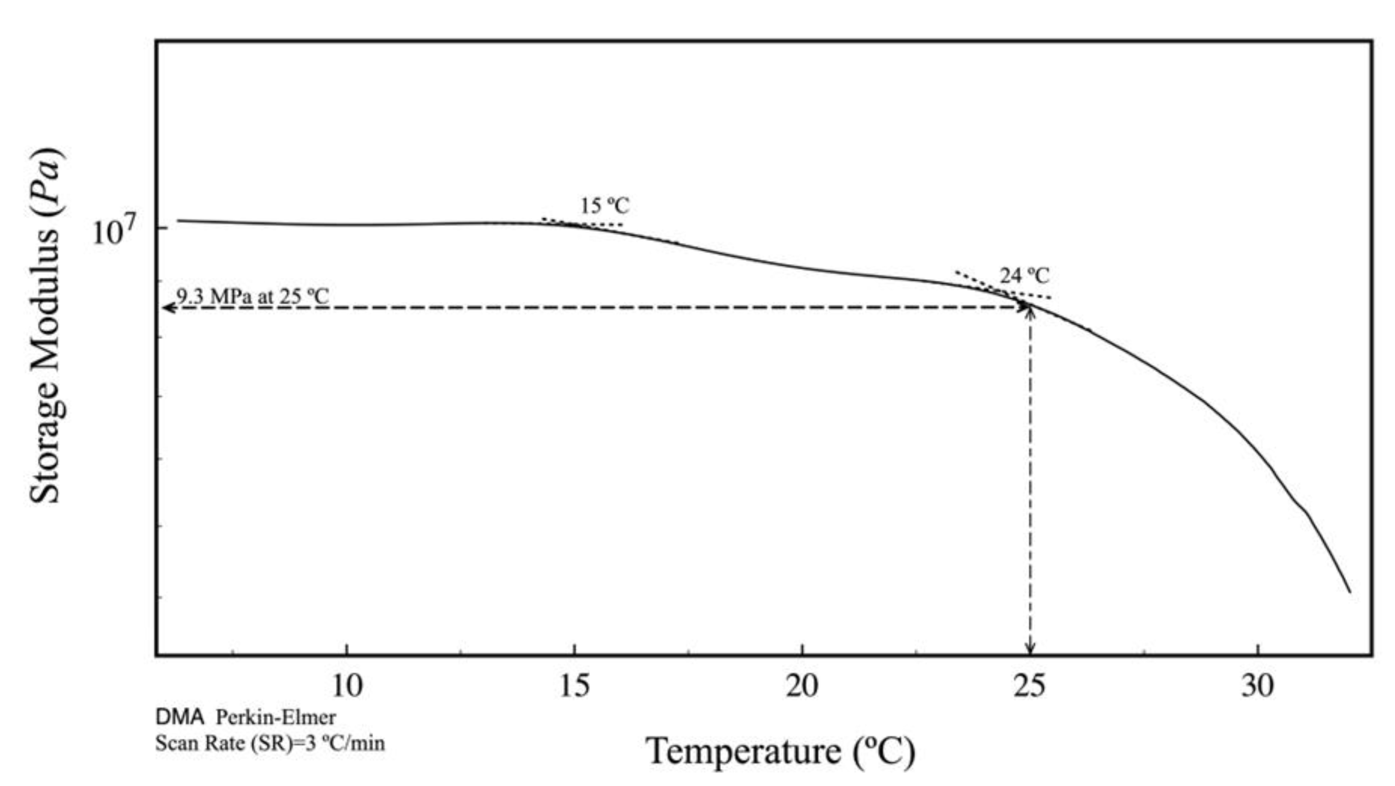
2.3.4. Tensile Mechanical Test
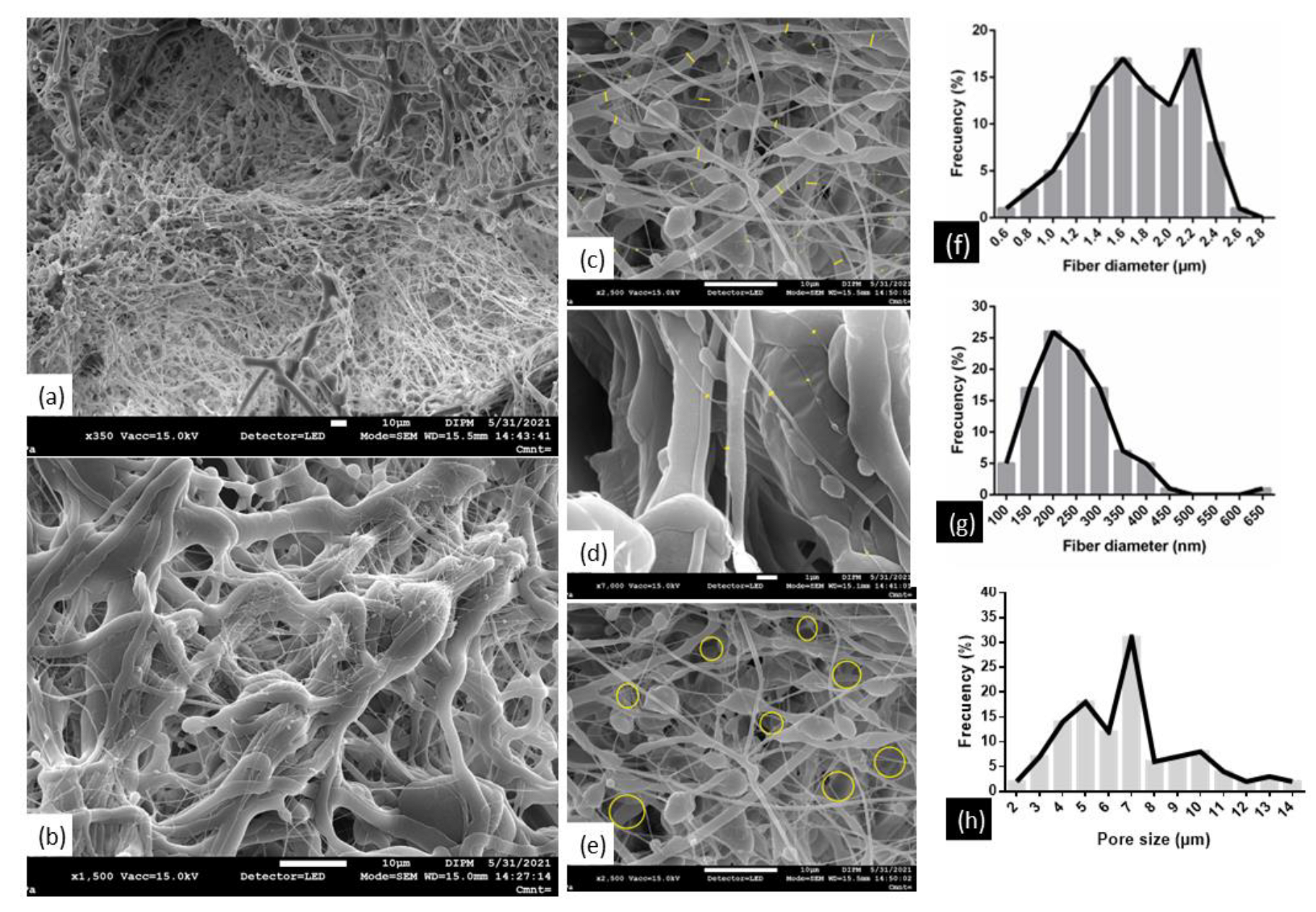
2.3.5. Microstructural Morphology by Scanning Electron Microscope (SEM)
2.4. In Vitro Assays
2.4.1. Isolation and Characterization of Dental Follicle Mesenchymal Stem Cells (DFMSCs)
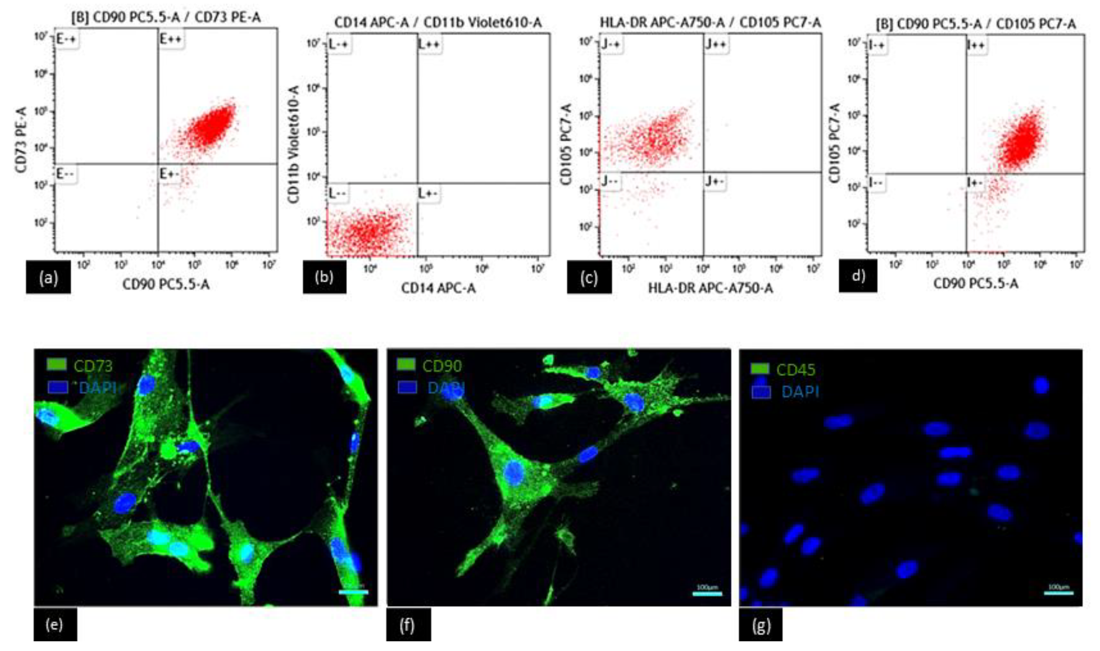
2.4.2. Phenotype Determination by Flow Cytometry
2.4.3. Phenotype Determination by Immunofluorescence
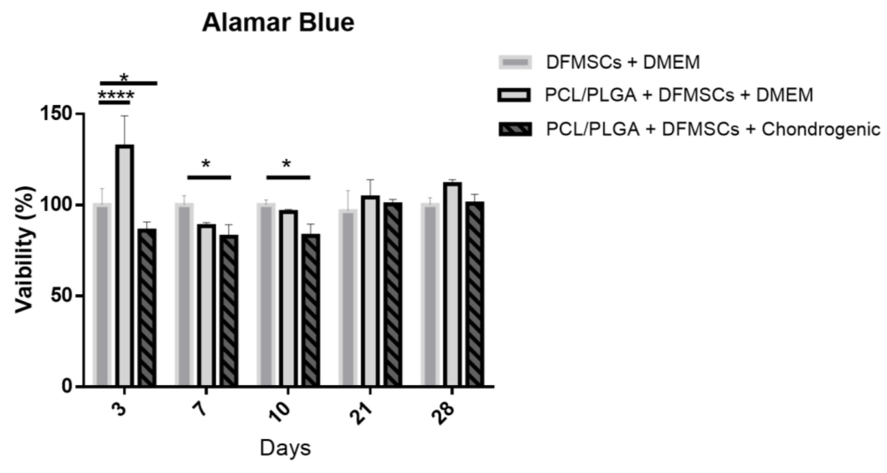
2.4.4. Alamar Blue Viability Assay
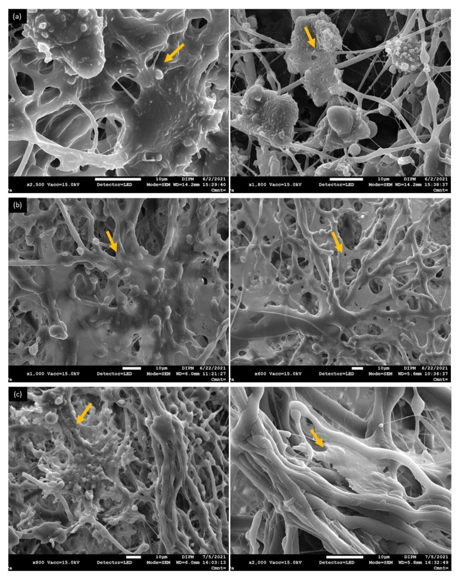
2.4.5. Adhesion DFMSCs Determined by SEM
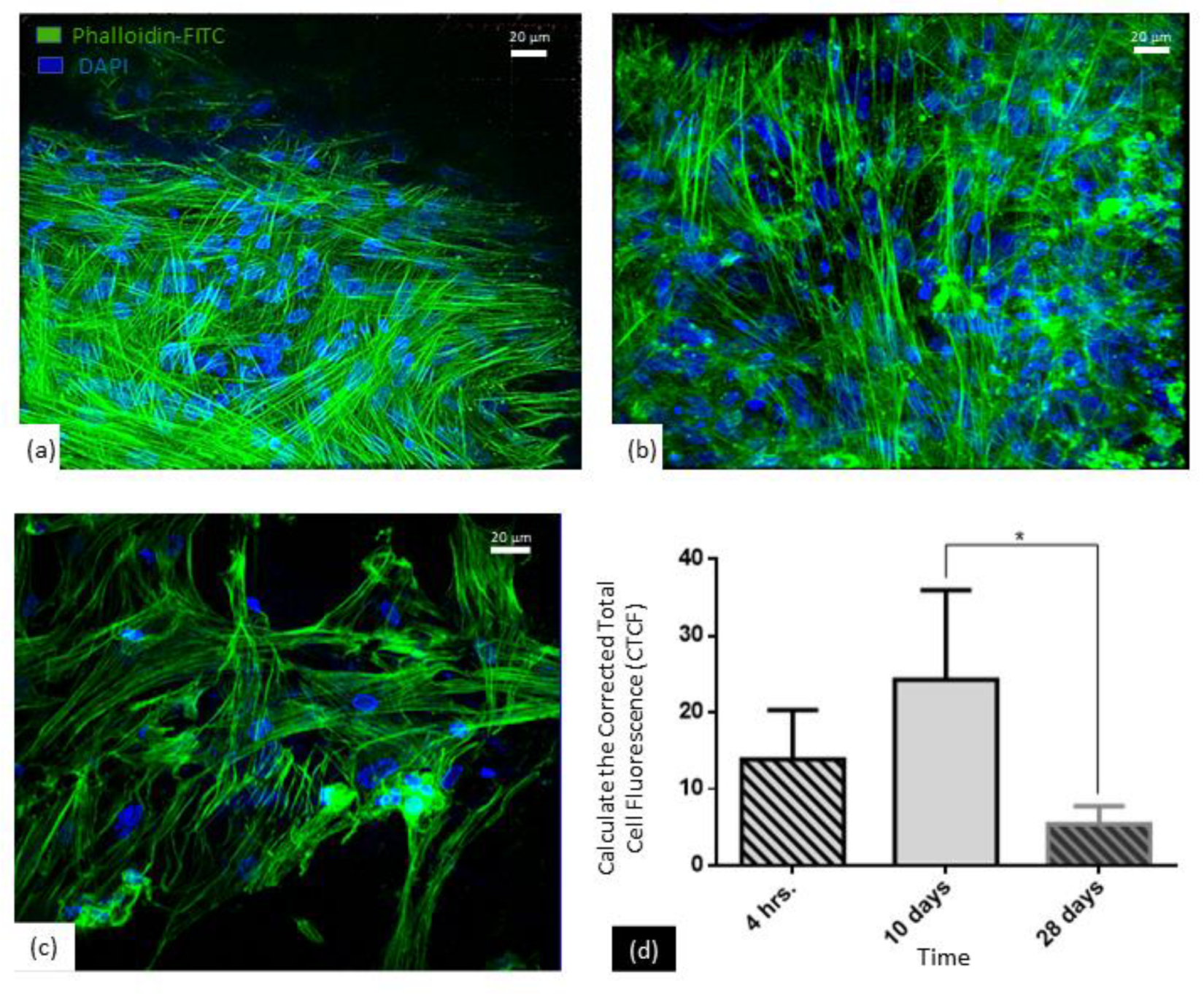
2.4.6. Cytoskeleton Organization
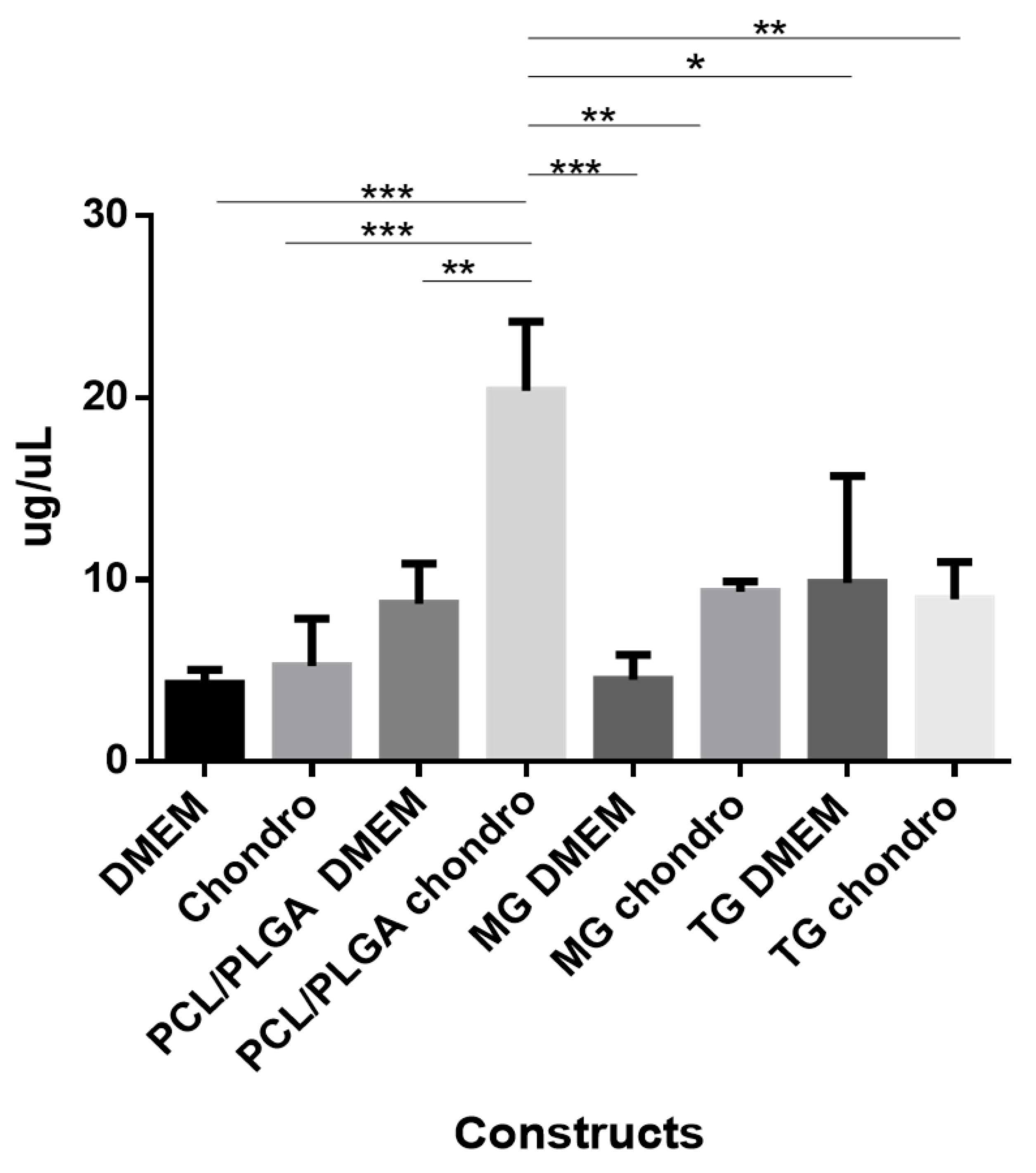
2.4.7. Quantification of Sulfated Glycosaminoglycans (GAGs)
2.5. In Vivo Assays
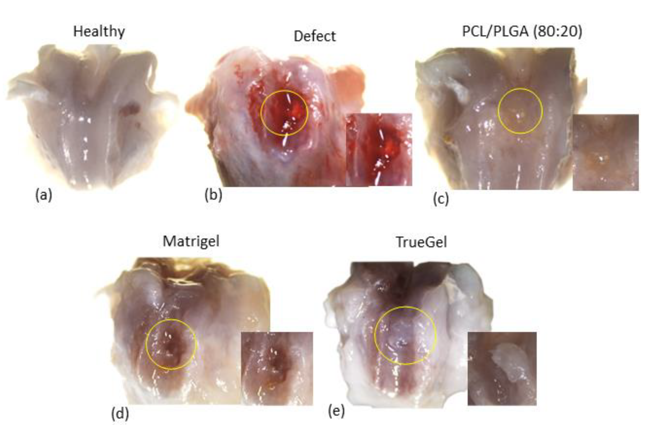
2.5.1. Articular Cartilage Defects and Scaffolds Implantation
2.5.2. Histological Analysis
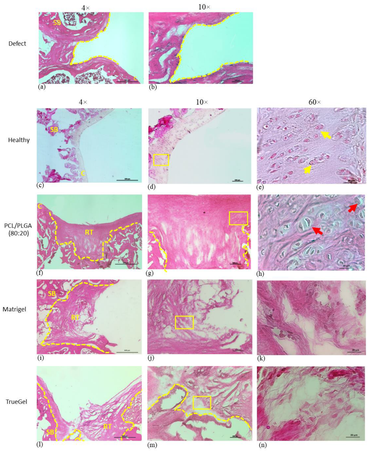
2.5.3. Hematoxylin and Eosin (H&E)
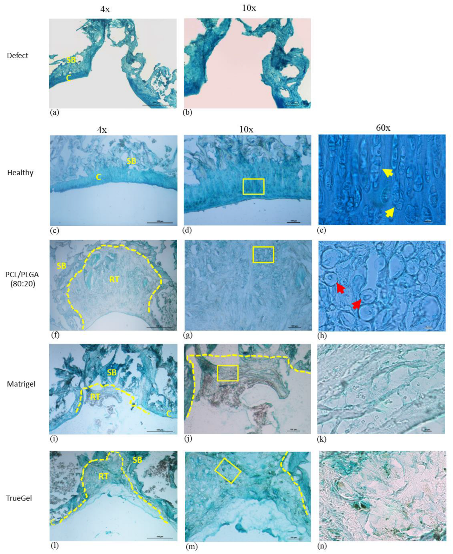
2.5.4. Alcian Blue
3. Results
3.1. Scaffold Characterization
3.1.1. Fourier Transform Infrared Spectroscopy (FTIR)
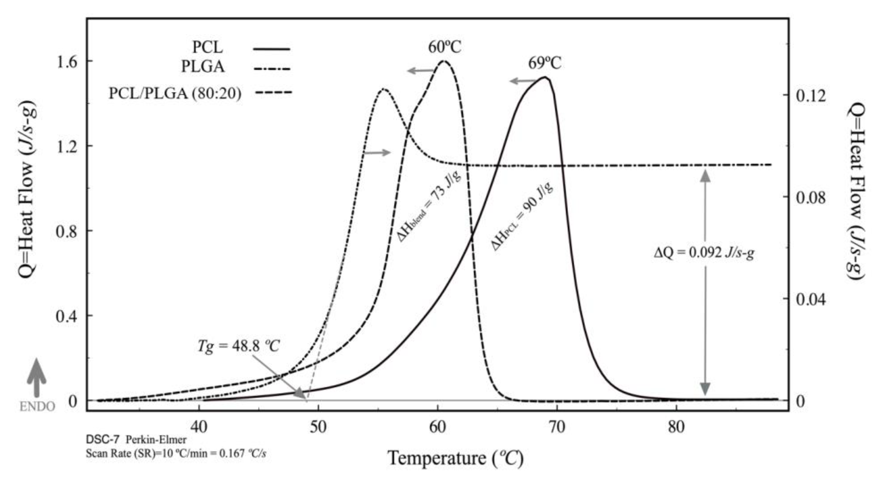
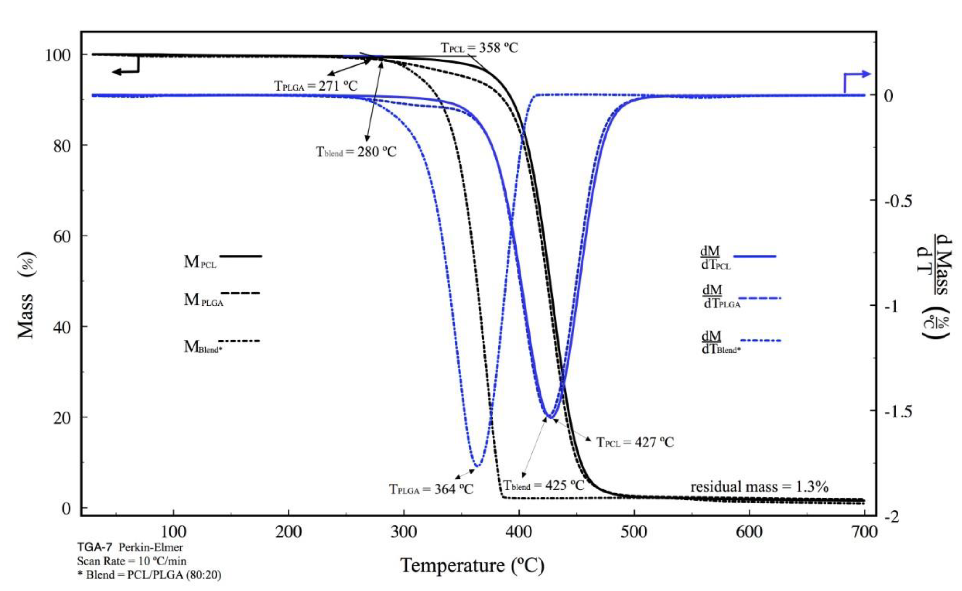
3.1.2. DSC and TGA
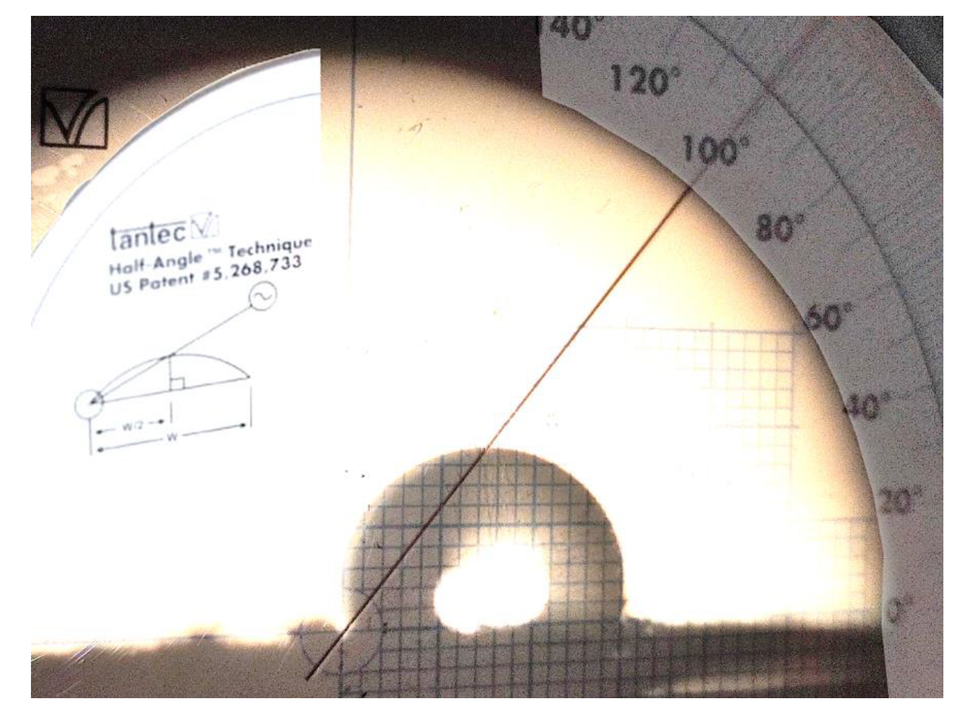
3.1.3. Evaluation of Wettability by Contact Angle
3.1.4. Tensile Mechanical Tests
3.1.5. Microstructural Morphology by Scanning Electron Microscope
3.2. In Vitro Assays
3.2.1. Isolation and Identification of DFMSCs by Flow Cytometry and Immunofluorescence
3.2.2. Alamar Blue Viability Assay
3.2.3. Adhesion DFMSCs by SEM
3.2.4. Cytoskeleton Organization
3.2.5. Quantitation of Sulfated Glycosaminoglycans
3.3. In Vivo Assay
Histology Evaluation
4. Discussion
Supplementary Materials
Author Contributions
Funding
Institutional Review Board Statement
Data Availability Statement
Acknowledgments
Conflicts of Interest
References
- Salgado, A.J.; Oliveira, J.M.; Martins, A.; Teixeira, F.G.; Silva, N.A.; Neves, N.M.; Sousa, N.; Reis, R.L. Tissue Engineering and Regenerative Medicine: Past, Present, and Future. Int. Rev. Neurobiol. 2013, 108, 1–33. [Google Scholar] [CrossRef]
- Lolli, A.; Colella, F.; De Bari, C.; van Osch, G.J.V.M. Targeting Anti-Chondrogenic Factors for the Stimulation of Chondrogenesis: A New Paradigm in Cartilage Repair. J. Orthop. Res. 2019, 37, 12–22. [Google Scholar] [CrossRef]
- Hunter, C.W.; Deer, T.R.; Jones, M.R.; Chang Chien, G.C.; D’Souza, R.S.; Davis, T.; Eldon, E.R.; Esposito, M.F.; Goree, J.H.; Hewan-Lowe, L.; et al. Consensus Guidelines on Interventional Therapies for Knee Pain (STEP Guidelines) from the American Society of Pain and Neuroscience. J. Pain Res. 2022, 15, 2683–2745. [Google Scholar] [CrossRef]
- Patel, J.M.; Dunn, M.G. 6-Cartilage Tissue Engineering. In Regenerative Engineering of Musculoskeletal Tissues and Interfaces; Nukavarapu, S.P., Freeman, J.W., Laurencin, C.T., Eds.; Woodhead Publishing: Sawston, UK, 2015; pp. 135–160. [Google Scholar] [CrossRef]
- Morouço, P.; Fernandes, C.; Santos-Rocha, R. Osteoarthritis, Exercise, and Tissue Engineering: A Stimulating Triad for Health Professionals. J. Aging Res. 2019, 2019, 1935806. [Google Scholar] [CrossRef]
- Stampoultzis, T.; Karami, P.; Pioletti, D.P. Thoughts on Cartilage Tissue Engineering: A 21st Century Perspective. Curr. Res. Transl. Med. 2021, 69, 103299. [Google Scholar] [CrossRef]
- Francis, S.L.; Di Bella, C.; Wallace, G.G.; Choong, P.F.M. Cartilage Tissue Engineering Using Stem Cells and Bioprinting Technology—Barriers to Clinical Translation. Front. Surg. 2018, 5, 70. [Google Scholar] [CrossRef]
- Pekovic, V.; Hutchison, C.J. Adult Stem Cell Maintenance and Tissue Regeneration in the Ageing Context: The Role for A-Type Lamins as Intrinsic Modulators of Ageing in Adult Stem Cells and Their Niches. J. Anat. 2008, 213, 5–25. [Google Scholar] [CrossRef]
- Pittenger, M.F.; Mackay, A.M.; Beck, S.C.; Jaiswal, R.K.; Douglas, R.; Mosca, J.D.; Moorman, M.A.; Simonetti, D.W.; Craig, S.; Marshak, D.R. Multilineage Potential of Adult Human Mesenchymal Stem Cells. Science 1999, 284, 143–147. [Google Scholar] [CrossRef]
- Pittenger, M.F.; Discher, D.E.; Péault, B.M.; Phinney, D.G.; Hare, J.M.; Caplan, A.I. Mesenchymal Stem Cell Perspective: Cell Biology to Clinical Progress. npj Regen. Med. 2019, 4, 22. [Google Scholar] [CrossRef]
- Li, B.; Ouchi, T.; Cao, Y.; Zhao, Z.; Men, Y. Dental-Derived Mesenchymal Stem Cells: State of the Art. Front. Cell Dev. Biol. 2021, 9, 654559. [Google Scholar] [CrossRef]
- Zhan, X.-S.; El-Ashram, S.; Luo, D.-Z.; Luo, H.-N.; Wang, B.-Y.; Chen, S.-F.; Bai, Y.-S.; Chen, Z.-S.; Liu, C.-Y.; Ji, H.-Q. A Comparative Study of Biological Characteristics and Transcriptome Profiles of Mesenchymal Stem Cells from Different Canine Tissues. Int. J. Mol. Sci. 2019, 20, 1485. [Google Scholar] [CrossRef] [PubMed]
- Wegmeyer, H.; Bröske, A.-M.; Leddin, M.; Kuentzer, K.; Nisslbeck, A.K.; Hupfeld, J.; Wiechmann, K.; Kuhlen, J.; von Schwerin, C.; Stein, C.; et al. Mesenchymal Stromal Cell Characteristics Vary Depending on Their Origin. Stem Cells Dev. 2013, 22, 2606–2618. [Google Scholar] [CrossRef] [PubMed]
- Zhou, L.; Liu, W.; Wu, Y.; Sun, W.; Dörfer, C.E.; Fawzy El-Sayed, K.M. Oral Mesenchymal Stem/Progenitor Cells: The Immunomodulatory Masters. Stem Cells Int. 2020, 2020, 1327405. [Google Scholar] [CrossRef]
- Mosaddad, S.A.; Rasoolzade, B.; Namanloo, R.A.; Azarpira, N.; Dortaj, H. Stem Cells and Common Biomaterials in Dentistry: A Review Study. J. Mater. Sci. Mater. Med. 2022, 33, 55. [Google Scholar] [CrossRef]
- Chen, G.; Chen, J.; Yang, B.; Li, L.; Luo, X.; Zhang, X.; Feng, L.; Jiang, Z.; Yu, M.; Guo, W.; et al. Combination of Aligned PLGA/Gelatin Electrospun Sheets, Native Dental Pulp Extracellular Matrix and Treated Dentin Matrix as Substrates for Tooth Root Regeneration. Biomaterials 2015, 52, 56–70. [Google Scholar] [CrossRef]
- Xu, Q.L.; Furuhashi, A.; Zhang, Q.Z.; Jiang, C.M.; Chang, T.-H.; Le, A.D. Induction of Salivary Gland-Like Cells from Dental Follicle Epithelial Cells. J. Dent. Res. 2017, 96, 1035–1043. [Google Scholar] [CrossRef]
- Wasyłeczko, M.; Sikorska, W.; Chwojnowski, A. Review of Synthetic and Hybrid Scaffolds in Cartilage Tissue Engineering. Membranes 2020, 10, 348. [Google Scholar] [CrossRef]
- Makadia, H.K.; Siegel, S.J. Poly Lactic-Co-Glycolic Acid (PLGA) as Biodegradable Controlled Drug Delivery Carrier. Polymers 2011, 3, 1377–1397. [Google Scholar] [CrossRef]
- Elmowafy, E.M.; Tiboni, M.; Soliman, M.E. Biocompatibility, Biodegradation and Biomedical Applications of Poly(Lactic Acid)/Poly(Lactic-Co-Glycolic Acid) Micro and Nanoparticles. J. Pharm. Investig. 2019, 49, 347–380. [Google Scholar] [CrossRef]
- Zhao, W.; Li, J.; Jin, K.; Liu, W.; Qiu, X.; Li, C. Fabrication of Functional PLGA-Based Electrospun Scaffolds and Their Applications in Biomedical Engineering. Mater. Sci. Eng. C Mater. Biol. Appl. 2016, 59, 1181–1194. [Google Scholar] [CrossRef]
- Gao, Y.; Callanan, A. Influence of Surface Topography on PCL Electrospun Scaffolds for Liver Tissue Engineering. J. Mater. Chem. B 2021, 9, 8081–8093. [Google Scholar] [CrossRef] [PubMed]
- Chou, S.-F.; Woodrow, K.A. Relationships between Mechanical Properties and Drug Release from Electrospun Fibers of PCL and PLGA Blends. J. Mech. Behav. Biomed. Mater. 2017, 65, 724–733. [Google Scholar] [CrossRef] [PubMed]
- Tiwari, S.K.; Tzezana, R.; Zussman, E.; Venkatraman, S.S. Optimizing Partition-Controlled Drug Release from Electrospun Core–Shell Fibers. Int. J. Pharm. 2010, 392, 209–217. [Google Scholar] [CrossRef] [PubMed]
- Alvim Valente, C.; Cesar Chagastelles, P.; Fontana Nicoletti, N.; Ramos Garcez, G.; Sgarioni, B.; Herrmann, F.; Pesenatto, G.; Goldani, E.; Zanini, M.L.; Campos, M.M.; et al. Design and Optimization of Biocompatible Polycaprolactone/Poly(l-Lactic-Co-Glycolic Acid) Scaffolds with and without Microgrooves for Tissue Engineering Applications: Design and Optimization of Biocompatible PCL/PLGA Scaffolds. J. Biomed. Mater. Res. 2018, 106, 1522–1534. [Google Scholar] [CrossRef]
- Ferreira, C.L.; Valente, C.A.; Zanini, M.L.; Sgarioni, B.; Ferreira Tondo, P.H.; Chagastelles, P.C.; Braga, J.; Campos, M.M.; Malmonge, J.A.; de Souza Basso, N.R. Biocompatible PCL/PLGA/Polypyrrole Composites for Regenerating Nerves. Macromol. Symp. 2019, 383, 1800028. [Google Scholar] [CrossRef]
- Franco, R.A.; Nguyen, T.H.; Lee, B.-T. Preparation and Characterization of Electrospun PCL/PLGA Membranes and Chitosan/Gelatin Hydrogels for Skin Bioengineering Applications. J. Mater. Sci. Mater. Med. 2011, 22, 2207–2218. [Google Scholar] [CrossRef]
- Tang, Z.G.; Callaghan, J.T.; Hunt, J.A. The Physical Properties and Response of Osteoblasts to Solution Cast Films of PLGA Doped Polycaprolactone. Biomaterials 2005, 26, 6618–6624. [Google Scholar] [CrossRef]
- Tang, Z.; Hunt, J. The Effect of PLGA Doping of Polycaprolactone Films on the Control of Osteoblast Adhesion and Proliferation in Vitro. Biomaterials 2006, 27, 4409–4418. [Google Scholar] [CrossRef]
- Hiep, N.T.; Lee, B.-T. Electro-Spinning of PLGA/PCL Blends for Tissue Engineering and Their Biocompatibility. J. Mater. Sci. Mater. Med. 2010, 21, 1969–1978. [Google Scholar] [CrossRef]
- Sánchez-Pech, J.C.; Rosales-Ibáñes, R.; Cauich-Rodriguez, J.V.; Carrillo-Escalante, H.J.; Rodríguez-Navarrete, A.; Avila-Ortega, A.; Hernández-Sánchez, F. Design, Synthesis, Characterization, and Cytotoxicity of PCL/PLGA Scaffolds through Plasma Treatment in the Presence of Pyrrole for Possible Use in Urethral Tissue Engineering. J. Biomater. Appl. 2020, 34, 840–850. [Google Scholar] [CrossRef]
- Stone, J.E.; Akhtar, N.; Botchway, S.; Pennock, C.A. Interaction of 1,9-Dimethylmethylene Blue with Glycosaminoglycans. Ann. Clin. Biochem. 1994, 31, 147–152. [Google Scholar] [CrossRef] [PubMed]
- Chung, J.Y.; Song, M.; Ha, C.-W.; Kim, J.-A.; Lee, C.-H.; Park, Y.-B. Comparison of Articular Cartilage Repair with Different Hydrogel-Human Umbilical Cord Blood-Derived Mesenchymal Stem Cell Composites in a Rat Model. Stem Cell Res. Ther. 2014, 5, 39. [Google Scholar] [CrossRef] [PubMed]
- Narayanan, G.; Shen, J.; Boy, R.; Gupta, B.S.; Tonelli, A.E. Aliphatic Polyester Nanofibers Functionalized with Cyclodextrins and Cyclodextrin-Guest Inclusion Complexes. Polymers 2018, 10, 428. [Google Scholar] [CrossRef] [PubMed]
- Paragkumar, N.T.; Edith, D.; Six, J.-L. Surface Characteristics of PLA and PLGA Films. Appl. Surf. Sci. 2006, 253, 2758–2764. [Google Scholar] [CrossRef]
- Gao, J.; Chen, S.; Tang, D.; Jiang, L.; Shi, J.; Wang, S. Mechanical Properties and Degradability of Electrospun PCL/PLGA Blended Scaffolds as Vascular Grafts. Trans. Tianjin Univ. 2019, 25, 152–160. [Google Scholar] [CrossRef]
- Sun, H.; Mei, L.; Song, C.; Cui, X.; Wang, P. The in Vivo Degradation, Absorption and Excretion of PCL-Based Implant. Biomaterials 2006, 27, 1735–1740. [Google Scholar] [CrossRef] [PubMed]
- Chou, S.-F.; Carson, D.; Woodrow, K.A. Current Strategies for Sustaining Drug Release from Electrospun Nanofibers. J. Control. Release 2015, 220, 584–591. [Google Scholar] [CrossRef] [PubMed]
- Licciardello, M.; Ciardelli, G.; Tonda-Turo, C. Biocompatible Electrospun Polycaprolactone-Polyaniline Scaffold Treated with Atmospheric Plasma to Improve Hydrophilicity. Bioengineering 2021, 8, 24. [Google Scholar] [CrossRef]
- Houchin, M.L.; Topp, E.M. Physical Properties of PLGA Films during Polymer Degradation. J. Appl. Polym. Sci. 2009, 114, 2848–2854. [Google Scholar] [CrossRef]
- Dias, J.R.; Sousa, A.; Augusto, A.; Bártolo, P.J.; Granja, P.L. Electrospun Polycaprolactone (PCL) Degradation: An In Vitro and In Vivo Study. Polymers 2022, 14, 3397. [Google Scholar] [CrossRef]
- Peng, C.; Zheng, J.; Chen, D.; Zhang, X.; Deng, L.; Chen, Z.; Wu, L. Response of HPDLSCs on 3D Printed PCL/PLGA Composite Scaffolds in Vitro. Mol. Med. Rep. 2018, 18, 1335–1344. [Google Scholar] [CrossRef] [PubMed]
- Critchley, S.; Sheehy, E.J.; Cunniffe, G.; Diaz-Payno, P.; Carroll, S.F.; Jeon, O.; Alsberg, E.; Brama, P.A.J.; Kelly, D.J. 3D Printing of Fibre-Reinforced Cartilaginous Templates for the Regeneration of Osteochondral Defects. Acta Biomater. 2020, 113, 130–143. [Google Scholar] [CrossRef] [PubMed]
- Zamanlui, S.; Mahmoudifard, M.; Soleimani, M.; Bakhshandeh, B.; Vasei, M.; Faghihi, S. Enhanced Chondrogenic Differentiation of Human Bone Marrow Mesenchymal Stem Cells on PCL/PLGA Electrospun with Different Alignments and Compositions. Int. J. Polym. Mater. Polym. Biomater. 2018, 67, 50–60. [Google Scholar] [CrossRef]
- Goonoo, N.; Bhaw-Luximon, A.; Jhurry, D. Drug Loading and Release from Electrospun Biodegradable Nanofibers. J. Biomed. Nanotechnol. 2014, 10, 2173–2199. [Google Scholar] [CrossRef] [PubMed]
- Caminal, M.; Peris, D.; Fonseca, C.; Barrachina, J.; Codina, D.; Rabanal, R.M.; Moll, X.; Morist, A.; García, F.; Cairó, J.J.; et al. Cartilage Resurfacing Potential of PLGA Scaffolds Loaded with Autologous Cells from Cartilage, Fat, and Bone Marrow in an Ovine Model of Osteochondral Focal Defect. Cytotechnology 2016, 68, 907–919. [Google Scholar] [CrossRef] [PubMed]
- Qian, Y.; Chen, H.; Xu, Y.; Yang, J.; Zhou, X.; Zhang, F.; Gu, N. The Preosteoblast Response of Electrospinning PLGA/PCL Nanofibers: Effects of Biomimetic Architecture and Collagen I. Int. J. Nanomed. 2016, 11, 4157–4171. [Google Scholar] [CrossRef]
- Indolfi, L.; Baker, A.B.; Edelman, E.R. The Role of Scaffold Microarchitecture in Engineering Endothelial Cell Immunomodulation. Biomaterials 2012, 33, 7019–7027. [Google Scholar] [CrossRef]
- Nicolas, J.; Magli, S.; Rabbachin, L.; Sampaolesi, S.; Nicotra, F.; Russo, L. 3D Extracellular Matrix Mimics: Fundamental Concepts and Role of Materials Chemistry to Influence Stem Cell Fate. Biomacromolecules 2020, 21, 1968–1994. [Google Scholar] [CrossRef]
- Chiquet, M.; Gelman, L.; Lutz, R.; Maier, S. From Mechanotransduction to Extracellular Matrix Gene Expression in Fibroblasts. Biochim. Biophys. Acta 2009, 1793, 911–920. [Google Scholar] [CrossRef]
- Ijima, H.; Nakamura, S.; Bual, R.; Shirakigawa, N.; Tanoue, S. Physical Properties of the Extracellular Matrix of Decellularized Porcine Liver. Gels 2018, 4, 39. [Google Scholar] [CrossRef]
- Brown, B.N.; Badylak, S.F. Extracellular Matrix as an Inductive Scaffold for Functional Tissue Reconstruction. Transl. Res. 2014, 163, 268–285. [Google Scholar] [CrossRef] [PubMed]
- Ishaug-Riley, S.L.; Okun, L.E.; Prado, G.; Applegate, M.A.; Ratcliffe, A. Human Articular Chondrocyte Adhesion and Proliferation on Synthetic Biodegradable Polymer Films. Biomaterials 1999, 20, 2245–2256. [Google Scholar] [CrossRef] [PubMed]
- Jelodari, S.; Ebrahimi Sadrabadi, A.; Zarei, F.; Jahangir, S.; Azami, M.; Sheykhhasan, M.; Hosseini, S. New Insights into Cartilage Tissue Engineering: Improvement of Tissue-Scaffold Integration to Enhance Cartilage Regeneration. BioMed Res. Int. 2022, 2022, e7638245. [Google Scholar] [CrossRef] [PubMed]
- Pawlik, J.; Łukowicz, K.; Cholewa-Kowalska, K.; Osyczka, A.M. New Insights into the PLGA and PCL Blending: Physico-Mechanical Properties and Cell Response. Mater. Res. Express 2019, 6, 085344. [Google Scholar] [CrossRef]
- Tanthaisong, P.; Imsoonthornruksa, S.; Ngernsoungnern, A.; Ngernsoungnern, P.; Ketudat-Cairns, M.; Parnpai, R. Enhanced Chondrogenic Differentiation of Human Umbilical Cord Wharton’s Jelly Derived Mesenchymal Stem Cells by GSK-3 Inhibitors. PLoS ONE 2017, 12, e0168059. [Google Scholar] [CrossRef] [PubMed]
- Li, L.; Yu, F.; Zheng, L.; Wang, R.; Yan, W.; Wang, Z.; Xu, J.; Wu, J.; Shi, D.; Zhu, L.; et al. Natural Hydrogels for Cartilage Regeneration: Modification, Preparation and Application. J. Orthop. Transl. 2019, 17, 26–41. [Google Scholar] [CrossRef]
- Wei, P.; Xu, Y.; Zhang, H.; Wang, L. Continued Sustained Insulin-Releasing PLGA Nanoparticles Modified 3D-Printed PCL Composite Scaffolds for Osteochondral Repair. Chem. Eng. J. 2021, 422, 130051. [Google Scholar] [CrossRef]


Disclaimer/Publisher’s Note: The statements, opinions and data contained in all publications are solely those of the individual author(s) and contributor(s) and not of MDPI and/or the editor(s). MDPI and/or the editor(s) disclaim responsibility for any injury to people or property resulting from any ideas, methods, instructions or products referred to in the content. |
© 2023 by the authors. Licensee MDPI, Basel, Switzerland. This article is an open access article distributed under the terms and conditions of the Creative Commons Attribution (CC BY) license (https://creativecommons.org/licenses/by/4.0/).
Share and Cite
González-González, A.M.; Cruz, R.; Rosales-Ibáñez, R.; Hernández-Sánchez, F.; Carrillo-Escalante, H.J.; Rodríguez-Martínez, J.J.; Velasquillo, C.; Talamás-Lara, D.; Ludert, J.E. In Vitro and In Vivo Evaluation of a Polycaprolactone (PCL)/Polylactic-Co-Glycolic Acid (PLGA) (80:20) Scaffold for Improved Treatment of Chondral (Cartilage) Injuries. Polymers 2023, 15, 2324. https://doi.org/10.3390/polym15102324
González-González AM, Cruz R, Rosales-Ibáñez R, Hernández-Sánchez F, Carrillo-Escalante HJ, Rodríguez-Martínez JJ, Velasquillo C, Talamás-Lara D, Ludert JE. In Vitro and In Vivo Evaluation of a Polycaprolactone (PCL)/Polylactic-Co-Glycolic Acid (PLGA) (80:20) Scaffold for Improved Treatment of Chondral (Cartilage) Injuries. Polymers. 2023; 15(10):2324. https://doi.org/10.3390/polym15102324
Chicago/Turabian StyleGonzález-González, Arely M., Raymundo Cruz, Raúl Rosales-Ibáñez, Fernando Hernández-Sánchez, Hugo J. Carrillo-Escalante, Jesús Jiovanni Rodríguez-Martínez, Cristina Velasquillo, Daniel Talamás-Lara, and Juan E. Ludert. 2023. "In Vitro and In Vivo Evaluation of a Polycaprolactone (PCL)/Polylactic-Co-Glycolic Acid (PLGA) (80:20) Scaffold for Improved Treatment of Chondral (Cartilage) Injuries" Polymers 15, no. 10: 2324. https://doi.org/10.3390/polym15102324