
Can 3D-Printed Bioactive Glasses Be the Future of Bone Tissue Engineering?

 , ,
, ,  ,
,  and
and
Abstract
:
1. Introduction
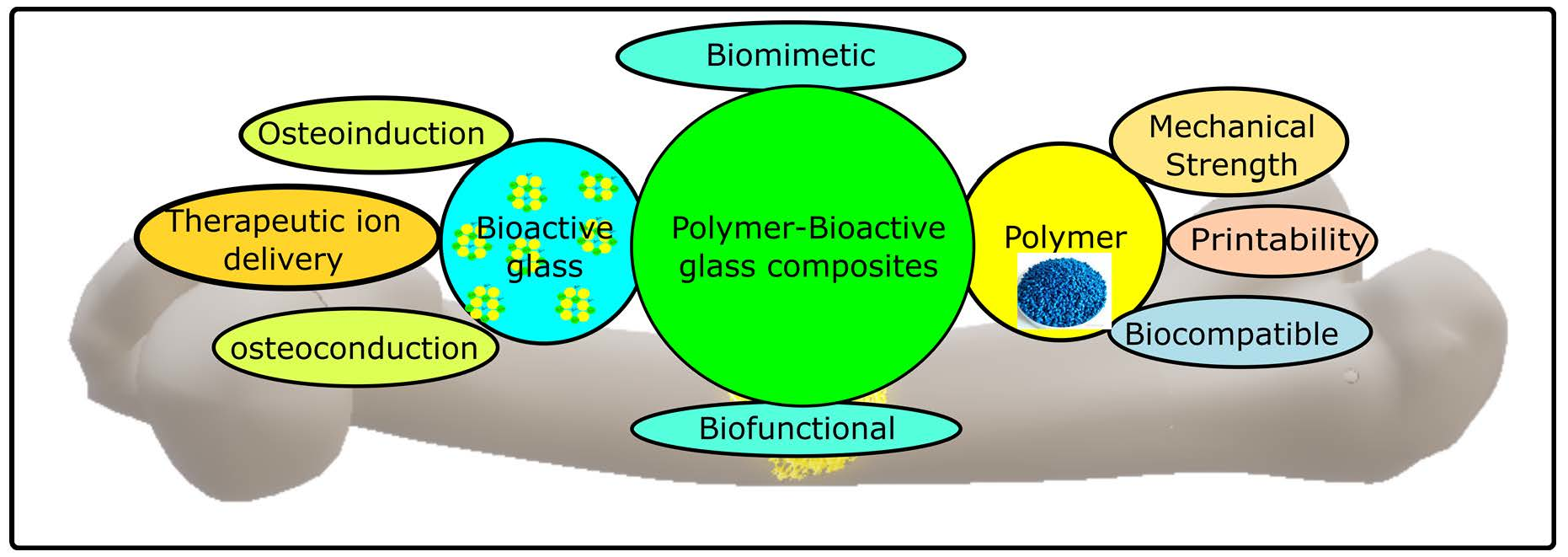
- Biocompatible: a scaffold should perform the function of cell recruitment and offer a conducive surface for proliferation. It should be porous to provide a large surface area for cell attachment and proliferation.
- Biodegradable: a scaffold should dissolve completely when bone regeneration is complete. Ideally, the rate of scaffold degradation should match the rate of new bone formation.
- Biomimetic: the human bone is a hierarchical structure comprising a ring-type matrix [21,22,23,24]. An ideal scaffold mimics the bone in terms of its hierarchical structure. The mechanical properties of human bones are highly anisotropic. An elastic modulus of cortical bone is 11,500 MPa in the transverse direction and 17,000 MPa in the longitudinal direction. The trabecular bone is MPa longitudinally [25]. The mechanical properties of an ideal scaffold should match the mechanical properties of the bone it is used in.
- Bio-functional: a scaffold should accelerate the healing process by delivering drugs and promoting growth factors to the defect site.
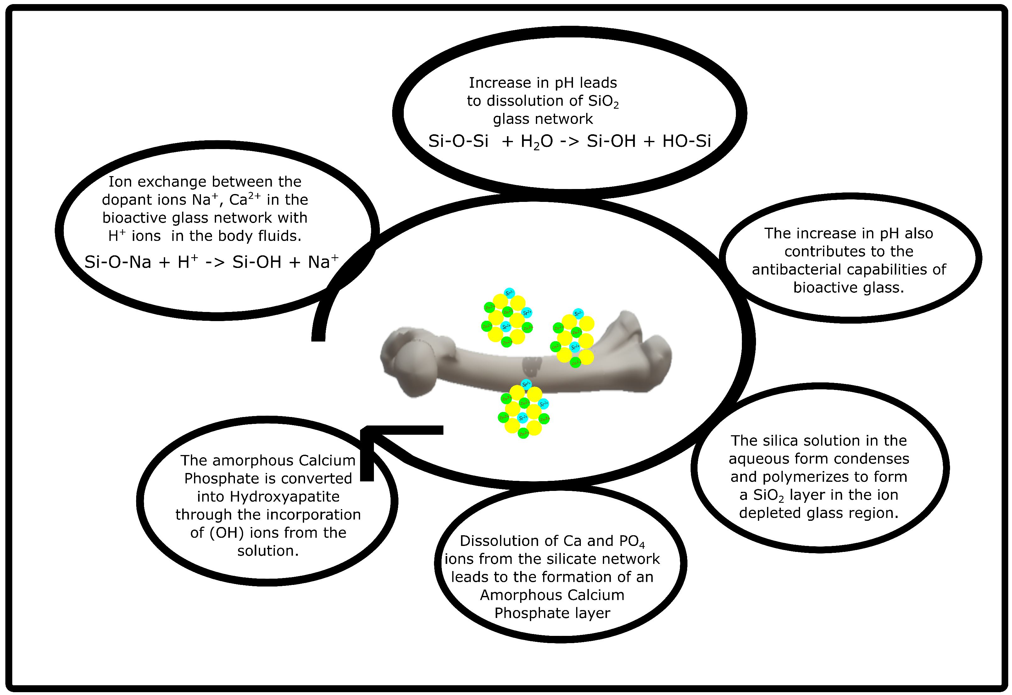
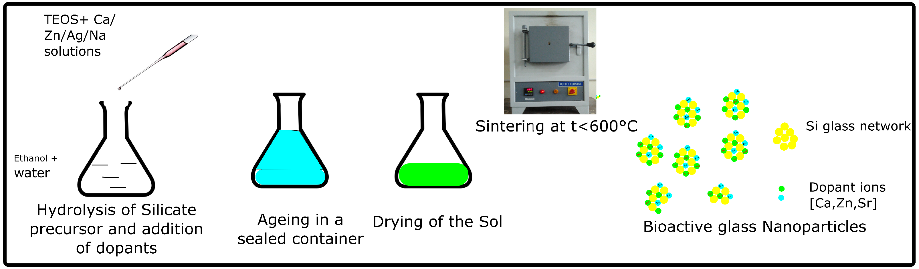
2. Bioactive Glass: An Active Bone Regeneration Material
3. 3D Printing of Polymer/Bioactive Glass Composites: An Overview
4. Synthetic Polymers Used in Bioactive Glass Composites
4.1. Thermoplastic Polymer/Bioactive Glass Composite
4.2. Thermoset Polymers/Bioactive Glass Composites
5. Natural Polymers Used for Bioactive Glass Composites
5.1. Protein-Based Polymer-Bioactive Glass Composites
5.2. Polysaccharide-Based Polymer-Bioactive Glass Composites
6. Conclusions
Author Contributions
Funding
Institutional Review Board Statement
Informed Consent Statement
Data Availability Statement
Acknowledgments
Conflicts of Interest
Abbreviations
| 3D | Three dimensional |
| µBG | micro bioactive glass |
| ALP | Alkaline phosphatase |
| BG | Bioactive glass |
| BMP-2 | Bone morphogenetic protein-2 |
| BNP | Bioactive nanoparticle |
| BSP | Bone sialoprotein |
| COL-1 | Collagen-1 |
| ECM | Extracellular matrix |
| G | Gelatin |
| hBM-MSCs | Human bone marrow-derived mesenchymal stem cells |
| MPa | Mega Pascal |
| nBG | nano bioactive glass |
| OCN | Osteocalcin |
| OPN | Osteopontin |
| PCL | Poly-caprolactone |
| rBM-MSCs | Rat bone marrow-derived mesenchymal stem cells |
| SA | Sodium alginate |
| SF | Silk fibroin |
| FDM | Fused Deposition Modelling |
| SLA | Stereolithography |
| PDM | Paste Deposition Modelling |
References
- Shegarfi, H.; Reikeras, O. Review article: Bone transplantation and immune response. J. Orthop. Surg. (Hong Kong) 2009, 17, 206–211. [Google Scholar] [CrossRef] [PubMed]
- Ben-David, D.; Kizhner, T.A.; Kohler, T.; Müller, R.; Livne, E.; Srouji, S. Cell-scaffold transplant of hydrogel seeded with rat bone marrow progenitors for bone regeneration. J. Cranio-Maxillo-Facial Surg. 2011, 39, 364–371. [Google Scholar] [CrossRef] [PubMed]
- Baht, G.S.; Vi, L.; Alman, B.A. The Role of the Immune Cells in Fracture Healing. Curr. Osteoporos. Rep. 2018, 16, 138–145. [Google Scholar] [CrossRef] [PubMed] [Green Version]
- Einhorn, T.A.; Gerstenfeld, L.C. Fracture healing: Mechanisms and interventions. Nat. Rev. Rheumatol. 2015, 11, 45–54. [Google Scholar] [CrossRef] [PubMed] [Green Version]
- Ghelich, P.; Kazemzadeh-Narbat, M.; Hassani Najafabadi, A.; Samandari, M.; Memić, A.; Tamayol, A. (Bio)manufactured Solutions for Treatment of Bone Defects with an Emphasis on US–FDA Regulatory Science Perspective. Adv. Nanobiomed Res. 2022, 2100073. [Google Scholar] [CrossRef]
- Cypher, T.J.; Grossman, J.P. Biological principles of bone graft healing. J. Foot Ankle Surg. 1996, 35, 413–417. [Google Scholar] [CrossRef]
- Albrektsson, T. Repair of bone grafts. A vital microscopic and histological investigation in the rabbit. Scand. J. Plast. Reconstr. Surg. 1980, 14, 1–12. [Google Scholar] [CrossRef]
- Stevenson, S. Biology of bone grafts. Orthop. Clin. N. Am. 1999, 30, 543–552. [Google Scholar] [CrossRef]
- Griffith, C.K.; Miller, C.; Sainson, R.C.A.; Calvert, J.W.; Jeon, N.L.; Hughes, C.C.W.; George, S.C. Diffusion limits of an in vitro thick prevascularized tissue. Tissue Eng. 2005, 11, 257–266. [Google Scholar] [CrossRef] [Green Version]
- Fu, Q.; Saiz, E.; Rahaman, M.N.; Tomsia, A.P. Bioactive glass scaffolds for bone tissue engineering: State of the art and future perspectives. Mater. Sci. Eng. C Mater. Biol. Appl. 2011, 31, 1245–1256. [Google Scholar] [CrossRef] [Green Version]
- Bauer, T.W. Bone graft substitutes. Skelet. Radiol. 2007, 36, 1105–1107. [Google Scholar] [CrossRef] [PubMed]
- Wang, W.; Yeung, K.W.K. Bone grafts and biomaterials substitutes for bone defect repair: A review. Bioact. Mater. 2017, 2, 224–247. [Google Scholar] [CrossRef] [PubMed]
- de Long, W.G.; Einhorn, T.A.; Koval, K.; McKee, M.; Smith, W.; Sanders, R.; Watson, T. Bone grafts and bone graft substitutes in orthopaedic trauma surgery. A critical analysis. J. Bone Jt. Surg. 2007, 89, 649–658. [Google Scholar] [CrossRef]
- Carson, J.S.; Bostrom, M.P.G. Synthetic bone scaffolds and fracture repair. Injury 2007, 38 (Suppl. 1), S33–S37. [Google Scholar] [CrossRef]
- Greenwald, A.S.; Boden, S.D.; Goldberg, V.M.; Khan, Y.; Laurencin, C.T.; Rosier, R.N. Bone-graft substitutes: Facts, fictions, and applications. J. Bone Jt. Surg. 2001, 83-A Pt 2 (Suppl. 2), 98–103. [Google Scholar] [CrossRef]
- Mauffrey, C.; Barlow, B.T.; Smith, W. Management of segmental bone defects. J. Am. Acad. Orthop. Surg. 2015, 23, 143–153. [Google Scholar] [CrossRef]
- Amini, A.R.; Laurencin, C.T.; Nukavarapu, S.P. Bone tissue engineering: Recent advances and challenges. Crit. Rev. Biomed. Eng. 2012, 40, 363–408. [Google Scholar] [CrossRef] [Green Version]
- Dimitriou, R.; Jones, E.; McGonagle, D.; Giannoudis, P.V. Bone regeneration: Current concepts and future directions. BMC Med. 2011, 9, 1–10. [Google Scholar] [CrossRef] [Green Version]
- Chan, B.; Leong, K. Scaffolding in tissue engineering: General approaches and tissue-specific considerations. Eur. Spine J. 2008, 17, 467–479. [Google Scholar] [CrossRef] [Green Version]
- Ghassemi, T.; Shahroodi, A.; Ebrahimzadeh, M.H.; Mousavian, A.; Movaffagh, J.; Moradi, A. Current concepts in scaffolding for bone tissue engineering. Arch. Bone Jt. Surg. 2018, 6, 90. [Google Scholar]
- Liu, Y.; Luo, D.; Wang, T. Hierarchical structures of bone and bioinspired bone tissue engineering. Small 2016, 12, 4611–4632. [Google Scholar] [CrossRef] [PubMed]
- Elsharkawy, S.; Mata, A. Hierarchical biomineralization: From nature’s designs to synthetic materials for regenerative medicine and dentistry. Adv. Healthc. Mater. 2018, 7, 1800178. [Google Scholar] [CrossRef] [PubMed]
- Fratzl, P.; Weinkamer, R. Nature’s hierarchical materials. Prog. Mater. Sci. 2007, 52, 1263–1334. [Google Scholar] [CrossRef] [Green Version]
- Reznikov, N.; Shahar, R.; Weiner, S. Bone hierarchical structure in three dimensions. Acta Biomater. 2014, 10, 3815–3826. [Google Scholar] [CrossRef]
- Hart, N.H.; Nimphius, S.; Rantalainen, T.; Ireland, A.; Siafarikas, A.; Newton, R. Mechanical basis of bone strength: Influence of bone material, bone structure and muscle action. J. Musculoskelet. Neuronal Interact. 2017, 17, 114. [Google Scholar]
- Alvarez, K.; Nakajima, H. Metallic scaffolds for bone regeneration. Materials 2009, 2, 790–832. [Google Scholar] [CrossRef]
- Dhandayuthapani, B.; Yoshida, Y.; Maekawa, T.; Kumar, D.S. Polymeric scaffolds in tissue engineering application: A review. Int. J. Polym. Sci. 2011, 2011, 290602. [Google Scholar] [CrossRef]
- Qu, H.; Fu, H.; Han, Z.; Sun, Y. Biomaterials for bone tissue engineering scaffolds: A review. RSC Adv. 2019, 9, 26252–26262. [Google Scholar] [CrossRef] [Green Version]
- Baino, F.; Novajra, G.; Vitale-Brovarone, C. Bioceramics and scaffolds: A winning combination for tissue engineering. Front. Bioeng. Biotechnol. 2015, 3, 202. [Google Scholar] [CrossRef] [Green Version]
- Gao, C.; Deng, Y.; Feng, P.; Mao, Z.; Li, P.; Yang, B.; Deng, J.; Cao, Y.; Shuai, C.; Peng, S. Current progress in bioactive ceramic scaffolds for bone repair and regeneration. Int. J. Mol. Sci. 2014, 15, 4714–4732. [Google Scholar] [CrossRef]
- Panseri, S.; Montesi, M.; Hautcoeur, D.; Dozio, S.M.; Chamary, S.; De Barra, E.; Tampieri, A.; Leriche, A. Bone-like ceramic scaffolds designed with bioinspired porosity induce a different stem cell response. J. Mater. Sci. Mater. Med. 2021, 32, 1–12. [Google Scholar] [CrossRef] [PubMed]
- Turnbull, G.; Clarke, J.; Picard, F.; Riches, P.; Jia, L.; Han, F.; Li, B.; Shu, W. 3D bioactive composite scaffolds for bone tissue engineering. Bioact. Mater. 2018, 3, 278–314. [Google Scholar] [CrossRef] [PubMed] [Green Version]
- Petretta, M.; Gambardella, A.; Boi, M.; Berni, M.; Cavallo, C.; Marchiori, G.; Maltarello, M.C.; Bellucci, D.; Fini, M.; Baldini, N.; et al. Composite scaffolds for bone tissue regeneration based on PCL and Mg-containing bioactive glasses. Biology 2021, 10, 398. [Google Scholar] [CrossRef] [PubMed]
- Hench, L.L. The story of Bioglass. J. Mater. Sci. Mater. Med. 2006, 17, 967–978. [Google Scholar] [CrossRef] [PubMed]
- Prem Ananth, K.; Shanmugam, S.; Jose, S.P.; Nathanael, A.J.; Oh, T.H.; Mangalaraj, D.; Ballamurugan, A.M. Structural and chemical analysis of silica-doped β-TCP ceramic coatings on surgical grade 316L SS for possible biomedical application. J. Asian Ceram. Soc. 2015, 3, 317–324. [Google Scholar] [CrossRef] [Green Version]
- Boskey, A.L. Bone composition: Relationship to bone fragility and antiosteoporotic drug effects. BoneKEy Rep. 2013, 2, 1–11. [Google Scholar] [CrossRef] [Green Version]
- Wei, S.; Ma, J.X.; Xu, L.; Gu, X.S.; Ma, X.L. Biodegradable materials for bone defect repair. Mil. Med Res. 2020, 7, 1–25. [Google Scholar] [CrossRef]
- Nathanael, A.J.; Mangalaraj, D.; Chen, P.C.; Ponpandian, N. Mechanical and photocatalytic properties of hydroxyapatite/titania nanocomposites prepared by combined high gravity and hydrothermal process. Compos. Sci. Technol. 2010, 70, 419–426. [Google Scholar] [CrossRef]
- Fiume, E.; Magnaterra, G.; Rahdar, A.; Verné, E.; Baino, F. Hydroxyapatite for Biomedical Applications: A Short Overview. Ceramics 2021, 4, 39. [Google Scholar] [CrossRef]
- Joseph Nathanael, A.; Mangalaraj, D.; Chi Chen, P.; Ponpandian, N. Enhanced mechanical strength of hydroxyapatite nanorods reinforced with polyethylene. J. Nanopart. Res. 2011, 13, 1841–1853. [Google Scholar] [CrossRef]
- Nathanael, A.J.; Hong, S.I.; Oh, T.H.; Seo, Y.H.; Singh, D.; Han, S.S. Enhanced cell viability of hydroxyapatite nanowires by surfactant mediated synthesis and its growth mechanism. RSC Adv. 2016, 6, 25070–25081. [Google Scholar] [CrossRef]
- Krishnan, V.; Lakshmi, T. Bioglass: A novel biocompatible innovation. J. Adv. Pharm. Technol. Res. 2013, 4, 78. [Google Scholar] [CrossRef] [PubMed]
- Boyan, B.; Baker, M.; Lee, C.; Raines, A.; Greenwald, A.; Olivares-Navarrete, R.; Schwartz, Z. Bone tissue grafting and tissue engineering concepts. In Comprehensive Biomaterials; Elsevier: Amsterdam, The Netherlands, 2011; pp. 237–255. [Google Scholar]
- Baino, F.; Hamzehlou, S.; Kargozar, S. Bioactive glasses: Where are we and where are we going? J. Funct. Biomater. 2018, 9, 25. [Google Scholar] [CrossRef] [PubMed] [Green Version]
- Hench, L.L.; Jones, J.R. Bioactive glasses: Frontiers and challenges. Front. Bioeng. Biotechnol. 2015, 3, 194. [Google Scholar] [CrossRef] [Green Version]
- Jones, J.R. Review of bioactive glass: From Hench to hybrids. Acta Biomater. 2013, 9, 4457–4486. [Google Scholar] [CrossRef]
- Mulchandani, N.; Katiyar, V. Bioactive Glasses: Prospects in Bone Tissue Engineering. In Advances in Sustainable Polymers; Springer: Singapore, 2019; pp. 67–83. [Google Scholar]
- Skallevold, H.E.; Rokaya, D.; Khurshid, Z.; Zafar, M.S. Bioactive glass applications in dentistry. Int. J. Mol. Sci. 2019, 20, 5960. [Google Scholar] [CrossRef] [Green Version]
- Crush, J.; Hussain, A.; Seah, K.; Khan, W.S. Bioactive Glass: Methods for Assessing Angiogenesis and Osteogenesis. Front. Cell Dev. Biol. 2021, 15239. [Google Scholar] [CrossRef]
- Fernandes, H.R.; Gaddam, A.; Rebelo, A.; Brazete, D.; Stan, G.E.; Ferreira, J.M. Bioactive glasses and glass-ceramics for healthcare applications in bone regeneration and tissue engineering. Materials 2018, 11, 2530. [Google Scholar] [CrossRef] [Green Version]
- Vichery, C.; Nedelec, J.M. Bioactive glass nanoparticles: From synthesis to materials design for biomedical applications. Materials 2016, 9, 288. [Google Scholar] [CrossRef] [Green Version]
- Wikipedia. Muffle Furnace—Wikipedia, The Free Encyclopedia. 2022. Available online: http://en.wikipedia.org/w/index.php?title=Muffle%20furnace&oldid=1060848408 (accessed on 19 February 2022).
- Haimi, S.; Gorianc, G.; Moimas, L.; Lindroos, B.; Huhtala, H.; Räty, S.; Kuokkanen, H.; Sándor, G.K.; Schmid, C.; Miettinen, S.; et al. Characterization of zinc-releasing three-dimensional bioactive glass scaffolds and their effect on human adipose stem cell proliferation and osteogenic differentiation. Acta Biomater. 2009, 5, 3122–3131. [Google Scholar] [CrossRef]
- Vitale-Brovarone, C.; Verné, E.; Robiglio, L.; Martinasso, G.; Canuto, R.A.; Muzio, G. Biocompatible glass–ceramic materials for bone substitution. J. Mater. Sci. Mater. Med. 2008, 19, 471–478. [Google Scholar] [CrossRef] [PubMed] [Green Version]
- Niu, H.; Ma, Y.; Wu, G.; Duan, B.; Wang, Y.; Yuan, Y.; Liu, C. Multicellularity-interweaved bone regeneration of BMP-2-loaded scaffold with orchestrated kinetics of resorption and osteogenesis. Biomaterials 2019, 216, 119216. [Google Scholar] [CrossRef] [PubMed]
- Lin, D.; Chai, Y.; Ma, Y.; Duan, B.; Yuan, Y.; Liu, C. Rapid initiation of guided bone regeneration driven by spatiotemporal delivery of IL-8 and BMP-2 from hierarchical MBG-based scaffold. Biomaterials 2019, 196, 122–137. [Google Scholar] [CrossRef] [PubMed]
- Duan, B.; Niu, H.; Zhang, W.; Ma, Y.; Yuan, Y.; Liu, C. Microporous density-mediated response of MSCs on 3D trimodal macro/micro/nano-porous scaffolds via fibronectin/integrin and FAK/MAPK signaling pathways. J. Mater. Chem. B 2017, 5, 3586–3599. [Google Scholar] [CrossRef] [PubMed]
- Liu, Y.; Ma, Y.; Zhang, J.; Xie, Q.; Wang, Z.; Yu, S.; Yuan, Y.; Liu, C. MBG-modified β-TCP scaffold promotes mesenchymal stem cells adhesion and osteogenic differentiation via a FAK/MAPK signaling pathway. ACS Appl. Mater. Interfaces 2017, 9, 30283–30296. [Google Scholar] [CrossRef]
- Bose, S.; Vahabzadeh, S.; Bandyopadhyay, A. Bone tissue engineering using 3D printing. Mater. Today 2013, 16, 496–504. [Google Scholar] [CrossRef]
- Kalirajan, C.; Dukle, A.; Nathanael, A.J.; Oh, T.H.; Manivasagam, G. A Critical Review on Polymeric Biomaterials for Biomedical Applications. Polymers 2021, 13, 3015. [Google Scholar] [CrossRef]
- Gebisa, A.W.; Lemu, H.G. Investigating Effects of Fused-Deposition Modeling (FDM) Processing Parameters on Flexural Properties of ULTEM 9085 using Designed Experiment. Materials 2018, 11, 500. [Google Scholar] [CrossRef] [Green Version]
- Prabhakar, P.; Sen, R.K.; Dwivedi, N.; Khan, R.; Solanki, P.R.; Srivastava, A.K.; Dhand, C. 3D-Printed Microfluidics and Potential Biomedical Applications. Front. Nanotechnol. 2021, 3, 1–16. [Google Scholar] [CrossRef]
- Annaji, M.; Ramesh, S.; Poudel, I.; Govindarajulu, M.; Arnold, R.D.; Dhanasekaran, M.; Babu, R.J. Application of extrusion-based 3D printed dosage forms in the treatment of chronic diseases. J. Pharm. Sci. 2020, 109, 3551–3568. [Google Scholar] [CrossRef]
- Kang, J.H.; Jang, K.J.; Sakthiabirami, K.; Oh, G.J.; Jang, J.G.; Park, C.; Lim, H.P.; Yun, K.D.; Park, S.W. Mechanical properties and optical evaluation of scaffolds produced from 45S5 bioactive glass suspensions via stereolithography. Ceram. Int. 2020, 46, 2481–2488. [Google Scholar] [CrossRef]
- Dizon, J.R.C.; Espera, A.H., Jr.; Chen, Q.; Advincula, R.C. Mechanical characterization of 3D-printed polymers. Addit. Manuf. 2018, 20, 44–67. [Google Scholar] [CrossRef]
- Altuntaş, E.; Özkan, B.; Yener, G. 3—Porous scaffolds. In Nanobiomaterials Science, Development and Evaluation; Razavi, M., Thakor, A., Eds.; Woodhead Publishing: Cambridge, UK, 2017; pp. 27–59. [Google Scholar] [CrossRef]
- Walker, J.; Santoro, M. 9—Processing and production of bioresorbable polymer scaffolds for tissue engineering. In Bioresorbable Polymers for Biomedical Applications; Perale, G., Hilborn, J., Eds.; Woodhead Publishing: Cambridge, UK, 2017; pp. 181–203. [Google Scholar] [CrossRef]
- Nommeots-Nomm, A.; Lee, P.; Jones, J. Direct ink writing of highly bioactive glasses. J. Eur. Ceram. Soc. 2017, 38. [Google Scholar] [CrossRef] [Green Version]
- Guduric, V.; Belton, N.; Richter, R.F.; Bernhardt, A.; Spangenberg, J.; Wu, C.; Lode, A.; Gelinsky, M. Tailorable zinc-substituted mesoporous bioactive glass/alginate-methylcellulose composite bioinks. Materials 2021, 14, 1225. [Google Scholar] [CrossRef] [PubMed]
- Bento, R.; Gaddam, A.; Oskoei, P.; Oliveira, H.; Ferreira, J.M. 3D Printing of Macro Porous Sol-Gel Derived Bioactive Glass Scaffolds and Assessment of Biological Response. Materials 2021, 14, 5946. [Google Scholar] [CrossRef]
- Domsta, V.; Seidlitz, A. 3D-Printing of drug-eluting implants: An overview of the current developments described in the literature. Molecules 2021, 26, 4066. [Google Scholar] [CrossRef]
- Lai, J.; Wang, C.; Wang, M. 3D printing in biomedical engineering: Processes, materials, and applications. Appl. Phys. Rev. 2021, 8, 021322. [Google Scholar] [CrossRef]
- Zhang, J.; Zhao, S.; Zhu, Y.; Huang, Y.; Zhu, M.; Tao, C.; Zhang, C. Three-dimensional printing of strontium-containing mesoporous bioactive glass scaffolds for bone regeneration. Acta Biomater. 2014, 10, 2269–2281. [Google Scholar] [CrossRef]
- Singhvi, M.; Gokhale, D. Biomass to biodegradable polymer (PLA). RSC Adv. 2013, 3, 13558–13568. [Google Scholar] [CrossRef]
- Olaiya, N.; Surya, I.; Oke, P.; Rizal, S.; Sadiku, E.; Ray, S.S.; Farayibi, P.; Hossain, M.S.; Abdul Khalil, H. Properties and characterization of a PLA–chitin–starch biodegradable polymer composite. Polymers 2019, 11, 1656. [Google Scholar] [CrossRef] [Green Version]
- Gregor, A.; Filová, E.; Novák, M.; Kronek, J.; Chlup, H.; Buzgo, M.; Blahnová, V.; Lukášová, V.; Bartoš, M.; Nečas, A.; et al. Designing of PLA scaffolds for bone tissue replacement fabricated by ordinary commercial 3D printer. J. Biol. Eng. 2017, 11, 1–21. [Google Scholar] [CrossRef] [PubMed]
- Santoro, M.; Shah, S.R.; Walker, J.L.; Mikos, A.G. Poly (lactic acid) nanofibrous scaffolds for tissue engineering. Adv. Drug Deliv. Rev. 2016, 107, 206–212. [Google Scholar] [CrossRef] [PubMed] [Green Version]
- Distler, T.; Fournier, N.; Grünewald, A.; Polley, C.; Seitz, H.; Detsch, R.; Boccaccini, A.R. Polymer-bioactive glass composite filaments for 3D scaffold manufacturing by fused deposition modeling: Fabrication and characterization. Front. Bioeng. Biotechnol. 2020, 552, 8. [Google Scholar] [CrossRef] [PubMed]
- Kolan, K.C.; Semon, J.A.; Bindbeutel, A.T.; Day, D.E.; Leu, M.C. Bioprinting with bioactive glass loaded polylactic acid composite and human adipose stem cells. Bioprinting 2020, 18, e00075. [Google Scholar] [CrossRef]
- Estrada, S.A.M.; Armendáriz, I.O.; García, A.T.; Paz, J.F.H.; González, C.A.R. Evaluation of in vitro bioactivity of 45S5 bioactive glass/poly lactic acid scaffolds produced by 3D printing. Int. J. Compos. Mater 2017, 7, 144–149. [Google Scholar]
- Avella, A.; Mincheva, R.; Raquez, J.M.; Lo Re, G. Substantial Effect of Water on Radical Melt Crosslinking and Rheological Properties of Poly (ε-Caprolactone). Polymers 2021, 13, 491. [Google Scholar] [CrossRef]
- Baier, R.V.; Raggio, J.I.C.; Giovanetti, C.M.; Palza, H.; Burda, I.; Terrasi, G.; Weisse, B.; De Freitas, G.S.; Nyström, G.; Vivanco, J.F.; et al. Shape fidelity, mechanical and biological performance of 3D printed polycaprolactone-bioactive glass composite scaffolds. Mater. Sci. Eng. C 2021, 132, 112540. [Google Scholar] [CrossRef]
- Daskalakis, E.; Huang, B.; Vyas, C.; Acar, A.A.; Fallah, A.; Cooper, G.; Weightman, A.; Koc, B.; Blunn, G.; Bartolo, P. Novel 3D Bioglass Scaffolds for Bone Tissue Regeneration. Polymers 2022, 14, 445. [Google Scholar] [CrossRef]
- Wu, C.; Luo, Y.; Cuniberti, G.; Xiao, Y.; Gelinsky, M. Three-dimensional printing of hierarchical and tough mesoporous bioactive glass scaffolds with a controllable pore architecture, excellent mechanical strength and mineralization ability. Acta Biomater. 2011, 7, 2644–2650. [Google Scholar] [CrossRef] [Green Version]
- Kleinfehn, A.P.; Lammel Lindemann, J.A.; Razvi, A.; Philip, P.; Richardson, K.; Nettleton, K.; Becker, M.L.; Dean, D. Modulating bioglass concentration in 3D printed poly (propylene fumarate) scaffolds for post-printing functionalization with bioactive functional groups. Biomacromolecules 2019, 20, 4345–4352. [Google Scholar] [CrossRef]
- Rai, R.; Tallawi, M.; Grigore, A.; Boccaccini, A.R. Synthesis, properties and biomedical applications of poly(glycerol sebacate) (PGS): A review. Prog. Polym. Sci. 2012, 37, 1051–1078. [Google Scholar] [CrossRef]
- Sha, D.; Wu, Z.; Zhang, J.; Ma, Y.; Yang, Z.; Yuan, Y. Development of modified and multifunctional poly (glycerol sebacate)(PGS)-based biomaterials for biomedical applications. Eur. Polym. J. 2021, 161, 110830. [Google Scholar] [CrossRef]
- Vogt, L.; Ruther, F.; Salehi, S.; Boccaccini, A.R. Poly (glycerol sebacate) in biomedical applications—A review of the recent literature. Adv. Healthc. Mater. 2021, 10, 2002026. [Google Scholar] [CrossRef] [PubMed]
- Wang, Y.; Wu, H.; Wang, Z.; Zhang, J.; Zhu, J.; Ma, Y.; Yang, Z.; Yuan, Y. Optimized synthesis of biodegradable elastomer pegylated poly (glycerol sebacate) and their biomedical application. Polymers 2019, 11, 965. [Google Scholar] [CrossRef] [Green Version]
- Yang, K.; Zhang, J.; Ma, X.; Ma, Y.; Kan, C.; Ma, H.; Li, Y.; Yuan, Y.; Liu, C. β-Tricalcium phosphate/poly (glycerol sebacate) scaffolds with robust mechanical property for bone tissue engineering. Mater. Sci. Eng. C 2015, 56, 37–47. [Google Scholar] [CrossRef]
- Touré, A.B.; Mele, E.; Christie, J.K. Multi-layer scaffolds of poly (Caprolactone), poly (glycerol sebacate) and bioactive glasses manufactured by combined 3d printing and electrospinning. Nanomaterials 2020, 10, 626. [Google Scholar] [CrossRef] [Green Version]
- de Oliveira, A.A.R.; de Carvalho, S.M.; de Fátima Leite, M.; Oréfice, R.L.; de Magalhães Pereira, M. Development of biodegradable polyurethane and bioactive glass nanoparticles scaffolds for bone tissue engineering applications. J. Biomed. Mater. Res. Part B Appl. Biomater. 2012, 100, 1387–1396. [Google Scholar] [CrossRef]
- Bjelić, D.; Finšgar, M. The Role of Growth Factors in Bioactive Coatings. Pharmaceutics 2021, 13, 1083. [Google Scholar] [CrossRef]
- Ma, Y.; Zhang, W.; Wang, Z.; Wang, Z.; Xie, Q.; Niu, H.; Guo, H.; Yuan, Y.; Liu, C. PEGylated poly (glycerol sebacate)-modified calcium phosphate scaffolds with desirable mechanical behavior and enhanced osteogenic capacity. Acta Biomater. 2016, 44, 110–124. [Google Scholar] [CrossRef]
- Shi, H.; Gan, Q.; Liu, X.; Ma, Y.; Hu, J.; Yuan, Y.; Liu, C. Poly (glycerol sebacate)-modified polylactic acid scaffolds with improved hydrophilicity, mechanical strength and bioactivity for bone tissue regeneration. RSC Adv. 2015, 5, 79703–79714. [Google Scholar] [CrossRef]
- Wang, Z.; Ma, Y.; Wang, Y.; Liu, Y.; Chen, K.; Wu, Z.; Yu, S.; Yuan, Y.; Liu, C. Urethane-based low-temperature curing, highly-customized and multifunctional poly (glycerol sebacate)-co-poly (ethylene glycol) copolymers. Acta Biomater. 2018, 71, 279–292. [Google Scholar] [CrossRef] [PubMed]
- Zheng, Z.; Eglin, D.; Alini, M.; Richards, G.R.; Qin, L.; Lai, Y. Visible Light-Induced 3D Bioprinting Technologies and Corresponding Bioink Materials for Tissue Engineering: A Review. Engineering 2021, 7, 966–978. [Google Scholar] [CrossRef]
- Ma, Y.; Zhang, C.; Wang, Y.; Zhang, L.; Zhang, J.; Shi, J.; Si, J.; Yuan, Y.; Liu, C. Direct three-dimensional printing of a highly customized freestanding hyperelastic bioscaffold for complex craniomaxillofacial reconstruction. Chem. Eng. J. 2021, 411, 128541. [Google Scholar] [CrossRef]
- Wang, C.; Meng, C.; Zhang, Z.; Zhu, Q. 3D printing of polycaprolactone/bioactive glass composite scaffolds for in situ bone repair. Ceram. Int. 2022, 48, 7491–7499. [Google Scholar] [CrossRef]
- Kolan, K.; Liu, Y.; Baldridge, J.; Murphy, C.; Semon, J.; Day, D.; Leu, M. Solvent based 3D printing of biopolymer/bioactive glass composite and hydrogel for tissue engineering applications. Procedia CIRP 2017, 65, 38–43. [Google Scholar] [CrossRef]
- Aráoz, B.; Karakaya, E.; González Wusener, A.; Detsch, R.; Bizzotto, J.; Gueron, G.; Boccaccini, A.R.; Hermida, É.B. 3D printed poly (hydroxybutyrate-co-hydroxyvalerate)—45S5 bioactive glass composite resorbable scaffolds suitable for bone regeneration. J. Mater. Res. 2021, 36, 4000–4012. [Google Scholar] [CrossRef]
- Fathi, A.; Kermani, F.; Behnamghader, A.; Banijamali, S.; Mozafari, M.; Baino, F.; Kargozar, S. Three-dimensionally printed polycaprolactone/multicomponent bioactive glass scaffolds for potential application in bone tissue engineering. Biomed. Glas. 2020, 6, 57–69. [Google Scholar] [CrossRef]
- Brovold, M.; Almeida, J.I.; Pla-Palacín, I.; Sainz-Arnal, P.; Sánchez-Romero, N.; Rivas, J.J.; Almeida, H.; Dachary, P.R.; Serrano-Aulló, T.; Soker, S.; et al. Naturally-Derived Biomaterials for Tissue Engineering Applications. Adv. Exp. Med. Biol. 2018, 1077, 421. [Google Scholar] [CrossRef]
- Boccaccini, A.R.; Erol, M.; Stark, W.J.; Mohn, D.; Hong, Z.; Mano, J.F. Polymer/bioactive glass nanocomposites for biomedical applications: A review. Compos. Sci. Technol. 2010, 70, 1764–1776. [Google Scholar] [CrossRef] [Green Version]
- Du, X.; Wei, D.; Huang, L.; Zhu, M.; Zhang, Y.; Zhu, Y. 3D printing of mesoporous bioactive glass/silk fibroin composite scaffolds for bone tissue engineering. Mater. Sci. Eng. C 2019, 103, 109731. [Google Scholar] [CrossRef]
- Bidgoli, M.R.; Alemzadeh, I.; Tamjid, E.; Khafaji, M.; Vossoughi, M. Fabrication of hierarchically porous silk fibroin-bioactive glass composite scaffold via indirect 3D printing: Effect of particle size on physico-mechanical properties and in vitro cellular behavior. Mater. Sci. Eng. C 2019, 103, 109688. [Google Scholar] [CrossRef] [PubMed]
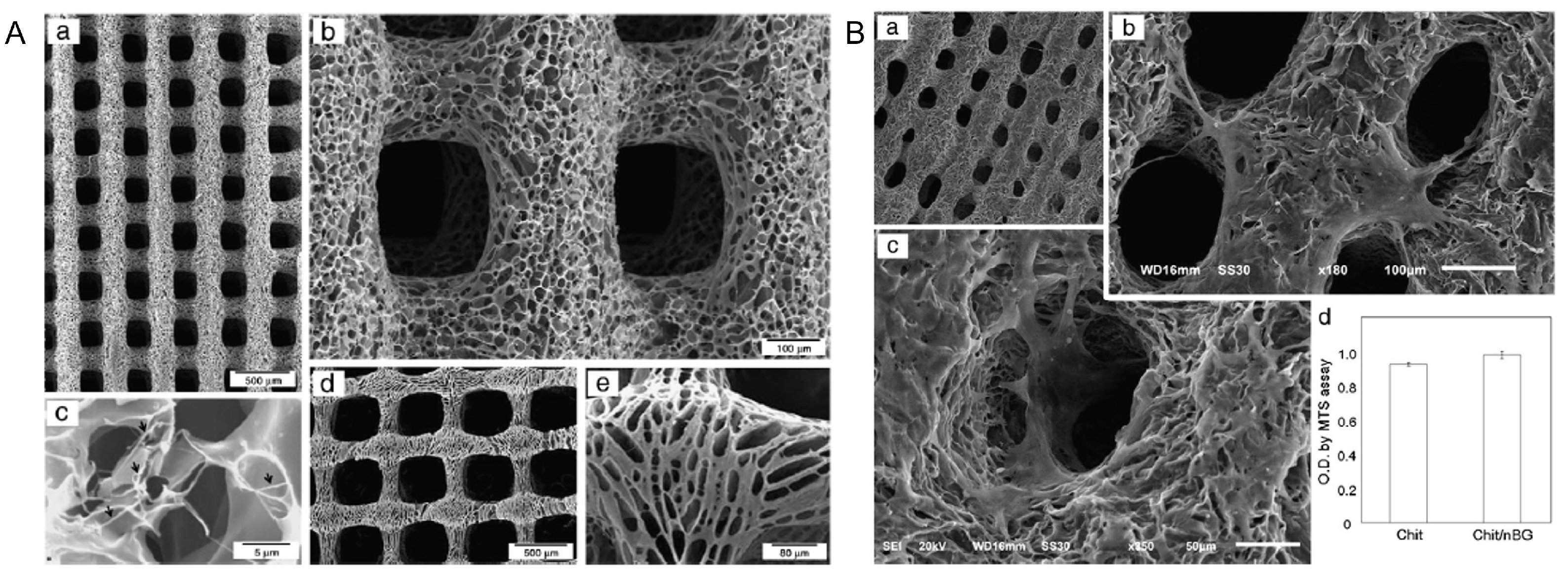
- Dorj, B.; Park, J.H.; Kim, H.W. Robocasting chitosan/nanobioactive glass dual-pore structured scaffolds for bone engineering. Mater. Lett. 2012, 73, 119–122. [Google Scholar] [CrossRef]
- Luo, G.; Ma, Y.; Cui, X.; Jiang, L.; Wu, M.; Hu, Y.; Luo, Y.; Pan, H.; Ruan, C. 13-93 bioactive glass/alginate composite scaffolds 3D printed under mild conditions for bone regeneration. RSC Adv. 2017, 7, 11880–11889. [Google Scholar] [CrossRef] [Green Version]
- Li, J.; Zhang, Y.; Enhe, J.; Yao, B.; Wang, Y.; Zhu, D.; Li, Z.; Song, W.; Duan, X.; Yuan, X.; et al. Bioactive nanoparticle reinforced alginate/gelatin bioink for the maintenance of stem cell stemness. Mater. Sci. Eng. C 2021, 126, 112193. [Google Scholar] [CrossRef]
- Ferreira, A.M.; Gentile, P.; Chiono, V.; Ciardelli, G. Collagen for bone tissue regeneration. Acta Biomater. 2012, 8, 3191–3200. [Google Scholar] [CrossRef]
- Moreira, C.D.; Carvalho, S.M.; Mansur, H.S.; Pereira, M.M. Thermogelling chitosan-collagen-bioactive glass nanoparticle hybrids as potential injectable systems for tissue engineering. Mater. Sci. Eng. C 2016, 58, 1207–1216. [Google Scholar] [CrossRef]
- Li, Z.; Ramay, H.R.; Hauch, K.D.; Xiao, D.; Zhang, M. Chitosan–alginate hybrid scaffolds for bone tissue engineering. Biomaterials 2005, 26, 3919–3928. [Google Scholar] [CrossRef]
- Anekar, N. Design and Analysis of FDM based 3D Printer. Int. J. Res. Eng. Sci. Manag. 2020, 2, 409–411. [Google Scholar]


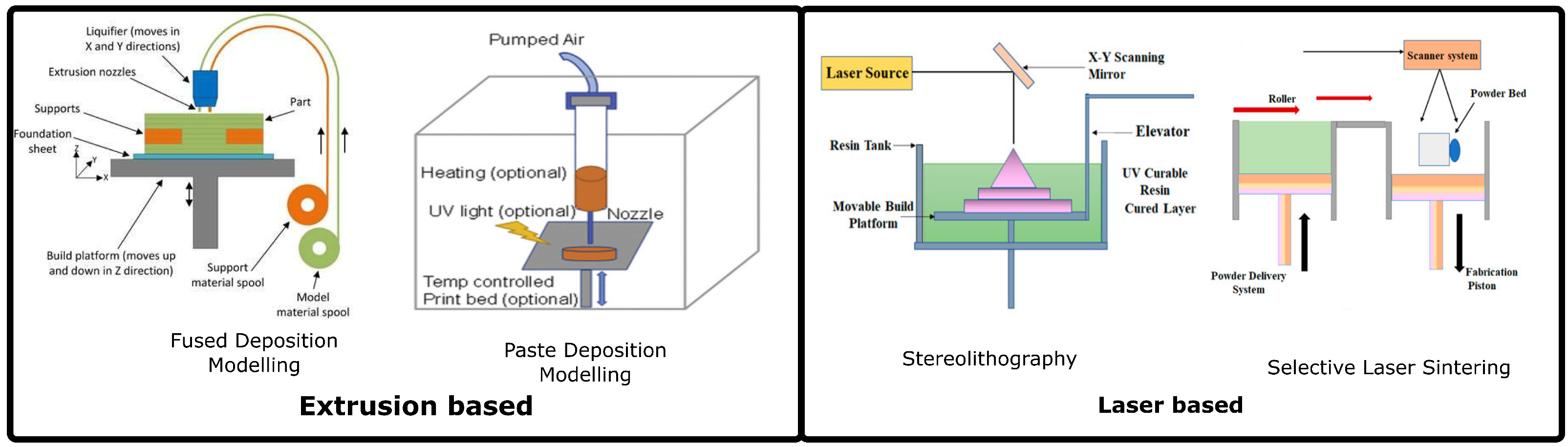
| Technology | Feedstock | Advantage(s) | Disadvantage(s) |
|---|---|---|---|
| Fused Deposition Modelling | Filament | Low cost High reproducibility. | Minimum resolution is 0.02 mm. |
| Paste Deposition modelling | Bioink/Paste | Resolution upto 1 µm. | Rheology of the bioink need to be tailored for efficient printing.Reproducibility depends on the rheology of bioink |
| Selective Laser Sintering | Granules of polymerand ceramic additives | Flexibility in geometries printed | Costly High temperature causes bioactive glass to lose amorphous nature. |
| Stereolithography | Vat of photopolymer inkand bioceramic | Resolution upto 170 nm | Toxicity of resins Exhaustive post processing steps are needed. |
| Polymer | Bioactive Glass Composition | Printing Technology | Summary of Results | Reference |
|---|---|---|---|---|
| PLA | 45S5 45 –24.5 –24.5 –6 wt.% | FDM | Filaments prepared by mixing PLA and bioactive glass in ratios 0, 1, 2.5, 5 and 10 wt.% using a filament maker. Scaffolds loaded with 0–2.5% (wt) BG showed mechanical properties mimicking those of cancellous bone of human proximal tibias. | [78] |
| PLA | 13-93B3 53% B2O3, 20% , 12% K2O, 6% Na2O, 5% MgO, 4% | PDM | PLA BG scaffold is seeded with human adipose Stem cells. The mechanical properties of the scaffold improved in BG-loaded composites. The cell viability was non-uniform with the top layer showing higher viability and reduced viability at the bottom. | [79] |
| PLA | 45S5 (45 –24.5 –24.5 -6 wt.%) | FDM | Confirmation of Hydrocarbonate Apatite layer formation when PLA/BG scaffold is immersed in Simulated Body Fluid. | [80] |
| PCL | 45 –24.5 –24.5 –6 wt.% | DIW | PCL dissolved in dichloromethane and loaded with bioactive glass. Loading of BG reduces the mechanical strength of the scaffolds. BG-loaded scaffold showed significantly higher cell proliferation. | [82] |
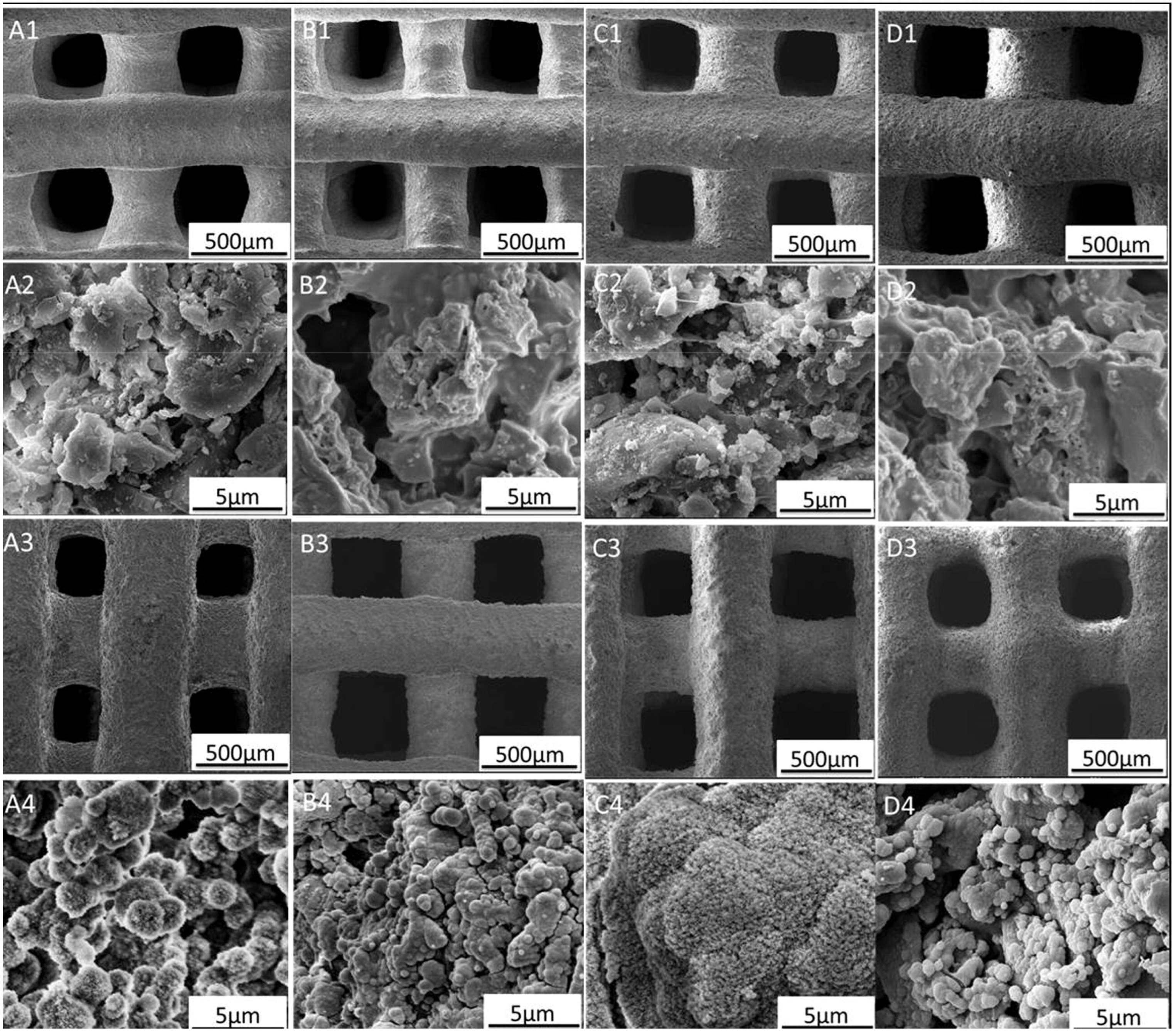
| PCL | 58S (mol%: 60–36–4 | FDM | PCL Scaffolds studied with 0, 5, 10, 20 wt.% Bioactive glass. The higher bioactive glass composition enhances osteogenic differentiation. The higher the content of bioactive glass in the scaffold, the slower the degradation rate of the scaffold. | [99] |
| PCL | 13-93B3 | PDM | Dual extruder 3D printing of PCL/BG and Pluronic F127 hydrogel as cell suspension medium. The scaffolds show the formationof hdroxyapatite layer formation and excellent bioactivity. | [100] |
| PHBV | 45S5 | FDM | There is an increase in mechanical strength with increase in the infill. The biological and mechanical properties match that of ECM of trabecular bone | [101] |
| PCL | Cobalt and strontium doped Bioactive glass | PDM | The scaffolds displayed hydrophilicity, bioactivity, cell viability, and apatite formation capabilities. | [102] |
| PCL | Mg-containing bioactive glass | Precision Entrusion Deposition (FDM) | Different compositions were studied. Scaffold with 50:50 composition was found to be most suitable. | [33] |
| PVA | MBG powder (Si/Ca/P molar ratio 80/15/5) | PDM | Mesoporous bioactive glass scaffolds with hierarchical pore architecture. PVA is used as a binder for 3D printing of BG paste. The scaffolds showed impressive apatite formation capability and sustained drug-release capabilities. | [84] |
| PVA | Sr-MBG | PDM | Scaffold showed excellent mechanical strength, bone forming capabilities and bioactivity. | [73] |
| PPF | DLP | Resin was prepared for DLP technology and tested for bioactivity. | [85] | |
| PGS-PCL | PDM+Electro-spinning | PGS-PCL mats are electrospun on PGS-PCL /bioactive glass composite 3D-printed grid. The scaffold displayed a pH-balancing system and superior mechanical properties compared to 3D-printed grids. | [91] |
| Natural Polymer | Bioactive Glass Composition | Printing Technology | Summary of Results | Reference |
|---|---|---|---|---|
| Silk fibroin | Mesoporous BG NPs | PDM | Porosity and the compression strength of SF-MBG of higher than PCL-MBG BMP-2 and BSP expression was higher in SF-BG scaffolds | [105] |
| Silk fibroin | 45S5 | PDM | Compressive modulus and compressive strength of SF-µBG were superior to SF-nBG SF-nBG would provide a good environment for hBM-MSCs for growth and differentiation | [106] |
| Chitosan | wt.% | PDM | Scaffolds showed higher cellular proliferation | [107] |
| Alginate | 13-93 | PDM | Porosity of these composites improved as the ratio of 13-93 BG increased in the scaffold | [108] |
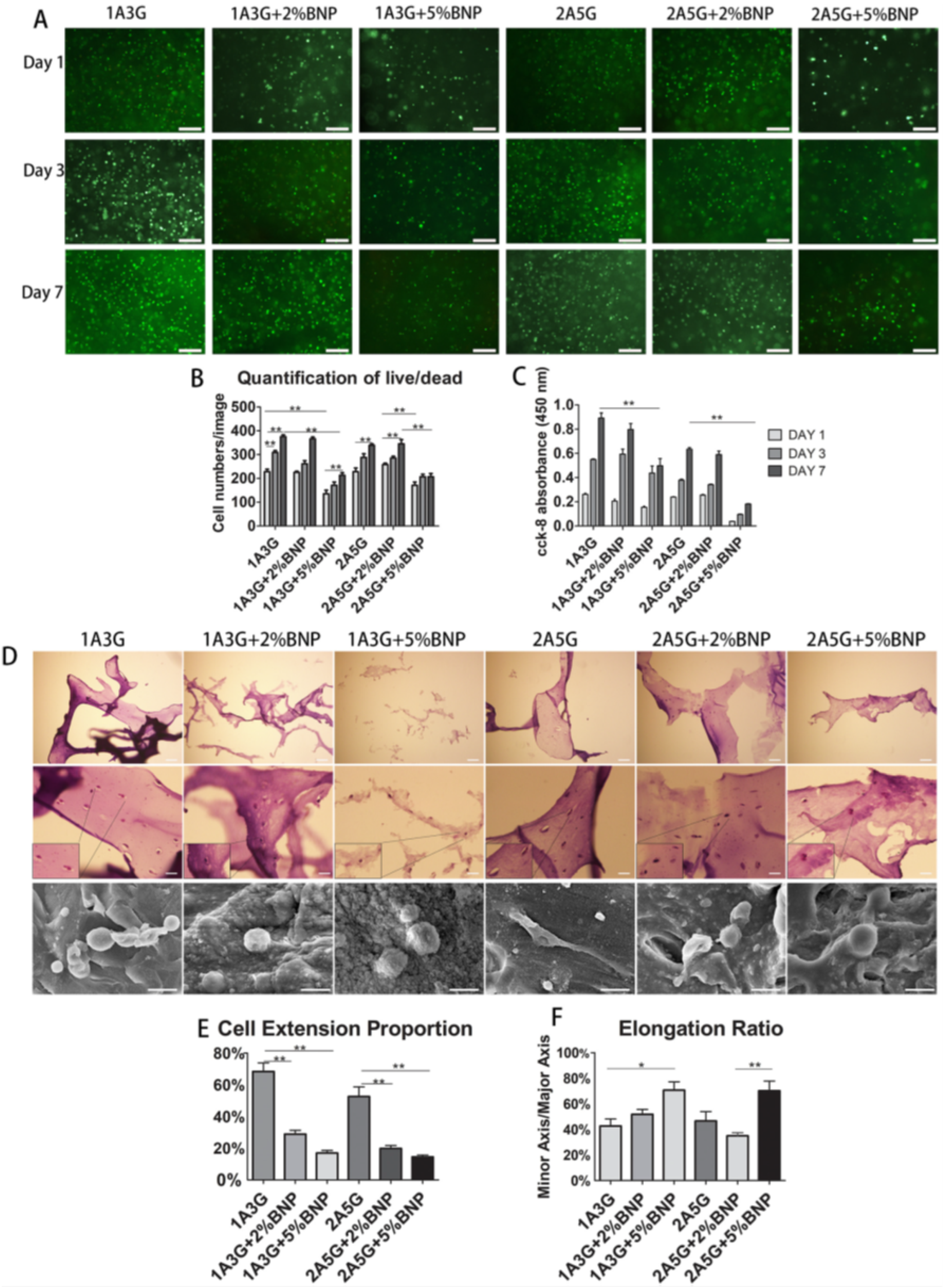
| Alginate-gelatin | BG NPs | PDM | Bioactive glass scaffolds with 2:5 (SA:G) ratio showed needle-like MSC cells with membrane protrusions | [109] |
Publisher’s Note: MDPI stays neutral with regard to jurisdictional claims in published maps and institutional affiliations. |
© 2022 by the authors. Licensee MDPI, Basel, Switzerland. This article is an open access article distributed under the terms and conditions of the Creative Commons Attribution (CC BY) license (https://creativecommons.org/licenses/by/4.0/).
Share and Cite
Dukle, A.; Murugan, D.; Nathanael, A.J.; Rangasamy, L.; Oh, T.-H. Can 3D-Printed Bioactive Glasses Be the Future of Bone Tissue Engineering? Polymers 2022, 14, 1627. https://doi.org/10.3390/polym14081627
Dukle A, Murugan D, Nathanael AJ, Rangasamy L, Oh T-H. Can 3D-Printed Bioactive Glasses Be the Future of Bone Tissue Engineering? Polymers. 2022; 14(8):1627. https://doi.org/10.3390/polym14081627
Chicago/Turabian StyleDukle, Amey, Dhanashree Murugan, Arputharaj Joseph Nathanael, Loganathan Rangasamy, and Tae-Hwan Oh. 2022. "Can 3D-Printed Bioactive Glasses Be the Future of Bone Tissue Engineering?" Polymers 14, no. 8: 1627. https://doi.org/10.3390/polym14081627