
Cytotoxicity Enhancement in MCF-7 Breast Cancer Cells with Depolymerized Chitosan Delivery of α-Mangostin


Abstract
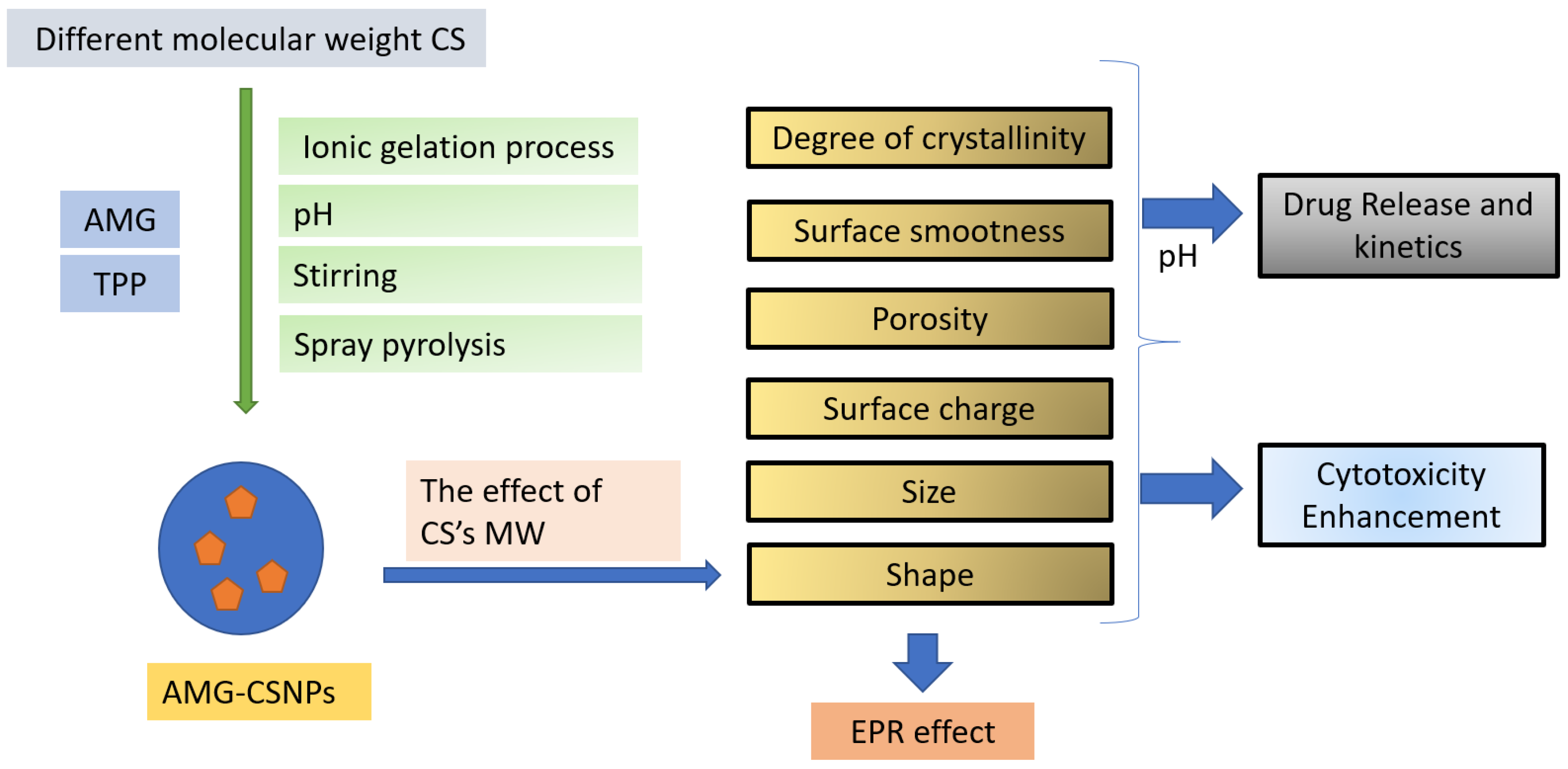
:1. Introduction
2. Materials and Methods
2.1. Materials
2.2. Preparation of Different Molecular Weights of Chitosan
2.3. Characterization CS–LMW
2.3.1. Characterization of CS DDA
2.3.2. CS Molecular Weight Characterization
2.3.3. Fourier-Transform Infrared Analysis
2.3.4. X-ray Diffraction Analysis
2.4. Preparation of CS NPs
2.5. Characterization of CS NPs
2.5.1. Particle Size (PS) and Zeta Potential (ZP)
2.5.2. Nanoparticles Morphology
2.5.3. Fourier-Transform Infrared Analysis
2.5.4. X-ray Diffraction Analysis
2.5.5. Encapsulation Efficiency and Drug-Loading Capacity
2.6. In Vitro Drug Release Study
2.7. In Vitro Cytotoxicity
2.8. Statistical Analysis
3. Results and Discussions
3.1. Characterization of CS–LMW
3.1.1. DDA and MW Characterization
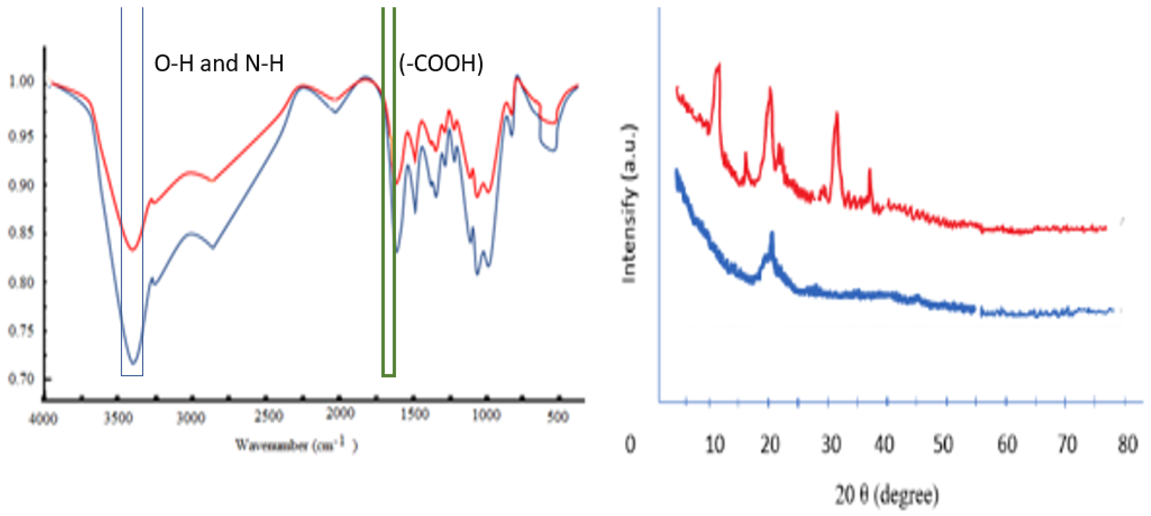
3.1.2. Fourier-Transform Infrared Analysis
3.1.3. X-ray Diffraction Analysis
3.2. Preparation of CS NPs
3.3. Characterization of CSNPs (HMW/LMW)
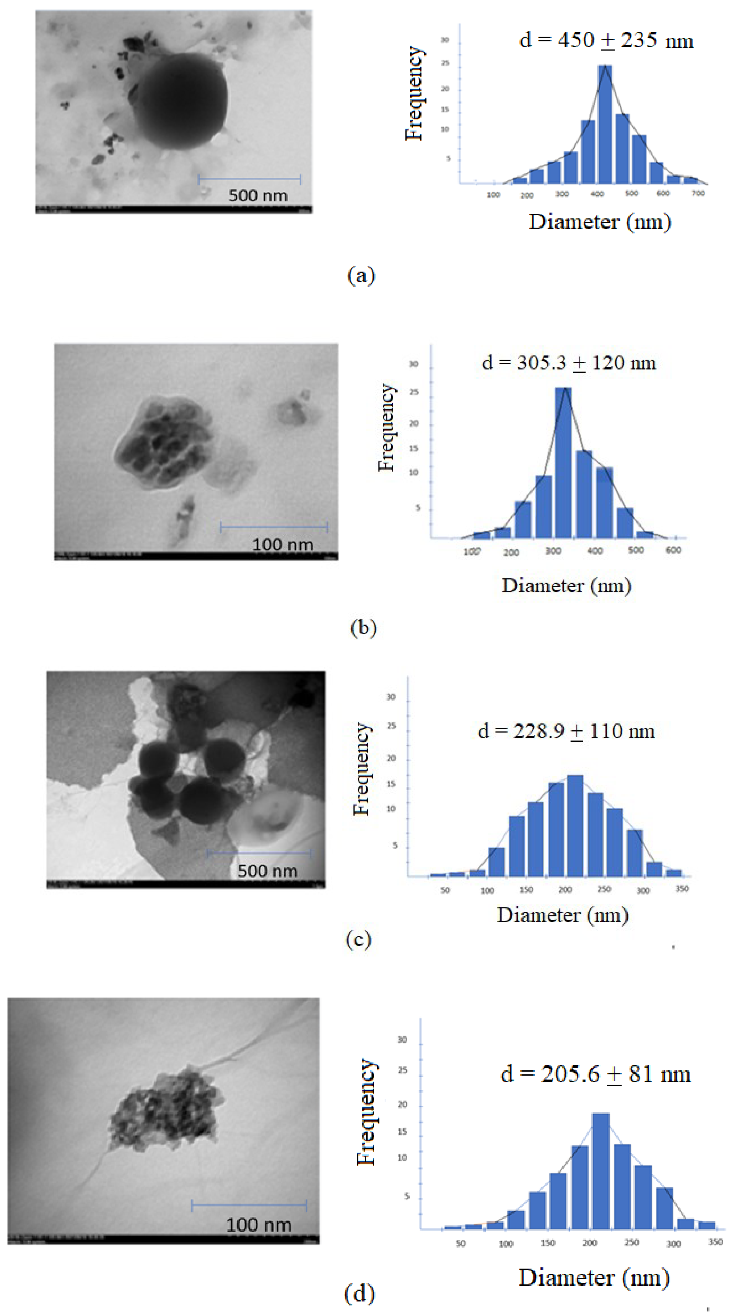
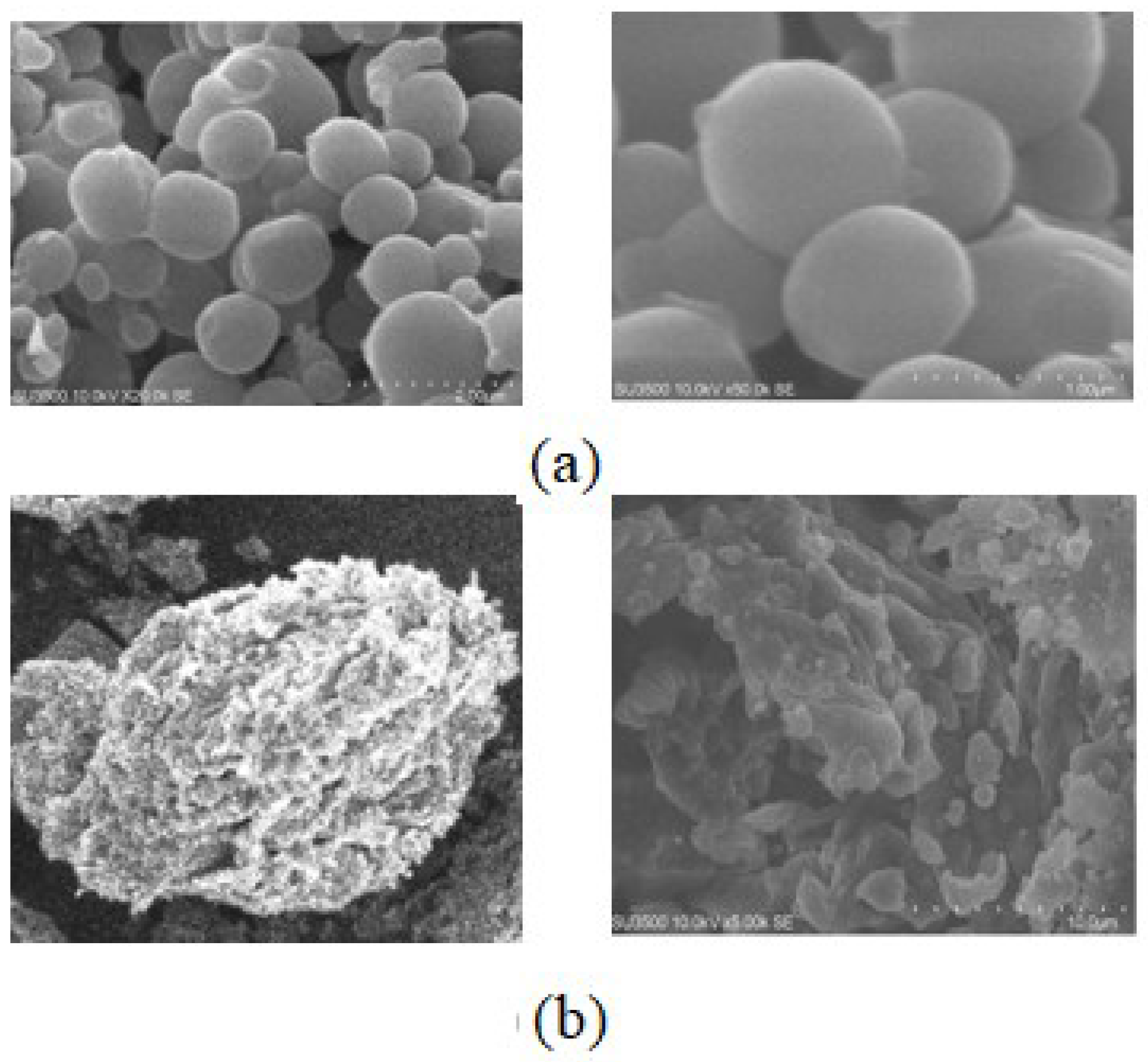
3.3.1. TEM, SEM, Zeta Potential (ZP), Particle Size (PS), Entrapment Efficiency (EE), Drug Loading (DL), and Poly Disperse Index (PDI)
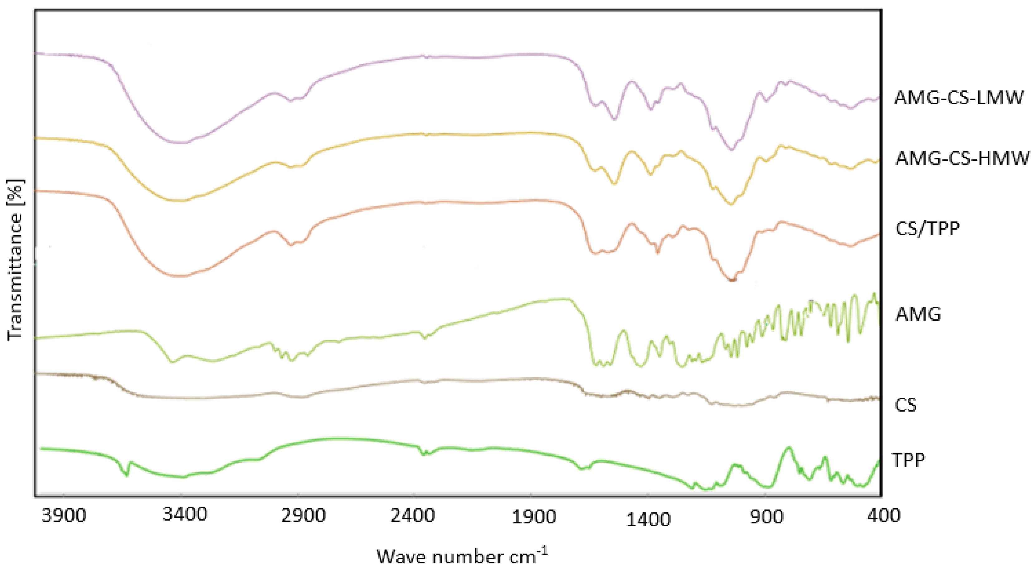
3.3.2. FTIR Analysis
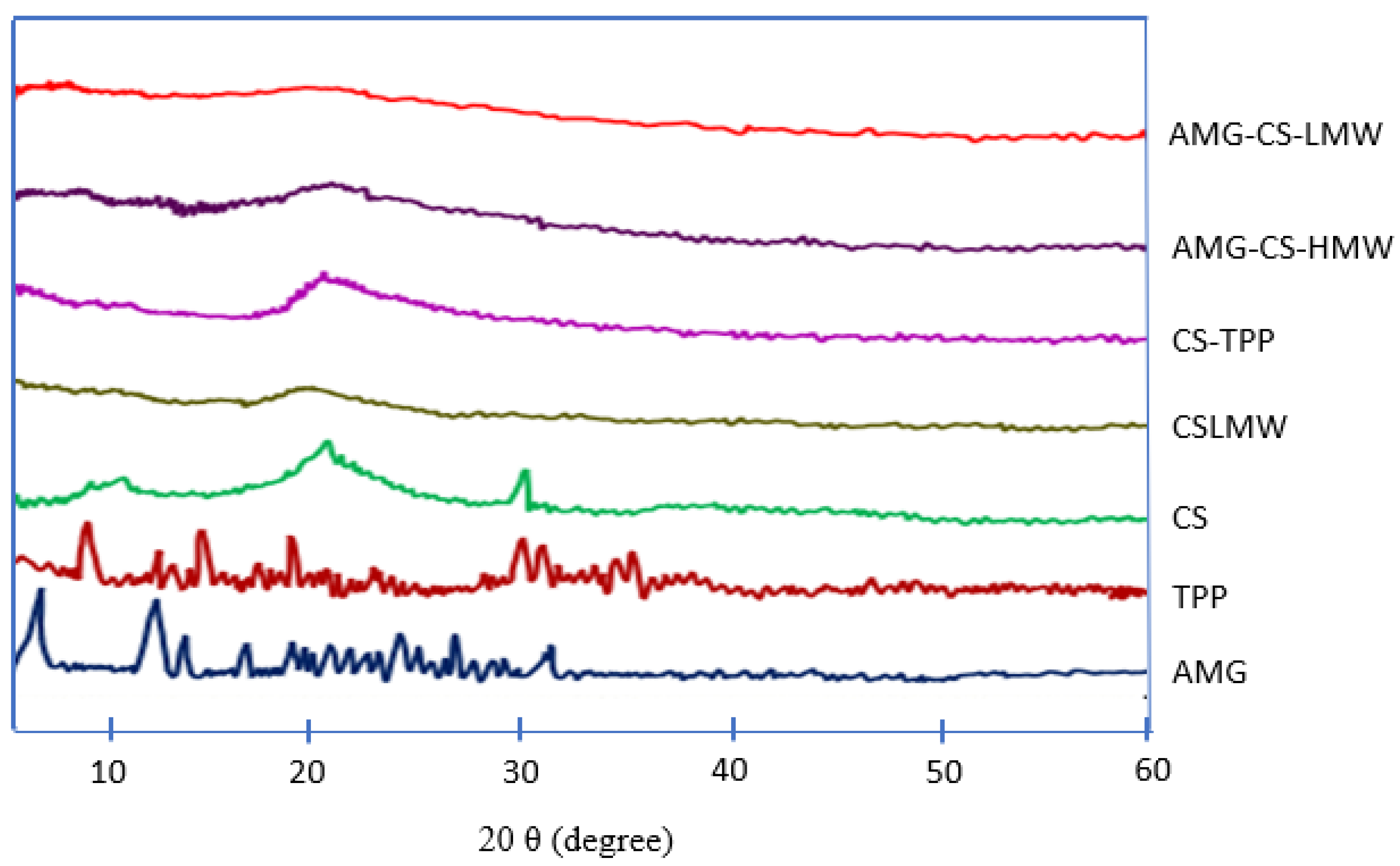
3.3.3. XRD Analysis
3.4. In Vitro Studies
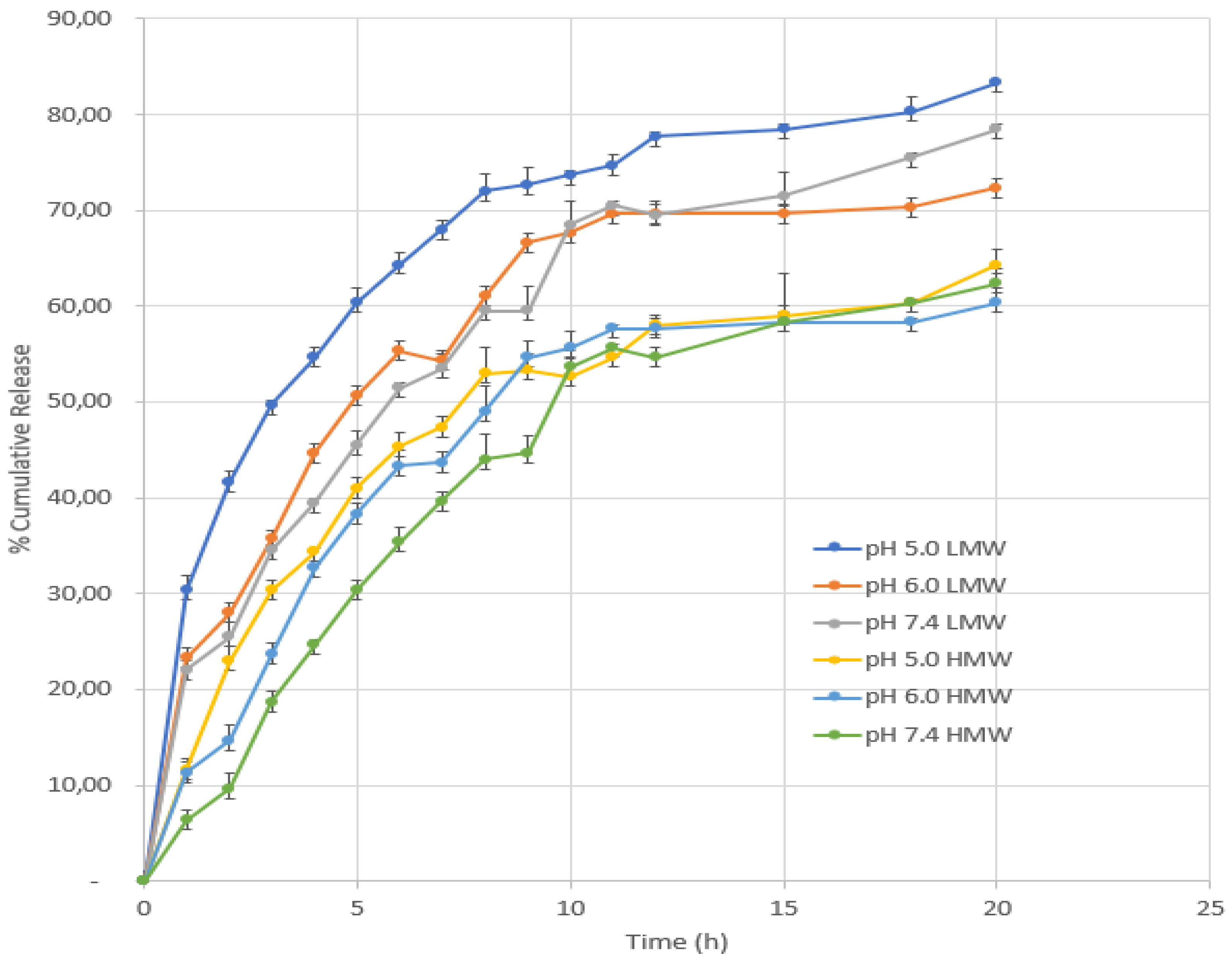
3.4.1. In Vitro Release
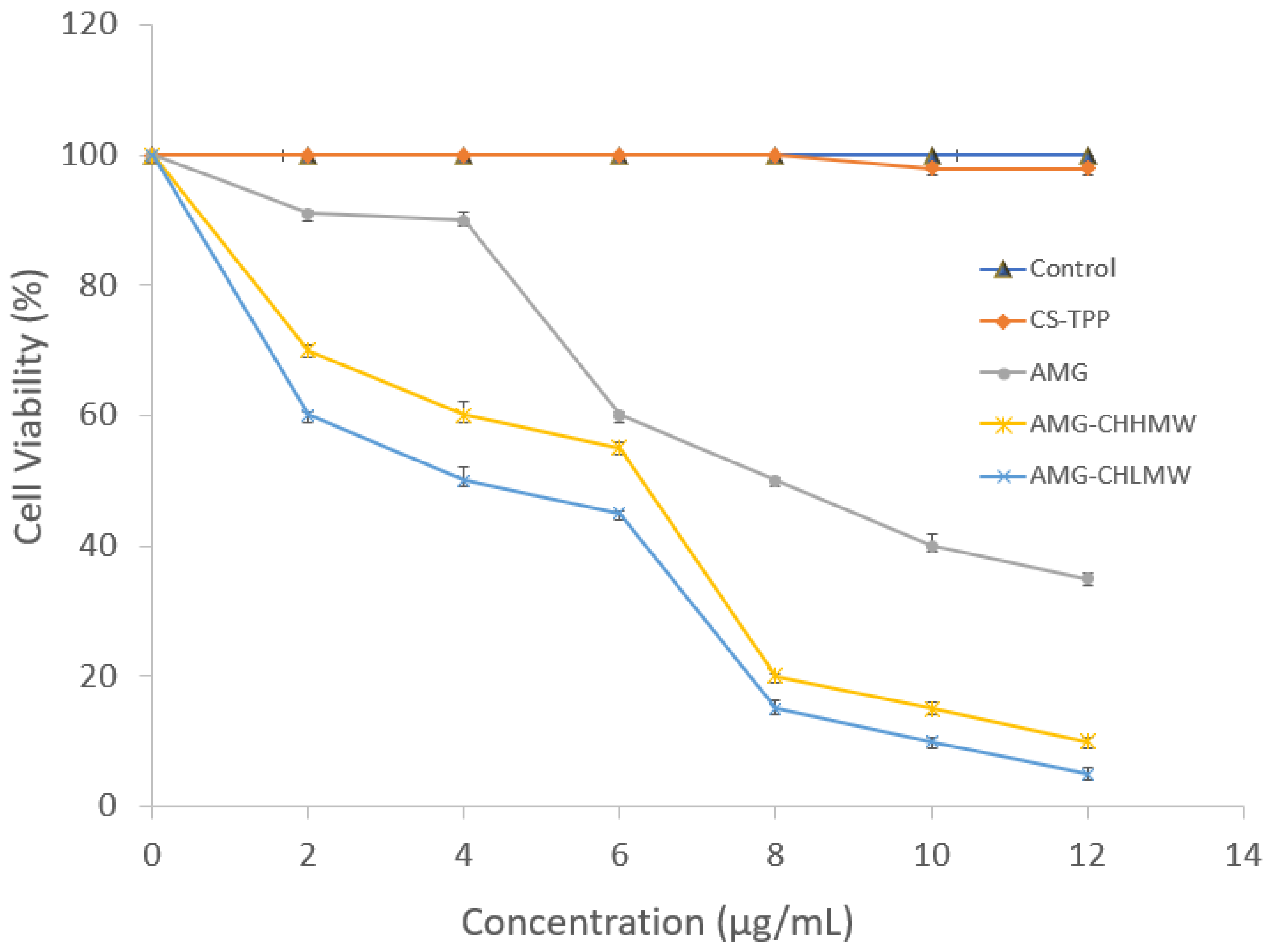
3.4.2. In Vitro Cytotoxicity
4. Conclusions
Author Contributions
Funding
Institutional Review Board Statement
Informed Consent Statement
Data Availability Statement
Acknowledgments
Conflicts of Interest
References
- Sharifi, E.; Bigham, A.; Yousefiasl, S.; Trovato, M.; Ghomi, M.; Esmaeili, Y.; Samadi, P.; Zarrabi, A.; Ashrafizadeh, M.; Sharifi, S.; et al. Mesoporous bioactive glasses in cancer diagnosis and therapy: Stimuli-responsive, toxicity, immunogenicity, and clinical translation. Adv. Sci. 2022, 9, 2102678. [Google Scholar] [CrossRef] [PubMed]
- Rabha, B.; Bharadwaj, K.K.; Pati, S.; Choudhury, B.K.; Sarkar, T.; Kari, Z.A.; Edinur, H.A.; Baishya, D.; Atanase, L.I. Development of polymer-based nanoformulations for glioblastoma brain cancer therapy and diagnosis: An update. Polymers 2021, 13, 4114. [Google Scholar] [CrossRef] [PubMed]
- Ferlay, J.; Colombet, M.; Soerjomataram, I.; Parkin, D.M.; Piñeros, M.; Znaor, A.; Bray, F. Cancer statistics for the year 2020: An overview. Int. J. Cancer 2021, 149, 778–789. [Google Scholar] [CrossRef] [PubMed]
- Lei, S.; Zheng, R.; Zhang, S.; Wang, S.; Chen, R.; Sun, K.; Zeng, H.; Zhou, J.; Wei, W. Global patterns of breast cancer incidence and mortality: A population-based cancer registry data analysis from 2000 to 2020. Cancer Commun. 2021, 41, 1183–1194. [Google Scholar] [CrossRef]
- Kritsanawong, S.; Innajak, S.; Imoto, M.; Watanapokasin, R. Antiproliferative and apoptosis induction of α-mangostin in T47D breast cancer cells. Int. J. Oncol. 2016, 48, 2155–2165. [Google Scholar] [CrossRef] [Green Version]
- Xu, S.; Liu, Y.; Zhang, T.; Zheng, J.; Lin, W.; Cai, J.; Zou, J.; Chen, Y.; Xie, Y.; Chen, Y.; et al. The global, regional, and national burden and trends of breast cancer from 1990 to 2019: Results from the global burden of disease study 2019. Front. Oncol. 2021, 11, 689562. [Google Scholar] [CrossRef] [PubMed]
- Chang, D.; Ma, Y.; Xu, X.; Xie, J.; Ju, S. Stimuli-responsive polymeric nanoplatforms for cancer therapy. Front. Bioeng. Biotechnol. 2021, 9, 707319. [Google Scholar] [CrossRef]
- Ashrafizadeh, M.; Delfi, M.; Hashemi, F.; Zabolian, A.; Saleki, H.; Bagherian, M.; Azami, N.; Farahani, M.V.; Sharifzadeh, S.O.; Hamzehlou, S.; et al. Biomedical application of chitosan-based nanoscale delivery systems: Potential usefulness in siRNA delivery for cancer therapy. Carbohydr. Polym. 2021, 260, 117809. [Google Scholar] [CrossRef] [PubMed]
- George, B.P.; Chandran, R.; Abrahamse, H. Role of phytochemicals in cancer chemoprevention: Insights. Antioxidants 2021, 10, 1455. [Google Scholar] [CrossRef] [PubMed]
- Aggarwal, N.; Yadav, J.; Chhakara, S.; Janjua, D.; Tripathi, T.; Chaudhary, A.; Chhokar, A.; Thakur, K.; Singh, T.; Bharti, A.C. Phytochemicals as potential chemopreventive and chemotherapeutic agents for emerging human papillomavirus–driven head and neck cancer: Current evidence and future prospects. Front. Pharmacol. 2021, 12, 699044. [Google Scholar] [CrossRef]
- Mohan Shankar, G.; Swetha, M.; Keerthana, C.K.; Rayginia, T.P.; Anto, R.J. Cancer chemoprevention: A strategic approach using phytochemicals. Front. Pharmacol. 2022, 12, 809308. [Google Scholar] [CrossRef]
- Herdiana, Y.; Wathoni, N.; Shamsuddin, S.; Muchtaridi, M. α-Mangostin nanoparticles cytotoxicity and cell death modalities in breast cancer cell lines. Molecules 2021, 26, 5119. [Google Scholar] [CrossRef] [PubMed]
- Hidayat, S.; Ibrahim, F.; Pratama, K.; Muchtaridi, M. The interaction of alpha-mangostin and its derivatives against main protease enzyme in COVID-19 using in silico methods. J. Adv. Pharm. Technol. Res. 2021, 12, 285–290. [Google Scholar] [CrossRef] [PubMed]
- Yao, L.; Gu, X.; Song, Q.; Wang, X.; Huang, M.; Hu, M.; Hou, L.; Kang, T.; Chen, J.; Chen, H.; et al. Nanoformulated alpha-mangostin ameliorates Alzheimer’s disease neuropathology by elevating LDLR expression and accelerating amyloid-beta clearance. J. Control. Release 2016, 226, 1–14. [Google Scholar] [CrossRef]
- Jingwen, L.; Jiang, S.; Xiaoting, L.; Yinghui, W.; Hangsheng, Z.; Chaofeng, M.; Wenjie, Y. Low molecular weight chitosan based conjugates for efficient Rhein oral delivery: Synthesis, characterization, and pharmacokinetics. Drug Dev. Ind. Pharm. 2018, 45, 96–104. [Google Scholar] [CrossRef]
- Patra, J.K.; Das, G.; Fraceto, L.F.; Campos, E.V.R.; Rodriguez-Torres, M.D.P.; Acosta-Torres, L.S.; Diaz-Torres, L.A.; Grillo, R.; Swamy, M.K.; Sharma, S.; et al. Nano based drug delivery systems: Recent developments and future prospects. J. Nanobiotechnol. 2018, 16, 1–34. [Google Scholar] [CrossRef] [Green Version]
- Zielinska, A.; Carreiró, F.; Oliveira, A.M.; Neves, A.; Pires, B.; Venkatesh, D.N.; Durazzo, A.; Lucarini, M.; Eder, P.; Silva, A.M.; et al. Polymeric nanoparticles: Production, characterization, toxicology and ecotoxicology. Molecules 2020, 25, 3731. [Google Scholar] [CrossRef]
- Herdiana, Y.; Wathoni, N.; Shamsuddin, S.; Joni, I.M.; Muchtaridi, M. Chitosan-based nanoparticles of targeted drug delivery system in breast cancer treatment. Polymers 2021, 13, 1717. [Google Scholar] [CrossRef]
- Bagheri, M.; Validi, M.; Gholipour, A.; Makvandi, P.; Sharifi, E. Chitosan nanofiber biocomposites for potential wound healing applications: Antioxidant activity with synergic antibacterial effect. Bioeng. Transl. Med. 2022, 7, e10254. [Google Scholar] [CrossRef]
- Narmani, A.; Jafari, S.M. Chitosan-based nanodelivery systems for cancer therapy: Recent advances. Carbohydr. Polym. 2021, 272, 118464. [Google Scholar] [CrossRef] [PubMed]
- Kuen, C.Y.; Masarudin, M.J. Chitosan nanoparticle-based system: A new insight into the promising controlled release system for lung cancer treatment. Molecules 2022, 27, 473. [Google Scholar] [CrossRef] [PubMed]
- Wathoni, N.; Meylina, L.; Rusdin, A.; Fouad, A.; Mohammed, A.; Tirtamie, D.; Herdiana, Y.; Motoyama, K.; Panatarani, C.; Joni, I.M.; et al. The potential cytotoxic activity enhancement of α-mangostin in chitosan-kappa carrageenan-loaded nanoparticle against MCF-7 cell line. Polymers 2021, 13, 1681. [Google Scholar] [CrossRef] [PubMed]
- Herdiana, Y.; Wathoni, N.; Shamsuddin, S.; Muchtaridi, M. Drug release study of the chitosan-based nanoparticles. Heliyon 2022, 8, e08674. [Google Scholar] [CrossRef]
- Lima, K.O.; Pinilla, C.M.B.; Alemán, A.; López-Caballero, M.E.; Gómez-Guillén, M.C.; Montero, P.; Prentice, C. Characterization, bioactivity and application of chitosan-based nanoparticles in a food emulsion model. Polymers 2021, 13, 3331. [Google Scholar] [CrossRef] [PubMed]
- Aibani, N.; Rai, R.; Patel, P.; Cuddihy, G.; Wasan, E.K. Chitosan nanoparticles at the biological interface: Implications for drug delivery. Pharmaceutics 2021, 13, 1686. [Google Scholar] [CrossRef]
- Fan, W.; Fan, W.; Yan, W.; Xu, Z.; Ni, H. Erythrocytes load of low molecular weight chitosan nanoparticles as a potential vascular drug delivery system colloids and surfaces B: Biointerfaces erythrocytes load of low molecular weight chitosan nanoparticles as a potential vascular drug delivery sy. Colloids Surf. B Biointerfaces 2018, 95, 258–265. [Google Scholar] [CrossRef] [PubMed]
- Vedula, S.S.; Yadav, G.D. Chitosan-based membranes preparation and applications: Challenges and opportunities. J. Indian Chem. Soc. 2021, 98, 100017. [Google Scholar] [CrossRef]
- Pita-López, M.L.; Fletes-Vargas, G.; Espinosa-Andrews, H.; Rodríguez-Rodríguez, R. Physically cross-linked chitosan-based hydrogels for tissue engineering applications: A state-of-the-art review. Eur. Polym. J. 2021, 145, 110176. [Google Scholar] [CrossRef]
- Garg, U.; Chauhan, S.; Nagaich, U.; Jain, N. Current advances in chitosan nanoparticles based drug delivery and targeting. Adv. Pharm. Bull. 2019, 7, 113–117. [Google Scholar] [CrossRef]
- Herdiana, Y.; Handaresta, D.F.; Joni, I.M.; Wathoni, N.; Muchtaridi, M. Synthesis of nano-α mangostin based on chitosan and Eudragit S 100. J. Adv. Pharm. Technol. Res. 2020, 11, 95–100. [Google Scholar] [CrossRef]
- Afzali, E.; Eslaminejad, T.; Yazdi Rouholamini, S.E.; Shahrokhi-Farjah, M.; Ansari, M. Cytotoxicity effects of curcumin loaded on chitosan alginate nanospheres on the KMBC-10 spheroids cell line. Int. J. Nanomed. 2021, 16, 579–589. [Google Scholar] [CrossRef] [PubMed]
- Sorasitthiyanukarn, F.N.; Muangnoi, C.; Thaweesest, W.; Rojsitthisak, P.; Rojsitthisak, P. Enhanced cytotoxic, antioxidant and anti-inflammatory activities of curcumin diethyl disuccinate using chitosan-tripolyphosphate nanoparticles. J. Drug Deliv. Sci. Technol. 2019, 53, 101118. [Google Scholar] [CrossRef]
- Yao, Q.; Liu, W.; Gou, X.-J.; Guo, X.-Q.; Yan, J.; Song, Q.; Chen, F.-Z.; Zhao, Q.; Chen, C.; Chen, T. Preparation, Characterization, and Cytotoxicity of Various Chitosan Nanoparticles. J. Nanomater. 2013, 2013, 1–6. [Google Scholar] [CrossRef] [Green Version]
- Filho, I.K.; Machado, C.S.; Diedrich, C.; Karam, T.K.; Nakamura, C.V.; Khalil, N.M.; Mainardes, R.M. Optimized chitosan-coated gliadin nanoparticles improved the hesperidin cytotoxicity over tumor cells. Braz. Arch. Biol. Technol. 2021, 64, 1–14. [Google Scholar] [CrossRef]
- Poerio, A.; Girardet, T.; Petit, C.; Fleutot, S.; Jehl, J.P.; Arab-Tehrany, E.; Mano, J.F.; Cleymand, F. Comparison of the physicochemical properties of chitin extracted from cicada orni sloughs harvested in three different years and characterization of the resulting chitosan. Appl. Sci. 2021, 11, 11278. [Google Scholar] [CrossRef]
- Lewandowska, K.; Szulc, M.; Sionkowska, A. Effect of solvent on the hydrodynamic properties of collagen. Polymers. 2021, 13, 3626. [Google Scholar] [CrossRef] [PubMed]
- Alhajj, N.; Zakaria, Z.; Naharudin, I.; Ahsan, F.; Wenji, L.; Wui, W.T. Critical physicochemical attributes of chitosan nanoparticles admixed lactose-PEG 3000 microparticles in pulmonary inhalation. Asian J. Pharm. Sci. 2019, 16, 374–384. [Google Scholar] [CrossRef]
- Mikušová, V.; Mikuš, P. Advances in chitosan-based nanoparticles for drug delivery. Int. J. Mol. Sci. 2021, 22, 9652. [Google Scholar] [CrossRef]
- Tan, S.L.J.; Billa, N. Improved bioavailability of poorly soluble drugs through gastrointestinal muco-adhesion of lipid nanoparticles. Pharmaceutics 2021, 13, 1817. [Google Scholar] [CrossRef]
- Mao, S.; Shuai, X.; Unger, F.; Simon, M.; Bi, D.; Kissel, T. The depolymerization of chitosan: Effects on physicochemical and biological properties. Int. J. Pharm. 2004, 281, 45–54. [Google Scholar] [CrossRef] [PubMed]
- Zheng, X.; Yin, Y.; Jiang, W.; Xing, L.; Pu, J. Synthesis and Characterization of Low Molecular Weight Chitosan. BioResources 2015, 10, 2338–2349. [Google Scholar] [CrossRef] [Green Version]
- Asasutjarit, R.; Meesomboon, T.; Adulheem, P. Physicochemical properties of alpha-mangostin loaded nanomeulsions prepared by ultrasonication technique. Heliyon 2019, 5, e02465. [Google Scholar] [CrossRef] [PubMed] [Green Version]
- Garms, B.C.; Poli, H.; Baggley, D.; Han, F.Y.; Whittaker, A.K.; Anitha, A.; Grøndahl, L. Evaluating the effect of synthesis, isolation, and characterisation variables on reported particle size and dispersity of drug loaded PLGA nanoparticles. Mater. Adv. 2021, 2, 5657–5671. [Google Scholar] [CrossRef]
- Lee, K.H.; Khan, F.N.; Cosby, L.; Yang, G.; Winter, J.O. Polymer Concentration Maximizes Encapsulation Efficiency in Electrohydrodynamic Mixing Nanoprecipitation. Front. Nanotechnol. 2021, 3, 92. [Google Scholar] [CrossRef]
- Cao, M.; Luo, X.; Wu, K.; He, X. Targeting lysosomes in human disease: From basic research to clinical applications. Signal Transduct. Target. Ther. 2021, 6, 379. [Google Scholar] [CrossRef]
- Lee, S.; Shanti, A. Effect of exogenous ph on cell growth of breast cancer cells. Int. J. Mol. Sci. 2021, 22, 9910. [Google Scholar] [CrossRef]
- Pérez-Herrero, E.; Fernández-Medarde, A. The reversed intra- and extracellular pH in tumors as a unified strategy to chemotherapeutic delivery using targeted nanocarriers. Acta Pharm. Sin. B 2021, 11, 2243–2264. [Google Scholar] [CrossRef]
- Caro León, F.J.; Lizardi-Mendoza, J.; Argüelles-Monal, W.; Carvajal-Millan, E.; López Franco, Y.L.; Goycoolea, F.M. Supercritical CO2 dried chitosan nanoparticles: Production and characterization. RSC Adv. 2017, 7, 30879–30885. [Google Scholar] [CrossRef] [Green Version]
- Lucio, D.; Martínez-Ohárriz, M.C. Chitosan: Strategies to increase and modulate drug release rate. In Biological Activities and Application of Marine Polysaccharides; IntechOpen: Rijeka, Croatia, 2017. [Google Scholar] [CrossRef] [Green Version]
- Abolhasani, M.H.; Safavi, M.; Goodarzi, M.T.; Kassaee, S.M.; Azin, M. Identification and anti-cancer activity in 2D and 3D cell culture evaluation of an Iranian isolated marine microalgae. DARU J. Pharm. Sci. 2018, 26, 105–116. [Google Scholar] [CrossRef] [PubMed]
- Caprifico, A.E.; Foot, P.J.S.; Polycarpou, E.; Calabrese, G. Overcoming the protein corona in chitosan-based nanoparticles. Drug Discov. Today 2021, 26, 1825–1840. [Google Scholar] [CrossRef]
- Qandil, A.M.; Marji, T.; Altaani, B.M.; Khaledd, A.H.; Badwan, A.A. Depolymerization of HMW into a predicted LMW chitosan and determination of the degree of deacetylation to guarantee its quality for research use. J. Excip. Food Chem. 2018, 9, 1–13. [Google Scholar]
- Mojumdar, A.; Kumar, A.; Raina, V.; Ray, L. A simple and rapid colorimetric method for the estimation of chitosan produced by microbial degradation of chitin waste. J. Microbiol. Methods 2019, 158, 66–70. [Google Scholar] [CrossRef] [PubMed]
- Almualla, M.A.; Mousa, M.N.; Sattar, M. Chemical modification and characterization of chitosan for pharmaceutical applications. Egypt. J. Chem. 2021, 64, 3635–3649. [Google Scholar] [CrossRef]
- Ang, L.F.; Por, L.Y.; Yam, M.F. Study on different molecular weights of chitosan as an immobilization matrix for a glucose biosensor. PLoS ONE 2013, 8, e70597. [Google Scholar] [CrossRef] [PubMed] [Green Version]
- Giri, T.K. Nanoarchitectured Polysaccharide-Based Drug Carrier for Ocular Therapeutics; Elsevier Inc.: Amsterdam, The Netherlands, 2016; ISBN 9780323477222. [Google Scholar]
- Chen, M.; Runge, T.; Wang, L.; Li, R.; Feng, J.; Shu, X.; Shi, Q. Hydrogen bonding impact on chitosan plasticization. Carbohydr. Polym. 2018, 200, 115–121. [Google Scholar] [CrossRef] [PubMed]
- Günseli, M.; Tilkan, Y.; Özdemir, N. Investigation of the parameters affecting the release of flurbiprofen from chitosan microspheres. Braz. J. Pharm. Sci. 2017, 53, 1–12. [Google Scholar]
- Hadji, H.; Bouchemal, K. Effect of micro- and nanoparticle shape on biological processes. J. Control. Release 2022, 342, 93–110. [Google Scholar] [CrossRef] [PubMed]
- Cheng, J.; Zhu, H.; Huang, J.; Zhao, J.; Ma, S.; Zhang, H.; Fan, D. The physicochemical properties of chitosan prepared by microwave heating. Food Sci. Nutr. 2020, 8, 1987–1994. [Google Scholar] [CrossRef]
- Liu, F.; Xu, J.; Wu, L.; Zheng, T.; Han, Q.; Liang, Y.; Zhang, L.; Li, G.; Yang, Y. The influence of the surface topographical cues of biomaterials on nerve cells in peripheral nerve regeneration: A review. Stem Cells Int. 2021, 2021. [Google Scholar] [CrossRef]
- De Farias, B.S.; Djenifer, D.; Grundmann, R.; Rizzi, F.Z.; Soares, N.; Martins, S.; Roberto, T.; Anna, S.; Junior, C.; Antonio, L.; et al. Production of low molecular weight chitosan by acid and oxidative pathways: E ff ect on physicochemical properties. Food Res. Int. 2019, 123, 88–94. [Google Scholar] [CrossRef]
- Ma, Z.; Wang, W.; Wu, Y.; He, Y.; Wu, T. Oxidative degradation of chitosan to the low molecular water-soluble chitosan over peroxotungstate as chemical scissors. PLoS ONE 2014, 9, e100743. [Google Scholar] [CrossRef]
- Kamaly, N.; Yameen, B.; Wu, J.; Farokhzad, O.C. Degradable controlled-release polymers and polymeric nanoparticles: Mechanisms of controlling drug release. Chem. Rev. 2016, 116, 2602–2663. [Google Scholar] [CrossRef] [PubMed] [Green Version]
- Gull, N.; Khan, S.M.; Zahid Butt, M.T.; Khalid, S.; Shafiq, M.; Islam, A.; Asim, S.; Hafeez, S.; Khan, R.U. In vitro study of chitosan-based multi-responsive hydrogels as drug release vehicles: A preclinical study. RSC Adv. 2019, 9, 31078–31091. [Google Scholar] [CrossRef] [Green Version]
- Popova, E.V.; Zorin, I.M.; Domnina, N.S.; Novikova, I.I.; Krasnobaeva, I.L. Chitosan–tripolyphosphate nanoparticles: Synthesis by the ionic gelation method, properties, and biological activity. Russ. J. Gen. Chem. 2020, 90, 1304–1311. [Google Scholar] [CrossRef]
- Pokharkar, V. Studies on effect of pH on cross-linking of chitosan with sodium tripolyphosphate: A technical note studies on effect of pH on cross-linking of chitosan with sodium tripolyphosphate: A technical note. AAPS PharmSciTech 2006, 7, E138–E143. [Google Scholar] [CrossRef]
- Fukumori, Y.; Takeuchi, H.; Ando, Y. Structural control of nanoparticles. In Nanoparticle Technology Handbook; Elsevier: Amsterdam, The Netherlands, 2018; ISBN 9780444641106. [Google Scholar]
- Hembram, K.C.; Prabha, S.; Chandra, R.; Nimesh, S.; Hembram, K.C.; Prabha, S.; Chandra, R.; Ahmed, B.; Nimesh, S. Advances in preparation and characterization of chitosan nanoparticles for therapeutics. Artif. Cells Nanomed. Biotechnol. 2016, 44, 305–314. [Google Scholar] [CrossRef] [PubMed]
- Dasgupta, S.; Auth, T.; Gompper, G. Shape and orientation matter for the cellular uptake of nonspherical particles. Nano Lett. 2014, 14, 687–693. [Google Scholar] [CrossRef] [PubMed]
- Ye, H.; Shen, Z.; Yu, L.; Wei, M.; Li, Y. Manipulating nanoparticle transport within blood flow through external forces: An exemplar of mechanics in nanomedicine. Proc. R. Soc. A Math. Phys. Eng. Sci. 2018, 474, 20170845. [Google Scholar] [CrossRef] [PubMed] [Green Version]
- Bruinsmann, F.A.; Pigana, S.; Aguirre, T.; Souto, G.D.; Pereira, G.G.; Bianchera, A.; Fasiolo, L.T.; Colombo, G. Chitosan-coated nanoparticles: Effect of chitosan molecular weight on nasal transmucosal delivery. Pharmaceutics 2019, 11, 86. [Google Scholar] [CrossRef] [Green Version]
- Abdullah, A.S.; El Sayed, I.E.T.; El-Torgoman, A.M.A.; Kalam, A.; Wageh, S.; Kamel, M.A. Green Synthesis of silymarin–chitosan nanoparticles as a new nano formulation with enhanced anti-fibrotic effects against liver fibrosis. Int. J. Mol. Sci. 2022, 23, 5420. [Google Scholar] [CrossRef] [PubMed]
- El-Naggar, N.E.A.; Saber, W.E.I.A.; Zweil, A.M.; Bashir, S.I. An innovative green synthesis approach of chitosan nanoparticles and their inhibitory activity against phytopathogenic Botrytis cinerea on strawberry leaves. Sci. Rep. 2022, 12, 3515. [Google Scholar] [CrossRef]
- Saraf, N.S. Formulation and evaluation of antifungal agent in a hydrogel containing nanoparticle of low molecular weight chitosan. Int. J. Res. Pharm. Sci. 2020, 11, 247–259. [Google Scholar]
- Mishra, A.; Chaturvedi, P.; Mishra, P.; Sudheesh, M. Comparative evaluation of amorphous polymers in solubility and bioavailability enhancement of famotidine through solid dispersion. EJPPS Eur. J. Parenter. Pharm. Sci. 2021, 262. [Google Scholar] [CrossRef]
- Li, Y.; Mann, A.K.P.; Zhang, D.; Yang, Z. Processing impact on in vitro and in vivo performance of solid dispersions—a comparison between hot-melt extrusion and spray drying. Pharmaceutics 2021, 13, 1307. [Google Scholar] [CrossRef] [PubMed]
- Sagita, E.; Syahdi, R.R.; Arrahman, A. Synthesis of polymer-drug conjugates using natural polymer: What, why and how? Pharm. Sci. Res. 2018, 5, 97–115. [Google Scholar]
- Motoyama, K.; Tanida, Y.; Hata, K.; Hayashi, T.; Higashi, T.; Ishitsuka, Y.; Kondo, Y.; Irie, T.; Kaneko, S.; Arima, H. Potential use of a megamolecular polysaccharide sacran as a hydrogel-based sustained release system. Chem. Pharm. Bull. 2014, 62, 636–641. [Google Scholar] [CrossRef] [Green Version]
- Wathoni, N.; Motoyama, K.; Higashi, T.; Okajima, M.; Kaneko, T.; Arima, H. Enhancement of curcumin wound healing ability by complexation with 2-hydroxypropyl-γ-cyclodextrin in sacran hydrogel film. Int. J. Biol. Macromol. 2017, 98, 268–276. [Google Scholar] [CrossRef]
- Ghasemi, M.; Turnbull, T.; Sebastian, S.; Kempson, I. The mtt assay: Utility, limitations, pitfalls, and interpretation in bulk and single-cell analysis. Int. J. Mol. Sci. 2021, 22, 12827. [Google Scholar] [CrossRef]
- Wang, Z.; Deng, X.; Ding, J.; Zhou, W.; Zheng, X.; Tang, G. Mechanisms of drug release in pH-sensitive micelles for tumour targeted drug delivery system: A review. Int. J. Pharm. 2018, 535, 253–260. [Google Scholar] [CrossRef]
- Parodi, A.; Buzaeva, P.; Nigovora, D.; Baldin, A.; Kostyushev, D.; Chulanov, V.; Savvateeva, L.V.; Zamyatnin, A.A. Nanomedicine for increasing the oral bioavailability of cancer treatments. J. Nanobiotechnol. 2021, 19, 354. [Google Scholar] [CrossRef]
- Sethi, A.; Ahmad, M.; Huma, T.; Khalid, I.; Ahmad, I. Evaluation of low molecular weight cross linked chitosan nanoparticles, to enhance the bioavailability of 5-flourouracil. Dose-Response 2021, 19, 15593258211025353. [Google Scholar] [CrossRef] [PubMed]


| Formulation | F1 | F2 | F3 | F4 |
|---|---|---|---|---|
| AMG (mg) | 20 | 20 | 20 | 20 |
| CS–HMW (mg) | 200 | 100 | ||
| CS–LMW (mg) | 200 | 100 | ||
| Sodium tripolyphosphate (mg) | 50 | 50 | 50 | 50 |
| CS | NaNO2 (mg/mL) | DDA | Mw (kDa) |
|---|---|---|---|
| CS–HMW | 0 | 80.0 | 300 |
| CS–LMW | 9 | 75.0 | 20 |
| Material | Result | Literature | Functional Groups Reference |
|---|---|---|---|
| CS–HMW | 350,954 | 347,868 | O–H stretch dan N–H stretch |
| 289,424 | 292,413 | C–H stretch | |
| 165,302 | 165,688 | C = O | |
| 1597 | 157,105 | N–H bend [38] | |
| 141,963 | 142,253 | C–H bend | |
| 137,816 | 137,816 | C–N | |
| 115,731 | 115,731 | C–O–C stretch | |
| 1079 | 102,518 | C–O | |
| CS–LMW | 350,954 | 347,868 | O–H stretch dan N–H stretch |
| 289,424 | 292,413 | C–H stretch | |
| 165,302 | 165,688 | C = O | |
| 1597 | 157,105 | N–H bend [38] | |
| 141,963 | 142,253 | C–H bend | |
| 137,816 | 137,816 | C–N | |
| 115,731 | 115,731 | C–O–C stretch | |
| 1079 | 102,518 | C–O |
| Formula | PS (nm) | ZP (mV) | Shape | Entrapment Efficiency (%) | Drug Loading (%) | PDI |
|---|---|---|---|---|---|---|
| F1 | 450.9 ± 235 | +51.56 ± 2.4 | Spherical | 90.45 ± 1.20 | 6.93 ± 0.87 | 0.487 ± 0.04 |
| F2 | 305.3 ± 120 | +15.69 ± 2.3 | Spherical | 89.35 ± 1.10 | 6.51 ± 0.54 | 0.394 ± 0.05 |
| F3 | 228.3 ± 110 | +25.56 ± 3.4 | Spherical | 88.36 ± 1.12 | 7.10 ± 1.70 | 0.435 ± 0.04 |
| F4 | 205.3 ± 81 | +10.56 ± 2.2 | Spherical | 85.35 ± 1.74 | 6.12 ± 0.86 | 0.375 ± 0.02 |
| Parameter | pH 7 | pH 6 | pH 4 | |||
|---|---|---|---|---|---|---|
| CS–HMW | CS–LMW | CS–HMW | CS–LMW | CS–HMW | CS–LMW | |
| Slope (%h−0.5) | 23.33 ± 0.21 | 23.79 ± 0.72 | 23.40 ± 0.23 | 23.65 ± 0.21 | 20.80 ± 0.20 | 21.02 ± 0.21 |
| Correlation coefficient (r) | 0.98 ± 0.02 | 0.98 ± 0.01 | 0.95 ± 0.01 | 0.96 ± 0.01 | 0.96 ± 0.01 | 0.96 ± 0.02 |
Publisher’s Note: MDPI stays neutral with regard to jurisdictional claims in published maps and institutional affiliations. |
© 2022 by the authors. Licensee MDPI, Basel, Switzerland. This article is an open access article distributed under the terms and conditions of the Creative Commons Attribution (CC BY) license (https://creativecommons.org/licenses/by/4.0/).
Share and Cite
Herdiana, Y.; Wathoni, N.; Shamsuddin, S.; Muchtaridi, M. Cytotoxicity Enhancement in MCF-7 Breast Cancer Cells with Depolymerized Chitosan Delivery of α-Mangostin. Polymers 2022, 14, 3139. https://doi.org/10.3390/polym14153139
Herdiana Y, Wathoni N, Shamsuddin S, Muchtaridi M. Cytotoxicity Enhancement in MCF-7 Breast Cancer Cells with Depolymerized Chitosan Delivery of α-Mangostin. Polymers. 2022; 14(15):3139. https://doi.org/10.3390/polym14153139
Chicago/Turabian StyleHerdiana, Yedi, Nasrul Wathoni, Shaharum Shamsuddin, and Muchtaridi Muchtaridi. 2022. "Cytotoxicity Enhancement in MCF-7 Breast Cancer Cells with Depolymerized Chitosan Delivery of α-Mangostin" Polymers 14, no. 15: 3139. https://doi.org/10.3390/polym14153139