Review

Cubosomes: Design, Development, and Tumor-Targeted Drug Delivery Applications

1
School of Pharmaceutical Science, Universiti Sains Malaysia, Minden 11800, Malaysia
2
Department of Nutrition Sciences, University of Management and Technology, Punjab 54770, Pakistan
*
Authors to whom correspondence should be addressed.
Polymers 2022, 14(15), 3118; https://doi.org/10.3390/polym14153118
Submission received: 28 June 2022 / Revised: 18 July 2022 / Accepted: 22 July 2022 / Published: 31 July 2022
(This article belongs to the Special Issue Biodegradable Polymers for Controlled Drug Release and Delivery)

Abstract

:
Because of the extraordinary advancements in biomedical nanotechnology over the last few decades, traditional drug delivery systems have been transformed into smart drug delivery systems that respond to stimuli. These well-defined nanoplatforms can boost therapeutic targeting efficacy while reducing the side effects/toxicities of payloads, which are crucial variables for enhancing patient compliance by responding to specific internal or external triggers. Cubosomes are lipid-based nano systems that are analogous to well-known vesicular systems, such as lipo- and niosomes. They could be used as part of a unique drug delivery system that includes hydro-, lipo-, and amphiphilic drug molecules. In this review, we critically analyze the relevant literature on cubosomesregarding theories of cubosomeself-assembly, composition, and manufacturing methods, with an emphasis on tumor-targeted drug delivery applications. Due to the bioadhesive and -compatible nature of cubosome dispersion, this review also focuses on a variety of drug delivery applications, including oral, ophthalmic and transdermal.

1. Introduction

The loss or diminution of cellular control and normal maturation pathways is a hallmark of cancer. Excessive cell development, undifferentiated cells and tissues, and the capacity to expand into surrounding tissues and metastasis are all characteristics of cancer. The excision of tumor and possibly neighboring tissues, chemo- and immunotherapy, radiation treatment, or combination of these are all options for treatment. However, some treatments have been linked to serious side effects. As a result, one of the most active fields of cancer research is the development of systems that might selectively deliver medication molecules to the diseased site without increasing their levels in the healthy parts of the organism [1]. A current trend in nanomedicine and drug delivery research is studies on improving the specificity of the treatment to increase efficacy and avoid side effects. Nanoparticles have played a large role in this trend because they can accumulate in target tissues either passively (via the so-called enhanced permeation and retention effect [2]) or actively (using surface-conjugated targeting ligands [3]).
Targeted drug delivery, also known as “drug targeting”, is used frequently and is quite different from “targeted therapy”, which is used in drug discovery [4]. Drug targeting can be defined as the accumulation of certain amounts of drugs at the specific or targeted site regardless of the route and method of drug administration. Targeted therapy is quite different and involves the interaction of the moiety with its receptors at a molecular level. Four components play integral roles in effective drug targeting, namely retain, evade, target, and release [5]. For intravenous administration, the drug is loaded to an efficient carrier for delivery, remains in circulation for sufficient time period, is retained within intended sites, and then is released for the effective functioning of the drug [6].
Drug targeting can be distinguished by two types, namely active and passive. These terms are usually used for in vitro analysis and do not correspond to the in vivo drug behavior. Passive targeting refers to the collection of the drug around the leaky vasculature of tumor sites, which is called the enhanced permeation and retention (EPR) effect. Passive targeting happens to almost all drug carriers, regard less if such targeting is required or not. The bulk (>95%) of the dispensed nanoparticles is believed to aggregate in the liver, spleen, and lungs; however, the EPR effect can be attained for the intravenous administration of nanoparticles. So, when >95% of an administered dose of a drug ends up in non-specific sites of the body, it cannot be called selective targeting. As such, it is evident that passive targeting is inappropriate—it is delivery of the drug by blood circulation in the body. Therefore, it should be replaced by the term extravasation or blood circulation because it is not selective and occurs after the majority of the drug administration process has concluded. Efficient “blood circulation and extravasation” is evident in locally activated delivery, where the drug remains at a specific site, such as around the tumor but not in other parts of the body [7].
“Active targeting” is specifically known as a ligand–receptor interaction and can also be narrated as the interaction of a drug/drug vector with the intended cells. Ligand–receptor interactions can only occur when the two moieties are in close proximity (<0.5 nm). The term “active targeting” resembles a cruise missile guiding the moiety to a specific target compared with traditional drug delivery systems, where the drug is mainly distributed by circulation and is retained at unintended sites [8,9,10]. Active targeting is a specific “ligand–receptor-type interaction” for intracellular localization. So, in order to achieve active (or effective) targeting, there should be enhanced blood circulation. PEGylation (the modification of nanoparticles with poly (ethylene glycol)) or enhancing the EPR effect can extend blood circulation time. The results also show that the presence of a tumor-targeting ligand does not necessarily lead to greater nanovector retention in tumors, implying that “active targeting” does not always imply “effective delivery to the entire tumor” [11,12]. Moreover, pegylated cubosomes exhibited an improved targeting of certain aggressive tumors, i.e., ovarian cancer cells [13].
The drug delivery facilitated by the lipid nanoparticle has proven fundamental in the development of liquid crystalline colloidal carriers, e.g., cubo-, spongo-, and hexosomes, and vesicles as indicated in Figure 1 [14]. Cubosomes are bi-continuous lipids containing nanoparticles, and have discrete, sub-micron, cubic liquid crystalline phases. The formulation of these nanocarriers involves a specific ratio of certain surfactants in order to form fine dispersion [15]. Cubosomes are nanoparticles or, more precisely, nanostructure particles, which are generated by the self-assembly of amphiphilic- or surfactant-like molecules in liquid crystalline phases with cubic crystallographic symmetry. Surfactant-controlled cubosome structuresare separated by two zones of water, and are also optically isotropic, viscous, and characterized by a solid lipid crystalline cubic symmetry that is comparable to honeycomb (cavernous) structures, and range in sizes from 100 to 500 nm. The continuous repeating and sophisticated 3D structures of cubic liquid crystalline phases provide excellent opportunities to entrap hydrophobic, hydrophilic, and amphiphilic components [16]. Bi-continuous water and oil channels are formed by amphiphilic molecules, with bi-continuous referring to two distinct (but non-intersecting) hydrophilic regions separated by the bilayer. The structure’s interconnectivity produces a clear, viscous gel with rheology, which looks similar to cross-linked polymer hydrogels [17].
The structure entirely distinguished cubosomes from hexosomes, which contains hexagons due to its inverse hexagonal phase. The cubosome assemblies present further advantages because the transformations between the different liquid crystalline organizations, e.g., Pn3m, Im3m, and Ia3d, in addition to the inverted hexagonal phases, can be tuned and controlled by changes in the temperature, ionic strength, or pH of the environment of the targeted application sites [18,19].
Cubosomes are isotropic and transparent in nature, and their cubic liquid crystals are physically stable when in excess of water. They are biodegradable, non-toxic, and have the ability to solubilize hydrophobic, hydrophilic, and amphiphilic compounds. Due to their thermodynamic stability, cubosomes last indefinitely [20]. Colloidal dispersions of cubosomes can be stabilized by the addition of polymers. They also possess the potential for the controlled delivery of actives, where diffusion is governed by the tortuous diffusion of the active through the “regular” channel structure of the cubic phase [21]. Due to their narrow pore sizes, cubosomes facilitate the controlled release of drugs and they are vital for maintaining the stability and efficacy of biologically active compounds, such as proteins and vitamins [22]. Compared with other liquid crystalline drug delivery systems, such as lipo- and hexosomes, cubosomes have lower viscosity and exist at any dilution in water and further cubic phases [23,24].
The cubic structure of cubosomes has the ability to entrap the moiety and then release agents based on different molecular weights and polarities, which follows the laws of Higuchi-diffusion-controlled kinetics [25,26].
Q = D m C d 2 A C d t ½
According to equation, the release (diffusion) of agents from the matrix depends on the square root of time. Q is the quantity of the agents released per unit area of the matrix, where Dm is the diffusion coefficient of the agents in the cubic matrix, Cd is the solubility of the agents in matrix, A is the primary quantity of the drug per unit volume of the matrix, and t is the time. From that equation, the quantity and rate of drug release can be estimated.
Nanostructured cubosomes, made by fragmenting bulk cubic phase gels or by lyotropic techniques, have the same cubic phase inner-structure but a substantially larger specific surface area and reduced viscosity. Cubosomes are regarded as promising vehicles for various routes of drug administration due to their unique properties, such as thermodynamic stability, bioadhesion, their ability to encapsulate hydrophilic, hydrophobic, and amphiphilic substances, and their potential for controlled release through functionalization. This review introduces cubosomes, concentrating on their structures, preparation methods, potential administration routes, and tumor-targeted drug delivery based on the most recent publications.

2. Types of Cubosomes

Cubosomes can be differentiated into liquid and powdered cubosome precursors depending upon the method used for formulation. Cubosomes can be spontaneously formed by the dilution of monoolein with a hydrotropicsolvent, such as ethanol. The nucleation process permits particles to form, which increases through crystallization and precipitation processes. The quid precursor technique allows for a faster cubosome preparation scale-up while avoiding bulk solids handling and possibly destructive high-energy operations. This method could be applied to produce cubosomes in situ [17].
In addition to the liquid cubosome precursors, powdered cubosomes can be prepared using dehydrated surface-active agents that are incorporated with a suitable polymer. By using the spray-drying technique, the powdered cubosomes can be formed after the encapsulation of particles from liquid droplets in emulsion and dispersion. This technique utilizes the coating of a waxy lipid on water-soluble and non-cohesive starch, which usually prevents agglomeration [27]. However, it is better to use cubosomes in a dry powder rather than liquid form in order to prevent the processing of bulk water.

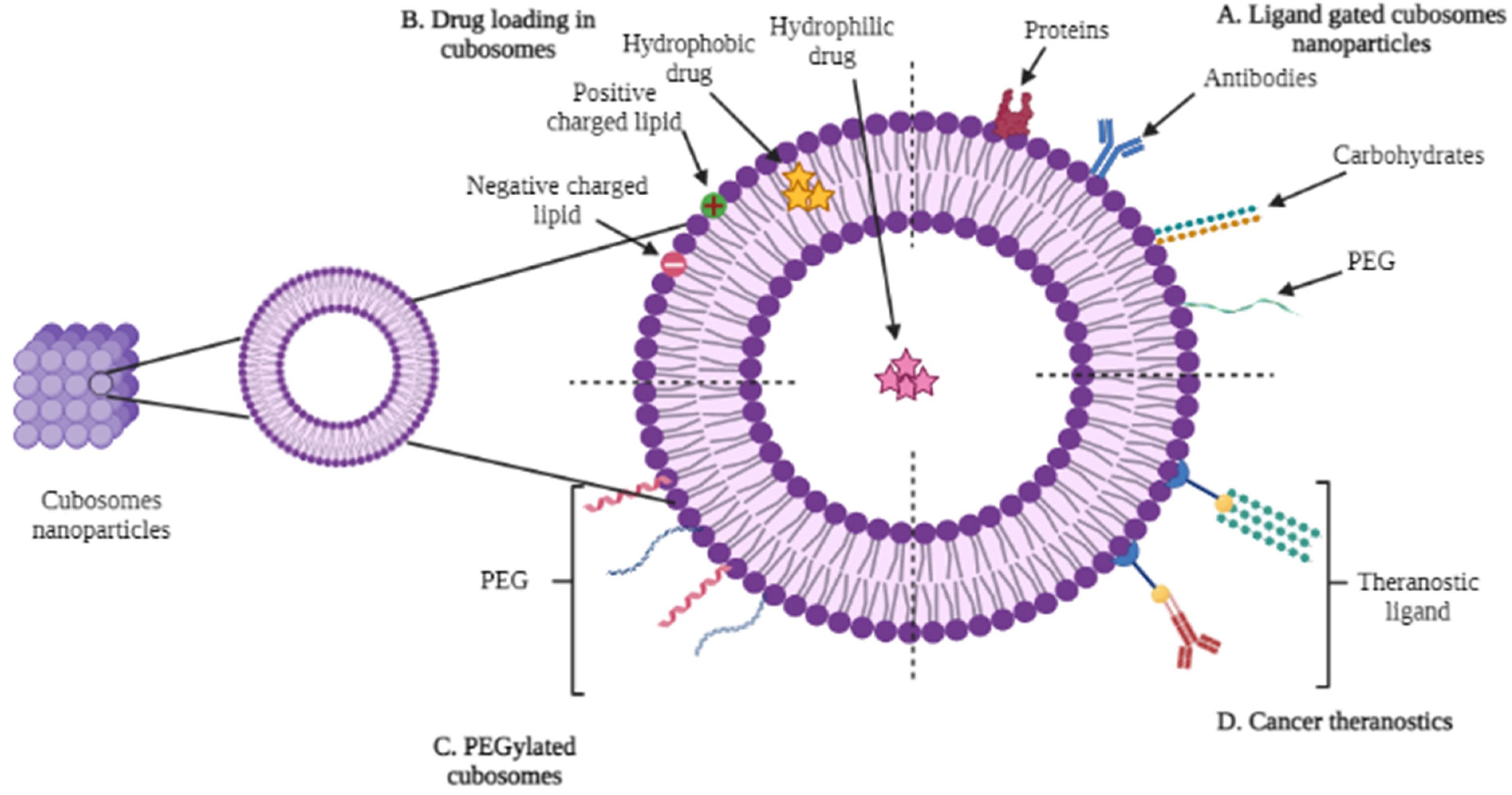
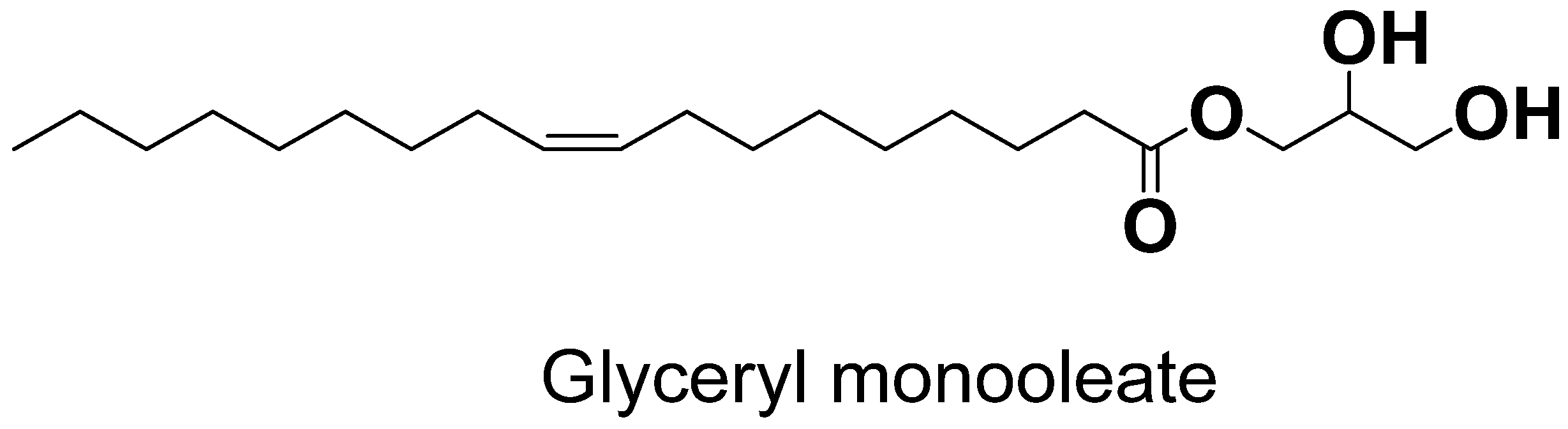
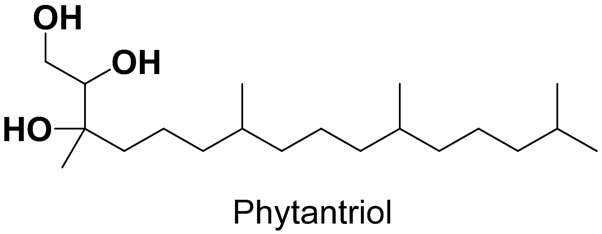
3. Components of Cubosomes

Amphiphilic lipids and stabilizers of various grades are the main components of cubosomes Table 1. Lipids are used to formulate cubic crystals based on their amphiphilic profile and these include monoglycerides, glycol- and phospholipids, and alkyl glycerates. Depending on the composition, lipid molecular structure, electrostatic interactions, and pressure and temperature, these amphiphilic lipids can self-assemble and form a wide range of nanostructures with distinct physicochemical properties and geometries [28].
The most common and widely used amphiphilic lipid for the formulation of cubosomes is glyceryl monooleate (GMO), which is also referred to as monoolein. The physical and chemical properties of GMO, which are significant for cubosome synthesis, include the polarity of the unsaturated monoglyceride, which has a melting point of 35–37 °C, storage temperature of 20–30 °C, and a HLB value of three, and it is usually colorless and clear in nature. In order to make bi-continuous cubic structures, GMO can be synthesized by a mixture of glycerides and estersfrom oleic and other fatty acids and mainly consists of monooleate [37].
Due to presence of hydroxyl groups in the head portion of the GMO structure, which contributed to the formation of a H-bond with water molecules and hydrocarbon chains in the tail, GMO acts as an amphiphile because it has hydrophilic and -phobic characteristics (Figure 2). In 1984, it was first recommended as a biocompatible encapsulating material. Being non-toxic and biocompatible and -degradable, it is used as an emulsifier in the food industry and in many pharmaceutical preparations [38].
Along with GMO, phytantriol is the other lipid used in cubosome formulation due to its increased penetration power and emulsification properties. Compared with monoglycerides, phytantriol gained more interest recently because of its characteristics, including better chemical stability due to absence of an ester group (Figure 3) and an improved moisture retention. Commercially available phytantriol has high purity (95%), while monoglycerides are present with different purities because they are synthesized from various sources. PHYT-based liquid crystalline matrices are considered a remarkable drug delivery system because they are able to release drugs at sustained rates, especially hydrophilic moieties [30,39].
Cubosomes are composed of bulk cubic aggregates that are thermodynamically stable but they are not kinetically stable due to the interaction of the hydrophobic portion with external hydrophilic aqueous media. So, in order to increase the stability of the formulation, there is a need to use a stabilizer in order to avoid the agglomeration of the dispersed particles into the parent bulk cubic structure. The stabilizer provides the electrostatic barrier between the particles in order to keep them dispersed and to avoid re-coalescence [40].
The stabilizer affects the dispersed particles’ structures and controls their phase behaviors. In particular, a sufficient concentration of P407 ensures the presence of the P-type cubic phase, which is in charge of forming a stable colloidal dispersion. The internal liquid crystalline structure and chemical makeup determine the frequency and nature of interactions between P407 and bulk liquid crystalline systems. For instance, P407 is integrated into the liquid crystalline structure of the GMO cubic phase as opposed to being adsorbed onto the surface of the PHYT cubic phase [41].
Pluronics are the most widely used stabilizing agents in the formation of cubosomes, especially the F127 (gold standard), which is also known as “Poloxamer 407”. These copolymers are composed of polyethylene oxide (PEO) and polypropylene (PPO) and are water soluble. The hydrophobic and -philic properties of Pluronics are because of the polypropylene and -ethylene oxide portions of Pluronics. The stabilizer can be used in different concentrations depending on the dispersed particles and types of lipids used in the formulation. In preparations containing GMO as a lipid, Pluronics are added up to 20% w/w depending on the total weight of the dispersion and the concentration of the stabilizer varies from 2.5% to 10% (w/w) [42].
The use of non-ionic molecules as a stabilizer has been popularized recently instead of using F127. The researchers found that the stabilizing effect of the stearate class of poly (ethylene oxide) is more effective than the F127 when used in the formulation of phytantriolcubosomes. In particular, product Myrj59®, which is 100 poly (ethylene oxide) units, was more effective in stabilizing phytantriolcubosomes when compared with the gold standard F127. The mechanism and reason for this enhanced stability are still unclear. On the other hand, polyvinyl alcohol (PVA) also showed great results in stabilizing the dispersion, and it can be used in cubosome preparations [33].

4. Methods of Preparation for Cubosomes

Techniques to form cubosomesinclude the bottom-up, top-down, spray-drying, and solvent evaporation methods. However, the main approaches for preparation are the top-down and bottom-up techniques. Both techniques require a suitable stabilizer, such as F127, to prevent cubosome dispersion aggregation, as previously described. The selection of the optimum method is to ensure stability, biocompatibility, and optimal drug release through the cubic matrix of the cubosomes [43].

4.1. Top-Down Approach

This is the most widely used method in the synthesis of cubosomes, especially when glyceryl monooleate (GMO) is used as a lipid polymer. This technique requires the application of high levels of energy in order prepare the fine dispersion of cubosomes. The steps involved are: first form the bulk cubic aggregates by adding a suitable stabilizer to a lipid; then, apply high energy through a homogenizer in order to form the dispersion. Fortunately, cubosomes prepared by this technique are stable for up to one year. However, the main drawback of this method is that when preparing large-scale batches, the application of high levels of energy is not viable when temperature-sensitive bioactive agents, especially peptides and proteins, are incorporated [44].

4.2. Bottom-Up Approach

The other method forcubosome preparation is the bottom-up approach, which is widely used when preparing cubosomes containing phytantriol as the lipid. This approach is also known as the solvent dilution method. This method requires a low energy input compared withthe top-down approach. This approach focused on forming cubosomes with a hydrotrope and stabilizer in an excess of water by applying minimal energy. In this technique, the hydrotrope is the key factor, as it controls the solubilization processto dissolve the water-insoluble lipids. It also prevents the formation of liquid crystals at high concentrations. The hydrotrope acts as a surface-active agent and is a molecule able to solubilize poorly soluble agents in aqueous media by hydrotropic solubilization. The bottom-up technique has advantages over the top-down approach because it requires low energy; thus, it can be safely used for the preparation of cubosomes incorporated with temperature-sensitive agents. Due to the uniform dispersion of stabilizers on the surface of the produced nanoparticles, this technique shows long-term formulation stability [45,46].

4.3. Spray-Drying Method

The spray-drying method is used in order to formulate powder precursors of cubosomes, and involves the use of organic solvents that evaporate upon the application of air. The lipid–surfactant solvent mixture is atomized with a wave of hot air, resulting in rapid solvent evaporation and the formation of the dry powder form of the cubosome precursor. It starts with mixing the lipid with the stabilizer and then dissolving them into ethanol (binary solutions can also be used). A separate mixture of aqueous phase, consisting of a hydrophilic solid carrier, i.e., sorbitol or dextran, is prepared, which is then combined with the previously prepared lipid mixture. Both mixtures are combined with continuous stirring, which leads to the formulation of a low viscosity emulsion. This technique is simple, cost effective, and normally easy to scale-up [47].

4.4. Solvent Evaporation Method

Solvent evaporation is another method of producing powder cubosomes by using a homogenizer or ultra-sonicator. The process is quite similar to the spray-drying method, except for the use of a high-energy sonicator. In this method, first, an organic solvent such as ethanol or chloroform is used to dissolve the lipids, which are then added dropwise to the other mixture containing a stabilizer, such as Pluronics inaqueous phase. The mixture is maintained at an elevated temperature under magnetic stirring. The drug can be dispersed in the lipid or the aqueous surfactant solution. Stirring under elevated temperatures removes the volatile organic solvent, and the mixture is homogenized by ultra-sonication or homogenizer, resulting in the formation of cubosomes [48]. Table 2 illustrates the benefits and drawbacks of these four techniques, which are usually used in the formation of cubosomes.

5. Physical–Chemical Characterizationsof Cubosomes

The physical and chemical characterizations of cubosomes include the evaluation of morphology using scanning electron or transmission electron microscopes. The particle size of a nanocarrier can be tested using dynamic light scattering (DLS). It also includes the evaluation of the zeta potential of the nanocarrier, and the polydispersity index (PDI) and drug encapsulation efficiency (EE%) of cubosomes. Table 3 describes the physical and chemical characterization of cubosomes.

6. Cubosomes as Tumor-Targeted Drug Delivery

Cancer is a huge threat to human life. Even after 50 years of research, the standard conventional treatment is not always effective in treating the disease. Only lymphocytic leukemia and Hodgkin’s lymphoma can be successfully treated with conventional therapy. The main drawback of conventional chemotherapy is that it delivers the toxic anticancer agent indiscriminately to tumors and normal organs and tissues. So, standard treatment isalso harmful to normal cells and tissues in the body. Therefore, there is a need to formulate a therapy that only targets the cancerous site, and does not affect the normal healthy tissues. This can only be established by delivering the antitumor drugs selectively to affected areas, thereby protecting the normal tissues. Exploiting the structural and pathophysiological anomalies of tumor tissues, particularly the tumor vasculature, and employing the increased permeability and retention (EPR) effect, is one of the most successful drug delivery techniques [57].
Anticancer drugs have a restricted clinical use due to their non-specific distribution in living creatures, thereby causing side effects in normal tissues. So, in order to overcome this, various drug carriers have been developed to deliver agents at specific sites. Nanocarriers are among the most effective to target the disease site, and is based on the enhanced permeation and retention effect (EPR effect). Targeting ligands that are complementary to specific or overexpressed receptors on cancer cells, for example, has been utilized to functionalize carrier surfaces, and an external magnetic field can also help in tumor medication delivery. The tumor microenvironment should be taken into consideration because it is heterogeneous in nature. Due to high interstitial pressures and a thick extracellular matrix (ECM), the delivery by nanoparticles can be compromised. However, by changing the size and improvising the microenvironment of the cancerous site, the accumulation and penetration of a nanocarrier can be enhanced. By normalization of tumor vasculature and deforming the ECM, the reflux of nanoparticles can be reduced, leading to high concentrations of the carrier at a specific tumor site. However, there are certain unanticipated side effects that should be considered regarding the disruption of ECM, which can enhance tumor migration and ultimately lead to tumor metastasis. The premature restoration of blood arteries may make it harder for nanoparticles to passively target tumor tissues via the EPR effect. The extent and timing of the microenvironment modification must be carefully balanced [58,59].
In the late 1960s, the concept of a bi-continuous liquid crystal phase was originally presented, and it was finally realized in the mid-1970s. The reverse bi-continuous cubic phases, especially those based on the monoolein–water system, are the most studied and fascinating of these phases. The applications of modern pharmaceutical nanotechnology in a variety of pharmaceutical sectors, including enzymes, antimuscarinic medicines, antibiotics, and analgesic administration, have been thoroughly researched and reviewed, thanks to the rapid advancement of modern pharmaceutical nanotechnology. The cubic liquid crystal, which could be diffused in water, has good biocompatibility and -adhesivity. Because of their promising qualities, these multifunctional delivery systems can be administered in a variety of methods, including intravenous, oral, percutaneous, and ocular injections. This new cancer treatment, which involves specifically delivering anticancer medicines to the tumor site while minimizing their buildup in healthy organs, has been hailed as a potential technique for reducing treatment side effects [60]. So, we provide a detailed review, which highlights the prospects of cubosomesbeing used in targeted drug delivery to various tumors Table 4.

6.1. Skin Cancer Therapy

Paclitaxel (PTX), a model medication, was encapsulated in this study using monoolein-based cubosomesstabilized by Pluronic F127 and polyethylene glycol polymers. The study was carried out to evaluate the in vivo tumor growth inhibition by paclitaxel (PTX)-loaded cubosomes (CB), and for that purpose, A431 tumor-bearing mice were randomly divided into three groups (five mice per group): Group 1—PBS control; Group 2—PTX control; and Group 3—PTX-CB. We administered 120 μL of the corresponding formulations, which contained a dose of PTX per 15 mg/kg body weight, to each animal. The study revealed that PTX inhibited the A431 tumor growth by lowering the tumor volume of 250 mm3 compared with 360 mm3 in the untreated PBS group after two weeks of treatment. Mice treated with PTX-loaded cubosomes lowered the tumor volume of 160 mm3, which exhibited a 0.7-fold reduction in the final tumor volume compared withthe PTX-free group. This might be due to the whole-body biodistribution study’s findings that PTX-CB accumulates preferentially at tumor locations compared with PTX-free [61].
Another study revealed there was an active targeting of cancer cells by incorporating paclitaxel (PX) to cubosomes that were designed to be monoolein-based nanocarriers. To do this, biotin, a targeted ligand with an affinity for the sodium-dependent multivitamin transporter (SMVT) that is overexpressed in the membrane of tumor cells, was coupled with PF108, a surfactant typically employed to maintain cubosomes. Biotinylated cubosomes with a high level of functionally active biotin on their surface significantly improved the anticancer activity of PX at a concentration of 1 µg/mL when compared with PX or the nontargeted cubosomes. Additionally, the biotin ligand encourages receptor-mediated endocytosis, which enhances the cancercell uptake of the formulation. These findings imply that the biotinylated-cubosome-targeting technique can lower PX toxicity while increasing PX efficacy against tumor cells [62].

6.2. Glioblastoma Multiforme Therapy

Utilizing a top-down method, glyceryl monooleate (GMO) and the surfactant Pluronic F-127 were used to create cubosomes that were loaded with AT101. Because of its capacity to accelerate apoptosis in tumor cells by autophagic cell death, AT101, the R-(-)-enantiomer of the cotton-seed-derived polyphenol gossypol, is a potential medication in glioblastoma multiforme (GBM) therapy. The main drawback of the drug is its low solubility in aqueous solutions and its low bioavailability, which hinder its response during treatment. To circumvent this limitation, AT101 is loaded on cubosomes to increase bioavailability and improve AT101’s anticancer capabilities. Cubosomes, as AT101 drug carriers in GBM cells, are first-time prepared and found to be effective in treatment [63]. The advantage of employing cubosomes while delivering hydrophobic moiety, such as AT101, is an enhancement in the drug’s bioavailability due to its greater solubility in the lipid membrane of the produced nanoparticles, and ultimately their dispersion in water-based media. The pronounced internalization linked to endocytic pathways may also play a role in the enhanced cytotoxicity response to GMO-AT101 cubosomes. Proteins with high binding affinities for AT101 molecules display lower AT101 activity. AT101 is unable to attach to the proteins in the culture medium due to cubosome-based carriers.

6.3. Lung Cancer Treatment

Inhalation medication delivery is extremely advantageous for treating non-small-cell lung cancer (NSCLC) because it requires less dosages and reduces systemic toxicity. An FDA-approved antituberculosis medicine called bedaquiline (BQ) has previously demonstrated outstanding anticancer activity. However, its transport via the lungs is constrained by weak water solubility. BQ-loaded cubosome (BQLC) nanocarriers were formulated that can be inhaled to treat NSCLC. The cubosomal nanocarriers used in the preparation of the BQLC had a particle size of 150.2 ± 5.1 nm, a zeta potential of (+) 35.4 ± 2.3 mV, and an encapsulation efficiency of 51.85 ± 4.83%. After 48 h of treatment, the BQLC showed an improved cellular internalization and cytotoxicity with a 3-fold lower IC50 compared with free BQ in NSCLC (A549) cells. The BQLC decreased colony growth and cancer spread in vitro by suppressing cell proliferation via the apoptotic route [51].

6.4. Colorectal Cancer Therapy

Cisplatin- and metformin-loaded cubosomes were prepared using GMO, F-127, and polyvinyl alcohol and tested against colorectal cancer. Colorectal cancer (CRC) is a dreadful tumor and remains a leading cause of death worldwide. Cisplatin has been found to be successful in treating cancer, although it is associated with serious side effects and drug resistance. Combining anticancer drug treatment with effective drug carriers is needed. That is why cisplatin and metformin are incorporated in the cubosomes formulation. When compared with unformulated cisplatin, cubosomal formulations had a stronger cytotoxic effect. During the treatment of HCT-116, the inhibitory concentration (IC50) of cisplatin was found to be 15 µM, while cisplatin-loaded nano cubosomes reduced the IC50 to 9.6 µM. Furthermore, including metformin into the cisplatin-loaded cubosomes reduced the IC30 cisplatin to 7 µM, inhibiting cell growth by 50%. Cisplatin-loaded nano cubosomes caused glucose and energy levels to drop, resulting in adenosine-monophosphate-activated protein kinase (AMPK) activation and mammalian target of rapamycin (mTOR) suppression. The aforementioned formulation resulted in a significant increase in ROS levels, as well as an increase in NADPH oxidase and caspase 3, and the inhibition of LDH. The cytotoxic effects of the drug-loaded nanoparticles were larger than the effects of the individual drug, as evidenced by the fraction of the cell survival values [64,65].

6.5. Liver Cancer Treatment

In the presence of Poloxamer 407 as a stabilizer, a cubic gel phase of monoolein and water was disrupted to create cubosomal dispersions. The goal of the study was to encapsulate the anticancer medication 5-fluorouracil (5-FU) in order to combat liver cancer. The monoolein and water cubosomal dispersions were made with Poloxamer 407 as a stabilizer. On formulations containing 5-FU, in vitro and in vivo experiments were conducted. The entrapment efficiency was determined to be 31.21% for the drug when compared with a 5-FU solution, revealing nanometer-sized particles with a narrow particle-size distribution. During the first hour of drug release from the cubosomes, nearly half of the entrapped drug was rapidly released, followed by a slower drug release. Rats given 5-FU-loaded cubosomes displayed severe dilatation and congestion to the central (CV) and portal veins (PV) and hepatic sinusoids (S). Additionally, it was discovered that some of the hepatocytes had fatty alterations, and inflammatory cells had infiltrated the space between the hepatocytes in the portal area. The free-5-FU-treated group displayed only minor congestion in the central and portal veins along with fatty alterations and ballooning degeneration in the hepatocytes at the periphery of the hepatic parenchyma, as opposed to the effects of 5-FU-loaded cubosomes. This pattern shows that the free 5-FU group has less hepatotoxicity than the 5-FU-loaded cubosome group. Lower dosages of 5-FU in cubosomal formulations for the treatment of hepatocellular carcinoma need to be further studied for hepatotoxicity and in vivo anticancer efficacy [66].

6.6. Ovarian Cancer Treatment

Icariin was loaded on cubosomes comprising GMO and P407 as a stabilizer. The potential of icariin (ICA)-loaded cubosomes in treating ovarian cancer was explored. The Box–Behnken statistical design was used to optimize the cubosome formulation. The drug entrapment efficiency of the formulations ranged from 78.3 to 97.3%, with particle sizes ranging from 73 to 183 nm. Against ovarian cancer cell lines (SKOV-3 and Caov 3), optimized ICA-loaded cubosomes (ICA-Cubs) showed better cytotoxicity and apoptotic capacity than ICA-raw. The optimized ICA-Cubs were found to be non-toxic to EA. hy926 endothelial cells. The analysis of cell cycle arrest showed a promising role for optimized cubs in the pre-G1 and G2/M phases in comparison with ICA-raw. The enhanced levels of the ICA’s apoptotic potential may be associated with this increased creation of ROS. Therefore, it is anticipated that treatment with ICA-Cubs has a stronger ability to decrease TNF production within the cytosol of the SKOV-3 cells, which may interfere with angiogenesis in the tumor microenvironment, as well as the growth, proliferation, invasion, and metastasis of cancer cells. The formulation strategy has a substantially greater impact because ICA-Cubs produce significantly more caspase-3 than ICA-raw. The enhanced internalization of ICA in the novel formulation may be the cause of this; the placebo formulation had no effect on the amount of caspase-3 in ovarian cancer cells. This function of caspase-3 in the therapy group may be related to p53 expression, which may have an impact on cancer cell growth and metastasis. Due to enhanced drug solubility and cellular permeability, it was concluded that ICA loaded cubosomes have the potential to suppress ovarian cancer cell development [67].

6.7. Cervical Carcinoma

Temozolomide and doxorubicin were mounted on cubosomes carrying miR-7-5p and comprising monoolein and F108. The effects of drug- and miRNA-loaded vehicles were investigated using molecular biology techniques, such as quantitative real-time PCR, anMTS-based cell proliferation test, flow cytometry, and spheroids formation assay. The anticancer effects were tested using cervical carcinoma-derived (HeLa) cells. MiR-7-5p enhances cell sensitivity to medicine, and nanoparticles containing both miRNA and pharmaceuticals have a better antitumor effect than drug treatment alone. The delivery of miRNA-7-5p in conjunction with a medication significantly reduced the ability of the treated cells to survive. Cancer cells treated with miR-7-5p have less chemoresistance. The gathered information shows a connection between the activation of MDR genes and the low intracellular level of miR-7-5p. In GB- (A172 and T98G), cervical-carcinoma- (HeLa), and PTC-derived (TPC-1) cells, it was demonstrated that a reconstitution of the miR-7-5p level led to a decrease in the expression of the PGP, BCRP, and MRP1 and 6 encoding genes [68].

6.8. Hepatocellular Carcinoma Therapy

ABZ-loaded cubosomal dispersions were prepared using GMO and P407 as a stabilizer. Albendazole (ABZ) has been discovered to be a potent inhibitor of numerous cancers, including hepatocellular carcinoma (HCC), which is the largest cause of cancer-related deaths globally. However, ABZ has a low bioavailability, and the processes behind its anticancer activities may be more complex than tubulin inhibition. We looked at the effects of ABZ suspension (i.p. and p.o.) and ABZ-loaded cubosomes (LC) on diethylnitrosamine-induced HCC in mice. The average particle size of ABZ-loaded nanoparticles was 48.17 ± 0.65 nm, with a 93.26 ± 2.48% entrapment effectiveness. When comparing ABZ-loaded cubs to aqueous ABZ suspension, the absorption analysis demonstrated a two-fold improvement in relative bioavailability. Reduced oxidative stress, enhanced liver function, and a lower necro-inflammation score were also discovered. ABZ effectively suppressed CD34 tissue, CD309 mRNA, and VEGF expressions at the protein level. MMP-9 and CXCR4 levels were also lowered, indicating an antimetastatic effectiveness. As revealed by the increased p53 mRNA expression, a greater Bax/BCL-2 ratio, and active caspase-3, ABZ showed a considerablelevel of apoptotic activity. Interrupting the ERK1/2-HIF-1-p300/CREB connection may thus be beneficial [69].
For the sustained release of anticancer drugs, such as cisplatin and paclitaxel, cubosomes coated with a layer of poly-ε-lysine were used. The dispersions of these formulations were investigated by using differential scanning calorimetry and X-ray diffractograms, and they were photographed using a transmission electron microscope. The medicine is completely disseminated in cubosomes according to in vitro tests. In addition, the zeta potential, in vitro release, and entrapment efficiency were assessed. Cubosomes have been discovered to be beneficial in avoiding an initial burst of anticancer medications followed by a steady release of anticancer chemicals. Using the human hepatoma HepG2 cell line, the coated, uncoated, and blank cubosomes were determined to be non-toxic. Impedance measurements and fluorescence imaging validated the cubosomes’ therapeutic efficacy against HeLa cells. Furthermore, the impairment of HeLa cells was demonstrated by the reduction in impedance in cells treated with coated combinatorial cubosomes, as validated by fluorescent microscopy [70].

6.9. Brain Tumor Therapy

Monoolein cubosomes were used to incorporate the anticancer agent doxorubicin (DOX). T98G glioblastoma cells and MTS cell proliferation assay were used to investigate the in vitro cytotoxicity of DOX-loaded cubosomes. Free DOX decreased T98G cell proliferation after 24 h at a dosage of 100 mg/mL but there was no inhibitory action at lower doses. Even after 72 h of incubation, the non-doped cubosome had no cytotoxic impact at a dosage of 90 mg/mL, and roughly 87% of metabolically active cells were present. In addition, after 24 h of incubation with T98G cells, the cytotoxic effect of DOX-loaded cubosomes with a DOX concentration of 2.3 mg/mL revealed that 70% of the viable cells were still present. The cytotoxicity of DOX integrated into cubic nanoparticles at a dosage of 2.3 mg/mL was higher than that of direct DOX delivery, according to our findings. Furthermore, a lower dosage of DOX was required to limit cancer cell proliferation than when free DOX was used [71].

6.10. Breast Cancer Therapy

The toxicity of blank cubosomes and cubosomes modified with folic acid is examined using MCF-7 cells. Blank cubosomes with GMO concentrations ranging from 10 to 1000 μg/mL were added to cells and incubated for 24 h, while FA-modified nanoparticles were used with a concentration of 1: 10 (100 μL/mL of medium). The MTT experiment revealed that neither the blank cubs nor the FAcubs are harmful to MCF-7 cells. The cytotoxicity of etoposide (ETP)-included cubosomes was then assessed using the MTT assay on the MCF-7 human breast cancer cell line, which has high amounts of folate receptors. When compared with free medication, ETP-Cubs had a significantly higher cytotoxicity. In MCF-7 cell lines, the IC50 values of the three groups increased in the sequence ETP > ETP-Cubs > ETP-Cubs-FA. The etoposide-loaded blank cubosomes’ tumor-targeting ability in this study was primarily a result of their EPR effects, whereas the folic-acid-modified cubosomes actively targeted the tumor through an interaction between the folate and its receptor, which overexpressed on the surface of MCF-7 breast cancer cells, resulting in a high level of tumor uptake [72].
Table 4. In vitro cytotoxicity of anticancer cubosomes.
TumorCubosomesAssayCell LineResultReference
Liver Cancer5-FU-loaded cubosomesMTT assayHep G2 cell lineCytotoxicity of 5-FU-loadedcubosomes is much higher than free drug only.[66]
Cisplatin- and paclitaxel-loaded cubosomesMTT assayHep G2 cell lineCytotoxicity of the uncoated-drug-loaded cubosomeswas more than the coated ones, which may be attributed to the faster release of drugs in the case of the uncoated ones.[70]
Colorectal CancerCisplatin and metformin nanocubosomesSulfo-rhodamine B (SRB) assayHCT-116The harmful effects of the drug-loaded nanoparticles were validated by the fraction of the cell survival values, which were higher than the effects of the individual drug.[65]
Breast CancerFolic-acid-modified etoposide cubosomesMTT assayMCF-7cell lines When compared with free medication, ETP-Cubs showed a significant increase in cytotoxicity.[72]
Brain CancerCubosomes loaded with temozolomide (TMZ) and doxorubicin (DOX)MTS cell proliferation assayT98G GB-derived cell linesThe viability of A172 and T98G cells was significantly reduced after cells were transfected with miR-7-5p and then treated with TMZ.[68]
DOX-loaded cubosomesMTS cell proliferation assayT98G glioblastoma cellsDOX incorporated into cubic nanoparticles at a concentration of 2.3µg/mL exerted higher cytotoxicity than direct DOX delivery.[71]
AT101-loaded cubosomesColorimetric WST-1 assayA172cell lines Encapsulated AT101 exhibited stronger cytotoxic effects and more extensive rearrangement of actin fibers in GBM cells than free AT101.[63]
Ovarian CancerIcariin cubosomesMTT assaySKOV-3 and Caov 3Indicated the cytotoxic potential of
ICA-Cubs stops cancer cells from multiplying.
[67]
Cervical CancerCubosomes loaded with doxorubicin labeled with 177LuMTS assayHeLa cellsThe cytotoxicity enhancement became statistically significant only after shorter incubation times.[73]

7. Miscellaneous Drug Delivery by Cubosomes

7.1. Ocular Applications

Many studies have been conducted recently in order to evaluate the efficiency of cubosomes for ocular drug delivery by utilizing their benefits, such as being biodegradable, able to encapsulate all three types of drug molecules, i.e., hydrophilic, hydrophobic, and amphiphilic, and their rendering of bioactive agents with targeted and controlled releases. Because they have a long residence time at the corneal surface, cubosomes proved to have a high ocular bioavailability with the incorporated drugs. GMO-containing cubosomes have mucoadhesive properties that are beneficial in enhancing corneal permeability, and they subsequently enhanced the bioavailability of drugs. Excellent results were obtained when cubosomes with topical ocular drug delivery systems were studied. Cubosomes loaded with dexamethasone are widely studied for their in vitro permeation through excised rabbit corneas. The apparent permeability coefficient was increased, while the precorneal residence time test and pharmacokinetic study of aqueous humor samples results showed that the preocular retention time is increased when compared with Dex-Na phosphate eye drop, and simultaneously there was an overall increase in dexamethasone concentration in the aqueous humor [74,75].

7.2. Dermatological Applications

Dermatological drugs are used to treat skin diseases, and in order to deliver drugs through the dermis, the stratum corneum acts as a front barrier because it is highly organized. It restricts the penetration of agents through the skin when applied topically. However, due to the unique structural configurations and properties of cubosomes, they provide the opportunity to act as a carrier for transdermal drug delivery. GMO-containing cubosomes can be effectively used in the mucosal and topical delivery of drugs due to the bioadhesive properties of GMO on the stratum corneum. Another promising topical application of cubosomes is vaccination through trans-cutaneous immunization (TCI). In this technique, microneedles (MNs) and cubosomes have been used successfully as a synergistic method for the delivery of vaccines via skin. It is revealed that the microneedle enhances the permeation of the peptide mixture in water through the skin layers, and cubosomes with peptide showed longer retention within the skin. Subsequently, the use of combined microneedle and cubosome approaches were proved to be anefficient system for the local delivery of antigens to the targeted cells in the skin [76,77].

7.3. Oral Delivery

Although oral drug delivery is most convenient and long-used method of drug delivery, it usually poses complications for the delivery of poorly water-soluble drugs—drugs that are poorly absorbed and with a large molecular weight. Cubosomes assist the absorption of orally administered drugs, possibly because of their interactions with the intestinal cell membrane or their induction of physiological secretions during lipid digestion in GI tract. It can be because of the bioadhesive properties of cubosomes that these agents are effectively administered orally. A study was conducted to orally administer cubosomes loaded with insulin to fasted streptozotocin-induced diabetic rats. In this study, water, emulsifier, and GMO were microfluidized at 80 °C and then cooled at room temperature. In order to keep insulin stable under harsh conditions, the formulation was prepared at room temperature and large aggregates were collected. The mucoadhesive nature of GMO and the biocompatibility of cubosomes, as well as the reproducible hypoglycemic effect and an enhanced adsorption on the intestinal epithelia, are achieved. In addition, cubosomes can be a vital vehicle for the oral delivery of poorly water-soluble agents. The oral delivery of drugs is incorporated in the solubilized form and within the lipid bilayer portion of their structure, which prevents drug precipitation in the gastro-intestinal tract and enhances intestinal adsorption due to the mucoadhesive nature of GMO [78].

8. Drugs Embedded in Cubosomes

As described earlier regarding the various applications of cubosomes in pharmaceutical drug delivery, Table 5 describes the list of drugs that researchers attached or incorporated into cubosomes for their effective and targeted delivery.

9. Challenges and Future Perspectives

Apart from all of these benefits and recent breakthroughs, there are some issues and areas of worry that need to be addressed. The most significant disadvantage to overcome is that large-scale production is sometimes difficult because of high viscosity, and there is a low trapping of water-soluble medicines due to the presence of huge volumes of water inside cubosomes [85]. The surface modification of cubosomes with certain polymers (such as poly-lysine) can minimize the burst release of hydrophilic medicines, such as cisplatin, in this regard. Unfortunately, there have been no convincing investigations on cubosomal oxaliplatin and carboplatin formulations as of yet. As a result, more research is needed to determine the anticancer effects of these formulations in vivo and in vitro [86].
They are also appealing nanovehicles for loading and delivering proteins and peptides but the reported studies are still at a foundational level, and the different aspects of the structural and morphological characteristics of these soft nanocarriers, as well as their loading capacity and release, should be addressed. Blood compatibility should be considered early in the development of cubosome-based intravenous nanomedicines in the future. In addition, little is known about their stability in biological fluids and the biological variables that regulate drug release from cubosomes; the structural transformations upon contact with biological fluids, such as plasma; interactions with cell membranes; and their infusion-related reactions, to mention a few. The use of cubosomes for intravenous drug delivery is ambitious; nevertheless, these nanocarriers may find expedited uses for the delivery of poorly water-soluble medicines via oral, ophthalmic, and topical routes, providing a cost-effective option in formulation science [87,88].
Cubosomes’ pharmaceutical uses are rapidly expanding; yet, the results presented in this study revealed that much more research is needed to fully comprehend the key features of its drug transportation capabilities, NP biological tissue interactions, and in vivo biodistribution. Such a lag in the pre-clinical development of cubosomes is reflected in the lack of globally commercialized products. While various liposome formulations are on the market or in clinical studies (Doxil® and Lipo-Dox®, to name a few), the pharmaceutical regulatory approval of non-lamellar LLC NPs has yet to be granted. The creation of multifunctional nanoplatforms has piqued interest as a promising way to integrating drug administration and imaging in the endeavor to efficiently create bioactive cubosomes and adapt them into clinical practice [89,90].

10. Conclusions

Cubosomes are a type of lipid-based nanovesicle that is distinguished by the liquid crystalline form of their nanostructure. They are made from amphiphilic lipids that self-assemble into cubosomes in the presence of a stabilizer. Several recent studies have demonstrated their potential as a new medication delivery mechanism. Cubosomes have been approved as an effective ocular drug delivery system, which has increased ocular residence time, bioavailability, and reduced eye irritation. Cubosomes were shown to be beneficial in increasing the absorption of weakly water-soluble medications, protecting the liable drug from enzymatic destruction, and delivering targeted drugs in an oral application. They offer a promising method for efficient transdermal medication administration, with improved skin penetration and less irritation. Cubosomes were used to administer anticancer medications, which resulted in fewer major side effects from the chemotherapeutic treatments and a more targeted drug delivery. Cubosomes’ pharmaceutical uses are rapidly expanding; yet, the results presented in this study revealed that much more research is needed to fully comprehend the key features of its drug transportation capabilities, NP biological tissue interactions, and in vivo biodistribution.

Author Contributions

Data curation, writing—original draft, H.U.; Supervision, conceptualization, funding acquisition, writing—review & editing, H.A.W.; Conceptualization, formal analysis, writing—review & editing, A.M.G.; Drawing the tables and figures, H.T.; Supervision, conceptualization, formal analysis, writing—review & editing, W.A. All authors have read and agreed to the published version of the manuscript.

Funding

This research was funded by the Ministry of Higher Education, Malaysia under the Fundamental Research Grant Scheme (FRGS), grant number FRGS/1/2020/SKK0/USM/01/4.

Institutional Review Board Statement

Not applicable.

Informed Consent Statement

Not applicable.

Data Availability Statement

Not applicable.

Acknowledgments

The authors thank the Ministry of Higher Education, Malaysia, for supporting this study under the Fundamental Research Grant Scheme (FRGS) [grant number FRGS/1/2020/SKK0/USM/01/4].

Conflicts of Interest

The authors declare no conflict of interest.

References

  1. Lübbe, A.S.; Alexiou, C.; Bergemann, C. Clinical applications of magnetic drug targeting. J. Surg. Res. 2001, 95, 200–206. [Google Scholar] [CrossRef] [PubMed]
  2. Iyer, A.K.; Khaled, G.; Fang, J.; Maeda, H. Exploiting the enhanced permeability and retention effect for tumor targeting. Drug Discov. Today 2006, 11, 812–818. [Google Scholar] [CrossRef] [PubMed]
  3. Torchilin, V.P. Passive and active drug targeting: Drug delivery to tumors as an example. Drug Deliv. 2010, 197, 3–53. [Google Scholar]
  4. Torchilin, V.P. Drug targeting. Eur. J. Pharm. Sci. 2000, 11, S81–S91. [Google Scholar] [CrossRef]
  5. Strebhardt, K.; Ullrich, A. Paul Ehrlich’s magic bullet concept: 100 years of progress. Nat. Rev. Cancer 2008, 8, 473–480. [Google Scholar] [CrossRef]
  6. Mills, J.K.; Needham, D. Targeted drug delivery. Expert Opin. Ther. Pat. 1999, 9, 1499–1513. [Google Scholar] [CrossRef]
  7. Theek, B.; Gremse, F.; Kunjachan, S.; Fokong, S.; Pola, R.; Pechar, M.; Deckers, R.; Storm, G.; Ehling, J.; Kiessling, F. Characterizing EPR-mediated passive drug targeting using contrast-enhanced functional ultrasound imaging. J. Control. Release 2014, 182, 83–89. [Google Scholar] [CrossRef] [Green Version]
  8. Béduneau, A.; Saulnier, P.; Hindré, F.; Clavreul, A.; Leroux, J.; Benoit, J. Design of targeted lipid nanocapsules by conjugation of whole antibodies and antibody Fab’fragments. Biomaterials 2007, 28, 4978–4990. [Google Scholar] [CrossRef]
  9. Hong, M.; Zhu, S.; Jiang, Y.; Tang, G.; Pei, Y. Efficient tumor targeting of hydroxycamptothecin loaded PEGylated niosomes modified with transferrin. J. Control. Release 2009, 133, 96–102. [Google Scholar] [CrossRef]
  10. Canal, F.; Vicent, M.J.; Pasut, G.; Schiavon, O. Relevance of folic acid/polymer ratio in targeted PEG–epirubicin conjugates. J. Control. Release 2010, 146, 388–399. [Google Scholar] [CrossRef]
  11. Kirpotin, D.B.; Drummond, D.C.; Shao, Y.; Shalaby, M.R.; Hong, K.; Nielsen, U.B.; Marks, J.D.; Benz, C.C.; Park, J.W. Antibody targeting of long-circulating lipidic nanoparticles does not increase tumor localization but does increase internalization in animal models. Cancer Res. 2006, 66, 6732–6740. [Google Scholar] [CrossRef] [Green Version]
  12. Mikhail, A.S.; Allen, C. Block copolymer micelles for delivery of cancer therapy: Transport at the whole body, tissue and cellular levels. J. Control. Release 2009, 138, 214–223. [Google Scholar] [CrossRef]
  13. Alavi, M.; Nokhodchi, A. Micro-and nanoformulations of paclitaxel based on micelles, liposomes, cubosomes, and lipid nanoparticles: Recent advances and challenges. Drug Discov. Today 2022, 27, 576–584. [Google Scholar] [CrossRef]
  14. Faria, A.R.; Silvestre, O.F.; Maibohm, C.; Adão, R.M.; Silva, B.F.; Nieder, J.B. Cubosome nanoparticles for enhanced delivery of mitochondria anticancer drug elesclomol and therapeutic monitoring via sub-cellular NAD (P) H multi-photon fluorescence lifetime imaging. Nano Res. 2019, 12, 991–998. [Google Scholar] [CrossRef]
  15. Chaudhary, K.; Sharma, D. Cubosomes: A Potential Drug Delivery System. Asian J. Pharm. Res. Dev. 2021, 9, 93–101. [Google Scholar] [CrossRef]
  16. Lee, K.W.; Nguyen, T.-H.; Hanley, T.; Boyd, B.J. Nanostructure of liquid crystalline matrix determines in vitro sustained release and in vivo oral absorption kinetics for hydrophilic model drugs. Int. J. Pharm. 2009, 365, 190–199. [Google Scholar] [CrossRef]
  17. Wörle, G.; Siekmann, B.; Koch, M.H.; Bunjes, H. Transformation of vesicular into cubic nanoparticles by autoclaving of aqueous monoolein/poloxamer dispersions. Eur. J. Pharm. Sci. 2006, 27, 44–53. [Google Scholar] [CrossRef]
  18. Fong, W.-K.; Negrini, R.; Vallooran, J.J.; Mezzenga, R.; Boyd, B.J. Responsive self-assembled nanostructured lipid systems for drug delivery and diagnostics. J. Colloid Interface Sci. 2016, 484, 320–339. [Google Scholar] [CrossRef]
  19. Barauskas, J.; Johnsson, M.; Joabsson, F.; Tiberg, F. Cubic phase nanoparticles (cubosome): Principles for controlling size, structure, and stability. Langmuir 2005, 21, 2569–2577. [Google Scholar] [CrossRef]
  20. Caboi, F.; Amico, G.S.; Pitzalis, P.; Monduzzi, M.; Nylander, T.; Larsson, K. Addition of hydrophilic and lipophilic compounds of biological relevance to the monoolein/water system. I. Phase behavior. Chem. Phys. Lipids 2001, 109, 47–62. [Google Scholar] [CrossRef]
  21. Garg, G.; Saraf, S.; Saraf, S. Cubosomes: An overview. Biol. Pharm. Bull. 2007, 30, 350–353. [Google Scholar] [CrossRef] [Green Version]
  22. Landau, E.M.; Rosenbusch, J.P. Lipidic cubic phases: A novel concept for the crystallization of membrane proteins. Proc. Natl. Acad. Sci. USA 1996, 93, 14532–14535. [Google Scholar] [CrossRef] [Green Version]
  23. Esposito, E.; Cortesi, R.; Drechsler, M.; Paccamiccio, L.; Mariani, P.; Contado, C.; Stellin, E.; Menegatti, E.; Bonina, F.; Puglia, C. Cubosome dispersions as delivery systems for percutaneous administration of indomethacin. Pharm. Res. 2005, 22, 2163–2173. [Google Scholar] [CrossRef]
  24. Rao, S.V.; Sravya, B.N.; Padmalatha, K. A review on cubosome: The novel drug delivery system. GSC Biol. Pharm. Sci. 2018, 5, 076–081. [Google Scholar]
  25. Higuchi, W.I. Diffusional models useful in biopharmaceutics: Drug release rate processes. J. Pharm. Sci. 1967, 56, 315–324. [Google Scholar] [CrossRef]
  26. Allen, T.M.; Mehra, T.; Hansen, C.; Chin, Y.C. Stealth liposomes: An improved sustained release system for 1-β-D-arabinofuranosylcytosine. Cancer Res. 1992, 52, 2431–2439. [Google Scholar]
  27. Spicer, P.T.; Small, W.B.; Lynch, M.L.; Burns, J.L. Dry powder precursors of cubic liquid crystalline nanoparticles (cubosomes). J. Nanoparticle Res. 2002, 4, 297–311. [Google Scholar] [CrossRef]
  28. Kaasgaard, T.; Drummond, C.J. Ordered 2-D and 3-D nanostructured amphiphile self-assembly materials stable in excess solvent. Phys. Chem. Chem. Phys. 2006, 8, 4957–4975. [Google Scholar] [CrossRef] [PubMed]
  29. Gustafsson, J.; Ljusberg-Wahren, H.; Almgren, M.; Larsson, K. Cubic lipid—Water phase dispersed into submicron particles. Langmuir 1996, 12, 4611–4613. [Google Scholar] [CrossRef]
  30. Dong, Y.-D.; Larson, I.; Hanley, T.; Boyd, B.J. Bulk and dispersed aqueous phase behavior of phytantriol: Effect of vitamin E acetate and F127 polymer on liquid crystal nanostructure. Langmuir 2006, 22, 9512–9518. [Google Scholar] [CrossRef] [PubMed]
  31. Yaghmur, A.; Laggner, P.; Almgren, M.; Rappolt, M. Self-assembly in monoelaidin aqueous dispersions: Direct vesicles to cubosomes transition. PLoS ONE 2008, 3, e3747. [Google Scholar] [CrossRef]
  32. Abraham, T.; Hato, M.; Hirai, M. Polymer-Dispersed Bicontinuous Cubic Glycolipid Nanoparticles. Biotechnol. Prog. 2005, 21, 255–262. [Google Scholar] [CrossRef]
  33. Chong, J.Y.; Mulet, X.; Waddington, L.J.; Boyd, B.J.; Drummond, C.J. Steric stabilisation of self-assembled cubic lyotropic liquid crystalline nanoparticles: High throughput evaluation of triblock polyethylene oxide-polypropylene oxide-polyethylene oxide copolymers. Soft Matter 2011, 7, 4768–4777. [Google Scholar] [CrossRef]
  34. Chong, J.Y.; Mulet, X.; Waddington, L.J.; Boyd, B.J.; Drummond, C.J. High-throughput discovery of novel steric stabilizers for cubic lyotropic liquid crystal nanoparticle dispersions. Langmuir 2012, 28, 9223–9232. [Google Scholar] [CrossRef]
  35. Rosa, M.; Rosa Infante, M.; Miguel, M.d.G.; Lindman, B. Spontaneous formation of vesicles and dispersed cubic and hexagonal particles in amino acid-based catanionic surfactant systems. Langmuir 2006, 22, 5588–5596. [Google Scholar] [CrossRef]
  36. Muller, F.; Salonen, A.; Glatter, O. Monoglyceride-based cubosomes stabilized by Laponite: Separating the effects of stabilizer, pH and temperature. Colloids Surf. A Physicochem. Eng. Asp. 2010, 358, 50–56. [Google Scholar] [CrossRef]
  37. Dumanli, I. Mechanistic Studies to Elucidate the Role of Lipid Vehicles on Solubility, Formulation and Bioavailability of Poorly Soluble Compounds; University of Rhode Island: Kingston, RI, USA, 2002. [Google Scholar]
  38. Milak, S.; Zimmer, A. Glycerol monooleate liquid crystalline phases used in drug delivery systems. Int. J. Pharm. 2015, 478, 569–587. [Google Scholar] [CrossRef]
  39. Rizwan, S.; Hanley, T.; Boyd, B.J.; Rades, T.; Hook, S. Liquid crystalline systems of phytantriol and glyceryl monooleate containing a hydrophilic protein: Characterisation, swelling and release kinetics. J. Pharm. Sci. 2009, 98, 4191–4204. [Google Scholar] [CrossRef]
  40. Spicer, P.T.; Hayden, K.L.; Lynch, M.L.; Ofori-Boateng, A.; Burns, J.L. Novel process for producing cubic liquid crystalline nanoparticles (cubosomes). Langmuir 2001, 17, 5748–5756. [Google Scholar] [CrossRef]
  41. Dong, Y.-D.; Larson, I.; Barnes, T.J.; Prestidge, C.A.; Allen, S.; Chen, X.; Roberts, C.J.; Boyd, B.J. Understanding the interfacial properties of nanostructured liquid crystalline materials for surface-specific delivery applications. Langmuir 2012, 28, 13485–13495. [Google Scholar] [CrossRef]
  42. Dan, Y.; Poo, M.-m. Spike timing-dependent plasticity of neural circuits. Neuron 2004, 44, 23–30. [Google Scholar] [CrossRef] [Green Version]
  43. Rizwan, S.B.; Boyd, B.J. Cubosomes: Structure, preparation and use as an antigen delivery system. In Subunit Vaccine Delivery; Springer: Berlin, Germany, 2015; pp. 125–140. [Google Scholar]
  44. Esposito, E.; Eblovi, N.; Rasi, S.; Drechsler, M.; Di Gregorio, G.M.; Menegatti, E.; Cortesi, R. Lipid-based supramolecular systems for topical application: A preformulatory study. Aaps Pharmsci. 2003, 5, 62–76. [Google Scholar] [CrossRef]
  45. Um, J.Y.; Chung, H.; Kim, K.S.; Kwon, I.C.; Jeong, S.Y. In vitro cellular interaction and absorption of dispersed cubic particles. Int. J. Pharm. 2003, 253, 71–80. [Google Scholar] [CrossRef]
  46. Mezzenga, R.; Meyer, C.; Servais, C.; Romoscanu, A.I.; Sagalowicz, L.; Hayward, R.C. Shear rheology of lyotropic liquid crystals: A case study. Langmuir 2005, 21, 3322–3333. [Google Scholar] [CrossRef] [PubMed]
  47. Nasr, M.; Dawoud, M. Sorbitol based powder precursor of cubosomes as an oral delivery system for improved bioavailability of poorly water soluble drugs. J. Drug Deliv. Sci. Technol. 2016, 35, 106–113. [Google Scholar] [CrossRef]
  48. Murgia, S.; Falchi, A.M.; Meli, V.; Schillén, K.; Lippolis, V.; Monduzzi, M.; Rosa, A.; Schmidt, J.; Talmon, Y.; Bizzarri, R. Cubosome formulations stabilized by a dansyl-conjugated block copolymer for possible nanomedicine applications. Colloids Surf. B Biointerfaces 2015, 129, 87–94. [Google Scholar] [CrossRef] [PubMed]
  49. Boge, L.; Hallstensson, K.; Ringstad, L.; Johansson, J.; Andersson, T.; Davoudi, M.; Larsson, P.T.; Mahlapuu, M.; Håkansson, J.; Andersson, M. Cubosomes for topical delivery of the antimicrobial peptide LL-37. Eur. J. Pharm. Biopharm. 2019, 134, 60–67. [Google Scholar] [CrossRef] [PubMed]
  50. Bessone, C.D.V.; Akhlaghi, S.P.; Tártara, L.I.; Quinteros, D.A.; Loh, W.; Allemandi, D.A. Latanoprost-loaded phytantriol cubosomes for the treatment of glaucoma. Eur. J. Pharm. Sci. 2021, 160, 105748. [Google Scholar] [CrossRef]
  51. Patil, S.M.; Sawant, S.S.; Kunda, N.K. Inhalable bedaquiline-loaded cubosomes for the treatment of non-small cell lung cancer (NSCLC). Int. J. Pharm. 2021, 607, 121046. [Google Scholar] [CrossRef]
  52. Al-Mahallawi, A.M.; Abdelbary, A.A.; El-Zahaby, S.A. Norfloxacin loaded nano-cubosomes for enhanced management of otitis externa: In vitro and in vivo evaluation. Int. J. Pharm. 2021, 600, 120490. [Google Scholar] [CrossRef]
  53. Rapalli, V.K.; Banerjee, S.; Khan, S.; Jha, P.N.; Gupta, G.; Dua, K.; Hasnain, M.S.; Nayak, A.K.; Dubey, S.K.; Singhvi, G. QbD-driven formulation development and evaluation of topical hydrogel containing ketoconazole loaded cubosomes. Mater. Sci. Eng. C 2021, 119, 111548. [Google Scholar] [CrossRef] [PubMed]
  54. Qiu, T.; Gu, P.; Wusiman, A.; Ni, H.; Xu, S.; Zhang, Y.; Zhu, T.; He, J.; Liu, Z.; Hu, Y. Immunoenhancement effects of chitosan-modified ginseng stem-leaf saponins-encapsulated cubosomes as an ajuvant. Colloids Surf. B Biointerfaces 2021, 204, 111799. [Google Scholar] [CrossRef] [PubMed]
  55. Sanjana, A.; Ahmed, M.G.; BH, J.G. Development and evaluation of dexamethasone loaded cubosomes for the treatment of vitiligo. Mater. Today: Proc. 2022, 50, 197–205. [Google Scholar] [CrossRef]
  56. Elsenosy, F.M.; Abdelbary, G.A.; Elshafeey, A.H.; Elsayed, I.; Fares, A.R. Brain Targeting of Duloxetine HCL via Intranasal Delivery of Loaded Cubosomal Gel: In vitro Characterization, ex vivo Permeation, and in vivo Biodistribution Studies. Int. J. Nanomed. 2020, 15, 9517–9537. [Google Scholar] [CrossRef]
  57. Leaf, C. Why we’re losing the war on cancer (and how to win it). Fortune (Eur. Ed.) 2004, 149, 42–55. [Google Scholar]
  58. Fan, C.; Gao, W.; Chen, Z.; Fan, H.; Li, M.; Deng, F.; Chen, Z. Tumor selectivity of stealth multi-functionalized superparamagnetic iron oxide nanoparticles. Int. J. Pharm. 2011, 404, 180–190. [Google Scholar] [CrossRef]
  59. Veiseh, O.; Gunn, J.W.; Zhang, M. Design and fabrication of magnetic nanoparticles for targeted drug delivery and imaging. Adv. Drug Deliv. Rev. 2010, 62, 284–304. [Google Scholar] [CrossRef] [Green Version]
  60. Miller, M.J.; Foy, K.C.; Kaumaya, P.T. Cancer immunotherapy: Present status, future perspective, and a new paradigm of peptide immunotherapeutics. Discov. Med. 2013, 15, 166–176. [Google Scholar]
  61. Zhai, J.; Tan, F.H.; Luwor, R.B.; Srinivasa Reddy, T.; Ahmed, N.; Drummond, C.J.; Tran, N. In Vitro and In Vivo Toxicity and Biodistribution of Paclitaxel-Loaded Cubosomes as a Drug Delivery Nanocarrier: A Case Study Using an A431 Skin Cancer Xenograft Model. ACS Appl. Bio Mater. 2020, 3, 4198–4207. [Google Scholar] [CrossRef]
  62. Aleandri, S.; Bandera, D.; Mezzenga, R.; Landau, E.M. Biotinylated cubosomes: A versatile tool for active targeting and codelivery of paclitaxel and a fluorescein-based lipid dye. Langmuir 2015, 31, 12770–12776. [Google Scholar] [CrossRef]
  63. Flak, D.K.; Adamski, V.; Nowaczyk, G.; Szutkowski, K.; Synowitz, M.; Jurga, S.; Held-Feindt, J. AT101-loaded cubosomes as an alternative for improved glioblastoma therapy. Int. J. Nanomed. 2020, 15, 7415–7431. [Google Scholar] [CrossRef]
  64. Yang, C.; Merlin, D. Lipid-based drug delivery nanoplatforms for colorectal cancer therapy. Nanomaterials 2020, 10, 1424. [Google Scholar] [CrossRef]
  65. Saber, M.M.; Al-Mahallawi, A.M.; Nassar, N.N.; Stork, B.; Shouman, S.A. Targeting colorectal cancer cell metabolism through development of cisplatin and metformin nano-cubosomes. BMC Cancer 2018, 18, 822. [Google Scholar] [CrossRef] [Green Version]
  66. Nasr, M.; Ghorab, M.K.; Abdelazem, A. In vitro and in vivo evaluation of cubosomes containing 5-fluorouracil for liver targeting. Acta Pharm. Sin. B 2015, 5, 79–88. [Google Scholar] [CrossRef] [Green Version]
  67. Fahmy, U.A.; Fahmy, O.; Alhakamy, N.A. Optimized Icariin Cubosomes Exhibit Augmented Cytotoxicity against SKOV-3 Ovarian Cancer Cells. Pharmaceutics 2021, 13, 20. [Google Scholar] [CrossRef]
  68. Gajda, E.; Godlewska, M.; Mariak, Z.; Nazaruk, E.; Gawel, D. Combinatory treatment with miR-7-5p and drug-loaded cubosomes effectively impairs cancer cells. Int. J. Mol. Sci. 2020, 21, 5039. [Google Scholar] [CrossRef]
  69. Saber, S.; Nasr, M.; Saad, A.S.; Mourad, A.A.; Gobba, N.A.; Shata, A.; Hafez, A.-M.; Elsergany, R.N.; Elagamy, H.I.; El-Ahwany, E. Albendazole-loaded cubosomes interrupt the ERK1/2-HIF-1α-p300/CREB axis in mice intoxicated with diethylnitrosamine: A new paradigm in drug repurposing for the inhibition of hepatocellular carcinoma progression. Biomed. Pharmacother. 2021, 142, 112029. [Google Scholar] [CrossRef]
  70. Zhang, L.; Li, J.; Tian, D.; Sun, L.; Wang, X.; Tian, M. Theranostic combinatorial drug-loaded coated cubosomes for enhanced targeting and efficacy against cancer cells. Cell Death Dis. 2020, 11, 1. [Google Scholar] [CrossRef] [Green Version]
  71. Nazaruk, E.; Majkowska-Pilip, A.; Bilewicz, R. Lipidic cubic-phase nanoparticles—cubosomes for efficient drug delivery to cancer cells. ChemPlusChem 2017, 82, 570–575. [Google Scholar] [CrossRef]
  72. Tian, Y.; Li, J.-c.; Zhu, J.-x.; Zhu, N.; Zhang, H.-m.; Liang, L.; Sun, L. Folic acid-targeted etoposide cubosomes for theranostic application of cancer cell imaging and therapy. Med. Sci. Monit. Int. Med. J. Exp. Clin. Res. 2017, 23, 2426–2435. [Google Scholar] [CrossRef] [Green Version]
  73. Cytryniak, A.; Nazaruk, E.; Bilewicz, R.; Górzyńska, E.; Żelechowska-Matysiak, K.; Walczak, R.; Mames, A.; Bilewicz, A.; Majkowska-Pilip, A. Lipidic cubic-phase nanoparticles (cubosomes) loaded with doxorubicin and labeled with 177Lu as a potential tool for combined chemo and internal radiotherapy for cancers. Nanomaterials 2020, 10, 2272. [Google Scholar] [CrossRef] [PubMed]
  74. Anbarasan, B.; Grace, X.F.; Shanmuganathan, S. An overview of cubosomes—Smart drug delivery system. Sri. Ramachandra J. Med. 2015, 8, 1–4. [Google Scholar]
  75. Gan, L.; Han, S.; Shen, J.; Zhu, J.; Zhu, C.; Zhang, X.; Gan, Y. Self-assembled liquid crystalline nanoparticles as a novel ophthalmic delivery system for dexamethasone: Improving preocular retention and ocular bioavailability. Int. J. Pharm. 2010, 396, 179–187. [Google Scholar] [CrossRef] [PubMed]
  76. Rattanapak, T.; Birchall, J.; Young, K.; Ishii, M.; Meglinski, I.; Rades, T.; Hook, S. Transcutaneous immunization using microneedles and cubosomes: Mechanistic investigations using Optical Coherence Tomography and Two-Photon Microscopy. J. Control. Release 2013, 172, 894–903. [Google Scholar] [CrossRef]
  77. Thadanki, M.; Kumari, P.S.; Prabha, K.S. Overview of cubosomes: A nano particle. Int. J. Res. Pharm. Chem. 2011, 1, 535–541. [Google Scholar]
  78. Chung, H.; Kim, J.-s.; Um, J.; Kwon, I.C.; Jeong, S. Self-assembled “nanocubicle” as a carrier for peroral insulin delivery. Diabetologia 2002, 45, 448–451. [Google Scholar] [CrossRef] [Green Version]
  79. Rarokar, N.R.; Saoji, S.D.; Raut, N.A.; Taksande, J.B.; Khedekar, P.B.; Dave, V.S. Nanostructured cubosomes in a thermoresponsive depot system: An alternative approach for the controlled delivery of docetaxel. AAPS Pharmscitech 2016, 17, 436–445. [Google Scholar] [CrossRef] [Green Version]
  80. Ali, Z.; Sharma, P.K.; Warsi, M.H. Fabrication and evaluation of ketorolac loaded cubosome for ocular drug delivery. J. Appl. Pharm. Sci. 2016, 6, 204–208. [Google Scholar] [CrossRef] [Green Version]
  81. Morsi, N.M.; Abdelbary, G.A.; Ahmed, M.A. Silver sulfadiazine based cubosome hydrogels for topical treatment of burns: Development and in vitro/in vivo characterization. Eur. J. Pharm. Biopharm. 2014, 86, 178–189. [Google Scholar] [CrossRef]
  82. Boyd, B.J.; Khoo, S.-M.; Whittaker, D.V.; Davey, G.; Porter, C.J. A lipid-based liquid crystalline matrix that provides sustained release and enhanced oral bioavailability for a model poorly water soluble drug in rats. Int. J. Pharm. 2007, 340, 52–60. [Google Scholar] [CrossRef]
  83. Elnaggar, Y.S.; Etman, S.M.; Abdelmonsif, D.A.; Abdallah, O.Y. Novel piperine-loaded Tween-integrated monoolein cubosomes as brain-targeted oral nanomedicine in Alzheimer’s disease: Pharmaceutical, biological, and toxicological studies. Int. J. Nanomed. 2015, 10, 5459–5473. [Google Scholar] [CrossRef] [Green Version]
  84. Cheng, M.-R.; Li, Q.; Wan, T.; He, B.; Han, J.; Chen, H.-X.; Yang, F.-X.; Wang, W.; Xu, H.-Z.; Ye, T. Galactosylated chitosan/5-fluorouracil nanoparticles inhibit mouse hepatic cancer growth and its side effects. World J. Gastroenterol. WJG 2012, 18, 6076–6087. [Google Scholar] [CrossRef]
  85. Karami, Z.; Hamidi, M. Cubosomes: Remarkable drug delivery potential. Drug Discov. Today 2016, 21, 789–801. [Google Scholar] [CrossRef]
  86. Alavi, M.; Webster, T.J. Nano liposomal and cubosomal formulations with platinum-based anticancer agents: Therapeutic advances and challenges. Nanomedicine 2020, 15, 2399–2410. [Google Scholar] [CrossRef]
  87. Angelova, A.; Angelov, B.; Drechsler, M.; Garamus, V.M.; Lesieur, S. Protein entrapment in PEGylated lipid nanoparticles. Int. J. Pharm. 2013, 454, 625–632. [Google Scholar] [CrossRef] [Green Version]
  88. Wibroe, P.P.; Azmi, I.D.M.; Nilsson, C.; Yaghmur, A.; Moghimi, S.M. Citrem modulates internal nanostructure of glyceryl monooleate dispersions and bypasses complement activation: Towards development of safe tunable intravenous lipid nanocarriers. Nanomed. Nanotechnol. Biol. Med. 2015, 11, 1909–1914. [Google Scholar] [CrossRef]
  89. Bozzuto, G.; Molinari, A. Liposomes as nanomedical devices. Int. J. Nanomed. 2015, 10, 975–999. [Google Scholar] [CrossRef] [Green Version]
  90. Murgia, S.; Biffi, S.; Mezzenga, R. Recent advances of non-lamellar lyotropic liquid crystalline nanoparticles in nanomedicine. Curr. Opin. Colloid Interface Sci. 2020, 48, 28–39. [Google Scholar] [CrossRef]
Figure 1. Cubosomes exhibiting internal and cubic structures with potential of drug delivery.
Polymers 14 03118 g001
Figure 2. Molecular structure of GMO.
Polymers 14 03118 g002
Figure 3. Structure of phytantriol.
Polymers 14 03118 g003
Table 1. Stabilizing agents and lipids used in preparation of cubosomes.
LipidsStabilizerReference
MonooleinPluronic F127[29]
PhytantriolPluronic F127[30]
MonoelaidinPluronic F127[31]
β-XP (1-O-phytanyl-β-d-xyloside)Pluronic F127[32]
Monoolein or phytantriolPluronic F108[33]
PhytantriolMyrj 59[34]
MonooleinModified starch[27]
Sodium octyl sulfate (SCS)Arginine-based cationicsurfactant[35]
MonooleinLaponite XLG[36]
Table 2. Comparison of techniques.
TechniquesBenefitDrawback
Top-down approachFormulation has stability against aggregation for up to one year.It requires high energy input to disperse the aggregates into cubosomes.
Bottom-up approachIt requires low energy input; thus, it can be safely used with temperature-sensitive agents.Preferable for only thermo-sensitive reactants, and preparations are stable for less time.
Spray-drying methodThe technique is a highly versatile, cheap, and scalable method. It is well-suited for drying labile products, such as vaccines and proteins.The mixture was difficult to spray-dry as a cubic phase is immediately formed upon hydration of monoolein.
Solvent evaporation methodCubosomes formed using solvent evaporation approach are smaller, with higher physical stability.High polydispersity of particle sizes is reported due to large-scale mixing of ethanol and water.
Table 3. Physical–chemical characterization of drugs-loaded cubosomes.
CubosomesComposition of CubosomesParticle Size (nm)Zeta Potential (mV)Encapsulation Efficiency (EE%)Polydispersity Index (PDI)Reference
Cubosomes loaded with antimicrobial peptidesGMO, Poloxamer 407, antimicrobial peptide LL-37191.7 ± 12.0 −24.8 ± 3.460.00.05 ± 0.02[49]
Latanoprost-loaded phytantriolcubosomesPhytantriol, F127, latanoprost209.3 ± 5.1−24.5 ± 0.694.00 ± 3.160.11 ± 0.01[50]
Inhalable-bedaquiline-loaded cubosomes-150.2 ± 5.135.4 ± 2.351.85 ± 4.830.24 ± 0.02[51]
Norfloxacin-loaded nanocubosomesGMO, F108, Cremophor216.8 ± 2.5−41.2 ± 2.394.3 ± 1.40.3 ± 0.0[52]
Ketoconazole-loaded cubosomesGMO, Poloxamer 407, PVA381 ± 2.082-2.22 ± 1.080.918 ± 0.0[53]
Chitosan-modified ginseng stem–leaf-saponins-encapsulated cubosomesGMO, Poloxamer 407, chitosan, ginseng stem–leaf saponins204.93 ± 5.8029.90 ± 0.55160.47 ± 4.720.160 ± 0.015[54]
Dexamethasone-loaded cubosomesGMO, Poloxamer 407, oleic acid250.40 nm−36.10 ± 2.56 93.8-[55]
Duloxetine-HCL-loaded cubosomalgelGMO, glycerol tripalmitate, Pluronic F68 and F127145.8 ± 4.81.6 ± 0.2198.57 ± 0.511 ± 0.1[56]
Table 5. Drugs embedded in cubosomes.
DrugsObjective of StudyOutcome of StudyReference
DocetaxelSynthesis and evaluation of controlled release of cubosomes incorporated with docetaxel as thermo-sensitive depot.The depot offered gradual drug release, preparation wasfree-flowing at room temperature, and changed to the depot at bodytemperature.[79]
Antimicrobial peptide LL-37The antimicrobial potential of cubosomal LL-37 was evaluated using in vitro andexvivo skin irritation models.The formulation provides superior protection to LL-37 against enzymatic degradation and significant bactericidal effects, and ensures a controlled release. Cubic nanoparticles reduce skin irritation due to LL-37.[49]
KetorolacMonoolein and poloxamer cubicnanoparticles for ocular delivery of ketorolac.Optimized cubosomes loaded with Ketorolac provided transcorneal permeation and retention.[80]
IndomethacinEvaluation of Indomethacin-fabricated cubosomes for anti-inflammatoryactivity.Homogenized-monoolein- andpoloxamer-containing-cubosomes prolonged the delivery of lipophilic drugthrough the skin.[23]
Flurbiprofen (FB)NSAID used for treatment of ocular inflammation.The formulation expressed less ocular irritation and enhanced trans-corneal permeation of FB.[80]
ErythromycinTreatment and prevention of several types of acne as a result of its bacteriostaticactivity againstPropionibacterium acnes.The formulation prevents the acne due to the topical application of erythromycin impregnated with cubosomes.[81]
InsulinTested against the C-Type-1-diabetic-induced rat (insulin-dependent diabetes).Cubosomes provide shield to insulin against proteolysis. It is found to be stable at normal temperature and controlled the hyperglycemia in a reproducible manner.[82]
20(S)protopanaxadiol
(PPD)
To improve the bioavailability of antitumor drug.Cubosomes enhanced the oral bioavailability of PPD as a result of enhanced absorption of sparinglywater-soluble drug.[83]
DacarbazineTo reduce the side effectsagainst melanoma.Dacarbazinedelivered through cubosomes decreases the side effects of intravenous delivery. It also enhanced drug efficacy, safety, and shelf life.[84]
Publisher’s Note: MDPI stays neutral with regard to jurisdictional claims in published maps and institutional affiliations.

