
Characterization of Gels and Films Produced from Pinhão Seed Coat Nanocellulose as a Potential Use for Wound Healing Dressings and Screening of Its Compounds towards Antitumour Effects

 , , ,
, , ,  , , and
Abstract
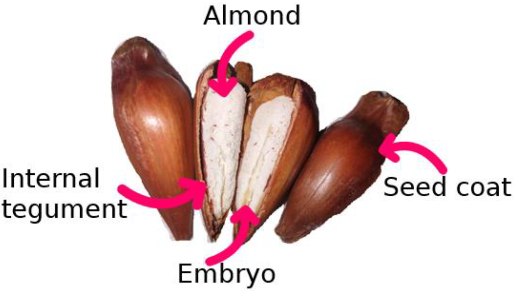
:1. Introduction
2. Materials and Methods
2.1. Materials
2.2. Micro-Fibrillated Cellulose (MFC) Preparation
2.3. PVA Solution and Film Formation
2.4. Scanning Electron Microscopy (SEM)
2.5. Fourier Transform Infrared Spectroscopy (FTIR)
2.6. Thermo and Mechanical Analysis
2.7. Swelling Test
2.8. Pinhão Bioactive Compounds Release
2.9. Antibacterial Evaluation
2.10. In Vitro Cytotoxicity Assay
2.11. In Vitro Cell Migration Test
2.12. Pre-Processing of Molecules Prior to Simulation
2.13. Docking Simulation
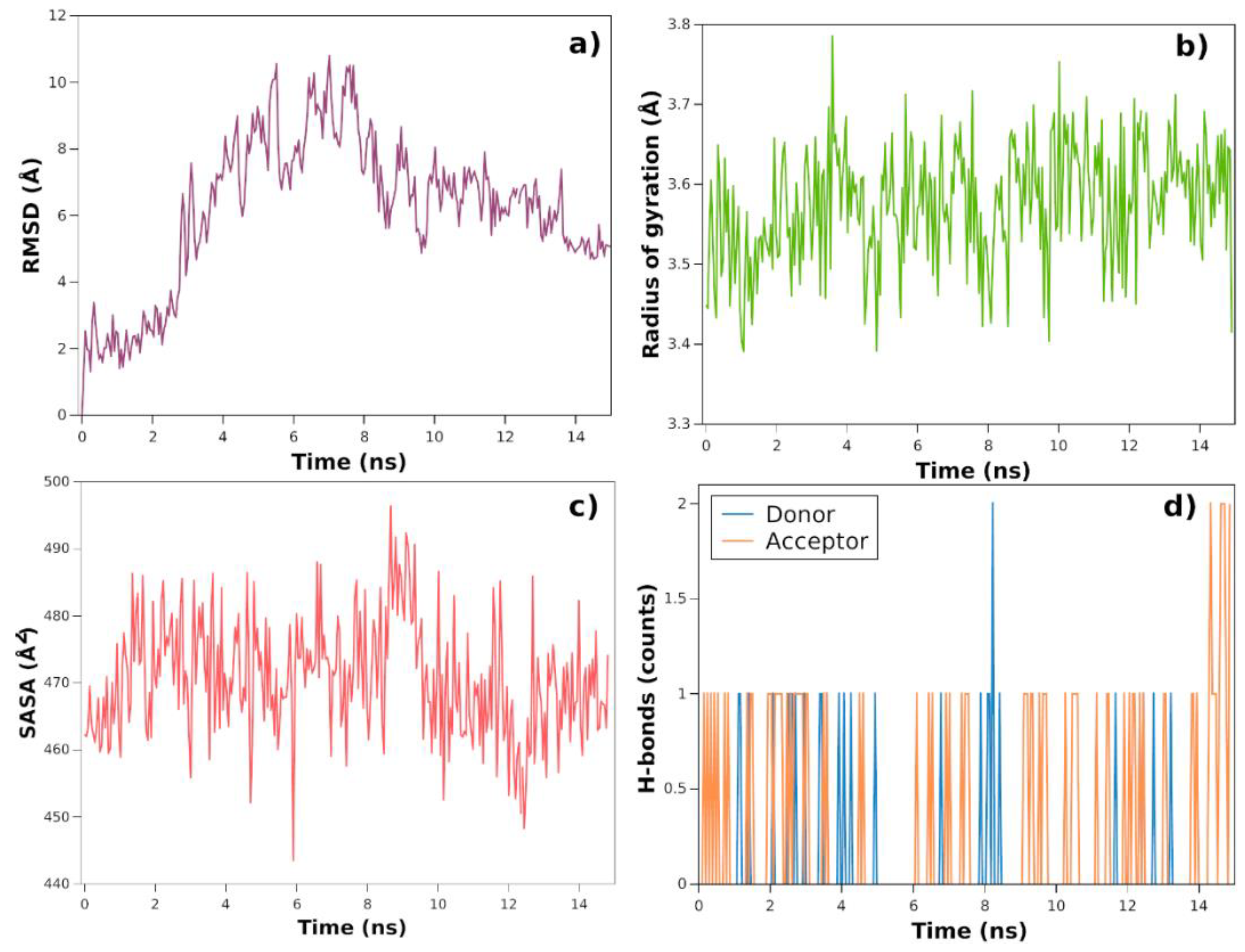
2.14. Molecular Dynamics Simulation
3. Results and Discussions
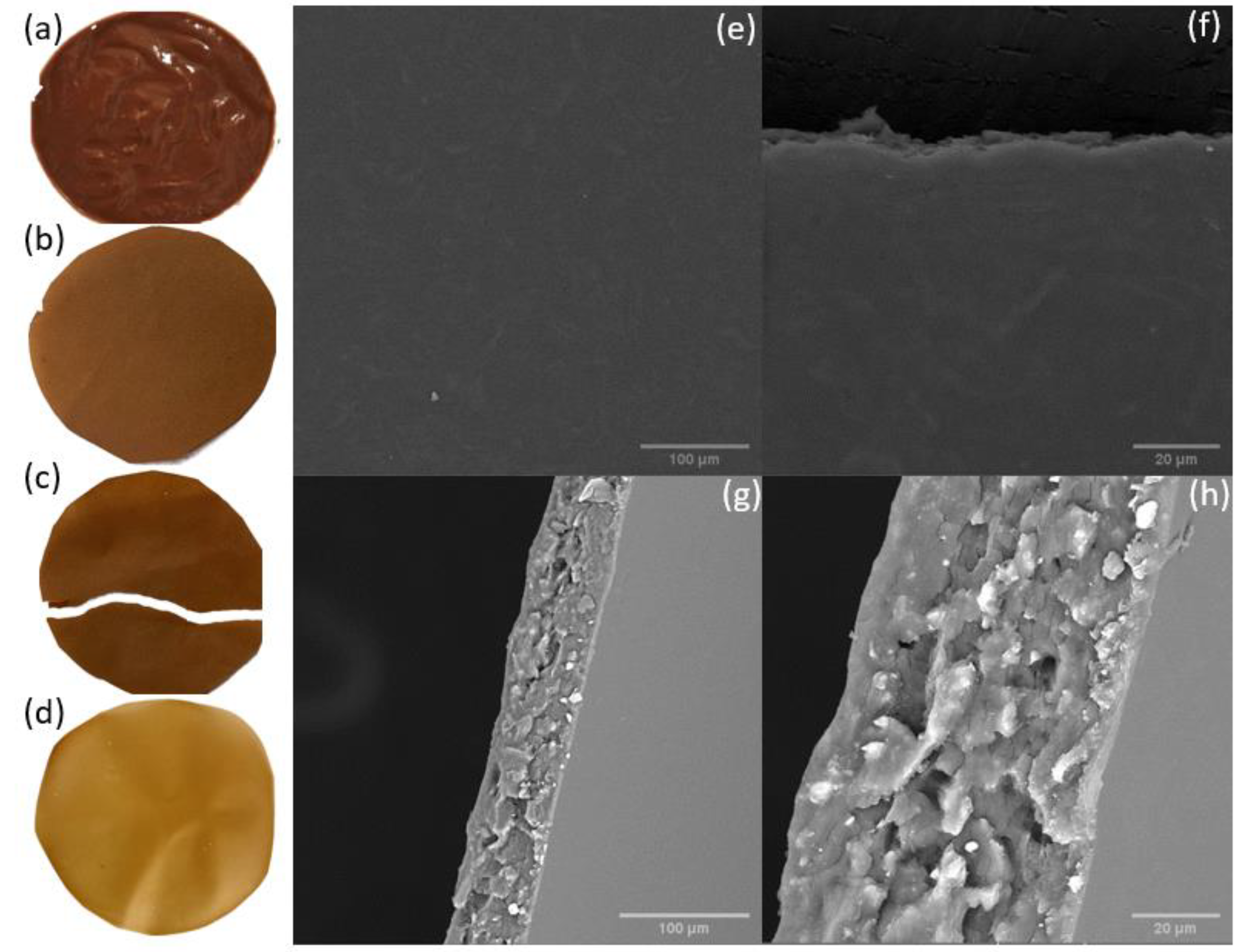
3.1. Scanning Electron Microscopy (SEM)
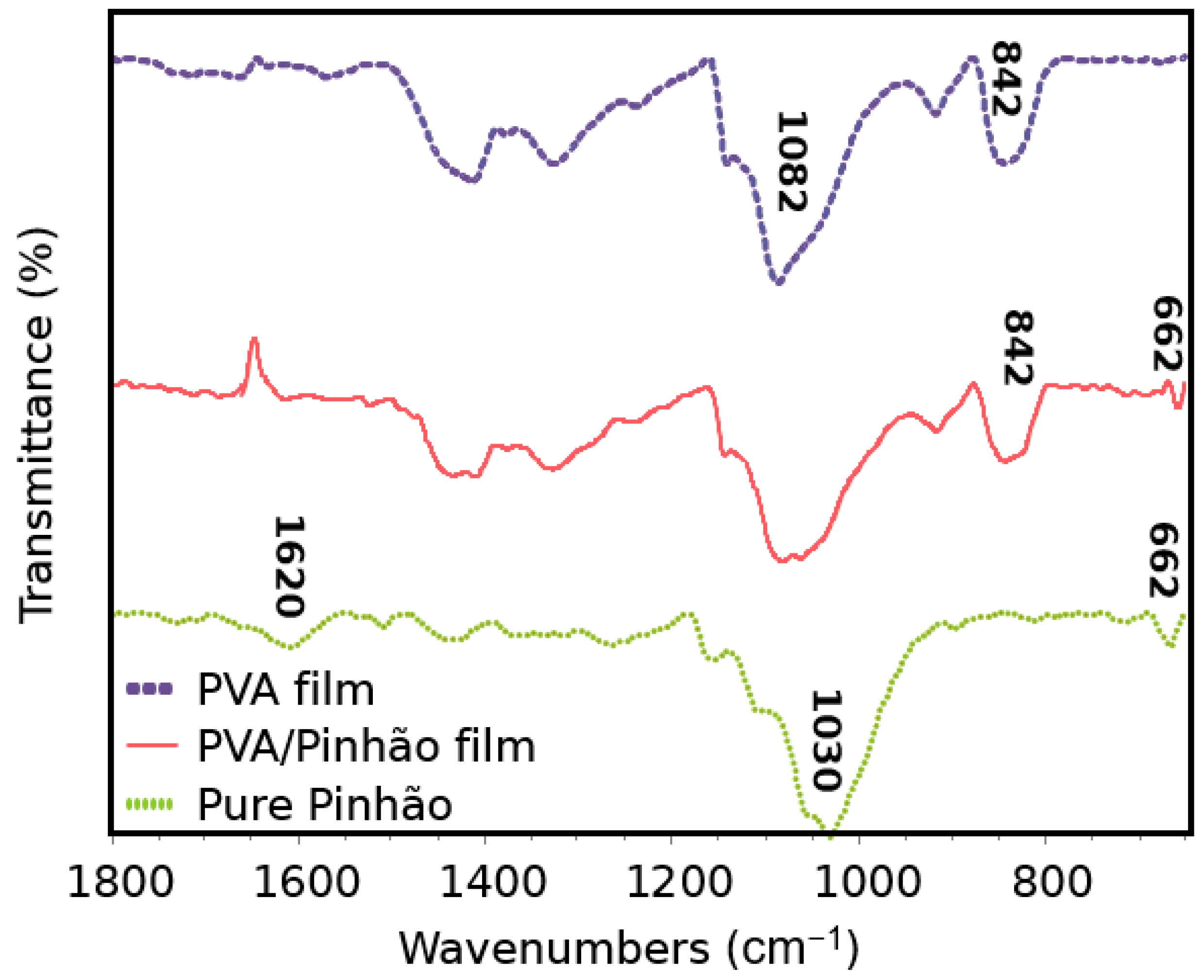
3.2. Fourier Transform Infrared Spectroscopy (FTIR)
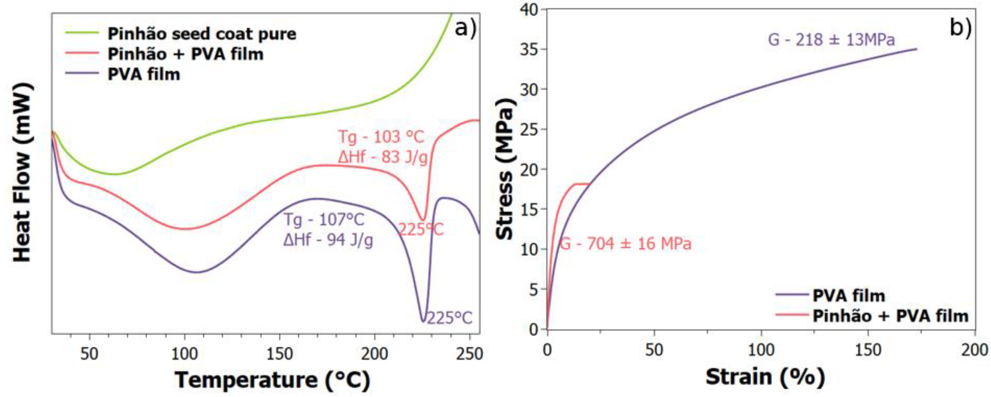
3.3. Thermal and Mechanical Analysis
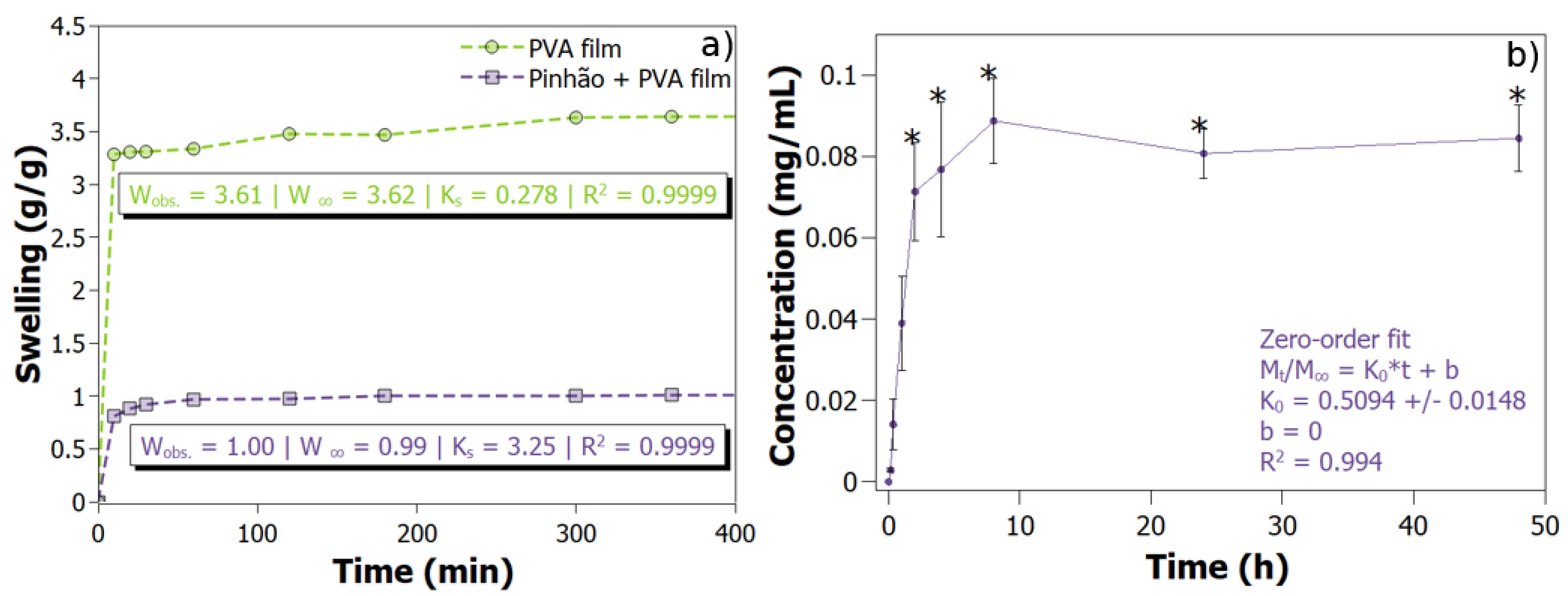
3.4. Swelling Test
3.5. Drug Release
3.6. Antibacterial Evaluation
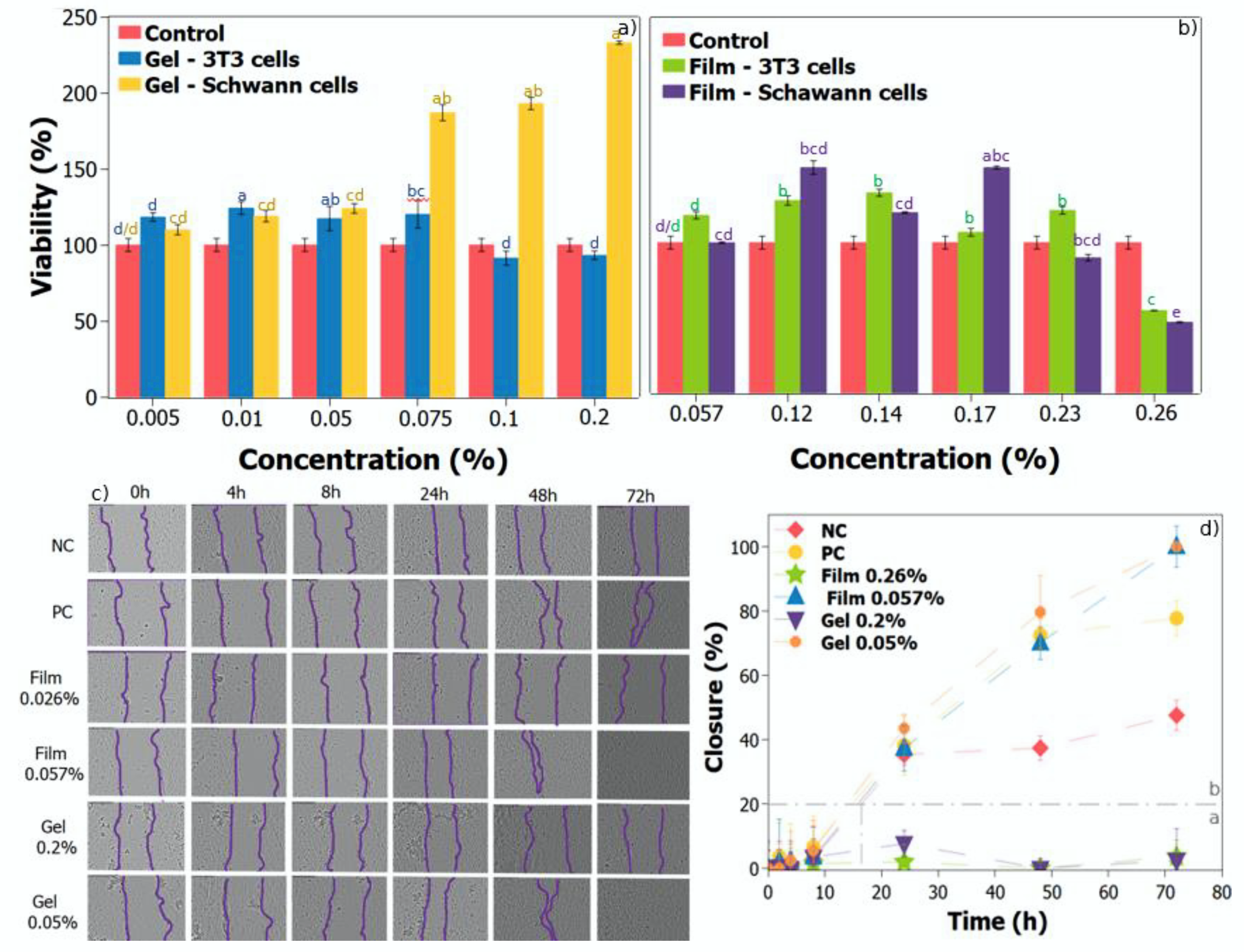
3.7. In Vitro Cytotoxicity Assay
3.8. In Vitro Cell Migration Test
3.9. Pinhão Seed Coat Component
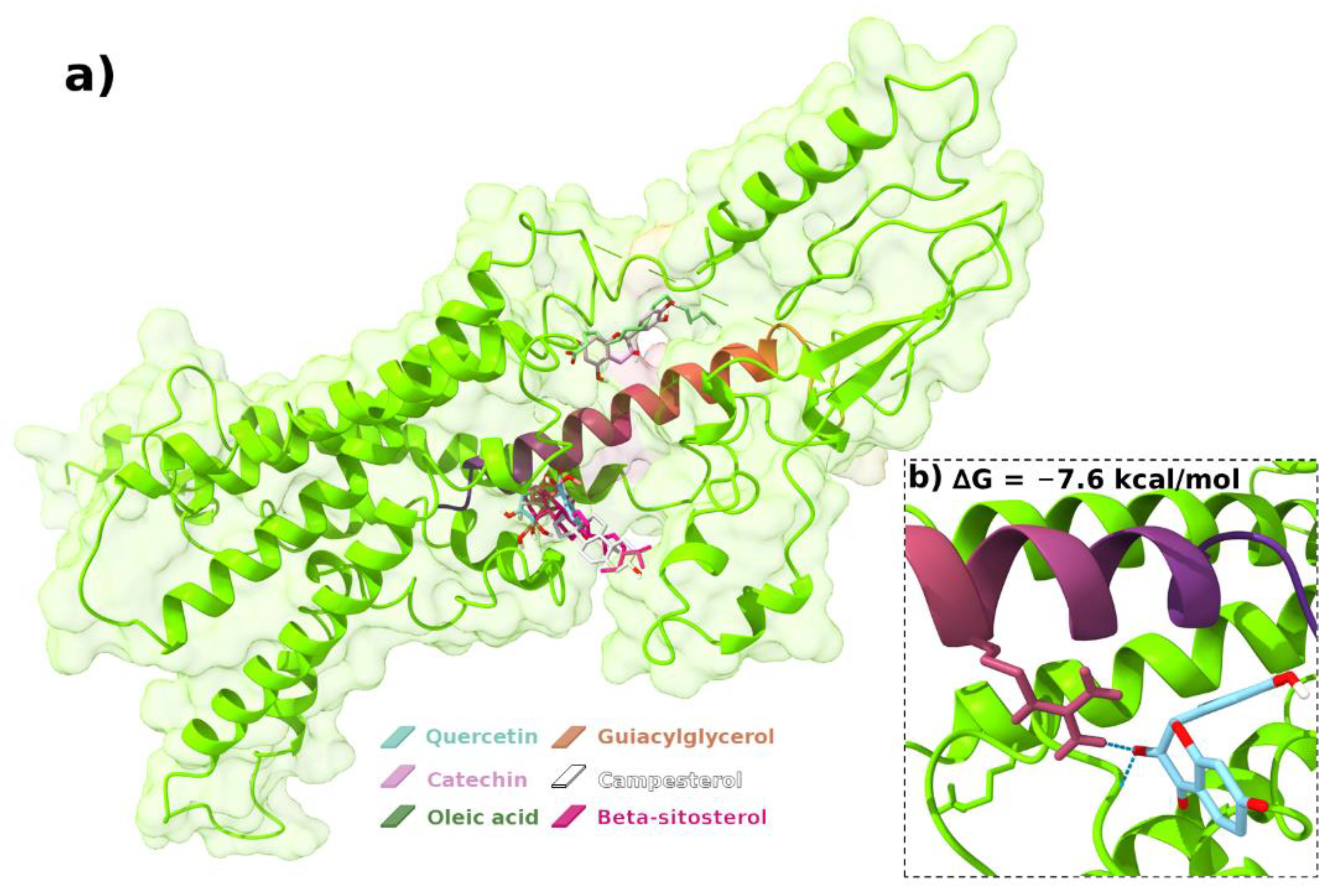
3.10. Molecular Docking
4. Conclusions
Supplementary Materials
Author Contributions
Funding
Institutional Review Board Statement
Informed Consent Statement
Data Availability Statement
Conflicts of Interest
Appendix A

| Compound | H-Bond Interactions |
|---|---|
| Quercetin | ARG12, ASN300, SER302, ASP301 |
| Catechin | SER11 |
| Oleic acid | CYS296, ASP298 |
| Guaiacylglycerol | ARG12, ASP8, ASN375, ASP301, SER302 |
| Campesterol | -- |
| Beta-sistosterol | -- |
| DONOUR | |||
|---|---|---|---|
| donour | acceptor | occupancy | protein |
| ASN375 | QUE | 2.91% | PAC1 |
| TRP306 | QUE | 2.27% | PAC1 |
| GLU374 | QUE | 0.32% | PAC1 |
| LYS310 | QUE | 1.29% | PAC1 |
| THR94 | QUE | 1.29% | PAC1 |
| LYS378 | QUE | 0.97% | PAC1 |
| ACCEPTOR | |||
| donour | acceptor | occupancy | protein |
| QUE | ASP8 | 2.27% | PACAP |
| QUE | PRO373 | 8.41% | PAC1 |
| QUE | SER372 | 0.65% | PAC1 |
| QUE | PHE371 | 0.65% | PAC1 |
| QUE | ASN375 | 0.97% | PAC1 |
| QUE | THR94 | 0.32% | PAC1 |
| QUE | SER9 | 4.53% | PACAP |
| QUE | GLU93 | 6.47% | PAC1 |
| QUE | LYS378 | 2.27% | PAC1 |
| QUE | GLU385 | 0.97% | PAC1 |
References
- Pinto, V.Z.; Moomand, K.; Vanier, N.L.; Colussi, R.; Villanova, F.A.; Zavareze, E.R.; Lim, L.-T.; Dias, A.R.G. Molecular structure and granule morphology of native and heat-moisture-treated pinhão starch. Int. J. Food Sci. Technol. 2015, 50, 282–289. [Google Scholar] [CrossRef]
- Tavares, F.F.d.C.; de Almeida, M.D.C.; da Silva, J.A.P.; Araújo, L.L.; Cardozo, N.S.M.; Santana, R.M.C. Thermal treatment of açaí (Euterpe oleracea) fiber for composite reinforcement. Polímeros 2020, 9, 1–9. [Google Scholar] [CrossRef]
- Fonseca, L.M.; Silva, F.T.; Bona, N.P.; Stefanello, F.M.; Borges, C.D.; Dias, A.R.G.; Zavareze, E.D.R. Aerogels from Native and Anionic Corn Starches Loaded with Pinhão (Araucaria angustifolia) Coat Extract: Anti-Tumor Activity in C6 Rat Glioma Cells and In Vitro Digestibility. Starch Stärke 2020, 72, 1900280. [Google Scholar] [CrossRef]
- Branco, C.d.S.; de Lima, É.D.; Rodrigues, T.S.; Scheffel, T.B.; Scola, G.; Laurino, C.C.F.C.; Moura, S.; Salvador, M. Mitochondria and redox homoeostasis as chemotherapeutic targets of Araucaria angustifolia (Bert.) O. Kuntze in human larynx HEp-2 cancer cells. Chem. Biol. Interact. 2015, 231, 108–118. [Google Scholar] [CrossRef] [PubMed] [Green Version]
- De Oliveira, A.; Moreira, T.F.M.; Pepinelli, A.L.S.; Costa, L.G.M.A.; Leal, L.E.; da Silva, T.B.V.; Gonçalves, O.H.; Ineu, R.P.; Dias, M.I.; Barros, L.; et al. Bioactivity screening of pinhão (Araucaria Angustifolia (Bertol.) Kuntze) seed extracts: The inhibition of cholinesterases and α-amylases, and cytotoxic and anti-inflammatory activities. Food Funct. 2021, 12, 9820–9828. [Google Scholar] [CrossRef] [PubMed]
- Fonseca, L.M.; da Silva, F.T.; Bruni, G.P.; Borges, C.D.; Zavareze, E.d.R.; Dias, A.R.G. Aerogels based on corn starch as carriers for pinhão coat extract (Araucaria angustifolia) rich in phenolic compounds for active packaging. Int. J. Biol. Macromol. 2021, 169, 362–370. [Google Scholar] [CrossRef] [PubMed]
- Da Cruz, E.P.; Fonseca, L.M.; Radünz, M.; da Silva, F.T.; Gandra, E.A.; Zavareze, E.D.R.; Borges, C.D. Pinhão coat extract encapsulated in starch ultrafine fibers: Thermal, antioxidant, and antimicrobial properties and in vitro biological digestion. J. Food Sci. 2021, 86, 2886–2897. [Google Scholar] [CrossRef]
- Spada, J.C.; Luchese, C.L.; Tessaro, I.C. Potential of pinhão Coat as Constituents of Starch Based Films Using Modification Techniques. J. Polym. Environ. 2018, 26, 2686–2697. [Google Scholar] [CrossRef]
- Osong, S.H.; Norgren, S.; Engstrand, P. Processing of wood-based microfibrillated cellulose and nanofibrillated cellulose, and applications relating to papermaking: A review. Cellulose 2016, 23, 93–123. [Google Scholar] [CrossRef]
- Timm, T.G.; de Lima, G.G.; Matos, M.; Magalhães, W.L.E.; Tavares, L.B.B.; Helm, C.V. Nanosuspension of pinhão seed coat development for a new high-functional cereal bar. J. Food Process. Preserv. 2020, 44, e14464. [Google Scholar] [CrossRef]
- Aliabadi, M.; Chee, B.S.; Matos, M.; Cortese, Y.J.; Nugent, M.J.D.; de Lima, T.A.M.; Magalhães, W.L.E.; de Lima, G.G.; Firouzabadi, M.D. Microfibrillated cellulose films containing chitosan and tannic acid for wound healing applications. J. Mater. Sci. Mater. Med. 2021, 32, 67. [Google Scholar] [CrossRef] [PubMed]
- Aliabadi, M.; Chee, B.S.; Matos, M.; Cortese, Y.J.; Nugent, M.J.D.; de Lima, T.A.M.; Magalhães, W.L.E.; de Lima, G.G. Yerba Mate Extract in Microfibrillated Cellulose and Corn Starch Films as a Potential Wound Healing Bandage. Polymers 2020, 12, 2807. [Google Scholar] [CrossRef] [PubMed]
- Tozluoglu, A.; Poyraz, B.; Candan, Z. Examining the efficiency of mechanic/enzymatic pretreatments in micro/nanofibrillated cellulose production. Maderas. Cienc. Tecnol. 2018, 18, 67–84. [Google Scholar] [CrossRef] [Green Version]
- De Lima, G.G.; Ferreira, B.D.; Matos, M.; Pereira, B.L.; Nugent, M.J.D.; Hansel, F.A.; Magalhães, W.L.E. Effect of cellulose size-concentration on the structure of polyvinyl alcohol hydrogels. Carbohydr. Polym. 2020, 245, 116612. [Google Scholar] [CrossRef] [PubMed]
- Zhang, Y.; Song, W.; Lu, Y.; Xu, Y.; Wang, C.; Yu, D.-G.; Kim, I. Recent Advances in Poly(α-L-glutamic acid)-Based Nanomaterials for Drug Delivery. Biomolecules 2022, 12, 636. [Google Scholar] [CrossRef]
- Zhang, Y.; Li, S.; Xu, Y.; Shi, X.; Zhang, M.; Huang, Y.; Liang, Y.; Chen, Y.; Ji, W.; Kim, J.R.; et al. Engineering of hollow polymeric nanosphere-supported imidazolium-based ionic liquids with enhanced antimicrobial activities. Nano Res. 2022, 15, 5556–5568. [Google Scholar] [CrossRef]
- Claro, F.C.; Jordão, C.; de Viveiros, B.M.; Isaka, L.J.E.; Junior, J.A.V.; Magalhães, W.L.E. Low cost membrane of wood nanocellulose obtained by mechanical defibrillation for potential applications as wound dressing. Cellulose 2020, 15, 10765–10779. [Google Scholar] [CrossRef]
- Schott, H. Swelling kinetics of polymers. J. Macromol. Sci. Part B 1992, 31, 1–9. [Google Scholar] [CrossRef]
- Reinhardt, L.S.; Chee, B.S.; Cao, Z.; Moura, D.J.; Nugent, M. Freeze-thaw electrospun PVA-dacarbazine nanoparticles: Preparation, characterization and anticancer evaluation. Int. J. Polym. Mater. Polym. Biomater. 2020, 69, 749–760. [Google Scholar] [CrossRef]
- Stewart, J.J.P. MOPAC: A semiempirical molecular orbital program. J. Comput. Aided Mol. Des. 1990, 4, 1–103. [Google Scholar] [CrossRef]
- Valdés-Tresanco, M.S.; Valdés-Tresanco, M.E.; Valiente, P.A.; Moreno, E. AMDock: A versatile graphical tool for assisting molecular docking with Autodock Vina and Autodock4. Biol. Direct 2020, 15, 12. [Google Scholar] [CrossRef] [PubMed]
- Jo, S.; Kim, T.; Iyer, V.G.; Im, W. CHARMM-GUI: A web-based graphical user interface for CHARMM. J. Comput. Chem. 2008, 29, 1859–1865. [Google Scholar] [CrossRef] [PubMed]
- Sterling, T.; Irwin, J.J. ZINC 15—Ligand Discovery for Everyone. J. Chem. Inf. Model. 2015, 55, 2324–2337. [Google Scholar] [CrossRef] [PubMed]
- Dolinsky, T.J.; Nielsen, J.E.; McCammon, J.A.; Baker, N.A. PDB2PQR: An automated pipeline for the setup of Poisson-Boltzmann electrostatics calculations. Nucleic Acids Res. 2004, 32, W665–W667. [Google Scholar] [CrossRef] [PubMed]
- Harris, R.; Olson, A.J.; Goodsell, D.S. Automated prediction of ligand-binding sites in proteins. Proteins Struct. Funct. Bioinform. 2007, 70, 1506–1517. [Google Scholar] [CrossRef] [PubMed]
- Trott, O.; Olson, A.J. AutoDock Vina: Improving the speed and accuracy of docking with a new scoring function, efficient optimization, and multithreading. J. Comput. Chem. 2009, 7, 455–461. [Google Scholar] [CrossRef] [Green Version]
- Pettersen, E.F.; Goddard, T.D.; Huang, C.C.; Meng, E.C.; Couch, G.S.; Croll, T.I.; Morris, J.H.; Ferrin, T.E. UCSF ChimeraX: Structure visualization for researchers, educators, and developers. Protein Sci. 2021, 30, 70–82. [Google Scholar] [CrossRef]
- Arantes, P.R.; Polêto, M.D.; Pedebos, C.; Ligabue-Braun, R. Making it Rain: Cloud-Based Molecular Simulations for Everyone. J. Chem. Inf. Model. 2021, 61, 4852–4856. [Google Scholar] [CrossRef]
- Eastman, P.; Swails, J.; Chodera, J.D.; McGibbon, R.T.; Zhao, Y.; Beauchamp, K.A.; Wang, L.-P.; Simmonett, A.C.; Harrigan, M.P.; Stern, C.D.; et al. OpenMM 7: Rapid development of high performance algorithms for molecular dynamics. PLoS Comput. Biol. 2017, 13, e1005659. [Google Scholar] [CrossRef]
- De Lima, G.G.; de Miranda, N.B.; Timm, T.G.; Matos, M.; de Lima, T.A.M.; Magalhães, W.L.E.; Tavares, L.B.B.; Hansel, F.A.; Helm, C.V. Characterisation and in vivo evaluation of Araucaria angustifolia pinhão seed coat nanosuspension as a functional food source. Food Funct. 2020, 11, 9820–9832. [Google Scholar] [CrossRef]
- Cordenunsi, B.R.; de Menezes, E.W.; Genovese, M.I.; Colli, C.; de Souza, A.G.; Lajolo, F.M. Chemical composition and glycemic index of Brazilian pine (Araucaria angustifolia) seeds. J. Agric. Food Chem. 2004, 52, 3412–3416. [Google Scholar] [CrossRef] [PubMed]
- Sampaio, D.A.; Garcia, R.A.; Lima, H.R.P. Anatomical and Physicochemical Characterization of the Araucaria angustifolia Seed Coat. Floresta e Ambient. 2019, 12, 1–12. [Google Scholar] [CrossRef]
- Krishnadev, P.; Subramanian, K.S.; Janavi, G.J.; Ganapathy, S.; Lakshmanan, A. Synthesis and characterization of nano-fibrillated cellulose derived from green Agave americana L. fiber. BioResources 2020, 15, 2442–2458. [Google Scholar] [CrossRef]
- Mihaela, D.; Nicoleta, A.; Ghiurea, M.; Ilie, C.; Radovici, C.; Doina, M. Properties of Polymer Composites with Cellulose Microfibrils. In Advances in Composite Materials—Ecodesign and Analysis; InTech: London, UK, 2011. [Google Scholar]
- Canillas, M.; De Lima, G.G.; Rodríguez, M.A.; Nugent, M.J.D.; Devine, D.M. Bioactive composites fabricated by freezing-thawing method for bone regeneration applications. J. Polym. Sci. Part B Polym. Phys. 2016, 54, 761–773. [Google Scholar] [CrossRef]
- Malucelli, L.C.; Matos, M.; Jordão, C.; Lacerda, L.G.; Filho, M.A.S.C.; Magalhães, W.L.E. Grinding severity influences the viscosity of cellulose nanofiber (CNF) suspensions and mechanical properties of nanopaper. Cellulose 2018, 25, 6581–6589. [Google Scholar] [CrossRef]
- Boufi, S.; Kaddami, H.; Dufresne, A. Mechanical Performance and Transparency of Nanocellulose Reinforced Polymer Nanocomposites. Macromol. Mater. Eng. 2014, 299, 560–568. [Google Scholar] [CrossRef]
- Das, B.; Gaur, S.S.; Katha, A.R.; Wang, C.-T.; Katiyar, V. Crosslinked poly(vinyl alcohol) membrane as separator for domestic wastewater fed dual chambered microbial fuel cells. Int. J. Hydrogen Energy 2021, 46, 7073–7086. [Google Scholar] [CrossRef]
- Gentile, G.; Cocca, M.; Avolio, R.; Errico, M.; Avella, M. Effect of Microfibrillated Cellulose on Microstructure and Properties of Poly(vinyl alcohol) Foams. Polymers 2018, 10, 813. [Google Scholar] [CrossRef] [Green Version]
- Tolve, R.; Galgano, F.; Condelli, N.; Cela, N.; Lucini, L.; Caruso, M.C. Optimization Model of Phenolics Encapsulation Conditions for Biofortification in Fatty Acids of Animal Food Products. Foods 2021, 10, 881. [Google Scholar] [CrossRef]
- Paulo, F.; Santos, L. New insights in the in vitro release of phenolic antioxidants: The case study of the release behavior of tyrosol from tyrosol-loaded ethylcellulose microparticles during the in vitro gastrointestinal digestion. Colloids Surf. B Biointerfaces 2020, 196, 111339. [Google Scholar] [CrossRef]
- Trojaike, G.H.; Biondo, E.; Padilha, R.L.; Brandelli, A.; Sant’Anna, V. Antimicrobial Activity of Araucaria angustifolia Seed (Pinhão) Coat Extract and its Synergism with Thermal Treatment to Inactivate Listeria monocytogenes. Food Bioprocess Technol. 2019, 12, 193–197. [Google Scholar] [CrossRef]
- Klein, M.I.; Biondo, E.; Kolchinski, E.M.; Padilha, R.L.; Brandelli, A.; Sant’Anna, V. Avaliacao da atividade antimicrobiana e alelopatica de extrato de casca de pinhao. In Proceedings of the 5 Simposio de Seguranca Alimentar—Alimentacao e Saude—Bento Goncalves, Rio Grande do Sul, Brazil, 15 August 2015. [Google Scholar]
- De Lima, G.G.; Elter, J.K.; Chee, B.S.; Magalhães, W.L.E.; Devine, D.M.; Nugent, M.J.D.; de Sá, M.J.C. A tough and novel dual-response PAA/P(NiPAAM-co-PEGDMA) IPN hydrogels with ceramics by photopolymerization for consolidation of bone fragments following fracture. Biomed. Mater. 2019, 14, 054101. [Google Scholar] [CrossRef] [PubMed]
- De Lima, G.G.G.G.; Chee, B.S.B.S.; Moritz, V.F.V.F.; Cortese, Y.J.Y.J.; Magalhães, W.L.E.W.L.E.; Devine, D.M.D.M.; Nugent, M.J.D.M.J.D. The production of a novel poly(vinyl alcohol) hydrogel cryogenic spheres for immediate release using a droplet system. Biomed. Phys. Eng. Express. 2019, 5, 045017. [Google Scholar] [CrossRef]
- Csepregi, R.; Temesfői, V.; Das, S.; Alberti, Á.; Tóth, C.A.; Herczeg, R.; Papp, N.; Kőszegi, T. Cytotoxic, Antimicrobial, Antioxidant Properties and Effects on Cell Migration of Phenolic Compounds of Selected Transylvanian Medicinal Plants. Antioxidants 2020, 9, 166. [Google Scholar] [CrossRef] [Green Version]
- Prado, L.G.; Arruda, H.S.; Araujo, N.M.P.; de Oliveira Braga, L.E.; Banzato, T.P.; Pereira, G.A.; Figueiredo, M.C.; Ruiz, A.L.T.G.; Eberlin, M.N.; de Carvalho, J.E.; et al. Antioxidant, antiproliferative and healing properties of araticum (Annona crassiflora Mart.) peel and seed. Food Res. Int. 2020, 133, 109168. [Google Scholar] [CrossRef]
- Lee, D.-Y.; Song, M.-C.; Yoo, K.-H.; Bang, M.-H.; Chung, I.-S.; Kim, S.-H.; Kim, D.-K.; Kwon, B.-M.; Jeong, T.-S.; Park, M.-H.; et al. Lignans from the fruits of Cornus kousa Burg. and their cytotoxic effects on human cancer cell lines. Arch. Pharm. Res. 2007, 30, 402–407. [Google Scholar] [CrossRef]
- Wang, H.; Geng, C.-A.; Xu, H.-B.; Huang, X.-Y.; Ma, Y.-B.; Yang, C.-Y.; Zhang, X.-M.; Chen, J.-J. Lignans from the Fruits of Melia toosendan and Their Agonistic Activities on Melatonin Receptor MT1. Planta Med. 2015, 81, 847–854. [Google Scholar] [CrossRef] [Green Version]
- Cadoná, F.C.; Machado, A.K.; Azzolin, V.F.; Barbisan, F.; Dornelles, E.B.; Glanzner, W.; Gonçalves, P.B.D.; Assmann, C.E.; Ribeiro, E.E.; Cruz, I.B.M. da Guaraná a Caffeine-Rich Food Increases Oxaliplatin Sensitivity of Colorectal HT-29 Cells by Apoptosis Pathway Modulation. Anticancer. Agents Med. Chem. 2016, 16, 1055–1065. [Google Scholar] [CrossRef]
- Sun, H.; Yin, M.; Hao, D.; Shen, Y. Anti-Cancer Activity of Catechin against A549 Lung Carcinoma Cells by Induction of Cyclin Kinase Inhibitor p21 and Suppression of Cyclin E1 and P–AKT. Appl. Sci. 2020, 10, 2065. [Google Scholar] [CrossRef] [Green Version]
- Lin, Y.-H.; Wang, C.-C.; Lin, Y.-H.; Chen, B.-H. Preparation of Catechin Nanoemulsion from Oolong Tea Leaf Waste and Its Inhibition of Prostate Cancer Cells DU-145 and Tumors in Mice. Molecules 2021, 26, 3260. [Google Scholar] [CrossRef]
- Liu, Y.; Tang, Z.-G.; Lin, Y.; Qu, X.-G.; Lv, W.; Wang, G.-B.; Li, C.-L. Effects of quercetin on proliferation and migration of human glioblastoma U251 cells. Biomed. Pharmacother. 2017, 92, 33–38. [Google Scholar] [CrossRef] [PubMed]
- Murakami, A.; Ashida, H.; Terao, J. Multitargeted cancer prevention by quercetin. Cancer Lett. 2008, 269, 315–325. [Google Scholar] [CrossRef] [PubMed]
- Al-Mutairi, A.A.; Mayson, H.; Alkhatib, H.M.G. Antitumor Activities of Co-loading Gemcitabine and Oxaliplatin into Oleic Acid-Based Solid Lipid Nanoparticle against Non-Small Cell Lung Cancer Cells. Biointerface Res. Appl. Chem. 2021, 12, 49–60. [Google Scholar] [CrossRef]
- Jiang, L.; Wang, W.; He, Q.; Wu, Y.; Lu, Z.; Sun, J.; Liu, Z.; Shao, Y.; Wang, A. Oleic acid induces apoptosis and autophagy in the treatment of Tongue Squamous cell carcinomas. Sci. Rep. 2017, 7, 11277. [Google Scholar] [CrossRef] [PubMed] [Green Version]
- Yuan, L.; Zhang, F.; Jia, S.; Xie, J.; Shen, M. Differences between phytosterols with different structures in regulating cholesterol synthesis, transport and metabolism in Caco-2 cells. J. Funct. Foods 2020, 65, 103715. [Google Scholar] [CrossRef]
- Bae, H.; Park, S.; Yang, C.; Song, G.; Lim, W. Disruption of Endoplasmic Reticulum and ROS Production in Human Ovarian Cancer by Campesterol. Antioxidants 2021, 10, 379. [Google Scholar] [CrossRef]
- Awad, A.B.; Chen, Y.C.; Fink, C.S.; Hennessey, T. beta-Sitosterol inhibits HT-29 human colon cancer cell growth and alters membrane lipids. Anticancer Res. 1996, 16, 2797–2804. [Google Scholar]
- Vundru, S.S.; Kale, R.K.; Singh, R.P. β-sitosterol induces G1 arrest and causes depolarization of mitochondrial membrane potential in breast carcinoma MDA-MB-231 cells. BMC Complement. Altern. Med. 2013, 13, 280. [Google Scholar] [CrossRef] [Green Version]
- Rajavel, T.; Packiyaraj, P.; Suryanarayanan, V.; Singh, S.K.; Ruckmani, K.; Devi, K.P. β-Sitosterol targets Trx/Trx1 reductase to induce apoptosis in A549 cells via ROS mediated mitochondrial dysregulation and p53 activation. Sci. Rep. 2018, 8, 2071. [Google Scholar] [CrossRef] [Green Version]
- Castorina, A.; Waschek, J.A.; Marzagalli, R.; Cardile, V.; Drago, F. PACAP Interacts with PAC1 Receptors to Induce Tissue Plasminogen Activator (tPA) Expression and Activity in Schwann Cell-Like Cultures. PLoS ONE 2015, 10, e0117799. [Google Scholar] [CrossRef]
- Castorina, A.; Tiralongo, A.; Giunta, S.; Carnazza, M.L.; Rasi, G.; D’Agata, V. PACAP and VIP prevent apoptosis in schwannoma cells. Brain Res. 2008, 1241, 29–35. [Google Scholar] [CrossRef] [PubMed]
- Castorina, A.; Scuderi, S.; D’Amico, A.G.; Drago, F.; D’Agata, V. PACAP and VIP increase the expression of myelin-related proteins in rat schwannoma cells: Involvement of PAC1/VPAC2 receptor-mediated activation of PI3K/Akt signaling pathways. Exp. Cell Res. 2014, 322, 108–121. [Google Scholar] [CrossRef]
- Murphy, D.J. Determination of accurate KI values for tight-binding enzyme inhibitors: An in silico study of experimental error and assay design. Anal. Biochem. 2004, 327, 61–67. [Google Scholar] [CrossRef] [PubMed]
- Adeoye, A.O.; Olanlokun, J.O.; Tijani, H.; Lawal, S.O.; Babarinde, C.O.; Akinwole, M.T.; Bewaji, C.O. Molecular docking analysis of apigenin and quercetin from ethylacetate fraction of Adansonia digitata with malaria-associated calcium transport protein: An in silico approach. Heliyon 2019, 5, e02248. [Google Scholar] [CrossRef] [PubMed] [Green Version]
- Maharani, M.G.; Lestari, S.R.; Lukiati, B. Molecular docking studies flavonoid (quercetin, isoquercetin, and kaempferol) of single bulb garlic (Allium sativum) to inhibit lanosterol synthase as anti-hypercholesterol therapeutic strategies. In AIP Conference Proceedings; AIP Publishing: Melville, NY, USA, 2020; p. 040021. [Google Scholar]
- Gu, Y.-Y.; Zhang, M.; Cen, H.; Wu, Y.-F.; Lu, Z.; Lu, F.; Liu, X.-S.; Lan, H.-Y. Quercetin as a potential treatment for COVID-19-induced acute kidney injury: Based on network pharmacology and molecular docking study. PLoS ONE 2021, 16, e0245209. [Google Scholar] [CrossRef] [PubMed]


| Pinhão Seed Coat Component (mg/kg) | Effective Doses/Concentrations | |
|---|---|---|
| Threo+erythro-guaiacylglycerol | 54.64 | IC50 (30.2 + 1.1 and 57.3 + 1.1μg mL−1) [48] 0.91 mM—agonistic rated (%) of 4.28, 0.64 and 6.04 [49] |
| Catechin | 254.51 | IC50 (0.43 μg mL−1) [50] IC50 (200; 400 and 600 μM) [51] IC50 (13.52 and 214.6 μg mL−1) [52] |
| Quercetin | 70.24 | 24 h IC50 (113.65 μg mL−1) or 48 h (IC50 of IC50 (48.61 μg ml−1) [53] IC50 (1.5 μM) [54] |
| 9-(Z)-Hexadecenoic acid | 16 | IC50 (3.125 to 100 µM) [55] IC50 (291~228 μM) [56] |
| 9,12-(Z,Z)-Octadecadienoic acid | 69.66 | |
| 9-(Z)-Octadecenoic acid | 189.09 | |
| 24-Methyl-cholest-5-en-3β-ol (campesterol) | 68.78 | IC50 > 200 μM [57] 125 µM [58] |
| Beta-Sitosterol | 449.72 | 16 μM [59] 90 μM [60] IC50 24.7 μM [61] |
| Compound | Affinity (kcal/mol) | Estimated Ki | Ki Units | Ligand Efficiency |
|---|---|---|---|---|
| Catechin | −7.6 | 2.69 | uM | −0.36 |
| Quercetin | −7.6 | 2.69 | uM | −0.36 |
| Guyacyl | −5.8 | 56.05 | uM | −0.39 |
| Oleic acid | −5.3 | 0.13 | mM | −0.27 |
| Campesterol | −6.8 | 10.37 | uM | −0.23 |
| Beta-sistosterol | −6.7 | 12.27 | uM | −0.22 |
| Quercetin | −7.6 | 2.69 | uM | −0.36 |
Publisher’s Note: MDPI stays neutral with regard to jurisdictional claims in published maps and institutional affiliations. |
© 2022 by the authors. Licensee MDPI, Basel, Switzerland. This article is an open access article distributed under the terms and conditions of the Creative Commons Attribution (CC BY) license (https://creativecommons.org/licenses/by/4.0/).
Share and Cite
de Lima, T.A.d.M.; de Lima, G.G.; Chee, B.S.; Henn, J.G.; Cortese, Y.J.; Matos, M.; Helm, C.V.; Magalhães, W.L.E.; Nugent, M.J.D. Characterization of Gels and Films Produced from Pinhão Seed Coat Nanocellulose as a Potential Use for Wound Healing Dressings and Screening of Its Compounds towards Antitumour Effects. Polymers 2022, 14, 2776. https://doi.org/10.3390/polym14142776
de Lima TAdM, de Lima GG, Chee BS, Henn JG, Cortese YJ, Matos M, Helm CV, Magalhães WLE, Nugent MJD. Characterization of Gels and Films Produced from Pinhão Seed Coat Nanocellulose as a Potential Use for Wound Healing Dressings and Screening of Its Compounds towards Antitumour Effects. Polymers. 2022; 14(14):2776. https://doi.org/10.3390/polym14142776
Chicago/Turabian Stylede Lima, Tielidy A. de M., Gabriel Goetten de Lima, Bor Shin Chee, Jeferson G. Henn, Yvonne J. Cortese, Mailson Matos, Cristiane V. Helm, Washington L. E. Magalhães, and Michael J. D. Nugent. 2022. "Characterization of Gels and Films Produced from Pinhão Seed Coat Nanocellulose as a Potential Use for Wound Healing Dressings and Screening of Its Compounds towards Antitumour Effects" Polymers 14, no. 14: 2776. https://doi.org/10.3390/polym14142776