
Development and Evaluation of Rifampicin Loaded Alginate–Gelatin Biocomposite Microfibers

 , , ,
, , , 
Abstract
:1. Introduction
2. Materials and Methods
2.1. Materials
2.2. Preparation of Rifampicin Loaded Biocomposite Microfibers
2.3. Characterization of Rifampicin Loaded Biocomposite Microfibers
2.3.1. Entrapment Efficiency
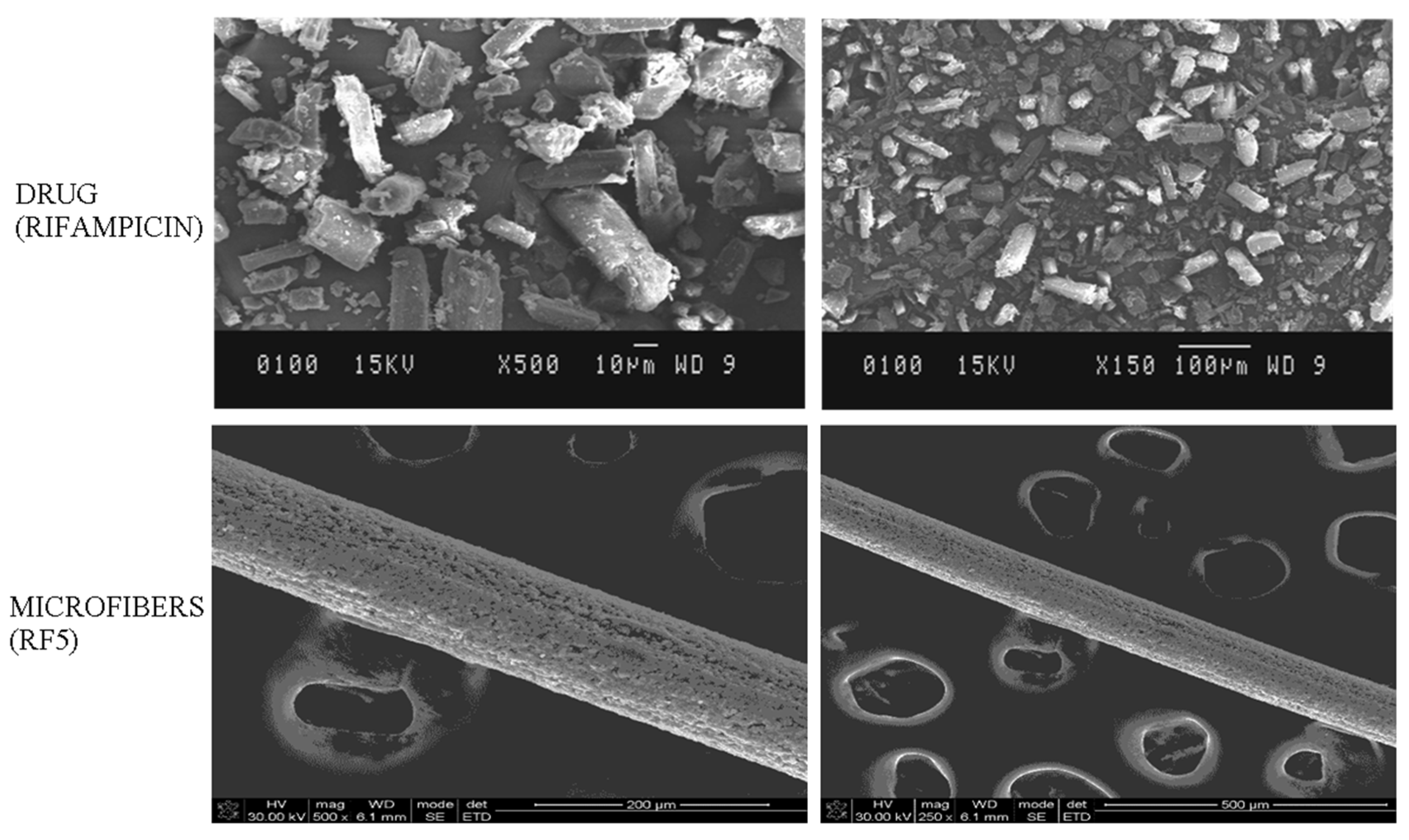
2.3.2. Morphological Analysis
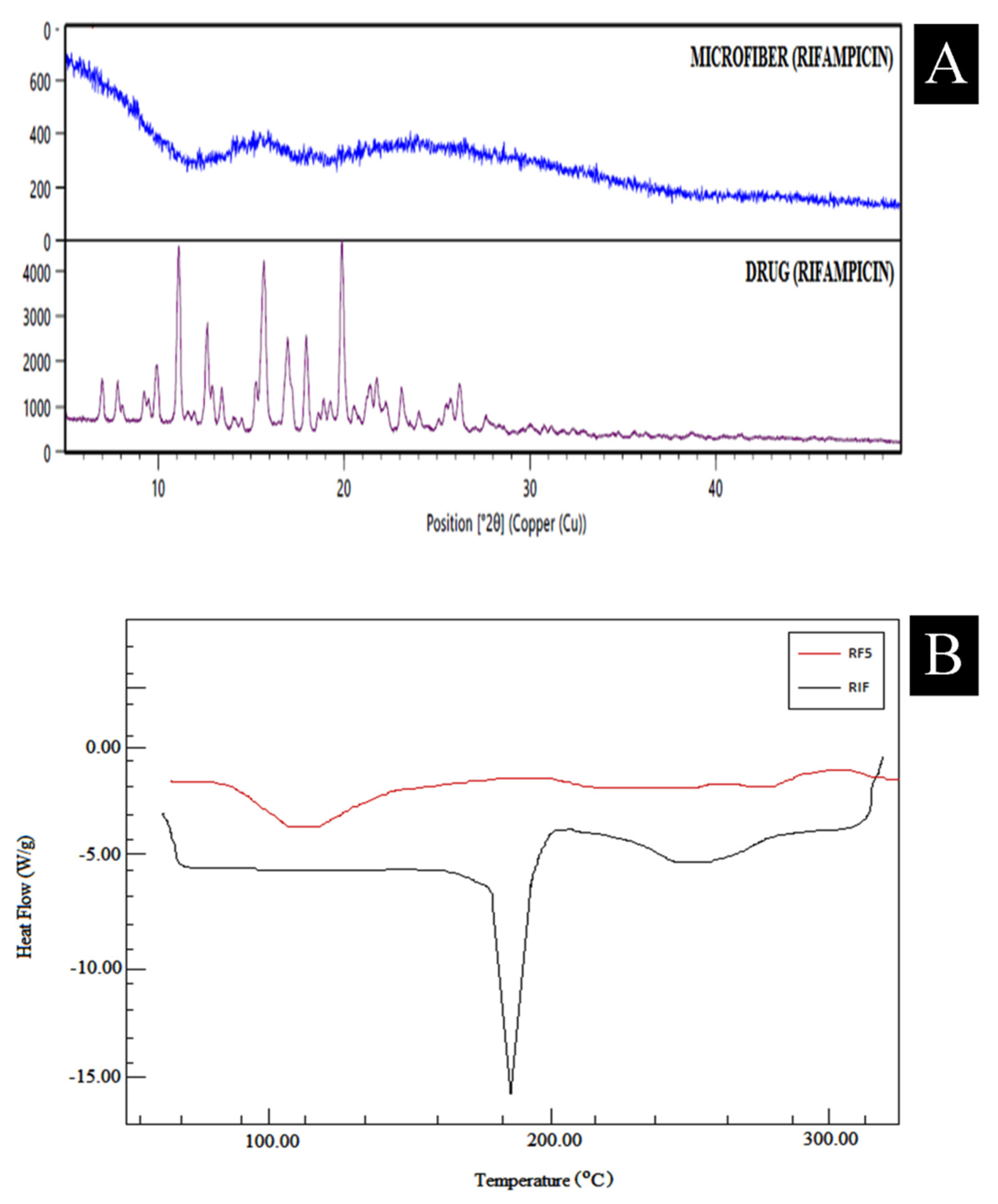
2.3.3. XRD
2.3.4. DSC
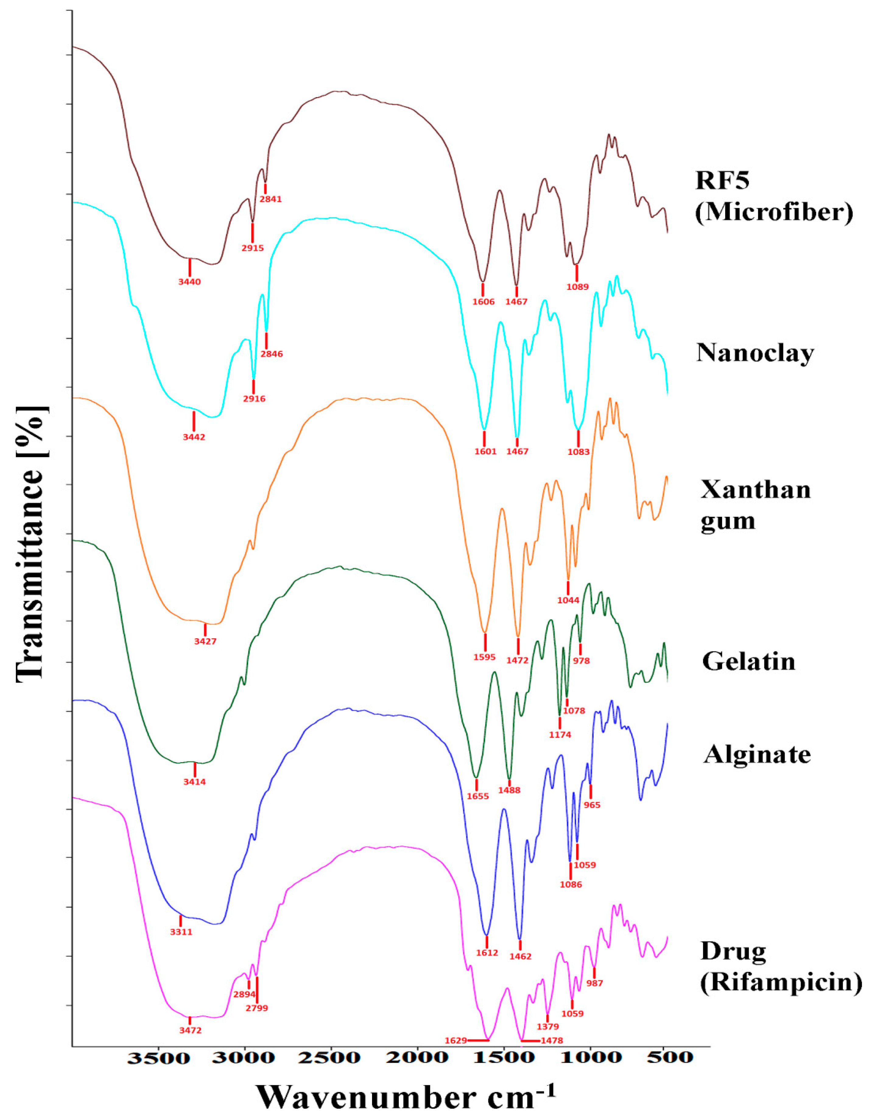
2.3.5. FTIR Studies
2.3.6. Water Uptake
2.3.7. Mechanical Properties
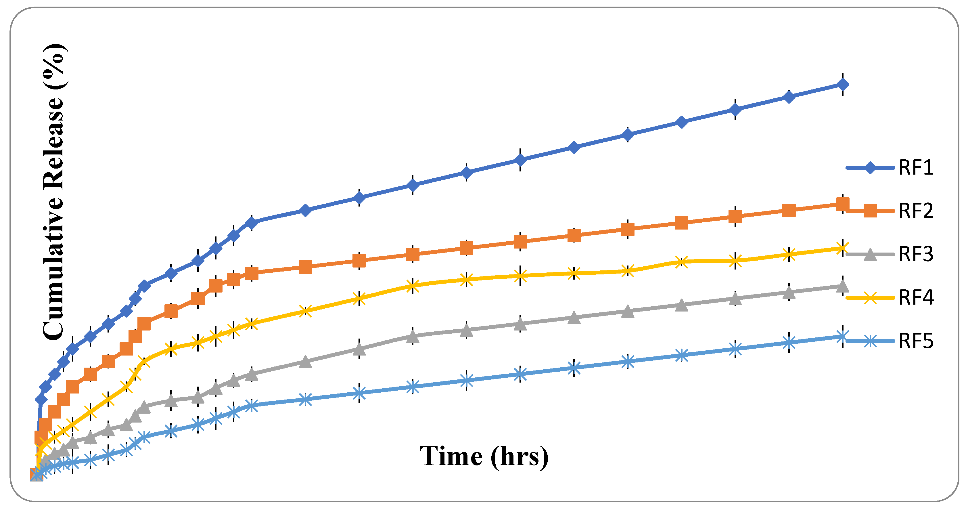
2.3.8. In Vitro Drug Release Studies
- Zero order—
- First order—
- Higuchi model—
- Hixson–Crowell model—
- Korsmeyer–Peppas model—
- Q = total amount of drug release at time t;
- Q0 = initial amount of drug;
- QR = amount of remaining drug at time t;
- QT = cumulative amount of drug release;
- k0, k1, kH, ks and kkp = kinetic constants for all respective models such as zero order, first order, Higuchi, Hixson–Crowell and Korsmeyer–Peppas models;
- n = release exponent.
2.3.9. Whole Blood Clotting
2.3.10. Antimicrobial Studies
3. Results and Discussion
4. Conclusions
Author Contributions
Funding
Institutional Review Board Statement
Informed Consent Statement
Data Availability Statement
Acknowledgments
Conflicts of Interest
References
- Bhardwaj, N.; Chouhan, D.; B Mandal, B. Tissue engineered skin and wound healing: Current strategies and future directions. Curr. Pharm. Des. 2017, 23, 3455–3482. [Google Scholar] [CrossRef]
- Sharma, A.; Puri, V.; Kumar, P.; Singh, I. Biopolymeric, nanopatterned, fibrous carriers for wound healing applications. Curr. Pharm. Des. 2020, 26, 4894–4908. [Google Scholar] [CrossRef]
- Manzini, B.M.; Machado, L.M.R.; Noritomi, P.Y.; da Silva, J.V.L. Advances in Bone tissue engineering: A fundamental review. J. Biosci. 2021, 46, 1–18. [Google Scholar] [CrossRef]
- Mazini, L.; Rochette, L.; Hamdan, Y.; Malka, G. Skin Immunomodulation during Regeneration: Emerging New Targets. J. Pers. Med. 2021, 11, 85. [Google Scholar] [CrossRef]
- Pyta, K.; Przybylski, P.; Klich, K.; Stefańska, J. A new model of binding of rifampicin and its amino analogues as zwitterions to bacterial RNA polymerase. Org. Biomol. Chem. 2012, 10, 8283–8297. [Google Scholar] [CrossRef] [PubMed]
- Sharma, A.; Puri, V.; Kumar, P.; Singh, I. Rifampicin-Loaded Alginate-Gelatin Fibers Incorporated within Transdermal Films as a Fiber-in-Film System for Wound Healing Applications. Membranes 2021, 11, 7. [Google Scholar] [CrossRef]
- Gilchrist, S.E.; Lange, D.; Letchford, K.; Bach, H.; Fazli, L.; Burt, H.M. Fusidic acid and rifampicin co-loaded PLGA nanofibers for the prevention of orthopedic implant associated infections. J. Control. Release 2013, 170, 64–73. [Google Scholar] [CrossRef] [PubMed]
- Vicosa, A.L.; Gomes, A.C.O.; Soares, B.G.; Paranhos, C.M. Effect of sepiolite on the physical properties and swelling behavior of rifampicin-loaded nanocomposite hydrogels. Express Polym. Lett. 2009, 3, 518–524. [Google Scholar] [CrossRef]
- Nathanael, A.J.; Oh, T.H. Biopolymer Coatings for Biomedical Applications. Polymers 2020, 12, 3061. [Google Scholar] [CrossRef]
- Álvarez-Suárez, A.S.; Dastager, S.G.; Bogdanchikova, N.; Grande, D.; Pestryakov, A.; García-Ramos, J.C.; Villarreal-Gómez, L.J. Electrospun fibers and sorbents as a possible basis for effective biocomposite wound dressings. Micromachines 2020, 11, 441. [Google Scholar] [CrossRef] [PubMed] [Green Version]
- Hazirah, M.N.; Isa, M.I.N.; Sarbon, N.M. Effect of xanthan gum on the physical and mechanical properties of gelatin-carboxymethyl cellulose film blends. Food Packag. Shelf Life 2016, 9, 55–63. [Google Scholar] [CrossRef]
- Alves, A.; Miguel, S.P.; Araujo, A.R.; de Jesús Valle, M.J.; Sánchez Navarro, A.; Correia, I.J.; Coutinho, P. Xanthan gum–Konjac glucomannan blend hydrogel for wound healing. Polymers 2020, 12, 99. [Google Scholar] [CrossRef] [PubMed] [Green Version]
- Noori, S.; Kokabi, M.; Hassan, Z.M. Nanoclay enhanced the mechanical properties of poly (vinyl alcohol)/chitosan/montmorillonite nanocomposite hydrogel as wound dressing. Procedia Mater. Sci. 2015, 11, 152–156. [Google Scholar] [CrossRef]
- Tian, H.; Wang, K.; Liu, D.; Yan, J.; Xiang, A.; Rajulu, A.V. Enhanced mechanical and thermal properties of poly (vinyl alcohol)/corn starch blends by nanoclay intercalation. Int. J. Biol. Macromol. 2017, 101, 314–320. [Google Scholar] [CrossRef]
- Bediako, J.K.; Lin, S.; Sarkar, A.K.; Zhao, Y.; Choi, J.W.; Song, M.H.; Yun, Y.S. Benignly-fabricated crosslinked polyethylenimine/calcium-alginate fibers as high-performance adsorbents for effective recovery of gold. J. Clean. Prod. 2020, 252, 119389. [Google Scholar] [CrossRef]
- Singh, P.; Singh, S.K.; Bajpai, J.; Bajpai, A.K.; Shrivastava, R.B. Iron crosslinked alginate as novel nanosorbents for removal of arsenic ions and bacteriological contamination from water. J. Mater Res. Tech. 2014, 3, 195–202. [Google Scholar] [CrossRef] [Green Version]
- Sharma, A.; Mittal, A.; Puri, V.; Kumar, P.; Singh, I. Curcumin-loaded, alginate–gelatin composite fibers for wound healing applications. 3 Biotech 2020, 10, 1–13. [Google Scholar] [CrossRef]
- Motiei, M.; Pleno de Gouveia, L.; Sopík, T.; Vícha, R.; Skoda, D.; Císar, J.; Khalili, R.; Domincová Bergerova, E.; Munster, L.; Fei, H.; et al. Nanoparticle-Based Rifampicin Delivery System Development. Molecules 2021, 26, 2067. [Google Scholar] [CrossRef]
- Varaprasad, K.; Nunez, D.; Ide, W.; Jayaramudu, T.; Sadiku, E.R. Development of high alginate comprised hydrogels for removal of Pb (II) ions. J. Mol. Liquids 2020, 298, 112087. [Google Scholar] [CrossRef]
- Mamidi, N.; Romo, I.L.; Leija Gutierrez, H.M.; Barrera, E.V.; Elías-Zúñiga, A. Development of forcespun fiber-aligned scaffolds from gelatin–zein composites for potential use in tissue engineering and drug release. MRS Commun. 2018, 8, 885–892. [Google Scholar] [CrossRef]
- Nejadmansouri, M.; Razmjooei, M.; Safdarianghomsheh, R.; Shad, E.; Delvigne, F.; Khalesi, M. Semi-continuous production of xanthan in biofilm reactor using Xanthomonas campestris. J. Biotechnol. 2021, 328, 1–11. [Google Scholar] [CrossRef] [PubMed]
- Chan, M.L.; Lau, K.T.; Wong, T.T.; Cardona, F. Interfacial bonding characteristic of nanoclay/polymer composites. Appl. Surf. Sci. 2011, 258, 860–864. [Google Scholar] [CrossRef]


| Formulation Batches | Sodium Alginate (%) | Gelatin (%) | Xanthan Gum (%) | Nanoclay (%) | Rifampicin (mg) |
|---|---|---|---|---|---|
| RF1 | 2 | - | - | - | 50 |
| RF2 | 2 | 2 | - | - | 50 |
| RF3 | 2 | 2 | 0.5 | - | 50 |
| RF4 | 2 | 2 | - | 0.5 | 50 |
| RF5 | 2 | 2 | 0.5 | 0.5 | 50 |
| Formulation Code | Entrapment Efficiency (%) | Water Uptake (%) | Tensile Strength (N/mm2) | Elongation to Break (%) |
|---|---|---|---|---|
| RF1 | 91.14 ± 3.23 | 36.42 ± 1.22 | 7.12 ± 0.25 | 15.20 ± 0.98 |
| RF2 | 94.34 ± 1.24 | 38.16 ± 2.18 | 9.44 ± 0.73 | 17.46 ± 0.42 |
| RF3 | 95.21 ± 2.31 | 47.11 ± 2.14 | 19.23 ± 0.72 | 32.27 ± 1.06 |
| RF4 | 94.97 ± 2.17 | 42.52 ± 2.64 | 18.21 ± 0.78 | 33.34 ± 1.07 |
| RF5 | 96.34 ± 1.76 | 47.24 ± 3.28 | 25.28 ± 0.76 | 42.09 ± 1.09 |
| Batches | Zero Order | First Order | Higuchi Model | Hixson Crowell Model | KorsmeyerPeppas Model | ||||||
|---|---|---|---|---|---|---|---|---|---|---|---|
| r2 | ko | r2 | k1 | r2 | kH | r2 | kHC | r2 | Kkp | N | |
| RF1 | 0.877 | 0.271 | 0.980 | −0.012 | 0.984 | 2.922 | 0.972 | −0.020 | 0.988 | 0.817 | 0.334 |
| RF2 | 0.780 | 0.190 | 0.970 | −0.014 | 0.942 | 2.131 | 0.939 | −0.019 | 0.981 | 0.616 | 0.388 |
| RF3 | 0.928 | 0.160 | 0.992 | −0.013 | 0.944 | 1.683 | 0.993 | −0.018 | 0.994 | 0.061 | 0.392 |
| RF4 | 0.832 | 0.177 | 0.989 | −0.015 | 0.965 | 1.947 | 0.965 | −0.020 | 0.977 | 0.388 | 0.424 |
| RF5 | 0.951 | 0.121 | 0.983 | −0.011 | 0.986 | 1.254 | 0.988 | −0.015 | 0.985 | 0.403 | 0.469 |
Publisher’s Note: MDPI stays neutral with regard to jurisdictional claims in published maps and institutional affiliations. |
© 2021 by the authors. Licensee MDPI, Basel, Switzerland. This article is an open access article distributed under the terms and conditions of the Creative Commons Attribution (CC BY) license (https://creativecommons.org/licenses/by/4.0/).
Share and Cite
Sharma, A.; Puri, V.; Kumar, P.; Singh, I.; Huanbutta, K. Development and Evaluation of Rifampicin Loaded Alginate–Gelatin Biocomposite Microfibers. Polymers 2021, 13, 1514. https://doi.org/10.3390/polym13091514
Sharma A, Puri V, Kumar P, Singh I, Huanbutta K. Development and Evaluation of Rifampicin Loaded Alginate–Gelatin Biocomposite Microfibers. Polymers. 2021; 13(9):1514. https://doi.org/10.3390/polym13091514
Chicago/Turabian StyleSharma, Ameya, Vivek Puri, Pradeep Kumar, Inderbir Singh, and Kampanart Huanbutta. 2021. "Development and Evaluation of Rifampicin Loaded Alginate–Gelatin Biocomposite Microfibers" Polymers 13, no. 9: 1514. https://doi.org/10.3390/polym13091514