Article

Light-Responsive Polymeric Micellar Nanoparticles with Enhanced Formulation Stability

1
Department of Chemistry, University of Colorado Denver, Denver, CO 80204, USA
2
TGel Bio, Co, LTD, Seoul 06185, Korea
3
Department of Biomedical Engineering, Rowan University, Glassboro, NJ 08028, USA
4
Division of Medical Oncology, Anschutz Medical Campus, University of Colorado, Aurora, CO 80045, USA
5
Department of Radiation Oncology, Anschutz Medical Campus, University of Colorado, Aurora, CO 80045, USA
6
Department of Bioengineering, University of Colorado Denver, Denver, CO 80204, USA
*
Author to whom correspondence should be addressed.
Polymers 2021, 13(3), 377; https://doi.org/10.3390/polym13030377
Submission received: 5 January 2021 / Revised: 17 January 2021 / Accepted: 22 January 2021 / Published: 26 January 2021
(This article belongs to the Special Issue Advanced Polymers for Cancer Therapy and Bioimaging)

Abstract

:
Light-sensitive polymeric micelles have recently emerged as promising drug delivery systems for spatiotemporally controlled release of payload at target sites. Here, we developed diazonaphthoquinone (DNQ)-conjugated micellar nanoparticles that showed a change in polarity of the micellar core from hydrophobic to hydrophilic under UV light, releasing the encapsulated anti-cancer drug, doxetaxel (DTX). The micelles exhibited a low critical micelle concentration and high stability in the presence of bovine serum albumin (BSA) solution due to the hydrophobic and π–π stacking interactions in the micellar core. Cell studies showed enhanced cytotoxicity of DTX-loaded micellar nanoparticles upon irradiation. The enhanced stability would increase the circulation time of the micellar nanoparticles in blood, and enhance the therapeutic effectiveness for cancer therapy.

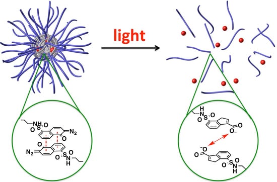
Graphical Abstract

1. Introduction

Polymeric micelles have emerged as promising drug delivery systems for cancer treatment because amphiphilic block copolymers enable micelles to improve drug solubility, control drug release, prolong circulation time in body and avoid the elimination of reticuloendothelial system (RES) [1,2,3,4,5,6,7,8]. Additionally, the prolonged circulation time makes polymeric micellar nanoparticles enhanced accumulation in solid tumors tissue with leaky vasculatures through the enhanced permeability and retention (EPR) effect [9,10,11,12]. Recently, stimulus-responsive polymeric micelles activated by pH, temperatures, redox, ultrasound, and light have been explored to provide spatial or temporal control of release of anti-cancer drugs to tumors [13,14,15,16,17,18]. Particularly, light-responsive drug delivery systems are attractive because they allow site-specific release of payload at desired disease sites with precisely controlled wavelength and intensity [19,20,21,22,23,24]. Light is already used in clinical practice in phototherapy (e.g., photodynamic and photothermal therapies) and optical imaging (e.g., fluorescence imaging and fluorescence guided surgery) [25,26].
In many light-responsive polymeric micelle systems, UV light triggers a change in polarity or a transition from hydrophobicity to hydrophilicity in domains bearing photochromic azobenzene, spiropyran, and 2-diazo-1,2-naphthoquinone (DNQ) [27,28,29,30,31,32,33], which leads to drug release [34,35,36,37,38]. However, conventional micelle systems have limited drug loading capacity (LC) and relatively high critical micelle concentrations (CMC), which impair particle stability resulting in decreased blood circulation time and release of drugs before the micelles accumulate in the tumor sites. To increase drug loading and lower CMC (i.e., to enhance the stability of micelles), micelles have been recently developed by supramolecular interactions such as π–π stacking, hydrogen bonding, stereocomplexation, and electrostatic interaction [39,40,41,42,43,44,45,46,47,48].
Here, we have developed a light-responsive polymeric micellar nanoparticle with high LC and stability using particularly π–π stacking. The light-responsiveness is imparted by poly(ethylene glycol)-block-poly-l-lysine (PEG-PLL) modified by the light-responsive DNQ group. Upon irradiation by UV or two-photon near infrared (NIR) light, hydrophobic DNQ can be converted to hydrophilic 3-indenecarboxylic acid by the Wolff rearrangement (Supplementary Materials Figure S2). Previously, photo-triggered dye release from micelles comprising a linear amphiphile, DNQ-terminated oligomer, has been reported [36]. However, these micelles had a high CMC (i.e., low stability) of 1.15 mg/mL in phosphate buffered saline (PBS). Alternative DNQ-based drug delivery systems have been developed to lower CMC, reduce the cytotoxicity, and increase biocompatibility, such as linear-dendritic PEO-b-G4 polyester conjugated with 16 DNQs [37], DNQ-modified dextran [34], and Janus-type dendritic polyamidoamine (PAMAM) amphiphiles with DNQ [35]. For in vivo drug delivery, there are still needs to enhance stability of polymeric micelle formulations, increase the drug LC, and develop new stimuli-responsive polymeric nano-sized particles for effective passive tumor targeting by the EPR effect [2,3,5]. Herein, we report a light-sensitive polymeric micellar nanoparticle activated under low power density of UV light with enhanced stability, LC, and drug retention by supramolecular hydrophobic and π–π stacking interactions between the copolymers and drugs. The micellar nanoparticles were formulated from the synthesized DNQs-conjugated PEG-PLL amphiphilic copolymers and we successfully obtained burst release of aromatic anticancer drugs, docetaxel (DTX), from the micellar nanoparticles under UV light irradiation.

2. Materials and Methods

2.1. Materials

Unless specified, chemicals were purchased from Sigma-Aldrich (St. Louis, MO, USA) and used without further purification. Methoxy-poly(ethylene glycol)-block-poly(l-lysine trifluoroacetate) (mPEG5K-b-PLKF10: Mw = 7400, 5k PEG conjugated with 10 lysine units, dispersity (Đ): 1.01–1.2) was purchased from Alamanda Polymers, Inc. (Huntsville, AL, USA) and 2-Diazo-1,2-naphthoquinone-5-sulfonyl chloride was purchased from ChemPacific (Baltimore, MD, USA). Docetaxel (DTX) was purchased from LC Laboratories (Woburn, MA, USA) and stored at −20 °C prior to use.

2.2. Characterizations

High performance liquid chromatography (HPLC) analysis was performed on a Hewlett Packard/Agilent series 1100 (Agilent, Santa Clara, CA, USA) equipped with an analytical C18 reverse phase column (Kinetex, 75 × 4.6 mm, 2.6 μm, Phenomenex, Torrance, CA, USA). Nuclear magnetic resonance (NMR) studies were performed on a Varian 500 system (400 MHz). The sizes and polydispersity of micelles were determined on a Delsa Nano C particle analyzer (Beckman Coulter, CA, USA). Fluorescence microscopy was conducted on a FSX 100 (Olympus, PA, USA). 8453 UV–Vis spectrophotometer (Agilent, CA, USA) was used to acquire UV–Vis spectra and Cary Eclipse fluorescence spectrophotometer was used to measure fluorescence. For transmission electron microscopy (TEM) image, a 2.0 μL aliquot of the micelle solution was deposited on a copper grid coated with a carbon film. The sample was dried at room temperature and then imaged on a Tecnai G2 Spirit BioTWIN transmission electron microscope, operating at 100 kV (note that this drying procedure employed in TEM analysis may cause considerable changes in the size and shape of the micelles since they only exist in solution).

2.3. Statistical Analysis

Due to the asymmetric distribution of some of our data, we did not assume a normal distribution, and data were analyzed by the non-parametric Mann–Whitney U-test. A p value < 0.05 was considered statistically significant unless stated otherwise. For multiple comparisons, Kruskal–Wallis tests were performed followed by Bonferroni corrections.

2.4. Synthesis of PEG-PLL-DNQ and PEG-PLL-Oct

Methoxy-poly(ethylene glycol)-block-poly(l-lysine trifluoroacetate) (PEG-PLL, 0.5 g, 0.068 mmol) and 2-diazo-1,2-naphthoquinone-5-sulfonyl chloride (sc-DNQ, 0.25 g, 0.93 mmol) or octanoyl chloride (c-Oct, 0.15 g, 0.93 mmol) were placed in a 100 mL round bottom flask and suspended in a CHCl3 (20 mL). Then, triethylamine (0.2 mL) was added slowly and the reaction mixture was stirred in the dark overnight. The solvent was evaporated under reduced pressure and the crude product was purified by size exclusion chromatography (SEC) with DMF. After evaporation of DMF under reduced pressure at 60 °C, PEG-PLL-DNQ or PEG-PLL-Oct was precipitated with ether/methanol and then centrifuged. The solvent was completely removed under high vacuum.

2.5. Formulation of the Micellar Nanoparticles with DTX or Nile Red (NR)

The thin-film hydration method was used to form drug-encapsulating micellar nanoparticles. Both PEG-PLL-DNQ and DTX (20% w/w) or NR (1% w/w) were dissolved in CHCl3 and mixed for 1 h. The organic solvent was then slowly removed using a rotary vacuum evaporation and further dried under a constant flow of nitrogen gas until a film formed in a round-bottomed flask. All organic solvents were removed in a high vacuum. After the drug-containing block copolymer film was formed, aqueous solution was added, heated up to 60 °C, and stirred to get a preliminary micellar nanoparticle solution. Subsequently, the solution was dialyzed in water for 24 h (molecular weight cutoff: 3.5 kDa). The final micellar nanoparticles were obtained by filtering through 0.22-μm filters. Similar procedures were used to formulate PEG-PLL-Oct-based micellar nanoparticles.

2.6. Determination of the Critical Micelle Concentration (CMC)

The pyrene fluorescence method was used to determine CMC of DNQ-conjugated micellar nanoparticles (NPDNQ) and Oct-conjugated micellar nanoparticles (NPOct). For the fluorescence measurements, 3.0 mL of solution of NPDNQ or NPOct containing 2.0 μM of pyrene was placed in a fluorescence quartz cuvette cell and the fluorescence emission spectra were measured in the range from 360 to 410 nm at 339 nm excitation. The concentrations of PEG-PLL-DNQ and PEG-PLL-Oct were varied from 10−4 to 1.0 mg/mL. The fluorescent intensities were recorded at the wavelengths corresponding to the first and third vibronic bands located at 373 nm and 383 nm. All fluorescence measurements were carried out at 25 °C.

2.7. Effect of Lyophilization/Reconstitution on Particle Size

1.0 mL of DTX-loaded NPDNQ (DTX-NPDNQ, 0.5 mg/mL) was frozen and lyophilized to obtain white powder and then the resulting white powder was reconstituted with 1.0 mL of distilled water followed by addition of a concentrated PBS solution (10×). Particle sizes of DTX-NPDNQ before and after lyophilization/reconstitution were measured by DLS. The reconstituted micellar nanoparticles were used for all data.

2.8. Stability Studies of NPs in the Presence of BSA

Nile Red-loaded NPDNQ (NR-NPDNQ) and NPOct (NR-NPOct) were incubated in bovine serum albumin solution (BSA, 10%) at 37 °C. At each time point, the micelle suspension was placed on a shaker with gentle agitation at room temperature, and then their emission spectra were recorded (excitation at 570 nm).

2.9. Irradiation of UV Light

To investigate the release of encapsulated DTX from DTX-NPDNQ, UV light at variable time periods was carried using a Mightex FCS fiber-coupled LED light (Mightex, Pleasanton, CA, USA) with the wavelength of 365 nm (50 mW/cm2).

2.10. Determination of DTX in DTX-NPDNQ

To determine the amount of DTX in DTX-NPDNQ, a PBS suspension of DTX-NPDNQ was place into Slide-A-Lyzer dialysis tubes (0.5 mL each tube) with a molecular weight cutoff at 3.5 kDa (Pierce, Rockford, IL, USA). These microtubes were individually dialyzed in 2.0 L of PBS buffer (1×). DTX-NPDNQ solutions from microtubes were collected separately, acetonitrile was added to dissolve the micelles, and DTX in each tube was measured by HPLC: Chromatography was performed in the isocratic mode. Mobile phase, eluent A: 0.1% orthophosphoric acid, 40%; eluent B: acetonitrile, 60%; the flow rate, 1 mL/min; the sample volume, 100 μL. The detection was carried out at 232 nm [49,50].

2.11. In Vitro Cytotoxicity

To compare cell proliferation by DTX, NPDNQ, and DTX-NPDNQ in normal cells (HUVECs) and breast cancer cells (MCF-7 cancer cells), HUVECs and MCF-7 cells were evaluated by (3-(4,5-dimethylthiazol-2-yl)-2,5-diphenyltetrazolium bromide) (MTT) based assay. 1 × 10⁴ cells per well were seeded into a 96-well plate and incubated at 37 °C in a humidified atmosphere with 5% CO2 for 24 h. Cell culture medium of HUVECs and MCF-7 cancer cells was used with EG2 media (Lonza, NJ, USA) with supplied various growth factors and RPMI 1640 supplemented with 10% FBS and 1% pen-strip, respectively. Each cell was treated with 200 μL of serially diluted samples with different concentrations (5-400 ng/mL) at 37 °C for 24 h. The negative control was treated with only cell culture media, and the empty NPs (NPDNQ) were prepared. After 24 h, they were rinsed twice with DPBS (pH 7.4, with Ca2+, Mg2+) to eliminate the remaining samples. 25 μL of the MTT reagent (5 mg/mL in media) was then added and incubated further for 4 h at 37 °C [51,52]. Thereafter, the purple precipitates (formazan crystals) were added and dissolved by adding 200 μL of DMSO. Absorbance at 570 nm was measured with a microplate reader (Gen5, Biotek, Winooski, VT, USA). The percentages of viable cells were determined through the reduction of MTT relative to the negative control.

2.12. Cytotoxicity of Light-Triggered DTX-NPDNQ

MCF-7 cells were grown six-well plates and various concentrations (e.g., 100, 200, and 400 ng/mL of DTX; the concentrations of NPs were adjusted to the concentration of DTX by UV-Vis spectroscopy) of DTX-NPDNQ were added. The cells were incubated for 2 h at 37 °C and then washed with fresh media (2×). Subsequently, UV light for 10 min was applied and the cells were further incubated for 24 h. The cytotoxicity of cells was determined by MTT assay.

2.13. Intracellular Localization of the NR-NPDNQ

To observe cellular behavior of DTX-NPDNQ in MCF-7 cancer cells, NR (hydrophobic fluorescence dye) loaded the core in NPDNQ. MCF-7 cancer cells were seeded on a 35 mm petri-dish and allowed to grow until a confluence of 60–80%. Then, they were washed twice with DPBS and the cells were incubated with 2 mL of NR-NPDNQ for up to 120 min at 37 °C. After predetermined incubation times (0, 30, 60, 90, and 120 min), the treated cells were rinsed twice with PBS and fixed by 4% paraformaldehyde for 2 h. Thereafter, 4, 6-diamidino-2-phenylindole (DAPI) (1 mg/mL in DI water) treated for nuclei staining for 3 min, and again washed twice with PBS. Fluorescent distribution in the cells was observed by a fluorescence microscope (Olympus FSX100, Olympus, Japan).

3. Results

3.1. Synthesis of the Light-Sensitive Block Copolymers and Formulation of the Micellar Nanoparticles

The synthetic route of the amphiphilic block copolymers, PEG-PLL-DNQ and PEG-PLL-Oct (Figure 1), is shown in Figure S1. PEG-PLL (Mn ~ 7400, 5k PEG conjugated with 10 lysine units) was simply coupled to DNQ-sulfonyl chloride (sc-DNQ) under a base condition to afford PEG-PLL-DNQ in 80% yield. The successful synthesis of PEG-PLL-DNQ was characterized by 1H NMR spectroscopy. As shown in Figure S2, the characteristic resonance signals of DNQ at 8.70, 8.47, 7.70, 7.60, and 5.29 ppm were observed in the spectrum of PEG-PLL-DNQ in DMSO-d6, which is evidence of the successful conjugation of DNQ groups on PEG-PLL. Octanoyl chloride (c-Oct) without aromatic groups was also conjugated to PEG-PLL in the same manner to form PEG-PLL-Oct that only provides hydrophobic interactions in the core of the micelle without π–π stacking interactions.
As previously reported, AB-type amphiphilic block copolymers composed of hydrophilic and hydrophobic components can self-assemble into micelles, where the hydrophobic core of the micelle can encapsulate hydrophobic drugs which will otherwise precipitate in aqueous solutions. In this study, we synthesized a new block copolymer, PEG-PLL-DNQ, containing hydrophobic light-sensitive DNQs which could generate additional π–π stacking interactions between the neighboring DNQs on the polymer backbone inside of the micelle core. Furthermore, these DNQs could also interact with DTX, which contains two aromatic phenyl groups that can form another π–π stacking in addition to hydrophobic interactions (Figure S1). These interactions will contribute to the higher formulation stability of the micelle as well as its increased loading capacity (LC) with aromatic drugs.
The DTX-loaded polymeric micellar nanoparticle (DTX-NPDNQ) was prepared by the thin-film hydration method. Dynamic light scattering (DLS) results showed that PEG-PLL-DNQ formed micelles with a volume average hydrodynamic diameter of 143 nm and TEM image revealed that DTX-NPDNQ were formed with a spherical morphology (Figure 2). For controlled experiments, DTX-NPOct was successfully formulated with 105 nm in diameter (Figure S3).

3.2. Measurements of CMC, LE and LC, and Stability

CMC is a key parameter to describe the self-assembly behavior of micelles. The CMC of NPDNQ was calculated by the pyrene fluorescence method [53]. Pyrene has a fluorescence profile that is dependent on its microenvironment. In hydrophobic environments, the fluorescent intensity ratio of the first (373 nm) to third (383 nm) emission peaks (Figure 3a), the I373/I383 ratio known as polarity parameter, is low (close to 1); whereas in hydrophilic environments, the I373/I383 ratio increases (close to 2). Using this method, we were able to determine whether pyrene was in the core of the micelle (hydrophobic environments) or in aqueous solution (hydrophilic environments). The polymer concentration at which the location of pyrene changed from solution to micelle is the CMC. Figure 3a shows the fluorescence emission spectra of pyrene encapsulated in NPDNQ and the change of the I373/I383 ratio at the various concentrations of PEG-PLL-DNQ at room temperature. The curve was almost flat at low polymer concentrations but rapidly decreased at higher polymer concentrations and eventually reached a plateau. The CMC value of NPDNQ was determined by the intersection of two straight lines and the CMC was about 7.1 μg/mL (Figure 3b), which is a relevantly low CMC compared to other reported amphiphilic diblock copolymers (>20 μg/mL) and four times lower than the control micelle system, NPOct (25 μg/mL, Figure 3c) due to π–π stacking interactions between neighboring DNQs on the polymer backbone leading to a more condensed core of NPDNQ. The reduced CMC could contribute to improved thermodynamic stability of the micelle, reduced toxicity toward cells, and increased circulating time in blood.
In addition to CMC, we evaluated the drug loading efficiency (LE) and capacity (LC) of DTX-NPDNQ and DTX-NPOct using HPLC (Table S1). The results show LCs of 13.5% and 7.9% for DTX-NPDNQ and DTX-NPOct, respectively. These were associated with LEs of 90.3% and 79.1%, respectively. NPDNQ encapsulated approximately two times more DTX than NPOct, which can be presumably ascribed that NPDNQ has a better compatibility of DTX due to π–π stacking interactions between DNQs and aromatic DTX.
Subsequently, we measured the stability of the micelles in blood plasma at 37 °C because the stability is greatly influenced by serum proteins. The hydrophobic Nile red dye (NR)-loaded micelles, NR-NPDNQ and NR-NPOct, were incubated in 10% bovine serum albumin (BSA) solution and the intensity of fluorescence emission was monitored. NR is a fluorescent indicator for the micropolarity; it shows strong fluorescence emission intensity in hydrophobic environments while it is quenched in a hydrophilic solution [54]. As shown in Figure 3d, NR-NPDNQ was stable in BSA solution retaining their intensity up to 2 days at 37 °C (>85%) while NR-NPOct showed a dramatic decrease in intensity, indicating release of NR into hydrophilic environments (Figure S4). These results indicate that the presence of DNQ on the polymer backbone increased the stability of the micelles.

3.3. Cellular Uptake Study and Cytotoxicity

The cell internalization of NR-NPDNQ was characterized by a fluorescence microscopy. As shown in Figure 4, NR-NPDNQ quickly entered into MCF-7 cells. After incubation with NR-NPDNQ for 30 min, most of MCF-7 cells showed red fluorescence in the cell and then the fluorescence intensity from the cells was saturated after 1 h. As shown in Figure 3d, less than 7% of hydrophobic NR was released from the micelle within 6 h. Thus, the saturation of fluorescence intensity after 1 h indicates that the cell uptake of NR-NPDNQ, not the free NR (e.g., NR fully stains cells with 10 min [55]), is the dominant factor of this fluorescence increase. Through the cellular uptake study, we confirmed that NR-NPDNQ efficiently entered into MCF-7 cells within 1 h.
Human umbilical vein endothelial cells (HUVECs) were used to evaluate the cytotoxicity. The viability of HUVECs was determined by MTT assay in corresponding culture medium after 24 h incubation with DTX, polymers and micelles. PEG-PLL-DNQ and PEG-PLL-Oct showed low cytotoxicity against HUVECs with the concentrations up to 0.5 mg/mL (>90% viability, Figure S5). Furthermore, the cytotoxicity of DTX, NPDNQ and DTX-NPDNQ showed that LC50 for DTX was 99 ng/mL and DTX-NPDNQ was 310 ng/mL in HUVECs (Figure 5a). The results show a lower cytotoxicity with DTX-NPDNQ than free drug DTX, against HUVECs, because NPDNQ strongly retains the hydrophobic aromatic DTX in the core of micelle due to hydrophobic and π–π stacking interactions.

3.4. Measurement of Light-Induced Destabilization of Micellar Nanoparticles

To investigate the light responsiveness of DTX-NPDNQ, the micellar nanoparticles were irradiated by 365 nm UV light, and DLS measurements were conducted to confirm the light-triggered destabilization process of DTX-NPDNQ. DLS results showed a change in the micellar diameter in response to UV light (Figure 5b). The irradiated DTX-NPDNQ (50 mW/cm2, 10 min) showed a decrease in its hydrodynamic size from 143 nm prior to irradiation to 18 nm and additionally, 63% of DTX was released into the solution during the light-triggered process; however, the DTX release profile after 1, 3, and 5 min of UV irradiation could not be obtained due to the destabilization of DTX-NPDNQ (i.e., partial IC from DNQ) during dialysis. These results revealed that the micelles dissociated partly under UV light, which was attributed to the Wolff rearrangement of hydrophobic DNQ moieties into hydrophilic IC molecules (Figure S2). The change in the hydrophilic–hydrophobic balance of NPDNQ then destroyed the structure of the micelles.
The light-triggered effect of the micelles was studied with MCF-7 cells (Figure 5c,d); Cells were incubated with DTX-NPDNQ for 2 h, washed with fresh media, irradiated by 365 nm UV light for 10 min (50 mW/cm2), and then further incubated for 24 h. Prior to irradiation, the cell viability of DTX-NPDNQ (400 ng/mL of DTX) was 55%. Upon irradiation with UV light, the cell viability was decreased to approximately 36%, similar to the cell viability of free DTX (25%), while there was no significant difference in the MCF-7 cell viability of NPDNQ with UV irradiation (Figure 5c). The study showed that DTX-NPDNQ was more cytotoxic than NPDNQ with UV irradiation because most of DTX-NPDNQ were disrupted by UV irradiation and then released DTX into MCF-7 cells.

4. Conclusions

In summary, we developed a photo-responsive micellar system with the DNQ-conjugated block copolymer. Hydrophobic aromatic DTX drugs were loaded into the micellar nanoparticles with enhanced stability and loading capacity, and then released into aqueous solution in a controlled manner under UV irradiation. Without light irradiation, the micellar nanoparticles were stable due to hydrophobic and π–π stacking interactions of DNQs with neighboring DNQs and DTX, making them suitable for biomedical applications within a living system. Investigations are underway on the potential use of these micellar nanoparticles for light-triggered intracellular delivery of hydrophobic aromatic anticancer drugs.

Supplementary Materials

The following are available online at https://www.mdpi.com/2073-4360/13/3/377/s1, Figure S1: Synthesis of PEG-PLL-DNQ and PEG-PLL-Oct, and the chemical structures of DTX and NR. Figure S2: Wolff Rearrangement of PEG-PLL-DNQ upon UV light irradiation. Figure S3: DLS size distribution and TEM image of DTX-NPoct. Figure S4: DLS measurement of size change of NR-NPOct in BSA solution. Figure S5: HUVECs Viability of Polymers (PEG-PLL-DNQ & PEG-PLL-Oct). Table S1: Measurement of Drug Loading Efficiency (LE) and Capacity (LC).

Author Contributions

Conceptualization, K.N.K. and J.-J.L.; methodology, K.N.K. and K.S.O.; formal analysis, K.N.K. and K.S.O.; resources, J.S., I.R.S., S.D.K. and J.-J.L.; writing—original draft preparation, K.N.K.; writing—review and editing, J.-J.L.; visualization, K.N.K. and K.S.O.; supervision, J.-J.L.; funding acquisition, J.-J.L. All authors have read and agreed to the published version of the manuscript.

Funding

This research was supported by ACS IRG #16-184-56 from the American Cancer Society to the University of Colorado Cancer Center, UC AMC Head and Neck Cancer Research Program Career Development Award, Cancer League of Colorado (No.173991), ACS-RSG-16-256-01, CLAS Research Innovation Award at CU Denver, the University of Colorado Cancer Center and Wings of Hope for Pancreatic Cancer Research, the Office of Research Services at CU Denver.

Institutional Review Board Statement

Not applicable.

Informed Consent Statement

Not applicable.

Data Availability Statement

Raw data are available from the corresponding author upon request.

Conflicts of Interest

The authors declare no conflict of interest.

References

  1. Gref, R.; Minamitake, Y.; Peracchia, M.T.; Trubetskoy, V.; Torchilin, V.; Langer, R. Biodegradable Long-Circulating Polymeric Nanospheres. Science 1994, 263, 1600–1603. [Google Scholar] [CrossRef] [PubMed] [Green Version]
  2. Tan, Q.; Liu, X.J.; Fu, X.Y.; Li, Q.L.; Dou, J.F.; Zhai, G.X. Current development in nanoformulations of docetaxel. Expert Opin. Drug Deliv. 2012, 9, 975–990. [Google Scholar] [CrossRef] [PubMed]
  3. Langer, R. Drug delivery and targeting. Nature 1998, 392, 5–10. [Google Scholar] [PubMed]
  4. Torchilin, V.P. Micellar nanocarriers: Pharmaceutical perspectives. Pharm. Res. 2007, 24, 1–16. [Google Scholar] [CrossRef] [PubMed]
  5. Langer, R. Polymer-Controlled Drug-Delivery Systems. Acc. Chem. Res. 1993, 26, 537–542. [Google Scholar] [CrossRef]
  6. Hruby, M.; Konak, C.; Ulbrich, K. Polymeric micellar pH-sensitive drug delivery system for doxorubicin. J. Control. Release 2005, 103, 137–148. [Google Scholar] [CrossRef]
  7. Langer, R.; Folkman, J. Polymers for Sustained-Release of Proteins and Other Macromolecules. Nature 1976, 263, 797–800. [Google Scholar] [CrossRef]
  8. Ganta, S.; Devalapally, H.; Shahiwala, A.; Amiji, M. A review of stimuli-responsive nanocarriers for drug and gene delivery. J. Control. Release 2008, 126, 187–204. [Google Scholar] [CrossRef]
  9. Jain, R.K.; Stylianopoulos, T. Delivering nanomedicine to solid tumors. Nat. Rev. Clin. Oncol. 2010, 7, 653–664. [Google Scholar] [CrossRef] [Green Version]
  10. Carmeliet, P.; Jain, R.K. Principles and mechanisms of vessel normalization for cancer and other angiogenic diseases. Nat. Rev. Drug Discov. 2011, 10, 417–427. [Google Scholar] [CrossRef]
  11. Yuan, F.; Chen, Y.; Dellian, M.; Safabakhsh, N.; Ferrara, N.; Jain, R.K. Time-dependent vascular regression and permeability changes in established human tumor xenografts induced by an anti-vascular endothelial growth factor vascular permeability factor antibody. Proc. Natl. Acad. Sci. USA 1996, 93, 14765–14770. [Google Scholar] [CrossRef] [Green Version]
  12. Yuan, F.; Dellian, M.; Fukumura, D.; Leunig, M.; Berk, D.A.; Torchilin, V.P.; Jain, R.K. Vascular-Permeability in a Human Tumor Xenograft—Molecular-Size Dependence and Cutoff Size. Cancer Res. 1995, 55, 3752–3756. [Google Scholar] [PubMed]
  13. Nakayama, M.; Okano, T.; Miyazaki, T.; Kohori, F.; Sakai, K.; Yokoyama, M. Molecular design of biodegradable polymeric micelles for temperature-responsive drug release. J. Control. Release 2006, 115, 46–56. [Google Scholar] [CrossRef] [PubMed]
  14. Liu, S.Y.; Weaver, J.V.M.; Tang, Y.Q.; Billingham, N.C.; Armes, S.P.; Tribe, K. Synthesis of shell cross-linked micelles with pH-responsive cores using ABC triblock copolymers. Macromolecules 2002, 35, 6121–6131. [Google Scholar] [CrossRef]
  15. Oishi, M.; Kataoka, K.; Nagasaki, Y. pH-responsive three-layered PEGylated polyplex micelle based on a lactosylated ABC triblock copolymer as a targetable and endosome-disruptive nonviral gene vector. Bioconjug. Chem. 2006, 17, 677–688. [Google Scholar] [CrossRef] [PubMed]
  16. Li, Y.W.; Tong, R.; Xia, H.S.; Zhang, H.J.; Xuan, J.A. High intensity focused ultrasound and redox dual responsive polymer micelles. Chem. Commun. 2010, 46, 7739–7741. [Google Scholar] [CrossRef]
  17. Zhao, Y. Photocontrollable block copolymer micelles: What can we control? J. Mater. Chem. 2009, 19, 4887–4895. [Google Scholar] [CrossRef]
  18. Schumers, J.M.; Fustin, C.A.; Gohy, J.F. Light-responsive block copolymers. Macromol. Rapid Commun. 2010, 31, 1588–1607. [Google Scholar] [CrossRef]
  19. Timko, B.P.; Kohane, D.S. Prospects for near-infrared technology in remotely triggered drug delivery. Expert Opin. Drug Deliv. 2014, 11, 1681–1685. [Google Scholar] [CrossRef] [Green Version]
  20. Liu, G.; Liu, W.; Dong, C.M. UV- and NIR-responsive polymeric nanomedicines for on-demand drug delivery. Polym. Chem. 2013, 4, 3431–3443. [Google Scholar] [CrossRef]
  21. Tong, R.; Chiang, H.H.; Kohane, D.S. Photoswitchable nanoparticles for in vivo cancer chemotherapy. Proc. Natl. Acad. Sci. USA 2013, 110, 19048–19053. [Google Scholar] [CrossRef] [PubMed] [Green Version]
  22. Tong, R.; Hemmati, H.D.; Langer, R.; Kohane, D.S. Photoswitchable Nanoparticles for Triggered Tissue Penetration and Drug Delivery. J. Am. Chem. Soc. 2012, 134, 8848–8855. [Google Scholar] [CrossRef] [PubMed]
  23. Jin, Q.A.; Liu, G.Y.; Liu, X.S.; Ji, J.A. Photo-responsive supramolecular self-assembly and disassembly of an azobenzene-containing block copolymer. Soft Matter 2010, 6, 5589–5595. [Google Scholar] [CrossRef]
  24. Tong, R.; Kohane, D.S. Shedding light on nanomedicine. Wiley Interdiscip. Rev. Nanomed. Nanobiotechnol. 2012, 4, 638–662. [Google Scholar] [CrossRef] [PubMed]
  25. Lovell, J.F.; Liu, T.W.B.; Chen, J.; Zheng, G. Activatable Photosensitizers for Imaging and Therapy. Chem. Rev. 2010, 110, 2839–2857. [Google Scholar] [CrossRef]
  26. Van Dam, G.M.; Themelis, G.; Crane, L.M.A.; Harlaar, N.J.; Pleijhuis, R.G.; Kelder, W.; Sarantopoulos, A.; de Jong, J.S.; Arts, H.J.G.; van der Zee, A.G.J.; et al. Intraoperative tumor-specific fluorescence imaging in ovarian cancer by folate receptor-alpha targeting: First in-human results. Nat. Med. 2011, 17, 1315–1319. [Google Scholar] [CrossRef]
  27. Kirmse, W. 100 years of the Wolff rearrangement. Eur. J. Org. Chem. 2002, 14, 2193–2256. [Google Scholar] [CrossRef]
  28. Hu, J.M.; Zhang, G.Q.; Liu, S.Y. Enzyme-responsive polymeric assemblies, nanoparticles and hydrogels. Chem. Soc. Rev. 2012, 41, 5933–5949. [Google Scholar] [CrossRef]
  29. Almstead, J.I.K.; Urwyler, B.; Wirz, J. Flash-Photolysis of Alpha-Diazonaphthoquinones in Aqueous-Solution—Determination of Rates and Equilibria for Keto-Enol Tautomerization of 1-Indene-3-Carboxylic Acid. J. Am. Chem. Soc. 1994, 116, 954–960. [Google Scholar] [CrossRef]
  30. Zhao, Y. Light-Responsive Block Copolymer Micelles. Macromolecules 2012, 45, 3647–3657. [Google Scholar] [CrossRef]
  31. Fomina, N.; Sankaranarayanan, J.; Almutairi, A. Photochemical mechanisms of light-triggered release from nanocarriers. Adv. Drug Del. Rev. 2012, 64, 1005–1020. [Google Scholar] [CrossRef] [PubMed] [Green Version]
  32. Kotharangannagari, V.K.; Sanchez-Ferrer, A.; Ruokolainen, J.; Mezzenga, R. Photoresponsive Reversible Aggregation and Dissolution of Rod-Coil Polypeptide Diblock Copolymers. Macromolecules 2011, 44, 4569–4573. [Google Scholar] [CrossRef]
  33. Hu, J.M.; Liu, S.Y. Responsive Polymers for Detection and Sensing Applications: Current Status and Future Developments. Macromolecules 2010, 43, 8315–8330. [Google Scholar] [CrossRef]
  34. Liu, G.Y.; Chen, C.J.; Li, D.D.; Wang, S.S.; Ji, J. Near-infrared light-sensitive micelles for enhanced intracellular drug delivery. J. Mater. Chem. 2012, 22, 16865–16871. [Google Scholar] [CrossRef]
  35. Sun, L.; Ma, X.F.; Dong, C.M.; Zhu, B.S.; Zhu, X.Y. NIR-Responsive and Lectin-Binding Doxorubicin-Loaded Nanomedicine from Janus-Type Dendritic PAMAM Amphiphiles. Biomacromolecules 2012, 13, 3581–3591. [Google Scholar] [CrossRef]
  36. Goodwin, A.P.; Mynar, J.L.; Ma, Y.Z.; Fleming, G.R.; Frechet, J.M.J. Synthetic micelle sensitive to IR light via a two-photon process. J. Am. Chem. Soc. 2005, 127, 9952–9953. [Google Scholar] [CrossRef]
  37. Mynar, J.L.; Goodwin, A.P.; Cohen, J.A.; Ma, Y.; Fleming, G.R.; Frechet, J.M.J. Two-photon degradable supramolecular assemblies of linear-dendritic copolymers. Chem. Commun. 2007, 20, 2081–2082. [Google Scholar] [CrossRef]
  38. Sun, L.; Yang, Y.; Dong, C.M.; Wei, Y. Two-Photon-Sensitive and Sugar-Targeted Nanocarriers from Degradable and Dendritic Amphiphiles. Small 2011, 7, 401–406. [Google Scholar] [CrossRef]
  39. Attia, A.B.E.; Yang, C.; Tan, J.P.K.; Gao, S.J.; Williams, D.F.; Hedrick, J.L.; Yang, Y.Y. The effect of kinetic stability on biodistribution and anti-tumor efficacy of drug-loaded biodegradable polymeric micelles. Biomaterials 2013, 34, 3132–3140. [Google Scholar] [CrossRef]
  40. Kim, B.S.; Park, S.W.; Hammond, P.T. Hydrogen-bonding layer-by-layer assembled biodegradable polymeric micelles as drug delivery vehicles from surfaces. ACS Nano 2008, 2, 386–392. [Google Scholar] [CrossRef]
  41. Song, B.; Wang, Z.Q.; Chen, S.L.; Zhang, X.; Fu, Y.; Smet, M.; Dehaen, W. The introduction of pi-pi stacking moieties for fabricating stable micellar structure: Formation and dynamics of disklike micelles. Angew. Chem. Int. Ed. 2005, 44, 4731–4735. [Google Scholar] [CrossRef] [PubMed]
  42. Zhang, P.; Huang, Y.X.; Liu, H.; Marquez, R.T.; Lu, J.Q.; Zhao, W.C.; Zhang, X.L.; Gao, X.; Li, J.; Venkataramanan, R.; et al. A PEG-Fmoc conjugate as a nanocarrier for paclitaxel. Biomaterials 2014, 35, 7146–7156. [Google Scholar] [CrossRef] [PubMed] [Green Version]
  43. Shi, Y.; van Steenbergen, M.J.; Teunissen, E.A.; Novo, L.; Gradmann, S.; Baldus, M.; van Nostrum, C.F.; Hennink, W.E. Pi-Pi Stacking Increases the Stability and Loading Capacity of Thermosensitive Polymeric Micelles for Chemotherapeutic Drugs. Biomacromolecules 2013, 14, 1826–1837. [Google Scholar] [CrossRef] [PubMed]
  44. Kang, N.; Perron, M.E.; Prud’homme, R.E.; Zhang, Y.B.; Gaucher, G.; Leroux, J.C. Stereocomplex block copolymer micelles: Core-shell nanostructures with enhanced stability. Nano Lett. 2005, 5, 315–319. [Google Scholar] [CrossRef] [PubMed]
  45. Zhang, J.; Wang, L.Q.; Wang, H.J.; Tu, K.H. Micellization phenomena of amphiphilic block copolymers based on methoxy poly(ethylene glycol) and either crystalline or amorphous poly(caprolactone-b-lactide). Biomacromolecules 2006, 7, 2492–2500. [Google Scholar] [CrossRef]
  46. Kim, Y.; Pourgholami, M.H.; Morris, D.L.; Stenzel, M.H. Effect of cross-linking on the performance of micelles as drug delivery carriers: A cell uptake study. Biomacromolecules 2012, 13, 814–825. [Google Scholar] [CrossRef]
  47. Lin, W.; He, Y.; Zhang, J.; Wang, L.; Wang, Z.; Ji, F.; Chen, S. Highly hemocompatible zwitterionic micelles stabilized by reversible cross-linkage for anti-cancer drug delivery. Colloids Surf. B Biointerfaces 2014, 115, 384–390. [Google Scholar] [CrossRef]
  48. Zarrintaj, P.; Ramsey, J.D.; Samadi, A.; Atoufi, Z.; Yazdi, M.K.; Ganjali, M.R.; Amirabad, L.M.; Zangene, E.; Farokhi, M.; Formela, K.; et al. Poloxamer: A versatile tri-block copolymer for biomedical applications. Acta Biomater. 2020, 110, 37–67. [Google Scholar] [CrossRef]
  49. Cao, C.; Zhao, J.; Lu, M.; Garvey, C.J.; Stenzel, M.H. Correlation between Drug Loading Content and Biological Activity: The Complexity Demonstrated in Paclitaxel-Loaded Glycopolymer Micelle System. Biomacromolecules 2019, 20, 1545–1554. [Google Scholar] [CrossRef]
  50. Sokol, M.B.; Nikolskaya, E.D.; Yabbarov, N.G.; Zenin, V.A.; Faustova, M.R.; Belov, A.V.; Zhunina, O.A.; Mollaev, M.D.; Zabolotsky, A.I.; Tereshchenko, O.G.; et al. Development of novel PLGA nanoparticles with co-encapsulation of docetaxel and abiraterone acetate for a highly efficient delivery into tumor cells. J. Biomed. Mater. Res. B Appl. Biomater. 2019, 107, 1150–1158. [Google Scholar] [CrossRef]
  51. Oh, K.S.; Song, J.Y.; Cho, S.H.; Lee, B.S.; Kim, S.Y.; Kim, K.; Jeon, H.; Kwon, I.C.; Yuk, S.H. Paclitaxel-loaded Pluronic nanoparticles formed by a temperature-induced phase transition for cancer therapy. J. Control. Release 2010, 148, 344–350. [Google Scholar] [CrossRef] [PubMed]
  52. Oh, K.S.; Yhee, J.Y.; Lee, D.E.; Kim, K.; Kwon, I.C.; Seo, J.H.; Kim, S.Y.; Yuk, S.H. Accurate sequential detection of primary tumor and metastatic lymphatics using a temperature-induced phase transition nanoparticulate system. Int. J. Nanomed. 2014, 9, 2955–2965. [Google Scholar] [CrossRef] [Green Version]
  53. Astafieva, I.; Zhong, X.F.; Eisenberg, A. Critical Micellization Phenomena in Block Polyelectrolyte Solutions. Macromolecules 1993, 26, 7339–7352. [Google Scholar] [CrossRef]
  54. Wang, W.P.; Chau, Y. Efficient and facile formation of two-component nanoparticles via aromatic moiety directed self-assembly. Chem. Commun. 2011, 47, 10224–10226. [Google Scholar] [CrossRef] [PubMed]
  55. Alemán-Nava, G.S.; Cuellar-Bermudez, S.P.; Cuaresma, M.; Bosma, R.; Muylaert, K.; Ritmann, B.E.; Parra, R. How to use Nile Red, a selective fluorescent stain for microalgal neutral lipids. J. Microbiol. Methods 2016, 128, 74–79. [Google Scholar] [CrossRef]
Figure 1. Structure of PEG-PLL-DNQ and PEG-PLL-Oct.
Polymers 13 00377 g001
Figure 2. (a) DLS size distribution of DTX-NPDNQ; (b) TEM image of DTX-NPDNQ (scale bar = 500 nm).
Polymers 13 00377 g002
Figure 3. (a) Normalized emission spectra of pyrene in solutions of PEG-PLL-DNQ in PBS; emission intensity of pyrene at 373 and 383 nm versus the concentrations of (b) PEG-PLL-DNQ and (c) PEG-PLL-Oct; (d) stability of NR-NPDNQ (d-blue) and NR-NPOct (d-red) in BSA solution. Data are means ± SD, N = 4, asterisks indicate p < 0.05.
Polymers 13 00377 g003
Figure 4. Fluorescence images of the internalization of NR-NPDNQ by MCF-7 cells for different time intervals: Nuclear staining with DAPI (blue color) and NR-NPDNQ (red color). (Scale bar: 20 μm).
Polymers 13 00377 g004
Figure 5. (a) Cytotoxicity of HUVECs with NPDNQ (green bar), DTX-NPDNQ (blue bar), and DTX (red bar); (b) DLS measurement of size change of DTX-NPDNQ upon UV light irradiation (365 nm, 50 mW/cm2, 10 min) in PBS; (c) MCF-7 Cells Viability with Light Irradiation (NPDNQ & DTX-NPDNQ); (d) Cytotoxicity of MCF-7 cells with different concentrations of DTX-NPDNQ (blue bar), DTX-NPDNQ with UV light irradiation (365 nm, 10 min, 50 mW/cm2) (green bar) and DTX (red bar). Data are means ± SD, N = 4, the concentrations of NPs were adjusted to the concentration of DTX by UV-Vis spectroscopy, asterisks indicate p < 0.05.
Polymers 13 00377 g005
Publisher’s Note: MDPI stays neutral with regard to jurisdictional claims in published maps and institutional affiliations.