
Effect of Multiple Enamel Surface Treatments on Micro-Shear Bond Strength

 ,
,
Abstract
:1. Introduction
2. Materials and Methods
2.1. Teeth Preparation
2.2. Surface Treatment
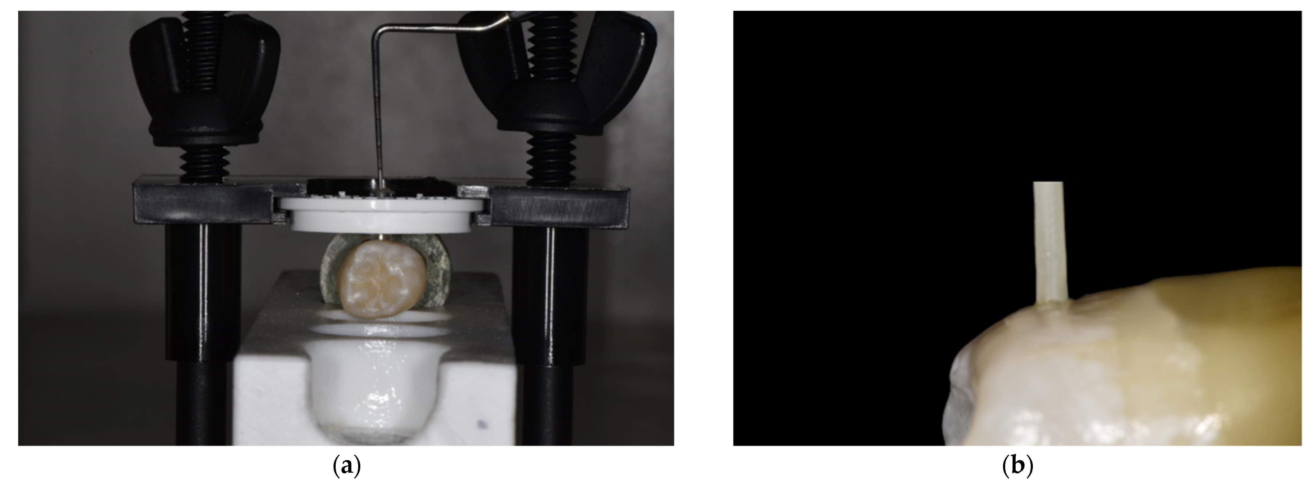
2.3. Micro-Shear Testing
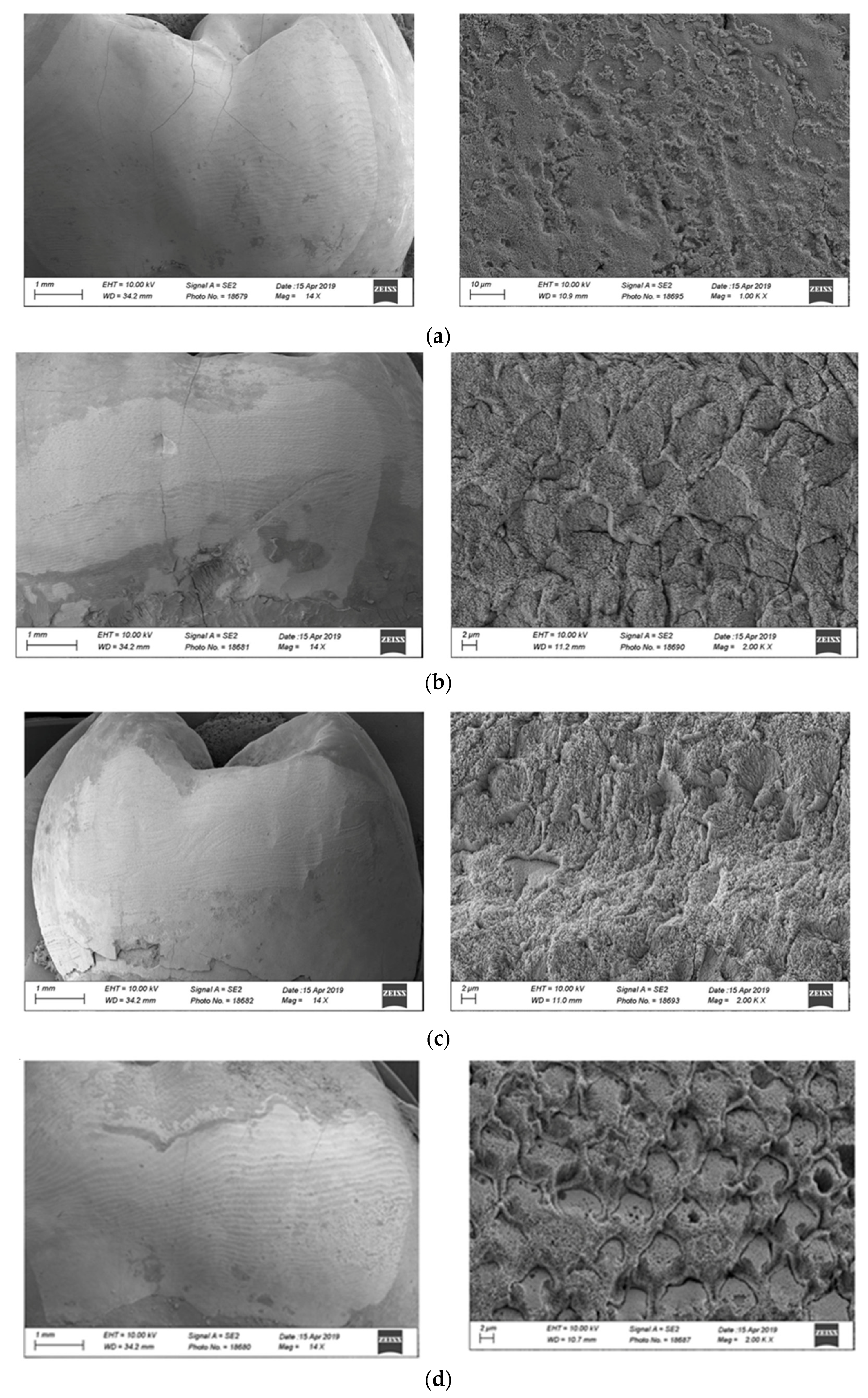
2.4. Morphological Analysis
2.5. Statistical Analysis
3. Results
4. Discussion
5. Conclusions
Author Contributions
Funding
Institutional Review Board Statement
Informed Consent Statement
Data Availability Statement
Conflicts of Interest
References
- Ardu, S.; Krejci, I. Biomimetic direct composite stratification technique for the restoration of anterior teeth. Quintessence Int. 2006, 37, 167–174, Erratum in: Quintessence Int. 2006, 37, 408. [Google Scholar]
- Dietschi, D.; Ardu, S.; Krejci, I. A new shading concept based on natural tooth color applied to direct composite restorations. Quintessence Int. 2006, 37, 91–102. [Google Scholar]
- Vanini, L. Light and color in anterior composite restorations. Pract. Periodontics Aesthet. Dent. 1996, 8, 673–682. [Google Scholar]
- Keys, W.; Carson, S.J. Rubber dam may increase the survival time of dental restorations. Evid. Based Dent. 2017, 18, 19–20. [Google Scholar] [CrossRef] [PubMed] [Green Version]
- Gao, S.S. The longevity of posterior restorations in primary teeth. Evid. Based Dent. 2018, 19, 44. [Google Scholar] [CrossRef] [PubMed]
- Soares, C.J.; Faria-E-Silva, A.L.; Rodrigues, M.P.; Vilela, A.B.F.; Pfeifer, C.S.; Tantbirojn, D.; Versluis, A. Polymerization shrinkage stress of composite resins and resin cements—What do we need to know? Braz. Oral Res. 2017, 28 (Suppl. 1), e62. [Google Scholar] [CrossRef] [PubMed] [Green Version]
- Ferracane, J.L. Resin composite—State of the art. Dent. Mater. 2011, 27, 29–38. [Google Scholar] [CrossRef] [PubMed]
- Krejci, I.; Lutz, F.; Lüscher, B.; Maffioli, E. Optimization of the marginal adaptation of posterior composite fillings using side reflected light wedges. Swiss Dent. J. 1986, 7, 47–52. [Google Scholar]
- Ivoclar Vivadent. Instructions for Use Total Etch. 2020. Available online: https://www.ivoclarvivadent.com/-en/p/dental-professional/products/adhesives/etchant/total-etch (accessed on 6 May 2020).
- 3M ESPE. Instructions for Use Scotchbond Etchant. 2020. Available online: https://media.dentalcompare.com/m/2-5/Downloads/Scotchbond%20Etchant%20Instructions%20for%20Use (accessed on 6 May 2020).
- Kerr. Instructions for Use Gel Etchant. 2020. Available online: https://www.kerrdental.com/resource-center/gel-etchant-instructions-use (accessed on 6 May 2020).
- Bisco. Instructions for Use Uni-Etch. 2020. Available online: https://www.bisco.com/uni-etch-w-bac (accessed on 6 May 2020).
- Heintze, S.D.; Rousson, V.; Hickel, R. Clinical effectiveness of direct anterior restorations—A meta-analysis. Dent. Mater. 2015, 31, 481–495. [Google Scholar] [CrossRef]
- Coelho-de-Souza, F.H.; Rocha Ada, C.; Rubini, A.; Klein-Júnior, C.A.; Demarco, F.F. Influence of adhesive system and bevel preparation on fracture strength of teeth restored with composite resin. Braz. Oral Res. 2010, 21, 327–331. [Google Scholar] [CrossRef] [Green Version]
- Mahn, E.; Rousson, V.; Heintze, S. Meta-Analysis of the Influence of Bonding Parameters on the Clinical Outcome of Tooth-colored Cervical Restorations. J. Adhes. Dent. 2015, 17, 391–403. [Google Scholar]
- da Veiga, A.M.; Cunha, A.C.; Ferreira, D.M.; da Silva Fidalgo, T.K.; Chianca, T.K.; Reis, K.R.; Maia, L.C. Longevity of direct and indirect resin composite restorations in permanent posterior teeth: A systematic review and meta-analysis. J. Dent. 2016, 54, 1–12. [Google Scholar] [CrossRef]
- Pashley, D.H.; Tay, F.R.; Breschi, L.; Tjäderhane, L.; Carvalho, R.M.; Carrilho, M.; Tezvergil-Mutluay, A. State of the art etch-and-rinse adhesives. Dent. Mater. 2011, 27, 1–16. [Google Scholar] [CrossRef] [Green Version]
- Breschi, L.; Mazzoni, A.; Ruggeri, A.; Cadenaro, M.; Di Lenarda, R.; De Stefano Dorigo, E. Dental adhesion review: Aging and stability of the bonded interface. Dent. Mater. 2008, 24, 90–101. [Google Scholar] [CrossRef]
- Ardu, S.; Perroud, R.; Krejci, I. Extended sealing of interproximal caries lesions. Quintessence Int. 2006, 37, 423–427. [Google Scholar] [PubMed]
- Fowler, C.; Lynch, R.J.M.; Shingler, D.; Walsh, D.; Carson, C.; Neale, A.; Willson, R.J.; Brown, A. A novel electron-microscopic method for measurement of mineral content in enamel lesions. Arch. Oral Biol. 2018, 94, 10–15. [Google Scholar] [CrossRef]
- Yoshihara, K.; Nagaoka, N.; Hayakawa, S.; Okihara, T.; Yasuhiro Yoshida, Y.; Van Meerbeek, B. Chemical interaction of glycero-phosphate dimethacrylate (GPDM) with hydroxyapatite and dentin. Dent. Mater. 2018, 34, 1072–1081. [Google Scholar] [CrossRef] [PubMed]
- Van Meerbeek, B.; Peumans, M.; Poitevin, A.; Mine, A.; Van Ende, A.; Neves, A.; De Munck, J. Relationship between bond-strength tests and clinical outcomes. Dent. Mater. 2010, 26, 100–121. [Google Scholar] [CrossRef]
- Armstrong, S.; Breschi, L.; Özcan, M.; Pfefferkorn, F.; Ferrari, M.; Van Meerbeek, B. Academy of Dental Materials guidance on in vitro testing of dental composite bonding effectiveness to dentin/enamel using micro-tensile bond strength (μTBS) approach. Dent. Mater. 2017, 33, 133–143. [Google Scholar] [CrossRef] [PubMed] [Green Version]
- Sirisha, K.; Rambabu, T.; Shankar, Y.R.; Ravikumar, P. Validity of bond strength tests: A critical review: Part I. J. Conserv. Dent. 2014, 17, 305–311. [Google Scholar] [CrossRef] [Green Version]
- Sirisha, K.; Rambabu, T.; Ravishankar, Y.; Ravikumar, P. Validity of bond strength tests: A critical review—Part II. J. Conserv. Dent. 2014, 17, 420–426. [Google Scholar] [CrossRef] [PubMed] [Green Version]


| Group | Surface Type | Etching Time | Bonding and Resin Composite Manufacturer |
|---|---|---|---|
| U15 | Unground enamel without primer application | H3PO4 15 s (Gel Etchant, Kerr) | Bond (OptibondTM FL Adhesive, Kerr) + Composite resin (Premise Kerr) |
| U30 | Unground enamel without primer application | H3PO4 30 s (Gel Etchant, Kerr) | Bond (OptibondTM FL Kerr) + Composite resin (Premise Kerr) |
| G15 | Ground enamel without primer application | H3PO4 15 s (Gel Etchant, Kerr) | Bond (OptibondTM FL Kerr) + Composite resin (Premise Kerr) |
| G30 | Ground enamel without primer application | H3PO4 30 s (Gel Etchant, Kerr) | Bond (OptibondTM FL Kerr) + Composite resin (Premise Kerr) |
| U15P | Unground enamel Primer application | H3PO4 15 s (Gel Etchant, Kerr) | Bond (OptibondTM FL Adhesive, Kerr) + Composite resin (Premise Kerr) |
| U30P | Unground enamel primer application | H3PO4 30 s (Gel Etchant, Kerr) | Bond (OptibondTM FL Adhesive, Kerr) + Composite resin (Premise Kerr) |
| G15P | Ground enamel Primer application | H3PO4 15 s (Gel Etchant, Kerr) | Bond (OptibondTM FL Adhesive, Kerr) + Composite resin (Premise Kerr) |
| G30P | Ground enamel Primer application | H3PO4 30 s (Gel Etchant, Kerr) | Bond (OptibondTM FL Adhesive, Kerr) + Composite resin (Premise Kerr) |
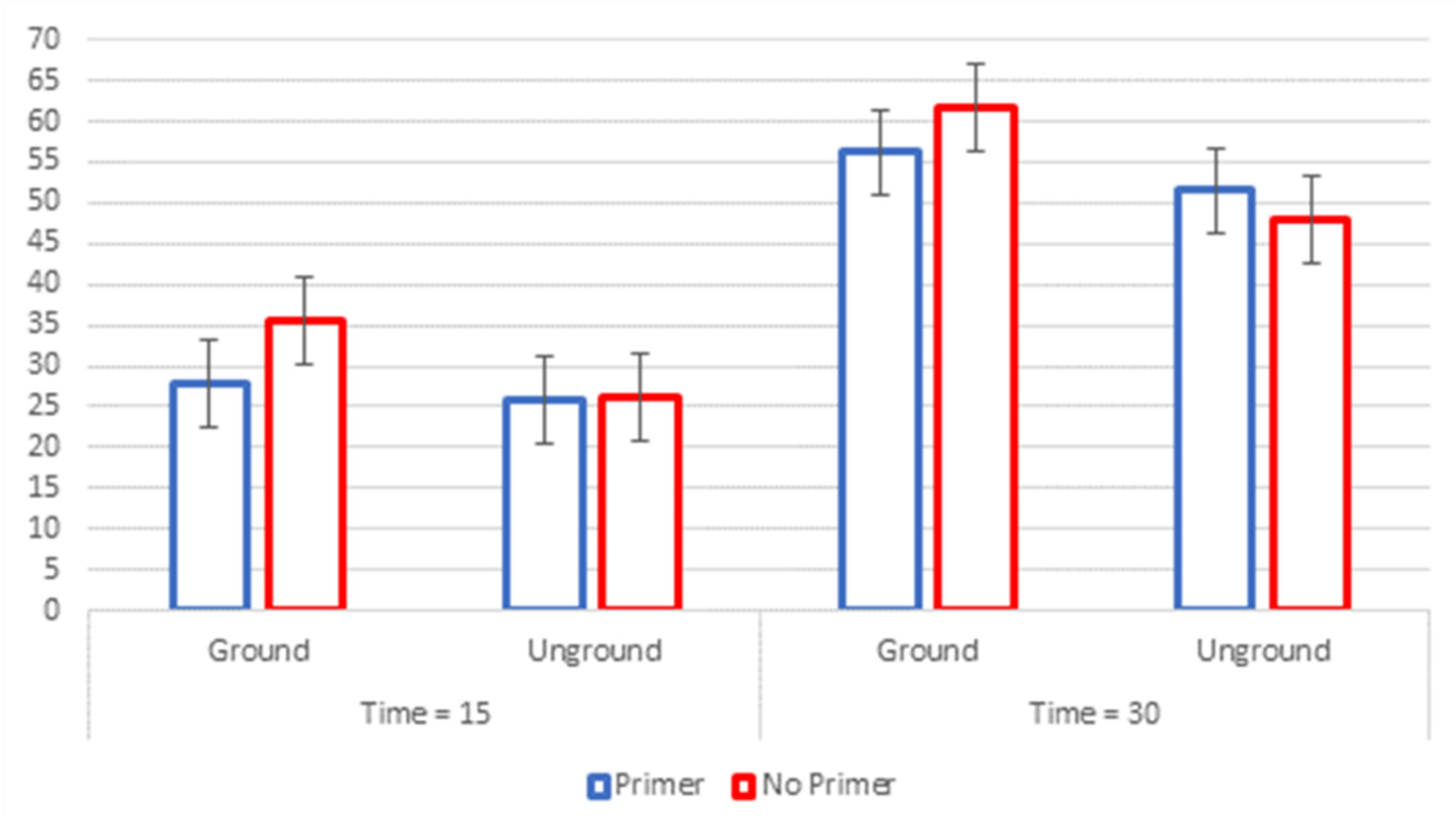
| Treatment Applied | Mean MPA | SD | Grouping | ||||
|---|---|---|---|---|---|---|---|
| G30 | 19.6 | 3.0 | A | ||||
| G30P | 17.9 | 1.7 | A | B | |||
| U30P | 16.4 | 1.7 | B | C | |||
| U30 | 15.2 | 2.7 | C | ||||
| G15 | 11.3 | 1.4 | D | ||||
| G15P | 8.8 | 3.0 | E | ||||
| U15 | 8.3 | 1.4 | E | ||||
| U15P | 8.2 | 2.1 | E | ||||
Publisher’s Note: MDPI stays neutral with regard to jurisdictional claims in published maps and institutional affiliations. |
© 2021 by the authors. Licensee MDPI, Basel, Switzerland. This article is an open access article distributed under the terms and conditions of the Creative Commons Attribution (CC BY) license (https://creativecommons.org/licenses/by/4.0/).
Share and Cite
Daher, R.; Krejci, I.; Mekki, M.; Marin, C.; Di Bella, E.; Ardu, S. Effect of Multiple Enamel Surface Treatments on Micro-Shear Bond Strength. Polymers 2021, 13, 3589. https://doi.org/10.3390/polym13203589
Daher R, Krejci I, Mekki M, Marin C, Di Bella E, Ardu S. Effect of Multiple Enamel Surface Treatments on Micro-Shear Bond Strength. Polymers. 2021; 13(20):3589. https://doi.org/10.3390/polym13203589
Chicago/Turabian StyleDaher, René, Ivo Krejci, Mustapha Mekki, Charlotte Marin, Enrico Di Bella, and Stefano Ardu. 2021. "Effect of Multiple Enamel Surface Treatments on Micro-Shear Bond Strength" Polymers 13, no. 20: 3589. https://doi.org/10.3390/polym13203589