
Synthesis and Antibacterial Activity of Metal-Containing Ultraviolet-Cured Wood Floor Coatings


Abstract
:1. Introduction
2. Materials and Methods
2.1. Materials
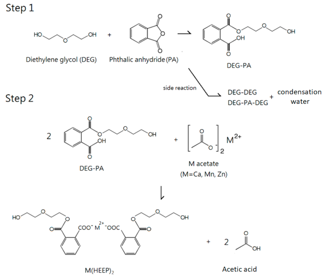
2.2. Manufacture of Mono(hydroxyethoxyethyl)phthalate [M(HEEP)2]
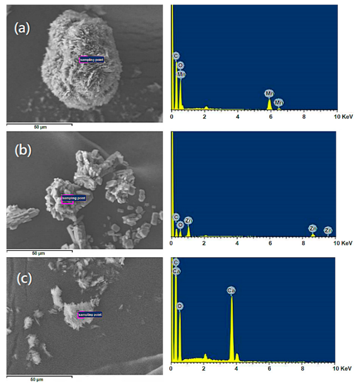
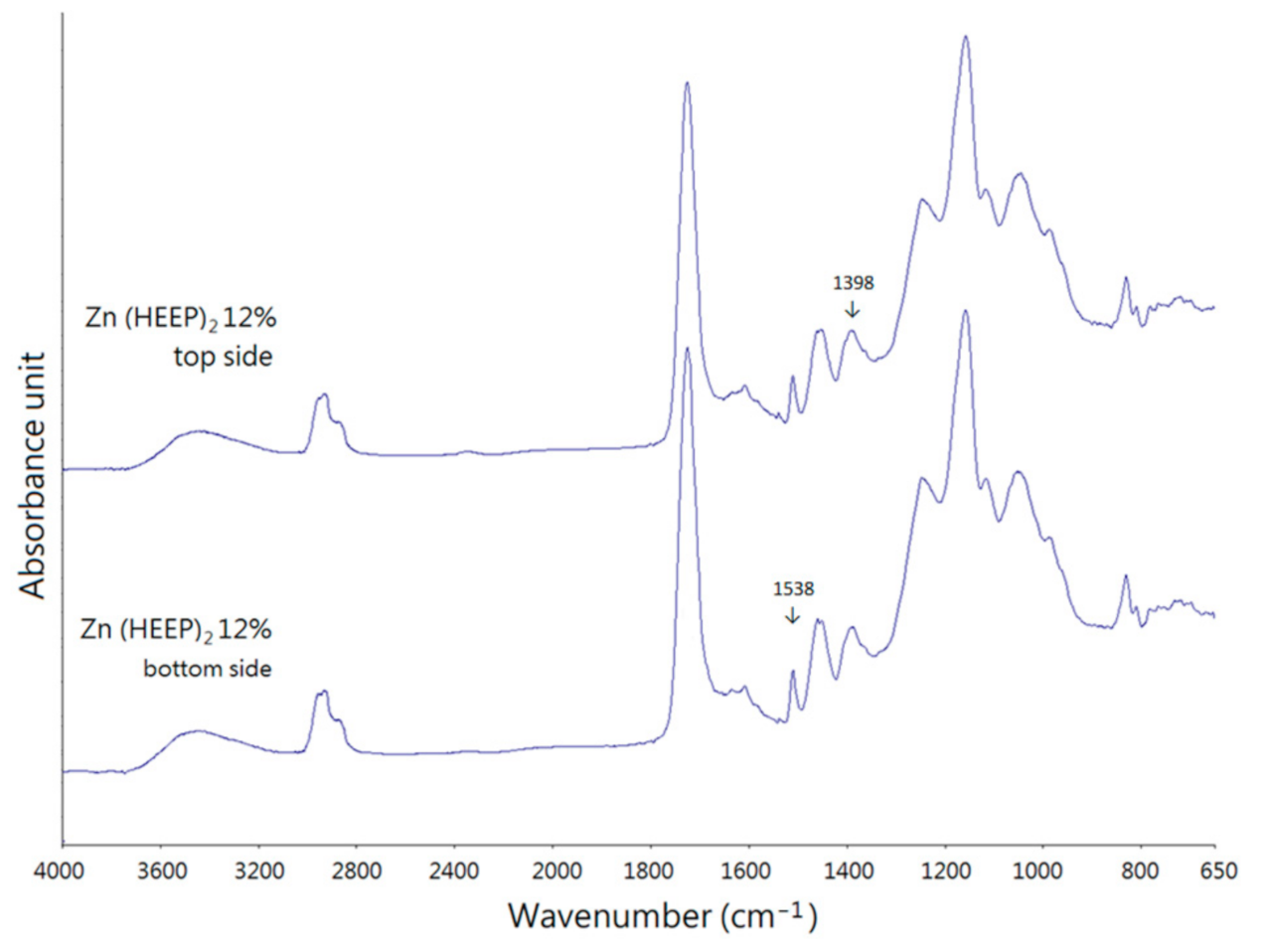
2.3. Identification of M(HEEP)2
2.4. Formulation of the UV Coatings
2.5. Antibacterial Activity Determination of UV Films
2.5.1. Preparation of Bacterial Strain
2.5.2. Antibacterial Activity Test
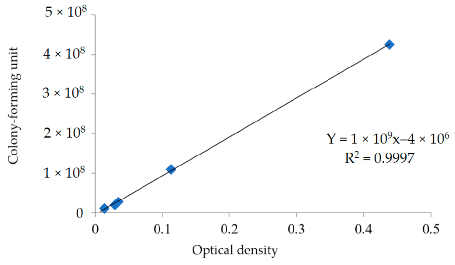
2.5.3. Determination of Antibacterial Activity
2.6. Determination of the UV Film Properties
3. Results
3.1. Manufacture and Identification of M(HEEP)2
3.2. Antibacterial Activity of UV Films with Different Zn(HEEP)2 Additions
4. Conclusions
Author Contributions
Funding
Institutional Review Board Statement
Informed Consent Statement
Data Availability Statement
Conflicts of Interest
References
- Pirmoradian, M.; Hooshmand, T. Remineralization and antibacterial capabilities of resin-based dental nanocomposites. In Applications of Nanocomposite Materials in Dentistry, 1st ed.; Abdullah, M.A., Inamuddin, D., Mohammad, A., Eds.; Chapter 15; Woodhead Publishing: Sawston, UK, 2019; pp. 237–269. [Google Scholar] [CrossRef]
- Balhaddad, A.A.; Ibrahim, M.S.; Weir, M.D.; Xu, H.H.; Melo, M.A.S. Concentration Dependence of Quaternary Ammonium Monomer on the Design of High-performance Bioactive Composite for Root Caries Restorations. Dent. Mater. 2020, 36, 266–278. [Google Scholar] [CrossRef]
- Balhaddad, A.A.; Ibrahim, M.S.; Garcia, I.M.; Collares, F.M.; Weir, M.D.; Xu, H.H.; Melo, M.A.S. Pronounced Effect of Antibacterial Bioactive Dental Composite on Microcosm Biofilms Derived from Patients with Root Carious Lesions. Front. Mater. 2020, 7, 583861. [Google Scholar] [CrossRef]
- Imazato, S.; McCabe, J.F. Influence of Incorporation of Antibacterial Monomer on Curing Behavior of a Dental Composite. J. Dent. Res. 1994, 73, 1641–1645. [Google Scholar] [CrossRef] [PubMed] [Green Version]
- Zafar, F.; Ashraf, S.M.; Ahmad, S. Studies on Zinc-containing Linseed Oil Based Polyesteramide. React. Funct. Polym. 2007, 67, 928–935. [Google Scholar] [CrossRef]
- Belkhir, K.; Pillon, C.; Cayla, A.; Campagne, C. Antibacterial Textile Based on Hydrolyzed Milk Casein. Materials 2021, 14, 251. [Google Scholar] [CrossRef]
- Belkhir, K.; Lacroix, M.; Jamshidian, M.; Salmieri, S.; Jegat, C.; Taha, M. Evaluation of Antibacterial Activity of Branched Quaternary Ammonium Grafted Green Polymers. Food Packag. Shelf Life 2017, 12, 28–41. [Google Scholar] [CrossRef]
- Sun, Q.; Zhang, L.; Bai, R.; Zhuang, Z.; Zhang, Y.; Yu, T.; Peng, L.; Xin, T.; Chen, S.; Han, B. Recent Progress in Antimicrobial Strategies for Resin-Based Restoratives. Polymers 2021, 13, 1590. [Google Scholar] [CrossRef]
- Cheng, S.S.; Lin, C.Y.; Chung, M.J.; Chen, Y.J.; Chang, S.T. Potential Source of Environmentally Benign Antifungal Agents from Cinnamomum Osmophloeum Leaves Against Phellinus Noxius. Plant Protect. Sci. 2019, 55, 43–53. [Google Scholar] [CrossRef] [Green Version]
- Chen, C.J.; Senthil Kumar, K.J.; Chen, Y.T.; Tsao, N.W.; Chien, S.C.; Chang, S.T.; Chu, F.H.; Wang, S.Y. Effect of hinoki and meniki essential oils on human autonomic nervous system activity and mood states. Nat. Prod. Commun. 2015, 10, 1305–1308. [Google Scholar] [CrossRef] [PubMed] [Green Version]
- Jayakurmar, R.; Nanjundan, S. Studies on Metal-Containing Co-Polyurethanes Based on Mono(Hydroxyethoxyethyl)phthalate. J. Macromol. Sci. 2006, 43, 945–954. [Google Scholar] [CrossRef]
- Jayakurmar, R.; Nanjundan, S.; Rajkumar, M.; Nagendran, R. Studies on Metal-Containing Polyurethane Based on Divalent Metal Salts of Mono (Hydroxylethoxyethyl)phthalate. J. Macromol. Sci. 2001, 38, 869–888. [Google Scholar] [CrossRef]
- Li, J.H.; Hong, R.Y.; Li, M.Y.; Li, H.Z.; Zheng, Y.; Ding, J. Effects of ZnO Nanoparticles on the Mechanical and Antibacterial Properties of Polyurethane Coatings. Prog. Org. Coat. 2009, 64, 504–509. [Google Scholar] [CrossRef]
- Hsu, S.H.; Tseng, H.J.; Lin, Y.C. The Biocompatibility and Antibacterial Properties of Waterborne Polyurethane-Silver Nanocomposites. Biomaterials 2010, 31, 6796–6808. [Google Scholar] [CrossRef] [PubMed]
- Choi, O.; Hu, Z. Size Dependent and Reactive Oxygen Species Related Nanosilver Toxicity to Nitrifying Bacteria. Environ. Sci. Technol. 2008, 42, 4583–4588. [Google Scholar] [CrossRef] [PubMed]
- Li, Q.; Mahendra, S.; Lyon, D.Y.; Brunet, L.; Liga, M.V.; Li, D.; Alvarez, P.J.J. Antimicrobial Nanomaterials for Water Disinfection and Microbial Control: Potential Applications and Implications. Water Res. 2008, 42, 4591–4602. [Google Scholar] [CrossRef] [PubMed]
- Díaz-Visurraga, J.; Gutiérrez, C.; Plessing, C.V.; García, A. Metal N-Nanostructures as Antibacterial Agents. In Science against Microbial Pathogens: Communicating Current Research and Technological Advances; Formatex Research Center: Badajoz, Spain, 2011; pp. 210–218. [Google Scholar]
- Gearhart, J.M.; Geiss, K.T.; Schlager, J.J.; Hussain, S.M.; Hess, K.L. In Vitro Toxicity of Nanoparticles in BRL 3a Rat Liver Cells. Toxicol. Vitro 2005, 19, 975–983. [Google Scholar] [CrossRef]
- Trop, M.; Rodl, N.S.; Hellbom, B.; Kroell, W.; Goessler, W. Silver Coated Dressing Acticoat Caused Raised Liver Enzymes and Argyria-Like Symptoms in Burn Patient. J. Trauma 2006, 60, 648–652. [Google Scholar] [CrossRef]
- Varner, K.E.; El-Badawy, A.; Feldhake, D.; Venkatapathy, R. State of the Science Review: Everything NanoSilver and More; EPA/600/R-10/084; U.S. Environmental Protection Agency: Washington, DC, USA, 2010.
- Miao, A.J.; Luo, Z.; Chen, C.S.; Chin, W.C.; Santschi, P.H.; Quigg, A. Intracellular Uptake: A Possible Mechanism for Silver Engineered Nanoparticle Toxicity to A Freshwater Alga Ochromonas Danica. PLoS ONE 2010, 5, e15196. [Google Scholar] [CrossRef] [Green Version]
- Blinova, I.; Niskanen, J.; Kajankari, P.; Kanarbik, L.; Käkinen, A.; Tenhu, H.; Penttinen, O.P.; Kahru, A. Toxicity of Two Types of Silver Nanoparticles to Aquatic Crustaceans Daphnia Magna and Thamnocephalus Platyurus. Environ. Sci. Pollut. Rest. Int. 2013, 20, 3456–3463. [Google Scholar] [CrossRef]
- Laban, G.; Nies, L.F.; Turco, R.F.; Bickham, J.W.; Sepúlveda, M.S. The Effects of Silver Nanoparticles on Fathead Minnow (Pimephales Promelas) Embryos. Ecotoxicology 2010, 19, 185–195. [Google Scholar] [CrossRef]
- Blaser, S.A.; Scheringer, A.M.; MacLeod, M.; Hungerbühler, K. Estimation of Cumulative Aquatic Exposure and Risk Due to Silver: Contribution of Nano-Functionalized Plastics and Textiles. Sci. Total Environ. 2008, 390, 396–409. [Google Scholar] [CrossRef] [PubMed]
- Faunce, T.; Watal, A. Nanosilver and Global Public Health: International Regulatory Issues. Nanomedicine 2010, 5, 617–632. [Google Scholar] [CrossRef] [PubMed] [Green Version]
- Lu, K.T.; Chang, J.P. Synthesis and Antimicrobial Activity of Metal-Containing Linseed Oil-Based Waterborne Urethane Oil Wood Coatings. Polymers 2020, 12, 663. [Google Scholar] [CrossRef] [PubMed] [Green Version]
- CNS. CNS 9007 Method of Test for Paints-Sampling and General Condition; CNS: Taipei, Taiwan, 1995. [Google Scholar]
- ISO. ISO 1522 Paints and Varnishes—Pendulum Damping Test; ISO: Geneva, Switzerland, 2007. [Google Scholar]
- Kelton, K.F.; Greer, A.L. Heterogeneous Nucleation. In Nucleation in Condensed Matter Pergamon, 1st ed.; Chapter 6, eBook; Pergamon: Oxford, UK, 2010; Volume 15, ISBN 9780080912646. [Google Scholar]
- Begot, C.; Desnier, I.; Daudin, J.D.; Labadie, J.C.; Lebert, A. Recommendations for Calculating Growth Parameters by Optical Density Measurements. J. Microbiol. Methods 1996, 25, 225–232. [Google Scholar] [CrossRef]
- Nasim, K.; Elsayed, S.; Pitout, J.D.D.; Conly, J.; Church, D.L.; Gregson, D.B. New Method for Laboratory Detection of Ampc Β-Lactamases in Escherichia Coli and Klebsiella Pneumoniae. J. Clin. Microbiol. 2004, 42, 4799–4802. [Google Scholar] [CrossRef] [Green Version]
- Abedi-moghaddam, N.; Bulić, A.; Henderson, L.; Lam, E. Survival of Escherichia Coli to UV Irradiation During Exponential and Stationary Phases of Growth. J. Exp. Microbiol. Immunol. 2004, 5, 44–49. [Google Scholar]
- Liu, Y.; He, L.; Mustapha, A.; Li, H.; Hu, Z.Q.; Lin, M. Antibacterial Activities of Zinc Oxide Nanoparticles against Escherichia Coli O157:H7. J. Appl. Microbiol. 2015, 107, 1193–1201. [Google Scholar] [CrossRef] [PubMed]
- Söderberg, T.; Sunzel, B.; Holm, S.; Elmros, T.; Hallmans, G.; Sjöberg, S. Antibacterial Effect of Zinc Oxide in Vitro. J. Plast. Surg. Hand. Surg. 1990, 24, 193–197. [Google Scholar] [CrossRef]
- Alekish, M.; Ismail, Z.B.; Albiss, B.; Nawasrah, S. In Vitro Antibacterial Effects of Zinc Oxide Nanoparticles on Multiple Drug-Resistant Strains of Staphylococcus Aureus and Escherichia Coli: An Alternative Approach for Antibacterial Therapy of Mastitis in Sheep. Vet World. 2018, 11, 1428–1432. [Google Scholar] [CrossRef] [Green Version]
- Sondi, I.; Salopek-Sondi, B. Silver Nanoparticles as Antimicrobial Agent: A Case Study on E. Coli as A Model for Gram-Negative Bacteria. J. Colloid Interface Sci. 2004, 275, 177–182. [Google Scholar] [CrossRef]
- Lok, C.N.; Ho, C.M.; Chen, R.; He, Q.Y.; Yu, W.Y.; Sun, H.; Tam, P.K.H.; Chiu, J.F.; Che, C.M. Silver Nanoparticles: Partial Oxidation and Antibacterial Activities. J. Biol. Inorg. Chem. 2007, 12, 527–534. [Google Scholar] [CrossRef] [PubMed]
- Balhaddad, A.A.; Mokeem, L.S.; Weir, M.D.; Xu, H.; Melo, M.A.S. Sustained Antibacterial Effect and Wear Behavior of Quaternary Ammonium Contact-Killing Dental Polymers after One-Year of Hydrolytic Degradation. Appl. Sci. 2021, 11, 3718. [Google Scholar] [CrossRef]
- Alsahafi, R.; Balhaddad, A.A.; Mitwalli, H.; Ibrahim, M.S.; Melo, M.A.S.; Oates, T.W.; Xu, H.H.; Weir, M.D. Novel Crown Cement Containing Antibacterial Monomer and Calcium Phosphate Nanoparticles. Nanomaterials 2020, 10, 2001. [Google Scholar] [CrossRef] [PubMed]
- Otts, D.B.; Urban, M.W. Heterogeneous Crosslinking of Waterborne Two-Component Polyurethanes (WB 2K-PUR): Stratification Processes and the Role of Water. Polymer 2005, 46, 2699–2709. [Google Scholar] [CrossRef]
- Ramezanzadeh, B.; Attar, M.M.; Farzam, M. A Study on the Anticorrosion Performance of the Epoxy–Polyamide Nanocomposites Containing ZnO Nanoparticles. Prog. Org. Coat. 2011, 72, 410–422. [Google Scholar] [CrossRef]
- Zuev, V.V.; Bertinic, F.; Audisio, G. Investigation on the Thermal Degradation of Acrylic Polymerswith Fluorinated Side-Chains. Polym. Degrad. Stab. 2006, 91, 512–516. [Google Scholar] [CrossRef]
- Hemalatha, K.S.; Parvatikar, N.; Rukmani, K. Influence of ZnO Nanoparticles on Thermal Behavior of Poly Vinyl Alcohol Films. Int. J. Adv. Sci. Technol. 2014, 5, 106–115. [Google Scholar] [CrossRef]


| Zn(HEEP)2 Additions (phr) | Bacterial Concentration 2 (CFU/mL, × 108) | OD Value 3 |
|---|---|---|
| Blank 1 | 2.70 | 0.400 |
| 0 | 2.55 | 0.340 |
| 4 | 1.52 | 0.234 |
| 8 | 1.53 | 0.239 |
| 12 | 1.58 | 0.244 |
| Zn(HEEP)2 Additions (phr) | Bacterial Concentration (CFU/mL, × 108) with Incubation Time (h) | |||
|---|---|---|---|---|
| 0 | 1 | 3 | 5 | |
| Blank 1 | 0.00 | 0.00 | 2.59 | 7.18 |
| 0 | 0.00 | 0.00 | 2.84 | 7.21 |
| 4 | 0.00 | 0.00 | 2.50 | 7.11 |
| 8 | 0.00 | 0.02 | 2.61 | 5.90 |
| 12 | 0.00 | 0.00 | 2.24 | 5.18 |
| Ag (12 phr) | 0.00 | 0.02 | 2.65 | 6.84 |
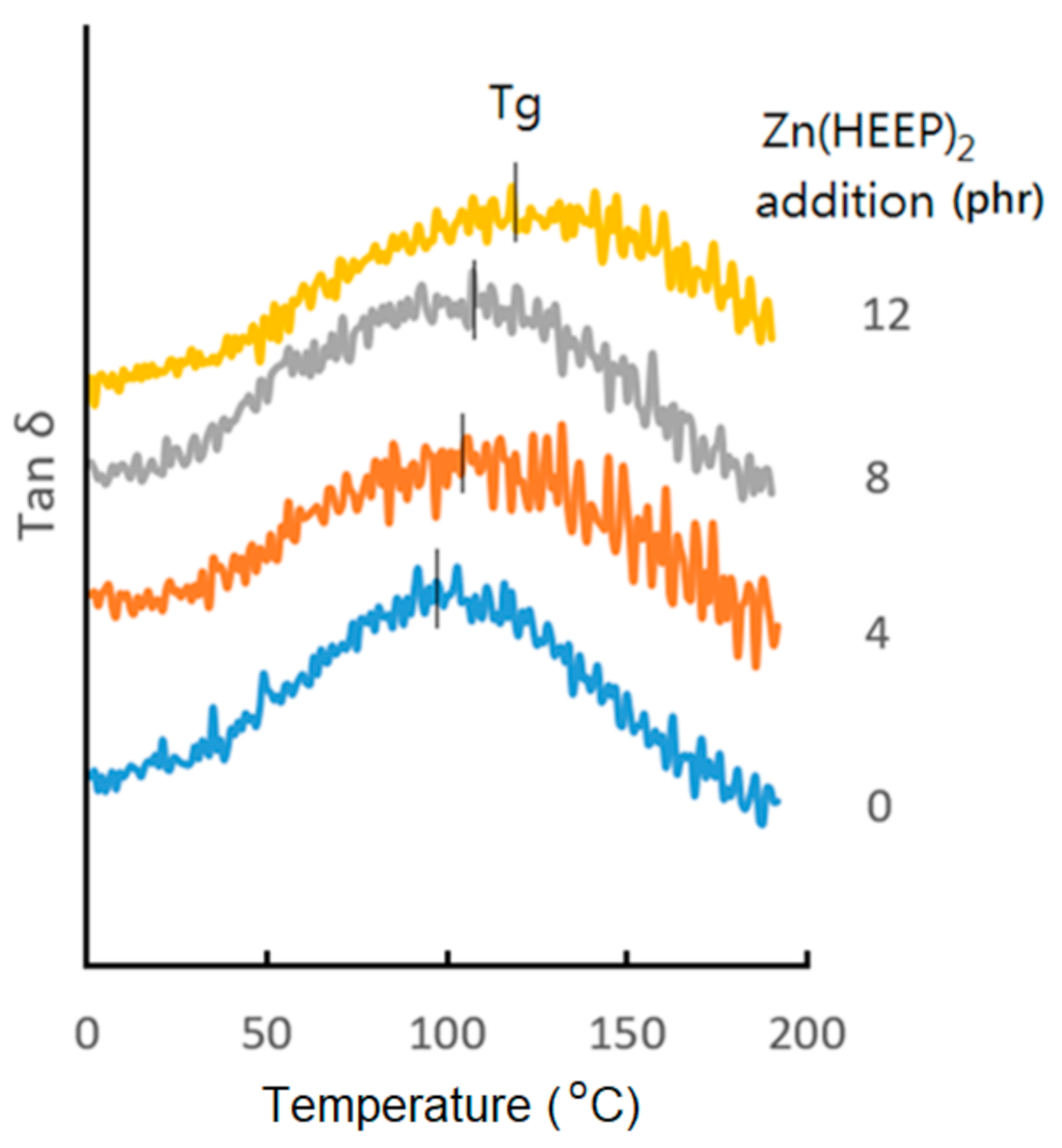
| Zn(HEEP)2 Additions (phr) | Hardness (König, sec) | Gloss (Gloss Units) | Abrasion Resistance (mg/1000 Cycles) | Mass Retention 1 (wt%) | Modulus 2 (GPa) | Tg 3 (°C) |
|---|---|---|---|---|---|---|
| 0 | 97 ± 1 | 84.4 ± 0.7 | 13.4 ± 0.4 | 72.8 ± 0.4 | 4.35 ± 0.02 | 96 |
| 4 | 97 ± 1 | 83.5 ± 1.3 | 13.9 ± 0.5 | 73.8 ± 0.2 | 4.70 ± 0.05 | 104 |
| 8 | 98 ± 2 | 78.8 ± 2.9 | 14.5 ± 1.1 | 73.6 ± 0.9 | 4.82 ± 0.08 | 111 |
| 12 | 103 ± 2 | 73.1 ± 4.7 | 17.1 ± 1.3 | 73.9 ± 0.5 | 4.76 ± 0.04 | 120 |
| Zn(HEEP)2 Additions (phr) | ΔL* | ΔE | ΔYI |
|---|---|---|---|
| 0 | −0.6 | 5.3 | 8.5 |
| 4 | −0.7 | 5.8 | 9.3 |
| 8 | −0.6 | 5.1 | 8.2 |
| 12 | −0.3 | 4.3 | 7.0 |
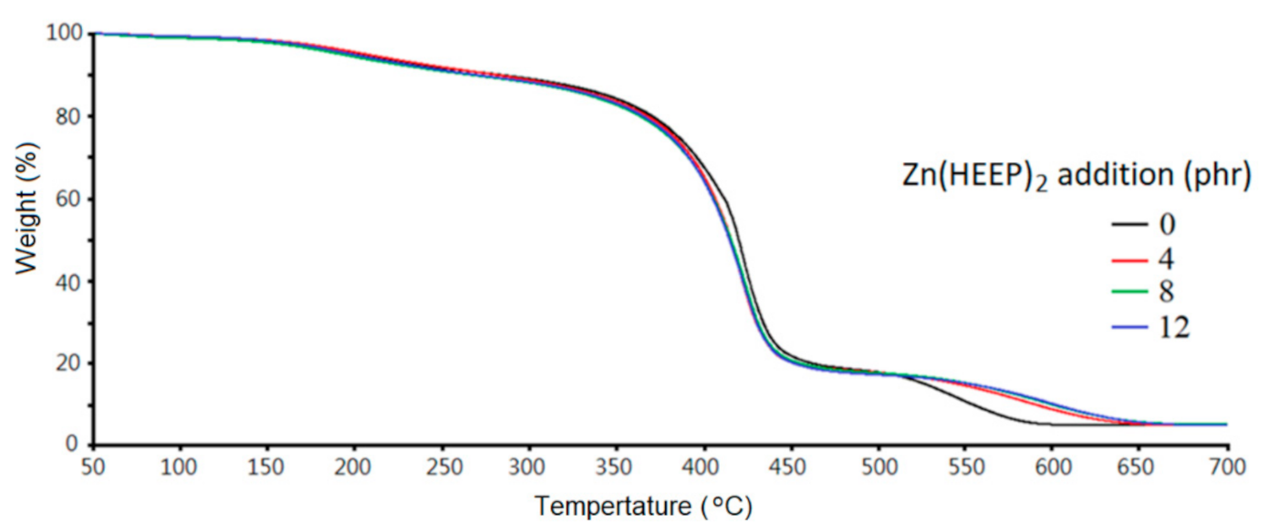
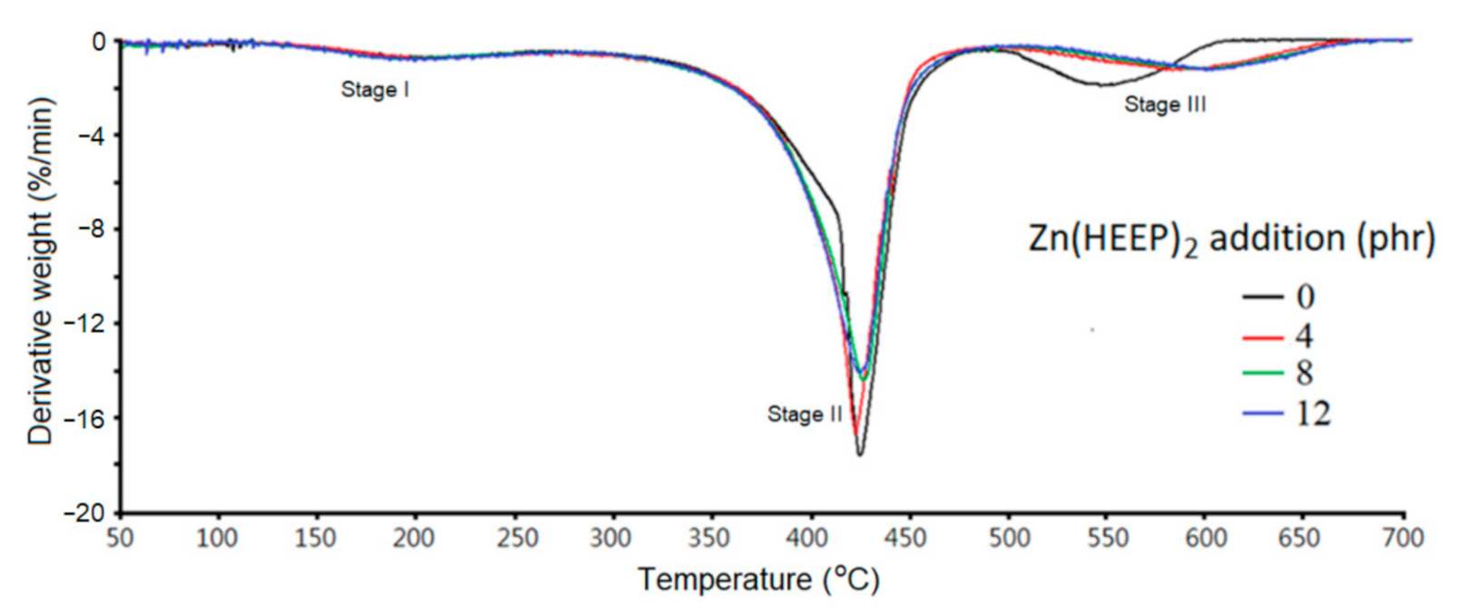
| Zn(HEEP)2 Additions (phr) | Stage II | Stage Ⅲ | Residual Weight at 700 °C (%) | ||||
|---|---|---|---|---|---|---|---|
| Onset (°C) | Derivative Weight Loss at Tdmax 1 (%/min) | Tdmax (°C) | Onset (°C) | Derivative Weight Loss at Tdmax (%/min) | Tdmax (°C) | ||
| 0 | 400 | −17.7 | 422 | 518 | −1.8 | 545 | 5.0 |
| 4 | 393 | −16.7 | 421 | 545 | −1.3 | 584 | 5.0 |
| 8 | 390 | −14.4 | 424 | 551 | −1.3 | 595 | 5.2 |
| 12 | 391 | −14.0 | 423 | 555 | −1.3 | 599 | 5.1 |
Publisher’s Note: MDPI stays neutral with regard to jurisdictional claims in published maps and institutional affiliations. |
© 2021 by the authors. Licensee MDPI, Basel, Switzerland. This article is an open access article distributed under the terms and conditions of the Creative Commons Attribution (CC BY) license (https://creativecommons.org/licenses/by/4.0/).
Share and Cite
Chang, C.-W.; Lu, K.-T. Synthesis and Antibacterial Activity of Metal-Containing Ultraviolet-Cured Wood Floor Coatings. Polymers 2021, 13, 3022. https://doi.org/10.3390/polym13183022
Chang C-W, Lu K-T. Synthesis and Antibacterial Activity of Metal-Containing Ultraviolet-Cured Wood Floor Coatings. Polymers. 2021; 13(18):3022. https://doi.org/10.3390/polym13183022
Chicago/Turabian StyleChang, Chia-Wei, and Kun-Tsung Lu. 2021. "Synthesis and Antibacterial Activity of Metal-Containing Ultraviolet-Cured Wood Floor Coatings" Polymers 13, no. 18: 3022. https://doi.org/10.3390/polym13183022