Thiolation of Biopolymers for Developing Drug Delivery Systems with Enhanced Mechanical and Mucoadhesive Properties: A Review


Abstract
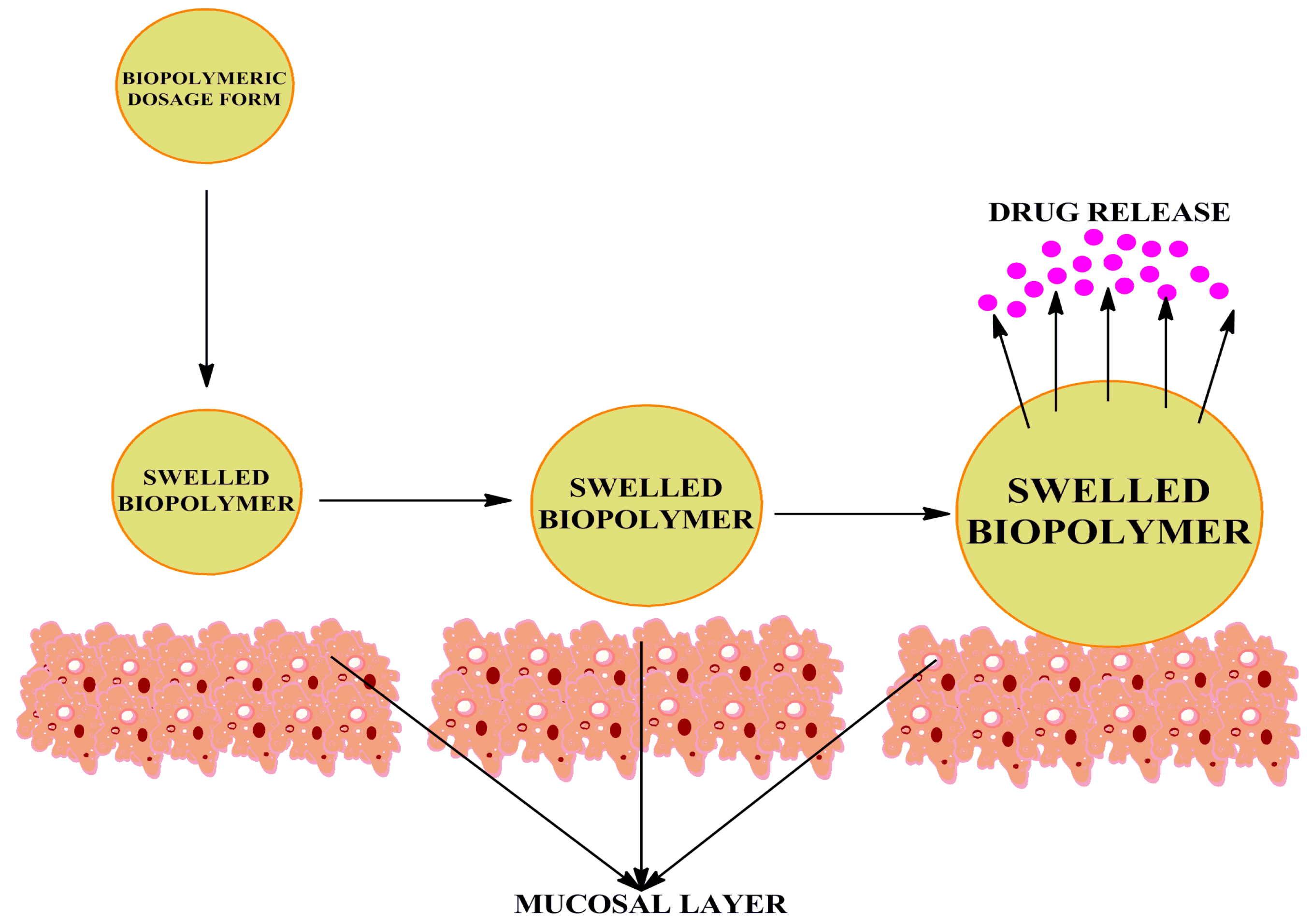
:1. Introduction
- (1)
- Cationic thiomers—the glucosamine contain a primary amino group at the second position that acts as the primary target for thiol group immobilization. The link between the two is formed covalently and the bond could either be amine or amidine. If the bond is amine, the carboxylic group of cysteine and thioglycolic acid reacts with the primary amine site of polymer. In case of amidine bond, 2-imidothiolane acts as a coupling agent. The advantage of using this is that the reaction occurs in a single step.
- (2)
- Anionic thiomers—these types of thiomers have carboxylic acid groups as the anionic target. They are advantageous as sulfhydryl moieties can easily attach to cysteine and homocysteine ligands with the help of amide linkage too. These bonds are mediated by carbodiamides.
2. Methods of Thiolation
2.1. Thiolation by Traut’s Reagent
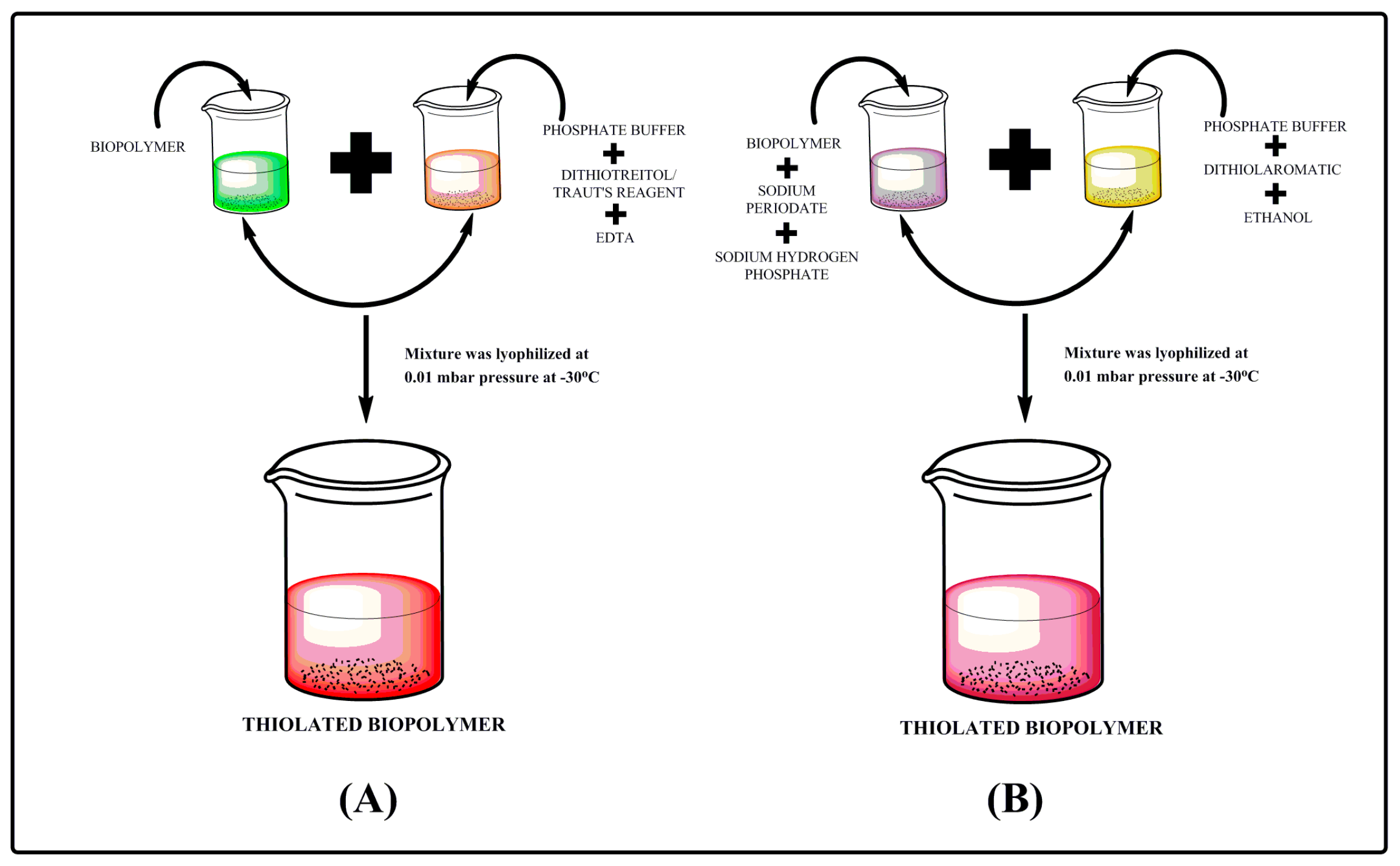
2.2. Thiolation by Dithiothreitol (DTT) Reduction
2.3. Thiolation by Dithiolaromatic (PEG6-CONHNH2)
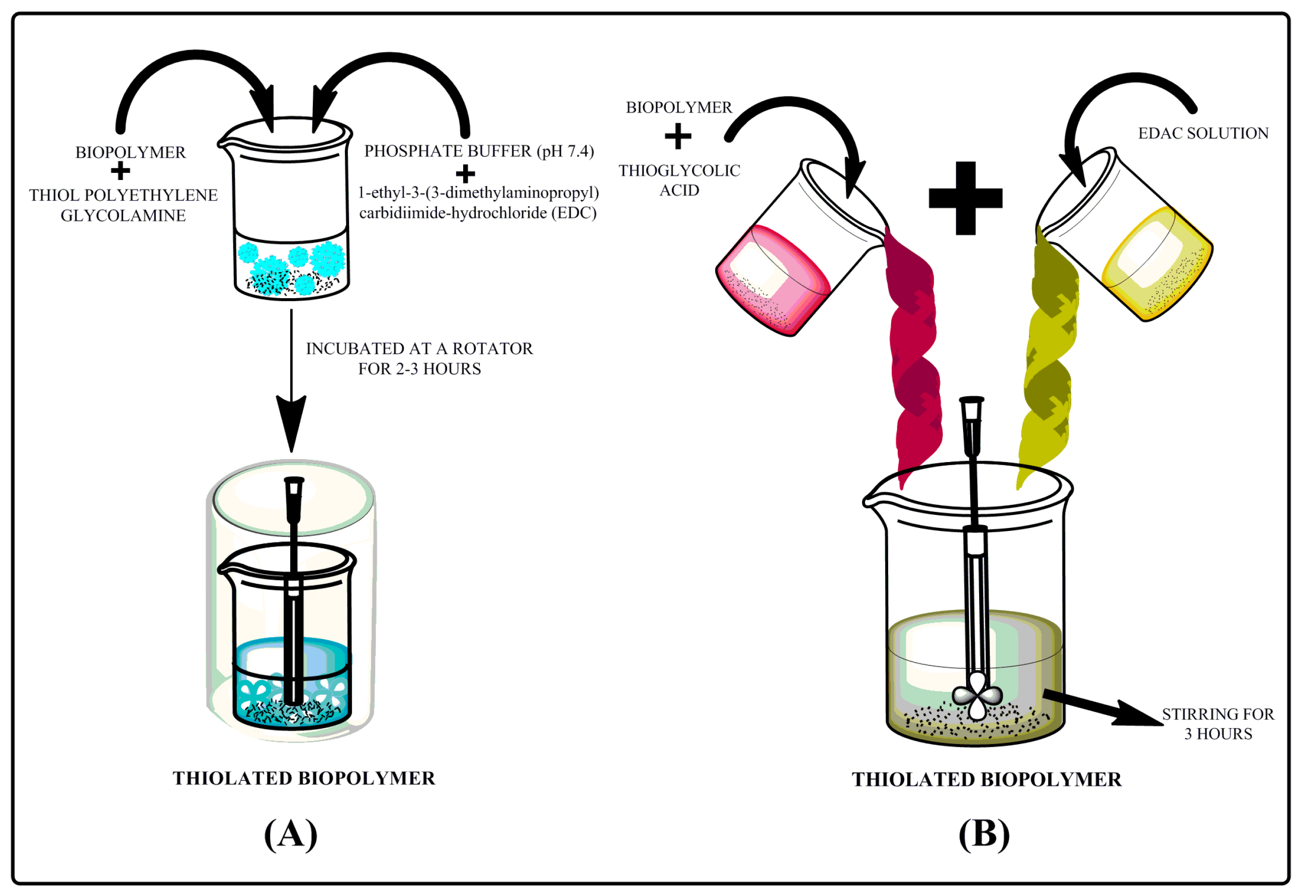
2.4. Thiolation by Thiol Polyethylene Glycolamine (SH-PEG-NH2)
2.5. Thiolation by Thioglycholic Acid
3. Characterization
3.1. Determination of Thiol Group Content
3.1.1. Determination of Thiol Group Content by Iodometry Titration Method
3.1.2. Determination of Thiol Group Content by Ellman’s Method
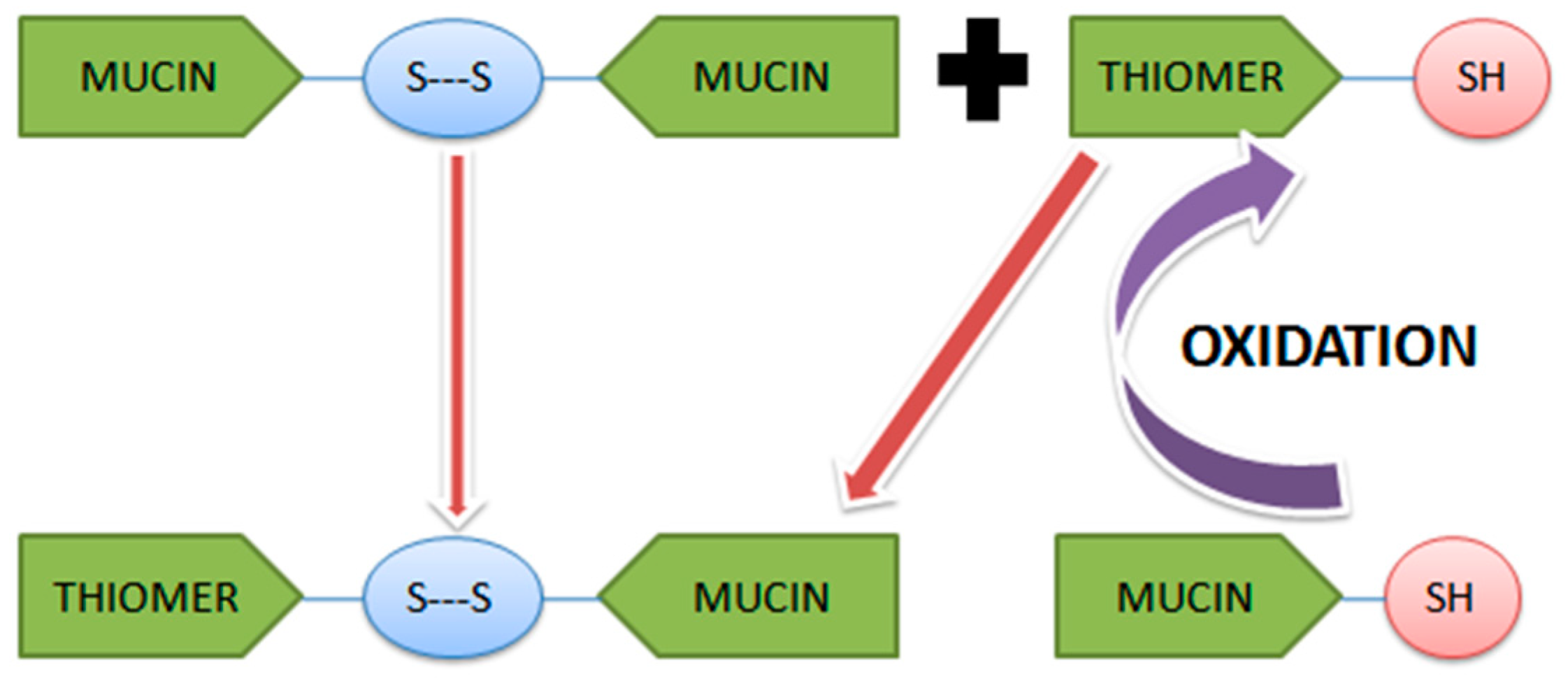
3.2. Disulfide Bond Formation
3.3. Swelling Behavior
3.4. Evaluation of Mucoadhesion
3.4.1. Atomic Force Microscopy
3.4.2. Mucin Adsorption Study
3.4.3. Polymer–Mucin Interaction Study
3.4.4. Ex Vivo
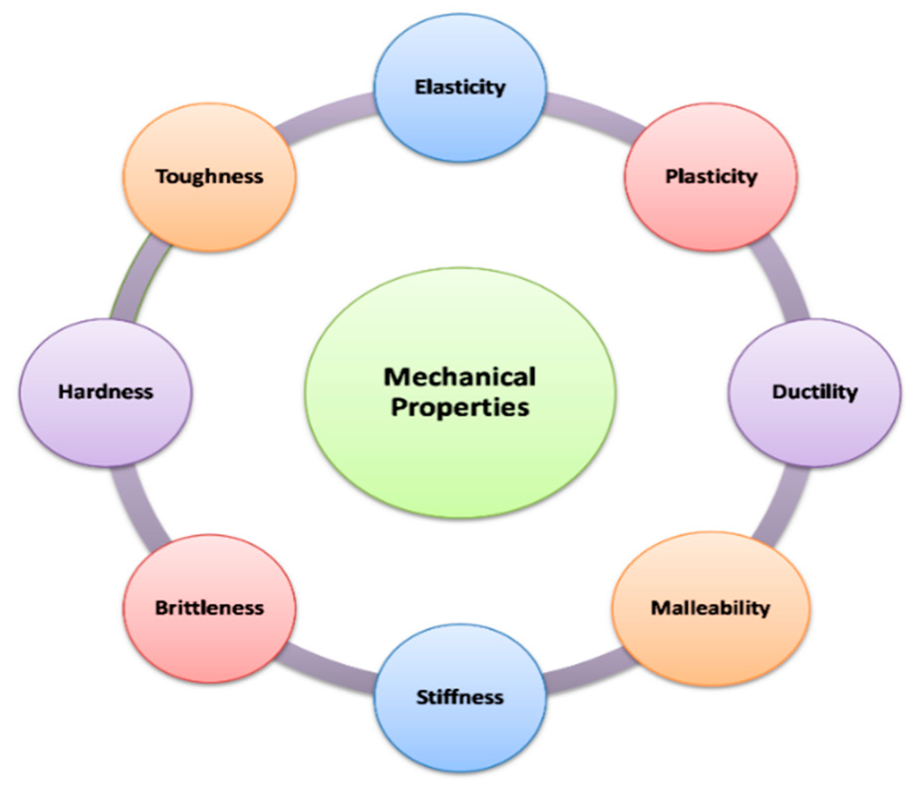
4. Mechanical Properties
4.1. Percent Elongation
4.2. Young’s Modulus
4.3. Toughness
4.4. Viscoelasticity
5. Mucoadhesive Properties and Dosage Forms
5.1. Tablets
5.2. Films
5.3. Fibers
5.4. Nanoparticles
5.5. Gels/Hydrogels
6. Conclusions
Author Contributions
Funding
Acknowledgments
Conflicts of Interest
References
- McClements, D.J.; Gumus, C.E. Natural emulsifiers—Biosurfactants, phospholipids, biopolymers, and colloidal particles, molecular and physicochemical basis of functional performance. Adv. Colloid Interface Sci. 2016, 234, 3–26. [Google Scholar] [CrossRef] [PubMed] [Green Version]
- Datta, L.P.; Manchineella, S.; Govindaraju, T. Biomolecules-derived biomaterials. Biomaterials 2019, 230, 119633. [Google Scholar] [CrossRef] [PubMed]
- Cojocaru, F.D.; Balan, V.; Popa, M.I.; Lobiuc, A.; Antoniac, A.; Antoniac, I.V.; Verestiuc, L. Biopolymers–calcium phosphates composites with inclusions of magnetic nanoparticles for bone tissue engineering. Int. J. Biol. Macromol. 2019, 125, 612–620. [Google Scholar] [CrossRef] [PubMed]
- Singh, I.; Rana, V. Techniques for the assessment of mucoadhesion in drug delivery systems: An overview. J. Adhes. Sci. Technol. 2012, 26, 2251–2267. [Google Scholar] [CrossRef]
- Puri, V.; Sharma, A.; Maman, P.; Rathore, N.; Singh, I. Overview of mucoadhesive biopolymers for buccal drug delivery systems. Int. J. App. Pharm. 2019, 11, 18–29. [Google Scholar] [CrossRef]
- Kumar, A.; Bali, V.; Kumar, M.; Pathak, K. Comparative evaluation of porous versus nonporous mucoadhesive films as buccal delivery system of glibenclamide. Aaps Pharmscitech 2013, 14, 1321–1332. [Google Scholar] [CrossRef] [Green Version]
- Laffleur, F.; Rottges, S. Mucoadhesive approach for buccal application, preactivated chitosan. Eur. Polym. J. 2019, 113, 60–66. [Google Scholar] [CrossRef]
- Estrellas, K.M.; Fiecas, M.; Azagury, A.; Laulicht, B.; Cho, D.Y.; Mancini, A.; Reineke, J.; Furtado, S.; Mathiowitz, E. Time-dependent mucoadhesion of conjugated bioadhesive polymers. Colloid. Surf. B 2019, 173, 454–469. [Google Scholar] [CrossRef]
- Sohail, M.; Minhas, M.U.; Khan, S.; Hussain, Z.; de Matas, M.; Shah, S.A.; Khan, S.; Kousar, M.; Ullah, K. Natural and synthetic polymer-based smart biomaterials for management of ulcerative colitis, A review of recent developments and future prospects. Drug Deliv. Transl. Res. 2019, 9, 595–614. [Google Scholar] [CrossRef]
- Duggan, S.; Cummins, W.; O’Donovan, O.; Hughes, H.; Owens, E. Thiolated polymers as mucoadhesive drug delivery systems. Eur. J. Pharm. Sci. 2017, 100, 64–78. [Google Scholar] [CrossRef]
- Ways, M.T.; Lau, W.; Khutoryanskiy, V. Chitosan and its derivatives for application in mucoadhesive drug delivery systems. Polymers 2018, 10, 267. [Google Scholar] [CrossRef] [PubMed] [Green Version]
- Shirvan, A.R.; Bashari, A.; Hemmatinejad, N. New insight into the fabrication of smart mucoadhesive buccal patches as a novel controlled-drug delivery system. Eur. Polym. J. 2019, 119, 541–550. [Google Scholar] [CrossRef]
- Bravo-Osuna, I.; Vauthier, C.; Farabollini, A.; Palmieri, G.F.; Ponchel, G. Mucoadhesion mechanism of chitosan and thiolated chitosan-poly (isobutyl cyanoacrylate) core-shell nanoparticles. Biomaterials 2007, 28, 2233–2243. [Google Scholar] [CrossRef] [PubMed]
- Van Vlierberghe, S.; Schacht, E.; Dubruel, P. Reversible gelatin-based hydrogels, finetuning of material properties. Eur. Polym. 2011, 47, 1039–1047. [Google Scholar] [CrossRef]
- Fu, Y.; Xu, K.; Zheng, X.; Giacomin, A.J.; Mix, A.W.; Kao, W.J. 3D cell entrapment in crosslinked thiolated gelatin-poly (ethylene glycol) diacrylate hydrogels. Biomaterials 2012, 33, 48–58. [Google Scholar] [CrossRef] [PubMed] [Green Version]
- Ding, J.; He, R.; Zhou, G.; Tang, C.; Yin, C. Multilayered mucoadhesive hydrogel films based on thiolated hyaluronic acid and polyvinylalcohol for insulin delivery. Acta Biomater. 2012, 8, 3643–3651. [Google Scholar] [CrossRef]
- Kafedjiiski, K.; Jetti, R.K.; Fager, F.; Hoyer, H.; Werle, M.; Hoffer, M.; Bernkop-Schnurch, A. Synthesis and in vitro evaluation of thiolated hyaluronic acid for mucoadhesive drug delivery. Int. J. Pharm. 2007, 343, 48–58. [Google Scholar] [CrossRef]
- Maculotti, K.; Genta, I.; Perugini, P.; Imam, M.; Bernkop-Schnurch, A.; Pavanetto, F. Preparation and in vitro evaluation of thiolated chitosan microparticles. J. Microencapsul. 2005, 22, 459–470. [Google Scholar] [CrossRef]
- Jindal, A.B.; Wasnik, M.N.; Nair, H.A. Synthesis of thiolated alginate and evaluation of mucoadhesiveness, cytotoxicity and release retardant properties. Indian J. Pharm. Sci. 2010, 72, 766. [Google Scholar]
- Xu, G.; Cheng, L.; Zhang, Q.; Sun, Y.; Chen, C.; Xu, H.; Chai, Y.; Lang, M. In situ thiolated alginate hydrogel, instant formation and its application in hemostasis. J. Biomater. Appl. 2016, 31, 721–729. [Google Scholar] [CrossRef]
- Martins, A.L.; de Oliveira, A.C.; do Nascimento, C.M.; Silva, L.A.; Gaeti, M.P.; Lima, E.M.; Taveira, S.F.; Fernandes, K.F.; Marreto, R.N. Mucoadhesive properties of thiolated pectin-based pellets prepared by extrusion-spheronization technique. J. Pharm. Sci. 2017, 106, 1363–1370. [Google Scholar] [CrossRef] [PubMed]
- Perera, G.; Hombach, J.; Bernkop-Schnurch, A. Hydrophobic thiolation of pectin with 4-aminothiophenol, synthesis and in vitro characterization. Aaps Pharmscitech 2010, 11, 174–180. [Google Scholar] [CrossRef] [PubMed] [Green Version]
- Suchaoin, W.; Bonengel, S.; Hussain, S.; Huck, C.W.; Ma, B.N.; Bernkop-Schnurch, A. Synthesis and in vitro evaluation of thiolated carrageenan. J. Pharm. Sci. 2015, 104, 2523–2530. [Google Scholar] [CrossRef] [PubMed]
- Bahulkar, S.S.; Munot, N.M.; Surwase, S.S. Synthesis, characterization of thiolated karaya gum and evaluation of effect of pH on its mucoadhesive and sustained release properties. Carbohyd. Polym. 2015, 130, 183–190. [Google Scholar] [CrossRef] [PubMed]
- Bhatia, M.; Ahuja, M.; Mehta, H. Thiol derivatization of Xanthan gum and its evaluation as a mucoadhesive polymer. Carbohyd. Polym. 2015, 131, 119–124. [Google Scholar] [CrossRef]
- Bhatia, M.; Ahuja, M. Thiol modification of psyllium husk mucilage and evaluation of its mucoadhesive applications. Sci. World J. 2013, 2013, 284182. [Google Scholar] [CrossRef]
- More, M.; Bhamare, M.S.; Bhavsar, C.J.; Patil, P.O.; Deshmukh, P.K. Development of novel thiolated carboxymethyl-gellan gum as potential mucoadhesive polymer, application of DoE. Adv. Mater. Sci. 2017, 2, 1–9. [Google Scholar]
- Grewal, P.; Mundlia, J.; Ahuja, M. Thiol modified Moringa gum–A potential bioadhesive polymer. Carbohyd. Polym. 2019, 209, 400–408. [Google Scholar] [CrossRef]
- Shah, K.U.; Shah, S.U.; Dilawar, N.; Khan, G.M.; Gibaud, S. Thiomers and their potential applications in drug delivery. Expert Opin. Drug Deliv. 2017, 14, 601–610. [Google Scholar] [CrossRef]
- Leitner, V.M.; Walker, G.F.; Bernkop-Schnürch, A. Thiolated polymers, evidence for the formation of disulphide bonds with mucus glycoproteins. Eur. J. Pharm. Biopharm. 2003, 56, 207–214. [Google Scholar] [CrossRef]
- Langoth, N.; Kalbe, J.; Bernkop-Schnürch, A. Development of buccal drug delivery systems based on a thiolated polymer. Int. J. Pharm. 2003, 252, 141–148. [Google Scholar] [CrossRef]
- Singh, I.; Rana, V. Enhancement of mucoadhesive property of polymers for drug delivery applications. Rev. Adhes. Adhes. 2013, 1, 271–290. [Google Scholar] [CrossRef]
- Kgesa, T.; Choonara, Y.E.; Tyagi, C.; Tomar, L.K.; Kumar, P.; du Toit Lisa, C.; Pillay, V. Disulphide-Thiol Chemisty, A multi-faceted tool for macromolecular design and synthesis of polyfunctional materials for specialized drug delivery. Curr. Drug Deliv. 2015, 12, 282–298. [Google Scholar] [CrossRef] [PubMed]
- Ijaz, M.; Bernkop-Schnürch, A. Preactivated thiomers, their role in drug delivery. Expert Opin. Drug Deliv. 2015, 12, 1269–1281. [Google Scholar] [CrossRef] [PubMed]
- Kaur, L.; Singh, I. Chitosan-catechol conjugates–a novel class of bioadhesive polymers, a critical review. Rev. Adhes. Adhes. 2019, 7, 51–67. [Google Scholar] [CrossRef]
- Duggan, S.; Hughes, H.; Owens, E.; Duggan, E.; Cummins, W.; O’Donovan, O. Synthesis and characterisation of mucoadhesive thiolated polyallylamine. Int. J. Pharm. 2016, 499, 368–375. [Google Scholar] [CrossRef]
- Kazemi, M.S.; Mohammadi, Z.; Amini, M.; Yousefi, M.; Tarighi, P.; Eftekhari, S.; Tehrani, M.R. Thiolated chitosan-lauric acid as a new chitosan derivative: Synthesis, characterization and cytotoxicity. Int. J. Biol. Macromol. 2019, 136, 823–830. [Google Scholar] [CrossRef]
- Griesser, J.; Hetényi, G.; Bernkop-Schnürch, A. Thiolated hyaluronic acid as versatile Mucoadhesive polymer: From the chemistry behind to product developments—What are the capabilities? Polymers 2018, 10, 243. [Google Scholar] [CrossRef] [Green Version]
- Garg, A.; Garg, S.; Kumar, M.; Kumar, S.; Shukla, A.K.; Kaushik, S.P. Applications of natural polymers in mucoadhesive drug delivery, An overview. Adv. Pharm. J. 2018, 3, 38–42. [Google Scholar] [CrossRef]
- Song, C.; Bioregen Biomedical (Changzhou) Co., Ltd. Thiol-Modified Macromolecule Derivatives and Cross-Linked Materials Thereof. US Patent 812,475,7B2, 28 February 2012. [Google Scholar]
- Leung, K.S.; Chau, Y.; Yu, Y. Induction of Chronic Elevation of Intraocular Pressure with Vinysulfonated Hyaluronic Acid (HA-VS) and Thiolated Hyaluronic Acid (HA-SH) Hydrogel. US Patent 989,539,4B2, 20 February 2018. [Google Scholar]
- Zarembinski, T.; Erickson, I.; Doty, N.; BioTime Inc., Assignee. Thiolated Hyaluronan-Based Hydrogels Cross-Linked Using Oxidized Glutathione. US Patent 1,013,719,9B2, 27 November 2018. [Google Scholar]
- Myntti, M.F.; Oliver, D.A.; Vaccaro, B.; Medtronic Inc. Rehydratable Thiolated Polysaccharide Particles and Sponge. US Patent 919,899,7B2, 1 December 2015. [Google Scholar]
- Trimnell, D.; Shasha, B.S.; Doane, W.M.; US Department of Agriculture. Thiolation of Polysaccharides. US Patent 39,142,14A, 21 October 1975. [Google Scholar]
- Reinhold, B.; Benesch, R.E.; Research Corp. Thiolation of Carbohydrates. US Patent 30,079,18A, 7 November 1961. [Google Scholar]
- Day, B.J.; Kachadourian, R.; Day Brian, J. Compounds and Methods for Thiol-Containing Compound Efflux and Cancer Treatment. US Patent 013,558,5A1, 7 September 2010. [Google Scholar]
- Johnson, R.H.; Rowe, E.L.; Upjohn Co. Medicinal Dosage Forms of Unpolymerized Thiolated Gelatin with a Cross-Linking Accelerating Agent Providing Slowly Released Medication from a Swollen Matrix. US Patent 35,748,20A, 13 April 1971. [Google Scholar]
- Cramail, H.; Boyer, A.; Cloutet, E.; Alfos, C.; Centre National de la Recherche Scientifique CNRS. Method for Preparing Polyols by Means of Thiolation and Products such as Those Obtained. US Patent 2,012,028,346,7A1, 16 September 2014. [Google Scholar]
- Meng, T.; Chao, Y. In-situ hydrogel capable of imitating extracellular matrix injection and preparation method and application thereof. CN Patent 1,056,882,84B, 6 December 2019. [Google Scholar]
- Bulpitt, P.C.; Sherwood, C.H.; Sadozai, K.K.; Anika Therapeutics Inc. Thiol-Modified Hyaluronan. US Patent 688,478,8B2, 26 April 2005. [Google Scholar]
- Prestwich, G.; Serban, M.; University of Utah. Thiolated Macromolecules and Methods of Making and Using Thereof. WO Patent 200,800,885,7A3, 8 April 2014. [Google Scholar]
- Sato, T.; Yamauchi, J.; Okaya, T.; Kuraray Co., Ltd. Block Copolymer Based on Polymer Having Thiol end Group and Linked by Divalent Sulfur. US Patent 46,999,50A, 13 October 1987. [Google Scholar]
- Bowman, C.; Anseth, K.; Hacioglu, B.; Nuttelman, C.; University of Colorado Boulder. Degradable Thiol-Ene Polymers. US Patent 1,018,995,2B2, 29 January 2019. [Google Scholar]
- Smith, J.D.; Reyn Pharma LLC. Method of administering hyaluronan formulation for preventing and ameliorating osteoarthritis. US Patent 9,492,381B1, 15 November 2016. [Google Scholar]
- Calabro, A.; Akst, L.; Alam, D.; Chan, J.; Darr, A.B.; Fukamachi, K.; Gross, R.A.; Haynes, D.; Kamohara, K.; Knott, D.P.; et al. Hydroxyphenyl cross-linked macromolecular network and applications thereof. US Patent 7,465,766B2, 16 December 2008. [Google Scholar]
- Fu, Y.; Wisconsin Alumni Research Foundation. Multifunctional in situ polymerized network via thiol-ene and thiol-maleimide chemistry. US Patent 8,980,295B2, 17 March 2015. [Google Scholar]
- Stanley, T.; Carmichael Andrew, N.; Clarkson Michael, D.; West. University of California Biotime Inc. Locally Released Growth Factors to Mediate Motor Recovery after Stroke. AU Patent 2,018,201,206, 9 April 2014. [Google Scholar]
- Wang, X.; Mei, Z.; Wang, Y.; Tang, L. Comparison of four methods for the biofunctionalization of gold nanorods by the introduction of sulfhydryl groups to antibodies. Beilstein J. Nanotechnol. 2017, 8, 372–380. [Google Scholar] [CrossRef] [Green Version]
- Ma, X.; Bussonniere, A.; Liu, Q. A facile sonochemical synthesis of shell-stabilized reactive microbubbles using surface-thiolated bovine serum albumin with the Traut’s reagent. Ultrason. Sonochem. 2017, 36, 454–465. [Google Scholar] [CrossRef] [PubMed]
- Roldo, M.; Hornof, M.; Caliceti, P.; Bernkop-Schnürch, A. Mucoadhesive thiolated chitosans as platforms for oral controlled drug delivery, synthesis and in vitro evaluation. Eur. J. Pharm. Biopharm. 2004, 57, 115–121. [Google Scholar] [CrossRef]
- Whitesides, G.M.; Houk, J.; Patterson, M.A. Activation parameters for thiolate-disulfide interchange reactions in aqueous solution. J. Org. Chem. 1983, 48, 112–115. [Google Scholar] [CrossRef]
- Brena, B.M.; Ovsejevi, K.; Luna, B.; Batista-Viera, F. Thiolation and reversible immobilization of sweet potato δ-amylase on thiolsulfonate-agarose. J. Mol. Catal. 1993, 84, 381–390. [Google Scholar] [CrossRef]
- Hauptstein, S.; Bonengel, S.; Griessinger, J.; Bernkop-Schnürch, A. Synthesis and characterization of pH tolerant and mucoadhesive (thiol–polyethylene glycol) chitosan graft polymer for drug delivery. J. Pharm. Sci. 2014, 103, 594–601. [Google Scholar] [CrossRef]
- Yadav, S.; Ahuja, M.; Kumar, A.; Kaur, H. Gellan–thioglycolic acid conjugate: Synthesis, characterization and evaluation as mucoadhesive polymer. Carbohydr. Polym. 2014, 99, 601–607. [Google Scholar] [CrossRef]
- Hanif, M.; Zaman, M.; Qureshi, S. Thiomers, a blessing to evaluating era of pharmaceuticals. Int. J. Polym. Sci. 2015, 2015, 146329. [Google Scholar] [CrossRef] [Green Version]
- Barbaric, M.; Kralj, M.; Marjanovic, M.; Husnjak, I.; Pavelic, K.; Filipovic-Grcic, J.; Zorc, D.; Zorc, B. Synthesis and in vitro antitumor effect of diclofenac and fenoprofen thiolated and nonthiolated polyaspartamide-drug conjugates. Eur. J. Med. Chem. 2007, 42, 20–29. [Google Scholar] [CrossRef]
- Bernkop-Schnürch, A.; Zarti, H.; Walker, G.F. Thiolation of polycarbophil enhances its inhibition of intestinal brush border membrane bound aminopeptidase N. J. Pharm. Sci. 2001, 90, 1907–1914. [Google Scholar] [CrossRef]
- Sharma, R.; Ahuja, M. Thiolated pectin, Synthesis, characterization and evaluation as a mucoadhesive polymer. Carbohyd. Polym. 2011, 85, 658–663. [Google Scholar] [CrossRef]
- Bermejo, D.; Azemar, A.; Podiyan, O.; Hilborn, J.; Varghese, O.P. Modulating thiol pKa promotes disulfide formation at physiological pH, An elegant strategy to design disulfide crosslinked hyaluronic acid hydrogels. Biomacromolecules 2019, 20, 1412–1420. [Google Scholar] [CrossRef] [PubMed]
- Zhang, P.; Zhang, N.; Wang, Q.; Wang, P.; Yuan, J.; Shen, J.; Fan, X. Disulfide bond reconstruction, A novel approach for grafting of thiolated chitosan onto wool. Carbohyd. Polym. 2019, 203, 369–377. [Google Scholar] [CrossRef] [PubMed]
- Bernkop-Schnürch, A.; Scholler, S.; Biebel, R.G. Development of controlled drug release systems based on thiolated polymers. J. Control. Release 2000, 66, 39–48. [Google Scholar] [CrossRef]
- Bernkop-Schnürch, A.; Egger, C.; Imam, M.E.; Krauland, A.H. Preparation and in vitro characterization of poly (acrylic acid)–cysteine microparticles. J. Control. Release 2003, 93, 29–38. [Google Scholar]
- Schmitz, T.; Grabovac, V.; Palmberger, T.F.; Hoffer, M.H.; Bernkop-Schnürch, A. Synthesis and characterization of a chitosan-N-acetyl cysteine conjugate. Int. J. Pharm. 2008, 347, 79–85. [Google Scholar] [CrossRef]
- Das, S.; Das, M.K. Synthesis and characterization of thiolated jackfruit seed starch as a colonic drug delivery carrier. Int. J. Appl. Pharm. 2019, 11, 53–62. [Google Scholar] [CrossRef]
- Tekade, M.; Maheshwari, N.; Youngren-Ortiz, S.R.; Pandey, V.; Chourasiya, Y.; Soni, V.; Deb, P.K.; Sharma, M.C. Thiolated-chitosan, a novel mucoadhesive polymer for better-targeted drug delivery. In Biomaterials and Bionanotechnology; Academic Press: Cambridge, MA, USA, 2019; pp. 459–493. [Google Scholar]
- Woertz, C.; Preis, M.; Breitkreutz, J.; Kleinebudde, P. Assessment of test methods evaluating mucoadhesive polymers and dosage forms, An overview. Eur. J. Pharm. Biopharm. 2013, 85, 843–853. [Google Scholar] [CrossRef]
- Cleary, J.; Bromberg, L.; Magner, E. Adhesion of polyether-modified poly (acrylic acid) to mucin. Langmuir 2004, 20, 9755–9762. [Google Scholar] [CrossRef]
- Dhawan, S.; Singla, A.K.; Sinha, V.R. Evaluation of mucoadhesive properties of chitosan microspheres prepared by different methods. Aaps Pharmscitech 2004, 5, 122–128. [Google Scholar] [CrossRef] [Green Version]
- Atyabi, F.; Talaie, F.; Dinarvand, R. Thiolated chitosan nanoparticles as an oral delivery system for amikacin, in vitro and ex vivo evaluations. J. Nanosci. Nanotechnol. 2009, 9, 4593–4603. [Google Scholar] [CrossRef]
- Thirawong, N.; Kennedy, R.A.; Sriamornsak, P. Viscometric study of pectin–mucin interaction and its mucoadhesive bond strength. Carbohyd. Polym. 2008, 71, 170–179. [Google Scholar] [CrossRef]
- Thirawong, N.; Nunthanid, J.; Puttipipatkhachorn, S.; Sriamornsak, P. Mucoadhesive properties of various pectins on gastrointestinal mucosa, an in vitro evaluation using texture analyzer. Eur. J. Pharm. Biopharm. 2007, 67, 132–140. [Google Scholar] [CrossRef] [PubMed]
- Zhang, Y.; Cho, U.R. Enhanced interfacial interactions of isocyanate-grafted graphene oxide/nitrile-butadiene rubber nanocomposites, mechanical and thermo-physical properties. Polym. Compos. 2019, 40, E1103–E1110. [Google Scholar] [CrossRef]
- Li, J.; Ma, J.; Chen, S.; He, J.; Huang, Y. Characterization of calcium alginate/deacetylated konjac glucomannan blend films prepared by Ca2+ crosslinking and deacetylation. Food Hydrocoll. 2018, 82, 363–369. [Google Scholar] [CrossRef]
- De Jesus, J.Y.; de Carvalho Dantas, E.S.; Serafini, M.R.; dos Passos Menezes, P.; Cardoso, J.C.; Albuquerque-Jr, R.L.; do Rosário Matos, J.; de Oliveira, J.F.; de Menezes, I.R.; da Silva, F.A.; et al. Development and physicochemical properties of extract of Morinda citrifolia Linn/pectin-based membranes. J. Anal. Calorim. 2016, 123, 2003–2012. [Google Scholar] [CrossRef]
- Li, Z.; Young, R.J.; Wilson, N.R.; Kinloch, I.A.; Vallés, C.; Li, Z. Effect of the orientation of graphene-based nanoplatelets upon the Young’s modulus of nanocomposites. Compos. Sci. Technol. 2016, 123, 125–133. [Google Scholar] [CrossRef]
- Rafiee, R.; Eskandariyun, A. Estimating Young’s modulus of graphene/polymer composites using stochastic multi-scale modeling. Compos. Part B Eng. 2019, 173, 106842. [Google Scholar] [CrossRef]
- Volinsky, A.A.; Vella, J.B.; Gerberich, W.W. Fracture toughness, adhesion and mechanical properties of low-K dielectric thin films measured by nanoindentation. Thin Solid Film 2003, 429, 201–210. [Google Scholar] [CrossRef]
- Safranski, D.L.; Gall, K. Effect of chemical structure and crosslinking density on the thermo-mechanical properties and toughness of (meth) acrylate shape memory polymer networks. Polymers 2008, 49, 4446–4455. [Google Scholar] [CrossRef] [Green Version]
- Li, X.; Zhang, M.; Wang, Y.; Prasad, A.; Chen, W.; Schadler, L.; Brinson, L.C. Rethinking interphase representations for modeling viscoelastic properties for polymer nanocomposites. Materialia 2019, 6, 100277. [Google Scholar] [CrossRef] [Green Version]
- Heggset, E.B.; Strand, B.L.; Sundby, K.W.; Simon, S.; Chinga-Carrasco, G.; Syverud, K. Viscoelastic properties of nanocellulose based inks for 3D printing and mechanical properties of CNF/alginate biocomposite gels. Cellulose 2019, 26, 581–595. [Google Scholar] [CrossRef]
- Sarangapani, P.S.; Weaver, J.; Parupudi, A.; Besong, T.M.; Adams, G.G.; Harding, S.E.; Manikwar, P.; Castellanos, M.M.; Bishop, S.M.; Pathak, J.A. Both reversible self-association and structural changes underpin molecular viscoelasticity of mAb solutions. J. Pharm. Sci. 2016, 105, 3496–3506. [Google Scholar] [CrossRef] [PubMed]
- Dosta, M.; Jarolin, K.; Gurikov, P. Modelling of mechanical behavior of biopolymer alginate aerogels using the bonded-particle model. Molecules 2019, 24, 2543. [Google Scholar] [CrossRef] [PubMed] [Green Version]
- Xie, K.; de Loubens, C.; Dubreuil, F.; Gunes, D.Z.; Jaeger, M.; Leonetti, M. Interfacial rheological properties of self-assembling biopolymer microcapsules. Soft Matter 2017, 13, 6208–6217. [Google Scholar] [CrossRef] [PubMed] [Green Version]
- Chakravartula, A.M.; Pruitt, L.A.; Komvopoulos, K. Viscoelastic properties of plasma-treated low-density polyethylene surfaces determined by nanoscale dynamic mechanical analysis. Mater. Res. Lett. 2019, 7, 320–326. [Google Scholar] [CrossRef] [Green Version]
- Dicharry, R.M.; Ye, P.; Saha, G.; Waxman, E.; Asandei, A.D.; Parnas, R.S. Wheat Gluten− Thiolated poly (vinyl alcohol) blends with improved mechanical properties. Biomacromolecules 2006, 7, 2837–2844. [Google Scholar] [CrossRef]
- Mangavel, C.; Barbot, J.; Gueguen, J.; Popineau, Y. Molecular determinants of the influence of hydrophilic plasticizers on the mechanical properties of cast wheat gluten films. J. Agric. Food Chem. 2003, 51, 1447–1452. [Google Scholar] [CrossRef]
- Cevher, E.; Sensoy, D.; Taha, M.A.; Araman, A. Effect of thiolated polymers to textural and mucoadhesive properties of vaginal gel formulations prepared with polycarbophil and chitosan. Aaps Pharmscitech 2008, 9, 953–965. [Google Scholar] [CrossRef]
- Cevher, E.; Taha, M.A.; Orlu, M.; Araman, A. Evaluation of mechanical and mucoadhesive properties of clomiphene citrate gel formulations containing carbomers and their thiolated derivatives. Drug Deliv. 2008, 15, 57–67. [Google Scholar] [CrossRef] [Green Version]
- Cramer, N.B.; Couch, C.L.; Schreck, K.M.; Boulden, J.E.; Wydra, R.; Stansbury, J.W.; Bowman, C.N. Properties of methacrylate–thiol–ene formulations as dental restorative materials. Dent. Mater. 2010, 26, 799–806. [Google Scholar] [CrossRef] [Green Version]
- Qin, H.; Zhang, T.; Li, H.N.; Cong, H.P.; Antonietti, M.; Yu, S.H. Dynamic Au-thiolate interaction induced rapid self-healing nanocomposite hydrogels with remarkable mechanical behaviors. Chem 2017, 3, 691–705. [Google Scholar] [CrossRef] [Green Version]
- Podgórski, M.; Becka, E.; Chatani, S.; Claudino, M.; Bowman, C.N. Ester-free thiol-X resins, new materials with enhanced mechanical behavior and solvent resistance. Polym. Chem. 2015, 6, 2234–2240. [Google Scholar] [CrossRef] [PubMed]
- Rakas, M.A.; Jacobine, A.F. Mechanical and dynamic mechanical properties of photocrosslinked norbornene-thiol copolymer films. J. Adhes. 1992, 36, 247–263. [Google Scholar] [CrossRef]
- Asati, S.; Jain, S.; Choubey, A. Bioadhesive or mucoadhesive drug delivery system, a potential alternative to conventional therapy. J. Drug Deliv. 2019, 9, 858–867. [Google Scholar]
- Grabovac, V.; Guggi, D.; Bernkop-Schnürch, A. Comparison of the mucoadhesive properties of various polymers. Adv. Drug Deliv. Rev. 2005, 57, 1713–1723. [Google Scholar] [CrossRef]
- Markl, D.; Strobel, A.; Schlossnikl, R.; Botker, J.; Bawuah, P.; Ridgway, C.; Rantanen, J.; Rades, T.; Gane, P.; Peiponen, K.E.; et al. Characterisation of pore structures of pharmaceutical tablets, A review. Int. J. Pharm. 2018, 538, 188–214. [Google Scholar] [CrossRef] [Green Version]
- Komati, S.; Swain, S.; Rao, M.E.; Jena, B.R.; Dasi, V. Mucoadhesive multiparticulate drug delivery systems, an extensive review of patents. Adv. Pharm. Bull. 2019, 9, 521. [Google Scholar] [CrossRef]
- Millotti, G.; Laffleur, F.; Perera, G.; Vigl, C.; Pickl, K.; Sinner, F.; Bernkop-Schnürch, A. In vivo evaluation of thiolated chitosan tablets for oral insulin delivery. J. Pharm. Sci. 2014, 103, 3165–3170. [Google Scholar] [CrossRef]
- Baloglu, E.; Senyigit, Z.A.; Karavana, S.Y.; Vetter, A.; Metin, D.Y.; Polat, S.H.; Guneri, T.; Bernkop-Schnurch, A. In vitro evaluation of mucoadhesive vaginal tablets of antifungal drugs prepared with thiolated polymer and development of a new dissolution technique for vaginal formulations. Chem. Pharm. Bull. 2011, 59, 952–958. [Google Scholar] [CrossRef] [Green Version]
- Madgulkar, A.; Bhalekar, M.; Rao, S.; Bedekar, S. Formulation development and optimization of vaginal tablet of clotrimazole using thiolated xyloglucan. Baoj Pharm. Sci. 2017, 3, 044. [Google Scholar]
- Naveen, N.R.; Gopinath, C.; Kurakula, M. Okra-Thioglycolic acid conjugate—Synthesis, characterization, and evaluation as a mucoadhesive polymer. Processes 2020, 8, 316. [Google Scholar] [CrossRef] [Green Version]
- Irfan, M.; Rabel, S.; Bukhtar, Q.; Qadir, M.I.; Jabeen, F.; Khan, A. Orally disintegrating films, A modern expansion in drug delivery system. Saudi Pharm. J. 2016, 24, 537–546. [Google Scholar] [CrossRef] [PubMed] [Green Version]
- Jalil, A.; Asim, M.H.; Le, N.M.; Laffleur, F.; Matuszczak, B.; Tribus, M.; Bernkop–Schnurch, A. S-protected gellan gum, Decisive approach towards mucoadhesive antimicrobial vaginal films. Int. J. Biol. Macromol. 2019, 130, 148–157. [Google Scholar] [CrossRef] [PubMed]
- Zaman, M.; Hanif, M.; Sultana, K. Synthesis of thiolated arabinoxylan and its application as sustained release mucoadhesive film former. Biomed. Mater. 2018, 13, 025019. [Google Scholar] [CrossRef] [PubMed]
- Naz, K.; Shahnaz, G.; Ahmed, N.; Qureshi, N.A.; Sarwar, H.S.; Imran, M.; Khan, G.M. Formulation and in vitro characterization of thiolated buccoadhesive film of fluconazole. Aaps Pharmscitech 2017, 18, 1043–1055. [Google Scholar] [CrossRef] [PubMed]
- Hanif, M.; Zaman, M. Thiolation of arabinoxylan and its application in the fabrication of controlled release mucoadhesive oral films. Daru J. Pharm. Sci. 2017, 25, 6. [Google Scholar] [CrossRef] [Green Version]
- Ahmad, Z.; Khan, M.I.; Siddique, M.I.; Sarwar, H.S.; Shahnaz, G.; Hussain, S.Z.; Bukhari, N.I.; Hussain, I.; Sohail, M.F. Fabrication and characterization of thiolated chitosan microneedle patch for transdermal delivery of tacrolimus. Aaps Pharmscitech 2020, 21, 1–12. [Google Scholar] [CrossRef]
- Goh, Y.; Shakir, I.; Hussain, R. Electrospun fibers for tissue engineering, drug delivery, and wound dressing. J. Mater. Sci. 2013, 48, 3027–3054. [Google Scholar] [CrossRef]
- Malik, R.; Garg, T.; Goyal, A.K.; Rath, G. Polymeric nanofibers, targeted gastro-retentive drug delivery systems. J. Drug Target 2015, 23, 109–124. [Google Scholar] [CrossRef]
- Asadian, M.; Onyshchenko, I.; Thiry, D.; Cools, P.; Declercq, H.; Snyders, R.; Morent, R.; de Geyter, N. Thiolation of polycaprolactone (PCL) nanofibers by inductively coupled plasma (ICP) polymerization, Physical, chemical and biological properties. Appl. Surf. Sci. 2019, 479, 942–952. [Google Scholar] [CrossRef]
- Dong, J.; Asandei, A.D.; Parnas, R.S. Aqueous electrospinning of wheat gluten fibers with thiolated additives. Polymers 2010, 51, 3164–3172. [Google Scholar] [CrossRef]
- Shanmuganathan, K.; Elliot, S.M.; Lane, A.P.; Ellison, C.J. Highly stretchable thermoset fibers and nonwovens using thiol–ene photopolymerization. Acs Appl. Mater. Interfaces 2014, 6, 14259–14265. [Google Scholar] [CrossRef] [PubMed]
- Polat, H.K.; Pehlivan, S.B.; Özkul, C.; Çalamak, S.; Öztürk, N.; Aytekin, E.; Fırat, A.; Ulubayram, K.; Kocabeyoğlu, S.; İrkeç, M.; et al. Development of besifloxacin HCl Loaded nanofibrous ocular inserts for the treatment of bacterial keratitis: In vitro, ex vivo and in vivo evaluation. Int. J. Pharm. 2020, 585, 119552. [Google Scholar] [CrossRef] [PubMed]
- Menzel, C.; Bonengel, S.; de Sousa, I.P.; Laffleur, F.; Prufert, F.; Bernkop-Schnurch, A. Preactivated thiolated nanoparticles, a novel mucoadhesive dosage form. Int. J. Pharm. 2016, 497, 123–128. [Google Scholar] [CrossRef]
- Esquivel, R.; Juarez, J.; Almada, M.; Ibarra, J.; Valdez, M.A. Synthesis and characterization of new thiolated chitosan nanoparticles obtained by ionic gelation method. Int. J. Polym. Sci. 2015, 2015, 502058. [Google Scholar] [CrossRef] [Green Version]
- Saremi, S.; Atyabi, F.; Akhlaghi, S.P.; Ostad, S.N.; Dinarvand, R. Thiolated chitosan nanoparticles for enhancing oral absorption of docetaxel, preparation, in vitro and ex vivo evaluation. Int. J. Nanomed. 2011, 6, 119–128. [Google Scholar]
- Sudhakar, S.; Chandran, S.V.; Selvamurugan, N.; Nazeer, R.A. Biodistribution and pharmacokinetics of thiolated chitosan nanoparticles for oral delivery of insulin in vivo. Int. J. Biol. Macromol. 2020, 150, 281–288. [Google Scholar] [CrossRef]
- Gajendiran, M.; Rhee, J.S.; Kim, K. Recent developments in thiolated polymeric hydrogels for tissue engineering applications. Tissue Eng. Part B Rev. 2018, 24, 66–74. [Google Scholar] [CrossRef]
- Asim, M.H.; Silberhumer, S.; Shahzadi, I.; Jalil, A.; Matuszczak, B.; Bernkop-Schnürch, A. S-protected thiolated hyaluronic acid: In-situ crosslinking hydrogels for 3D cell culture scaffold. Carbohydr. Polym. 2020, 237, 116092. [Google Scholar] [CrossRef]
- Hennink, W.E.; van Nostrum, C.F. Novel crosslinking methods to design hydrogels. Adv. Drug Deliv. Rev. 2012, 64, 223–236. [Google Scholar] [CrossRef]
- Yu, H.; Wang, Y.; Yang, H.; Peng, K.; Zhang, X. Injectable self-healing hydrogels formed via thiol/disulfide exchange of thiol functionalized F127 and dithiolane modified PEG. J. Mater. Chem. B 2017, 5, 4121–4127. [Google Scholar] [CrossRef] [PubMed]


| Polymer | Source | Solubility | Charge | Structure |
|---|---|---|---|---|
| Gelatin | Animal collagen of bones, tendons and skin | Soluble in water or some alcohol | Positive or negative (the isoelectric point depends on its extraction procedure from collagen) |  |
| Hyaluronic acid | Connective, epithelial and neural tissues | Soluble in water, Slightly soluble in organic solvent | Negative charge |  |
| Chitin | Exoskeletons of arthropods, shells of crustaceans and cell walls of yeast and fungi | Dilute acidic medium | Positive charge |  |
| Alginate | Cell wall of brown seaweed | Water Soluble | Negative charge |  |
| Pectin | Inner rind of citrus peel (Cirtusaurantium) | Soluble in hot water | Negative charge |  |
| Carrageenan | Red seaweed (also called Irish moss) | Iota and kappa sodium salts are soluble in water at 20 °C | Negative charge |  |
| Karaya gum | Stems and branches of strains of: Sterculia urens (Roxburgh) and other species of Sterculia | Soluble in alkali solvents | Positive or negative (ionic charge) |  |
| Xanthan gum | Derived by fermentation of Gram-negative bacteria Xanthomonas campestris | Soluble in both cold and hot water | Negative charge |  |
| Psyllium mucilage | Seed coat of Planta goovata | Soluble in water | Negative charge |  |
| Gellan gum | Produced by the bacterium Sphingomonas elodea | Soluble in water | Negative charge |  |
| Moringa gum | Oils are made from the seeds, while powders can be made from the leaves and roots | Soluble in water | Negative charge |  |
| Sr. No. | Biopolymer | Thiolation Moiety | Thiolation Formulation | Remarks | Reference |
|---|---|---|---|---|---|
| 1. | Gelatin | Traut’sreagent (2-Iminothiolane) | Hydrogel |
| [13] |
| 2. | Gelatin | l-cysteine | Hydrogel |
| [14] |
| 3. | Hyaluronic acid | Dithiobis (propanoic dihydozide) and Dithiobis(butyric dihydrozide) | Hydrogels |
| [15] |
| 4. | Hyaluronic acid | l-cysteine | Tablets |
| [16] |
| 5. | Chitosan | 3-mercaptopropionic acid | Nanoparticles |
| [17] |
| 6. | Chitosan | Traut’s reagent (2-Iminothiolane) | Microparticles |
| [18] |
| 7. | Alginate | Cysteine hydrochloride monohydrate | Tablets |
| [19] |
| 8. | Alginate | Cysteine methyl ester hydrochloride | Hydrogel |
| [20] |
| 9. | Pectin | Thioglycolic acid | Beads |
| [21] |
| 10. | Pectin | 4-Aminothiophenol | Hydrogels |
| [22] |
| 11. | Carrageenan | Thiourea | Tablets |
| [23] |
| 12. | Karaya Gum | Thioglycolic acid | Tablets |
| [24] |
| 13. | Xanthan Gun | Thioglycolic acid and mercaptopropionic acid | Buccal Pellets |
| [25] |
| 14. | Psyllium Mucilage | Thioglycolic acid | Gel |
| [26] |
| 15. | Gellan Gum | Thioglycolic acid | Gel |
| [27] |
| 16. | Moringa Gum | Thioglycolic acid | Tablets |
| [28] |
| Patent Number | Title | Inventor | Original Assignee | Reference |
|---|---|---|---|---|
| US8124757B2 | Thiol-modified macromolecule derivatives and cross-linked materials thereof | Chan Song | Bioregen Biomedical (Chang zhou) Co., Ltd. | [40] |
| US9895394B2 | Induction of chronic elevation of intraocular pressure with vinysulfonated hyaluronic acid (HA-VS) and thiolated hyaluronic acid (HA-SH)hydrogel | Kai-shun Christopher Leung, Ying Chau, Yu Yu | Kai-shun Christopher Leung, Ying Chau, Yu Yu | [41] |
| US10137199B2 | Thiolated hyaluronan-based hydrogels cross-linked using oxidized glutathione | Thomas Zarembinski, Isaac Erickson, Nathaniel Doty | BioTime Inc. | [42] |
| US9198997B2 | Rehydratable thiolated polysaccharide particles and sponge | Matthew Franco Myntti, Dana A. Oliver, Brian Vaccaro | Medtronic Inc. | [43] |
| US3914214A | Thiolation of polysaccharides | Donald Trimnell, Baruch S Shasha, William M Doane | US Department of Agriculture | [44] |
| US3007918A | Thiolation of carbohydrates | Benesch Reinhold, Ruth E Benesch | Research Corp. | [45] |
| US20060135585A1 | Compounds and methods for thiol-containing compound efflux and cancer treatment | Brian J. Day, Remy Kachadourian | National Jewish Health Co. | [46] |
| US3574820A | Medicinal dosage forms of unpolymerized thiolated gelatin with a cross-linking accelerating agent providing slowly released medication from a swollen matrix | Richard H Johnson, Englebert L Rowe | Upjohn Co. | [47] |
| US20120283467A1 | Method for preparing polyols by means of thiolation and products such as those obtained | Henri Cramail, Aurelie Boyer, Eric Cloutet, Carine Alfos | Centre National de la Recherche Scientifique CNRS | [48] |
| CN105688284B | In-situ hydrogel capable of imitating extracellular matrix injection and preparation method and application thereof | Meng, T.; Chao, Y. | - | [49] |
| US6884788B2 | Thiol-modified hyaluronan | Paul C. A. Bulpitt, Charles H. Sherwood, Khalid K. Sadozai | Anika Therapeutics Inc. | [50] |
| WO2008008857A3 | Thiolated macromolecules and methods of making and using thereof | Glenn D. Prestwich, Monica Serban | Glenn D. Prestwich Monica Serban Univ Utah Res Found | [51] |
| US4699950A | Block copolymer based on polymer having thiol end group and linked by divalent sulfur | Toshiaki Sato, Junnosuke Yamauchi, TakujiOkaya | Kuraray Co., Ltd. | [52] |
| US10189952B2 | Degradable thiol-ene polymers | Christopher Bowman, Kristi Anseth, Bilge Hacioglu, Charlie Nuttelman | University of Colorado Boulder | [53] |
| US9492381B1 | Method of administering hyaluronan formulation for preventing and ameliorating osteoarthritis | James D. Smith | Bi Investment LLC | [54] |
| US7465766B2 | Hydroxyphenyl cross-linked macromolecular network and applications thereof | Anthony Calabro, Lee Akst, Daniel Alam, James Chan, Aniq B. Darr, Kiyotaka Fukamachi, Richard A. Gross, David Haynes, Keiji Kamohara, Daniel P. Knott, Hilel Lewis, Alex Melamud, Anthony Miniaci, Marshall Strome | Cleveland Clinic Foundation | [55] |
| US8980295B2 | Multifunctional in situ polymerized network via thiol-ene and thiol-maleimide chemistry | Weiyuan J. KAO, Yao Fu | Wisconsin Alumni Research Foundation | [56] |
| AU2018201206 | Locally released growth factors to mediate motor recovery after stroke | Stanley T. Carmichael, Andrew N. Clarkson, Michael D. West | University of California Biotime Inc. | [57] |
© 2020 by the authors. Licensee MDPI, Basel, Switzerland. This article is an open access article distributed under the terms and conditions of the Creative Commons Attribution (CC BY) license (http://creativecommons.org/licenses/by/4.0/).
Share and Cite
Puri, V.; Sharma, A.; Kumar, P.; Singh, I. Thiolation of Biopolymers for Developing Drug Delivery Systems with Enhanced Mechanical and Mucoadhesive Properties: A Review. Polymers 2020, 12, 1803. https://doi.org/10.3390/polym12081803
Puri V, Sharma A, Kumar P, Singh I. Thiolation of Biopolymers for Developing Drug Delivery Systems with Enhanced Mechanical and Mucoadhesive Properties: A Review. Polymers. 2020; 12(8):1803. https://doi.org/10.3390/polym12081803
Chicago/Turabian StylePuri, Vivek, Ameya Sharma, Pradeep Kumar, and Inderbir Singh. 2020. "Thiolation of Biopolymers for Developing Drug Delivery Systems with Enhanced Mechanical and Mucoadhesive Properties: A Review" Polymers 12, no. 8: 1803. https://doi.org/10.3390/polym12081803