Evaluation of the Rheologic and Physicochemical Properties of a Novel Hyaluronic Acid Filler Range with eXcellent Three-Dimensional Reticulation (XTR™) Technology


Abstract
:
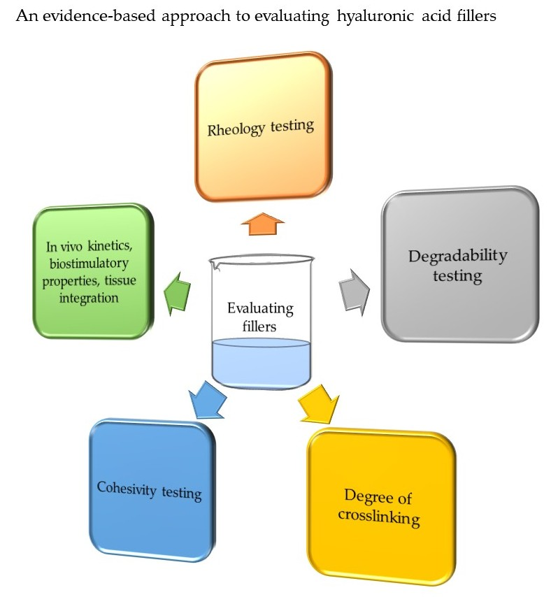
1. Introduction
2. Methods
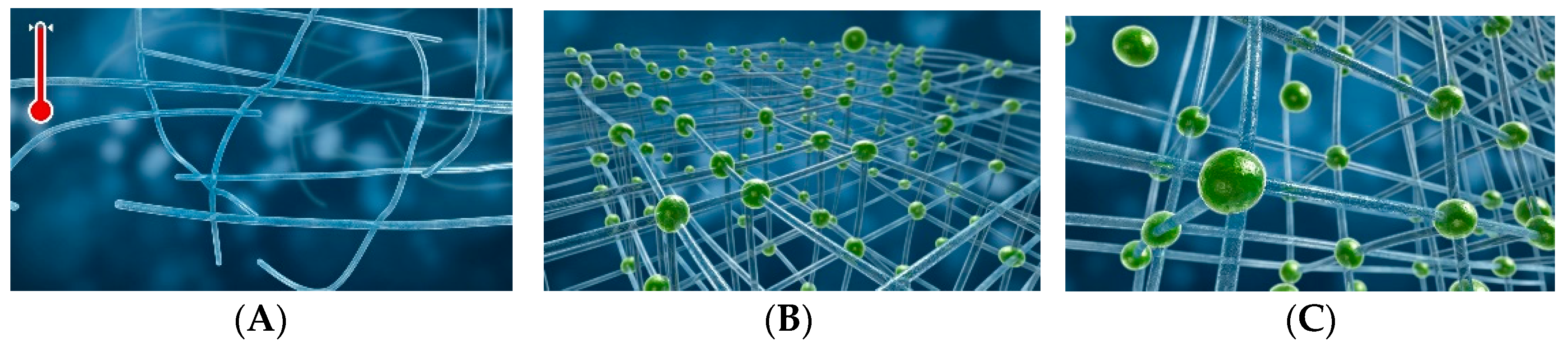
2.1. The XTR™ Technology Manufacturing Process
2.2. Rheological Evaluation
2.3. Degree of Crosslinking
- The degree of modification (MoD) is the stoichiometric ratio between the sum of mono- and double-linked BDPE residues and HA disaccharide units. The more crosslink modifications seen when compared with the acetyl group, the higher the MoD%.
- The crosslinker ratio (CrR) indicates the fraction of double-linked crosslinker residues compared to all linked crosslinkers and this represents the measure of crosslinker efficiency.
- The degree of substitution (DS) is the proportion of the HA disaccharides that are substituted.
- The degree of crosslinking (CrD) is the stoichiometric ratio between BDPE residues that are double-linked and HA disaccharide units.
- The degree of crosslinking (DC) is the number of HA disaccharides involved in crosslinking in relation to the total number of HA disaccharides.
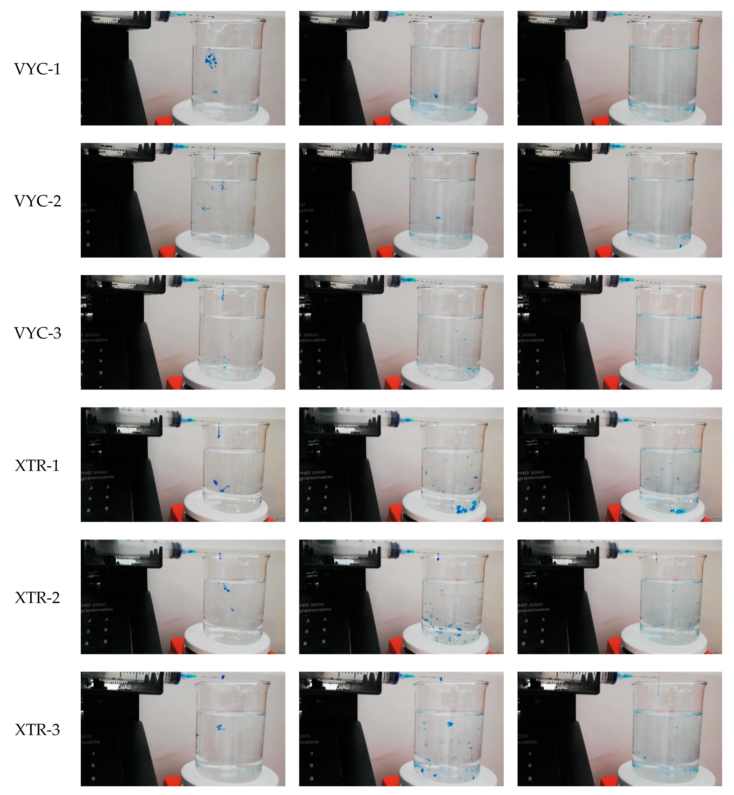
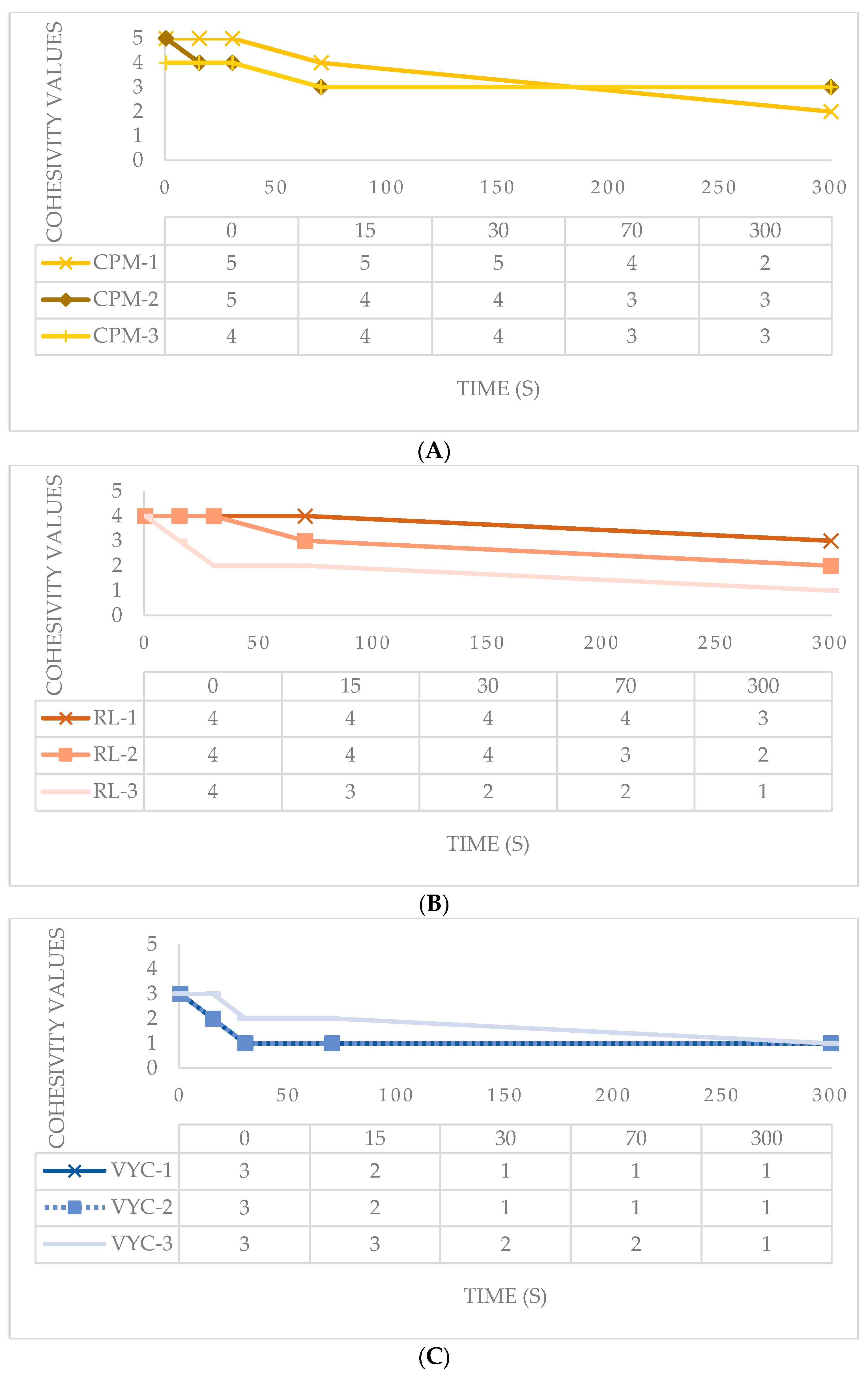
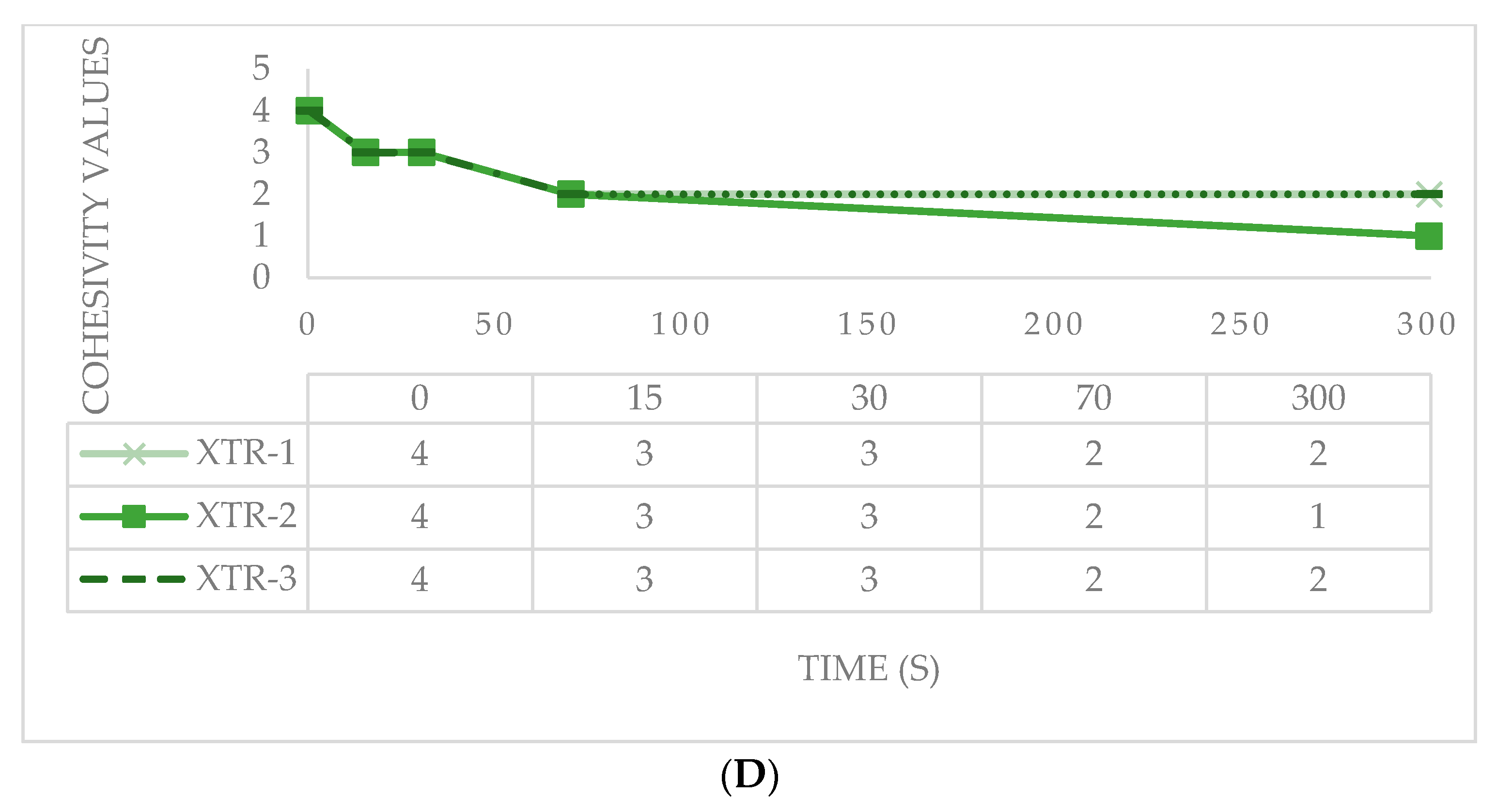
2.4. Cohesivity
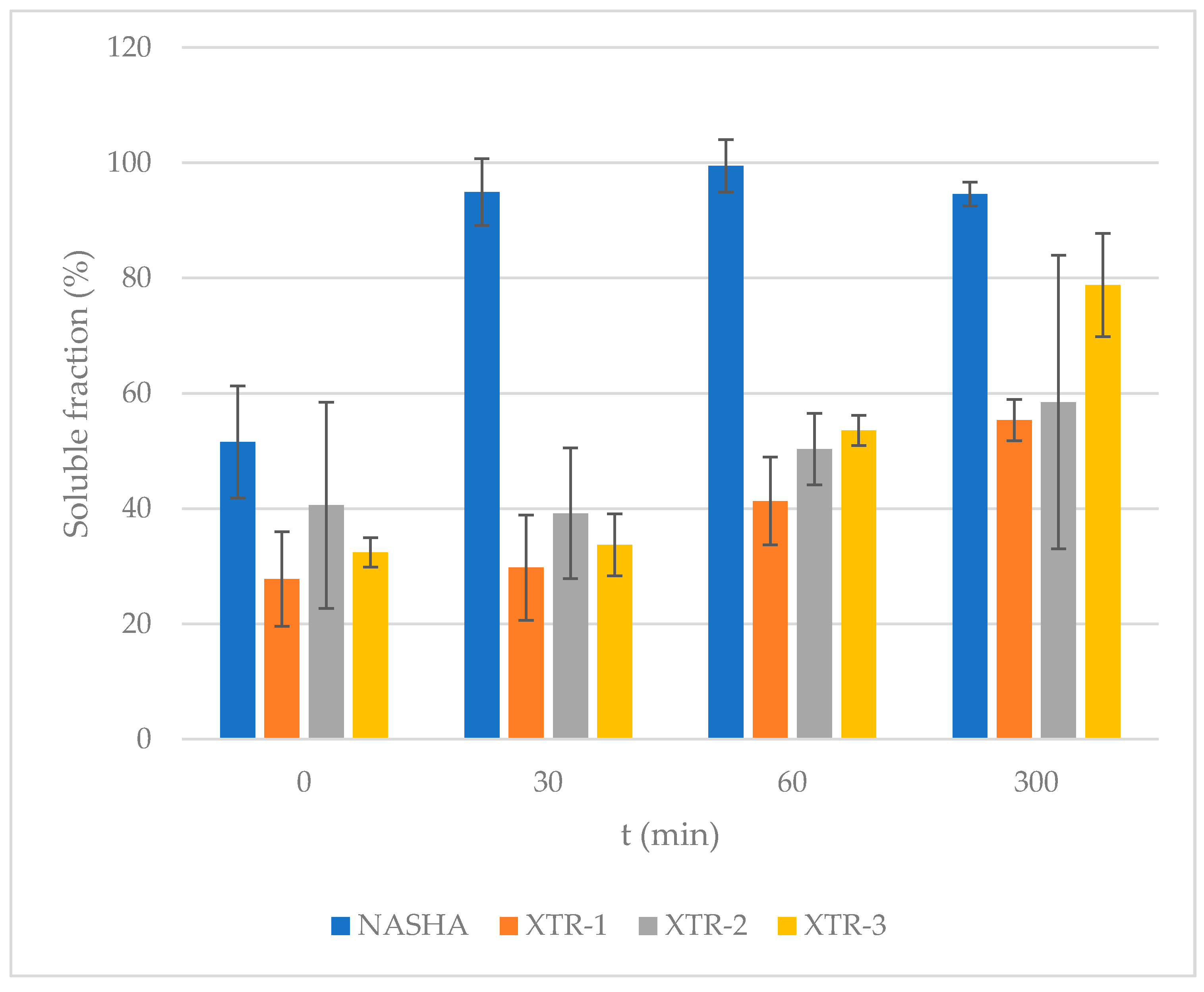
2.5. Degradability
3. Results
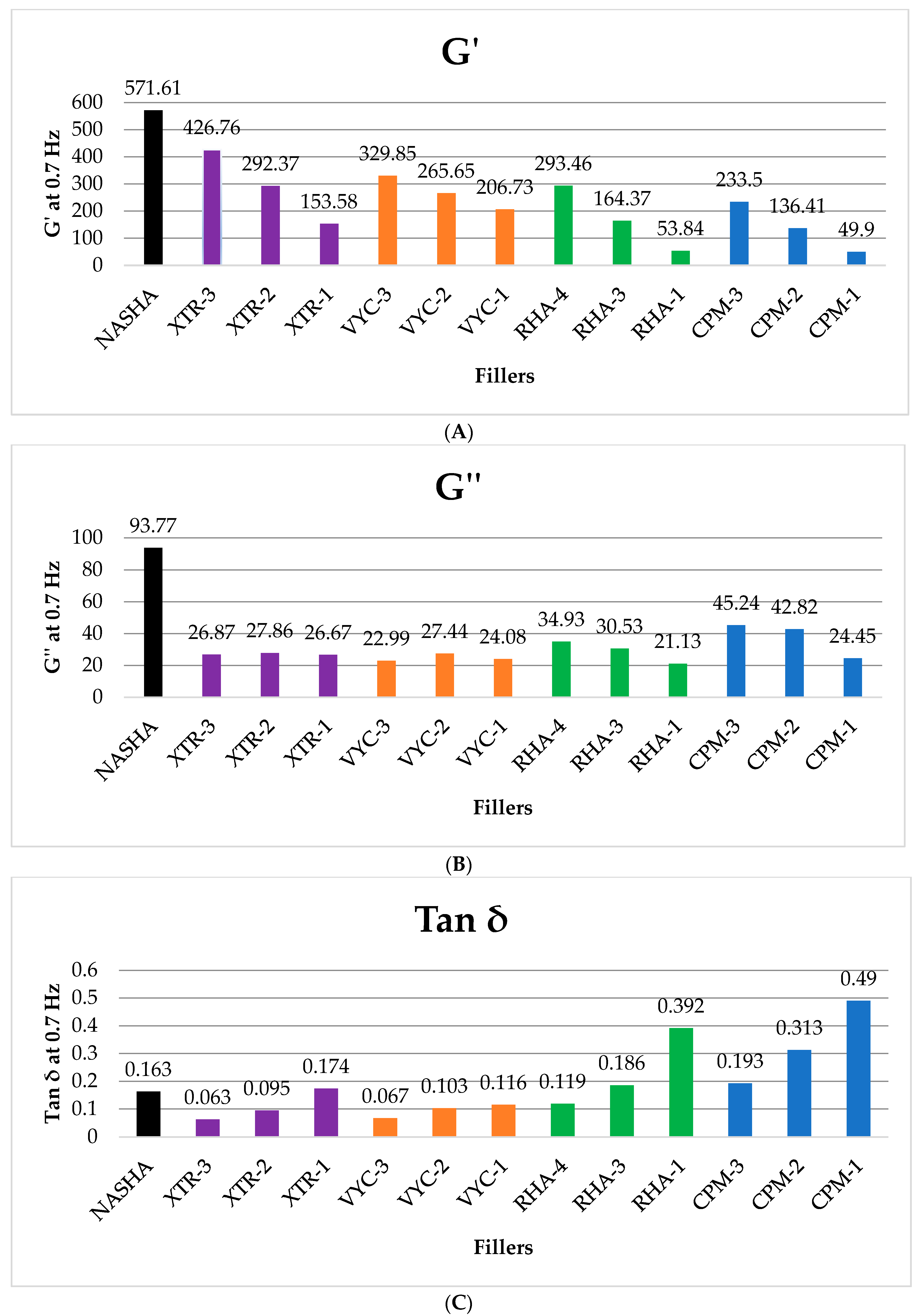
3.1. Rheological Evaluation
3.2. Degree of Crosslinking
3.3. Cohesivity
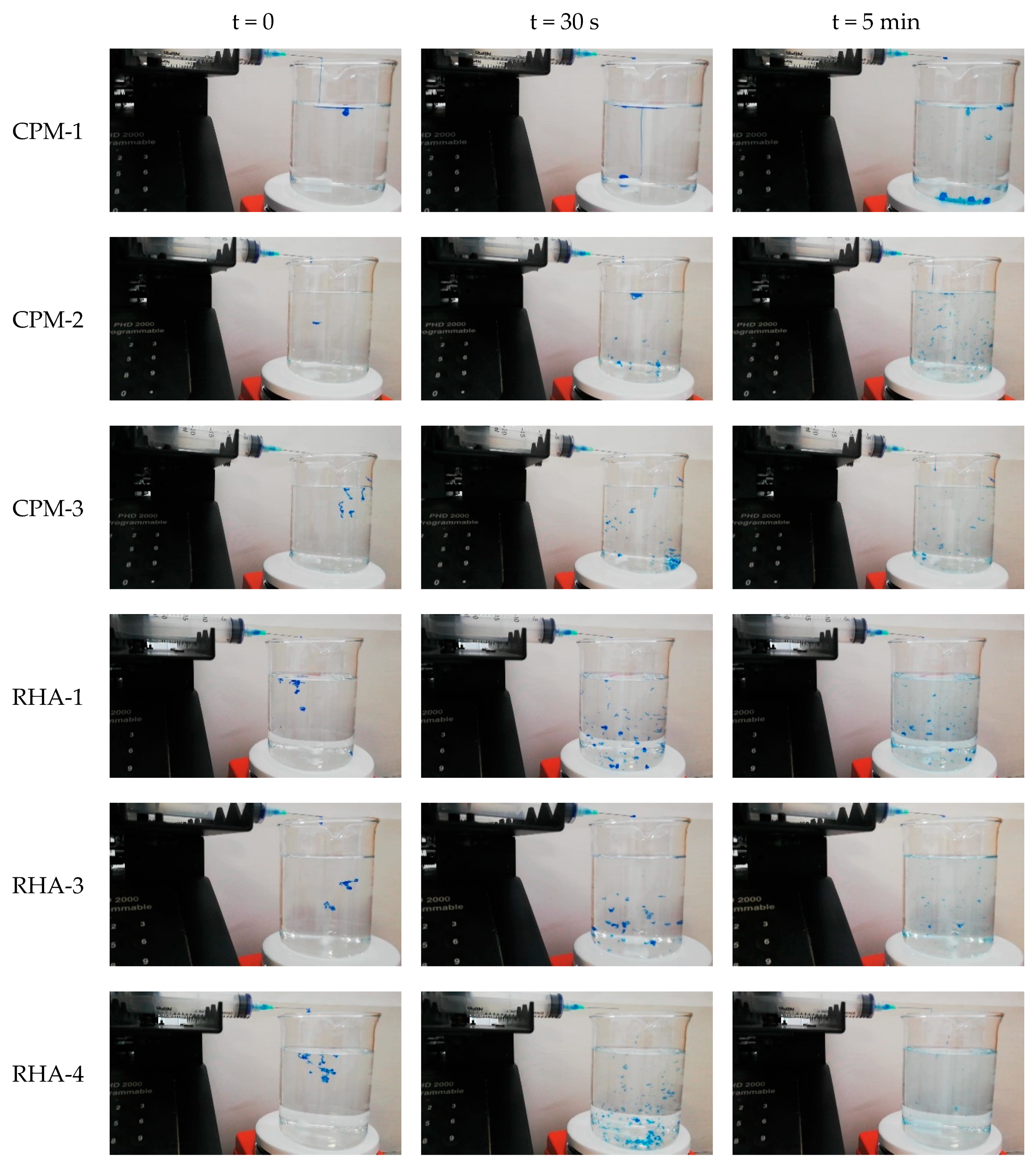
3.4. Degradability
4. Discussion
4.1. Rheologic Properties
4.2. Crosslinking Properties
4.3. Cohesivity
4.4. Degradability
4.5. Clinical Application, Limitations and Recommendations
5. Conclusions
Author Contributions
Funding
Acknowledgments
Conflicts of Interest
References
- La Gatta, A.; Salzillo, R.; Catalano, C.; D’Agostino, A.; Pirozzi AV, A.; De Rosa, M.; Schiraldi, C. Hyaluronan-based hydrogels as dermal fillers: The biophysical properties that translate into a “volumetric” effect. PLoS ONE 2019, 14, 1–17. [Google Scholar] [CrossRef] [PubMed] [Green Version]
- Smith, L.; Cockerham, K. Hyaluronic acid dermal fillers: Can adjunctive lidocaine improve patient satisfaction without decreasing efficacy or duration? Patient Prefer. Adherence 2011, 5, 133–139. [Google Scholar] [PubMed] [Green Version]
- Salti, G. Meet the Innovative XTRTM (eXcellent Three-dimensional Reticulation) Technology of DefinisseTM Filler. Prime J. 2020, 10, 84–85. [Google Scholar]
- Borrell, M.; Leslie, D.B.; Tezel, A. Lift capabilities of hyaluronic acid fillers. J. Cosmet. Laser Ther. 2011, 13, 21–27. [Google Scholar] [CrossRef] [PubMed]
- Micheels, P.; Sarazin, D.; Tran, C.; Salomon, D. Effect of Different Crosslinking Technologies on Hyaluronic Acid Behavior: A Visual and Microscopic Study of Seven Hyaluronic Acid Gels. J. Drugs Derm. 2016, 15, 600–606. [Google Scholar]
- Gavard Molliard, S.; Bon Bétemps, J.; Hadjab, B.; Topchian, D.; Micheels, P.; Salomon, D. Key rheological properties of hyaluronic acid fillers: From tissue integration to product degradation. Plast. Aesthetic Res. 2018, 5, 17. [Google Scholar] [CrossRef] [Green Version]
- Lee, W.; Yoon, J.-H.; Koh, I.-S.; Oh, W.; Kim, K.-W.; Yang, E.-J. Clinical application of a new hyaluronic acid filler based on its rheological properties and the anatomical site of injection. Biomed. Dermatol. 2018, 2, 1–5. [Google Scholar] [CrossRef]
- Rohrich, R.J.; Bartlett, E.L.; Dayan, E. Practical Approach and Safety of Hyaluronic Acid Fillers. Plast. Reconstr. Surg. Glob. Open 2019, 7, e2172. [Google Scholar] [CrossRef]
- Buhren, B.A.; Schrumpf, H.; Bölke, E.; Kammers, K.; Gerber, P.A. Standardized in vitro analysis of the degradability of hyaluronic acid fillers by hyaluronidase. Eur. J. Med. Res. 2018, 23, 1–6. [Google Scholar] [CrossRef]
- Cavallini, M.; Papagni, M.; Trocchi, G. Sensitivity of Hyaluronic Acid Fillers to Hyaluronidase: An in vitro Analysis. J. Clin. Exp. 2020, 11, 1–6. [Google Scholar] [CrossRef]
- Park, S.; Park, K.Y.; Yeo, I.K.; Cho, S.Y.; Ah, Y.C.; Koh, H.J.; Park, W.S.; Kim, B.J. Investigation of the degradation-retarding effect caused by the low swelling capacity of a novel hyaluronic acid filler developed by solid-phase crosslinking technology. Ann. Dermatol. 2014, 26, 357–362. [Google Scholar] [CrossRef] [PubMed] [Green Version]
- Rigano Laboratories. Analysis of Different Types of Injectable Cross-Linked Hyaluronic Acid Gels: Determination of Degree of Cross-Linking in Hyaluronic Acid-Based Filler; Rigano Laboratories S.r.l.: Milan, Italy, 2019. [Google Scholar]
- Stern, R.; Kogan, G.; Jedrzejas, M.J.; Soltes, L. The many ways to cleave hyaluronan. Biotechnol. Adv. 2007, 25, 537–557. [Google Scholar] [CrossRef] [PubMed]
- Tokita, Y.; Okamoto, A. Degradation of hyaluronic acid—Kinetic study and thermodynamics. Eur. Polym. J. 1996, 32, 1011–1014. [Google Scholar] [CrossRef]
- Lekander, M.; Troncoso, J.F.; Idjbara, A.; Karlsson, I.; Lindgren, T.; Ström, S. A kinetic study of the degradation of hyaluronic acid at high concentrations of sodium hydroxide. Mater. Sci. 2016, 1, 1–32, URN: urn:nbn:se:uu:diva-301379. [Google Scholar]
- Tran, C.; Carraux, P.; Micheels, P.; Kaya, G.; Salomon, D. In vivo bio-integration of three hyaluronic acid fillers in human skin: A histological study. Dermatology 2014, 228, 47–54. [Google Scholar] [CrossRef]
- De Boulle, K.; Glogau, R.; Kono, T.; Nathan, M.; Tezel, A.; Roca-Martinez, J.-X.; Paliwal, S.; Stroumpoulis, D. A review of the metabolism of 1,4-butanediol diglycidyl ether-crosslinked hyaluronic acid dermal fillers. Dermatol. Surg. 2013, 39, 1758–1766. [Google Scholar] [CrossRef] [Green Version]
- Guarise, C.; Barbera, C.; Pavan, M.; Panfilo, S.; Beninatto, R.; Galesso, D. HA-based dermal filler: Downstream process comparison, impurity quantitation by validated HPLC-MS analysis, and in vivo residence time study. J. Appl. Biomater. Funct. Mater. 2019, 17, 2280800019867075. [Google Scholar] [CrossRef]
- US NIH. Restylane + Lidocaine and Restylane Lyft for the Treatment of Nasolabial Folds. Available online: https://clinicaltrials.gov/ct2/show/NCT04174131 (accessed on 17 March 2020).
- Woodward, J.A. Periocular fillers and related anatomy. Cutis 2016, 98, 330–335. [Google Scholar]
- Kühne, U.; Esmann, J.; Von Heimburg, D.; Imhof, M.; Weissenberger, P.; Sattler, G.; Heinz, M.; Kliebe-Frisch, C. Safety and performance of cohesive polydensified matrix hyaluronic acid fillers with lidocaine in the clinical setting—An open-label, multicenter study. Clin. Cosmet. Investig. Dermatol. 2016, 9, 373–381. [Google Scholar] [CrossRef] [Green Version]
- Rigano Laboratories. Determination of the Viscoelastic Properties of HA Fillers; Rigano Laboratories S.r.l.: Milan, Italy, 2019. [Google Scholar]
- Pierre, S.; Liew, S.; Bernardin, A. Basics of dermal filler rheology. Dermatol. Surg. 2015, 41 (Suppl. 1), S120–S126. [Google Scholar] [CrossRef]
- Schanté, C.E.; Zuber, G.; Herlin, C.; Vandamme, T.F. Improvement of hyaluronic acid enzymatic stability by the grafting of amino-acids. Carbohydr. Polym. 2012, 87, 2211–2216. [Google Scholar] [CrossRef]
- Wende, F.J.; Gohil, S.; Nord, L.I.; Helander Kenne, A.; Sandström, C. 1D NMR methods for determination of degree of cross-linking and BDDE substitution positions in HA hydrogels. Carbohydr. Polym. 2017, 157, 1525–1530. [Google Scholar] [CrossRef] [PubMed] [Green Version]
- Kenne, L.; Gohil, S.; Nilsson, E.M.; Karlsson, A.; Ericsson, D.; Helander Kenne, A.; Nord, L.I. Modification and cross-linking parameters in hyaluronic acid hydrogels--definitions and analytical methods. Carbohydr. Polym. 2013, 91, 410–418. [Google Scholar] [CrossRef] [PubMed] [Green Version]
- Sundaram, H.; Rohrich, R.J.; Liew, S.; Sattler, G.; Talarico, S.; Trevidic, P.; Molliard, S.G. Cohesivity of Hyaluronic Acid Fillers: Development and Clinical Implications of a Novel Assay, Pilot Validation with a Five-Point Grading Scale, and Evaluation of Six U.S. Food and Drug Administration-Approved Fillers. Plast. Reconstr. Surg. 2015, 136, 678–686. [Google Scholar] [CrossRef]
- Rigano Laboratories. Analysis of Different Types of Injectable Cross-Linked Hyaluronic Acid Gels: Cohesivity of Cross-Linked Hyaluronic Acid; Rigano Laboratories S.r.l.: Milan, Italy, 2019. [Google Scholar]
- La Gatta, A.; De Rosa, M.; Frezza, M.A.; Catalano, C.; Meloni, M.; Schiraldi, C. Biophysical and biological characterization of a new line of hyaluronan-based dermal fillers: A scientific rationale to specific clinical indications. Mater. Sci. Eng. C. Mater. Biol. Appl. 2016, 68, 565–572. [Google Scholar] [CrossRef]
- Rigano Laboratories. Analysis of Different Types of Injectable Cross-Linked Hyaluronic Acid Gels; Rigano Laboratories S.r.l.: Milan, Italy, 2020. [Google Scholar]
- Fagien, S.; Bertucci, V.; von Grote, E.; Mashburn, J.H. Rheologic and Physicochemical Properties Used to Differentiate Injectable Hyaluronic Acid Filler Products. Plast. Reconstr. Surg. 2019, 143, 707e–720e. [Google Scholar] [CrossRef]
- Stocks, D.; Sundaram, H.; Michaels, J.; Durrani, M.J.; Wortzman, M.S.; Nelson, D.B. Rheological evaluation of the physical properties of hyaluronic acid dermal fillers. J. Drugs Dermatol. 2011, 10, 974–980. [Google Scholar]
- Edsman, K.; Nord, L.I.; Ohrlund, A.; Lärkner, H.; Kenne, A.H. Gel properties of hyaluronic acid dermal fillers. Dermatol. Surg. 2012, 38, 1170–1179. [Google Scholar] [CrossRef]
- Lorenc, Z.P.; Öhrlund, Å.; Edsman, K. Factors Affecting the Rheological Measurement of Hyaluronic Acid Gel Fillers. J. Drugs Dermatol. 2017, 16, 876–882. [Google Scholar]
- Kablik, J.; Monheit, G.D.; Yu, L.; Chang, G.; Gershkovich, J. Comparative physical properties of hyaluronic acid dermal fillers. Dermatol. Surg. 2009, 35 (Suppl. 1), 302–312. [Google Scholar] [CrossRef]
- Flynn, T.C.; Sarazin, D.; Bezzola, A.; Terrani, C.; Micheels, P. Comparative histology of intradermal implantation of mono and biphasic hyaluronic acid fillers. Dermatol. Surg. 2011, 37, 637–643. [Google Scholar] [CrossRef]
- Sundaram, H.; Voigts, B.; Beer, K.; Meland, M. Comparison of the rheological properties of viscosity and elasticity in two categories of soft tissue fillers: Calcium hydroxylapatite and hyaluronic acid. Dermatol. Surg. 2010, 36 (Suppl. 3), 1859–1865. [Google Scholar] [CrossRef] [PubMed]
- Santoro, S.; Russo, L.; Argenzio, V.; Borzacchiello, A. Rheological properties of cross-linked hyaluronic acid dermal fillers. J. Appl. Biomater. Biomech. 2011, 9, 127–136. [Google Scholar] [CrossRef] [PubMed]
- Öhrlund, J.Å.; Edsman, K.L.M. The Myth of the ‘Biphasic’ Hyaluronic Acid Filler. Dermatol. Surg. 2015, 41 (Suppl. 1), S358–S364. [Google Scholar]
- Sundaram, H. Going with the Flow: An Overview and Clinical Discussion of The Rheology of Soft Tissue Fillers, Part 1 of 2. Pract. Dermatol. 2010, 11, 21–24. Available online: https://practicaldermatology.com/articles/2010-nov/cosmetics-challenge-going-with-the-flow-an-overview-and-clinical-discussion-of-the-rheology-of-soft-tissue-fillers-part-1-of-2/pdf (accessed on 1 July 2020).
- Sundaram, H.; Liew, S.; Signorini, M.; Vieira Braz, A.; Fagien, S.; Swift, A.; De Boulle, K.L.; Raspaldo, H.; Trindade De Almeida, A.R.; Monheit, G. Global Aesthetics Consensus: Hyaluronic Acid Fillers and Botulinum Toxin Type A-Recommendations for Combined Treatment and Optimizing Outcomes in Diverse Patient Populations. Plast. Reconstr. Surg. 2016, 137, 1410–1423. [Google Scholar] [CrossRef] [Green Version]
- Funt, D.; Pavicic, T. Dermal fillers in aesthetics: An overview of adverse events and treatment approaches. Clin. Cosmet. Investig. Dermatol. 2013, 6, 295–316. [Google Scholar] [CrossRef]
- Lee, D.Y.; Cheon, C.; Son, S.; Kim, Y.Z.; Kim, J.T.; Jang, J.W.; Kim, S.S. Influence of molecular weight on swelling and elastic modulus of hyaluronic acid dermal fillers. Polymer 2015, 39, 976–980. [Google Scholar] [CrossRef]
- Edsman, K.L.M.; Wiebensjö, Å.M.; Risberg, A.M.; Öhrlund, J.Å. Is There a Method That Can Measure Cohesivity? Cohesion by Sensory Evaluation Compared With Other Test Methods. Dermatol. Surg. 2015, 41 (Suppl. 1), S365–S372. [Google Scholar] [CrossRef]
- Niamtu, J. 10—Injectable Fillers: Lip Augmentation, Lip Reduction, and Lip Lift. Cosmet. Facial Surg. In Cosmetic Facial Surgery; Elsevier: Amsterdam, The Netherlands, 2018. [Google Scholar] [CrossRef]


| Filler Identifier | Crosslinking Technology [20] | Hyaluronic Acid Concentration (mg/mL) [19,20,21] |
|---|---|---|
| NASHA | NASHA® | 20 |
| CPM-1 | CPM® | 22.5 |
| CPM-2 | 25 | |
| CPM-3 | 26 | |
| RHA-1 | RHA® | 15 |
| RHA-3 | 23 | |
| RHA-4 | 23 | |
| VYC-1 | Vycross® | 15 |
| VYC-2 | 17.5 | |
| VYC-3 | 20 | |
| XTR-1 | XTR™ | 23 |
| XTR-2 | 23 | |
| XTR-3 | 25 |
| Product | G′ | G′′ | Tan δ |
|---|---|---|---|
| XTR-1 | 153.58 | 26.67 | 0.174 |
| CPM-1 | 49.9 | 24.45 | 0.49 |
| VYC-1 | 206.73 | 24.08 | 0.116 |
| RHA-1 | 53.84 | 21.13 | 0.392 |
| XTR-2 | 292.37 | 27.86 | 0.095 |
| CPM-2 | 136.41 | 42.82 | 0.313 |
| VYC-2 | 265.65 | 27.44 | 0.103 |
| RHA-3 | 164.37 | 30.53 | 0.186 |
| XTR-3 | 426.76 | 26.87 | 0.063 |
| CPM-3 | 233.5 | 45.24 | 0.193 |
| RHA-4 | 293.46 | 34.93 | 0.119 |
| VYC-3 | 329.85 | 22.99 | 0.067 |
| Product | mg/mL HA | MoD% | CrR | CrD% |
|---|---|---|---|---|
| CPM-1 | 22.5 | 7.50 | 0.48 | 3.57 |
| CPM-2 | 25.5 | 9.80 | 0.11 | 1.06 |
| CPM-3 | 26 | 15.90 | 0.03 | 0.41 |
| RHA-1 | 15 | 3.16 | 0.25 | 0.80 |
| RHA-3 | 23 | 6.02 | 0.31 | 1.87 |
| RHA-4 | 23 | 6.85 | 0.09 | 0.64 |
| VYC-1 | 15 | 6.61 | 0.43 | 2.85 |
| VYC-2 | 17.5 | 7.73 | 0.14 | 1.08 |
| VYC-3 | 20 | 7.36 | 0.45 | 3.31 |
| XTR-1 | 23 | 7.01 | 0.15 | 1.05 |
| XTR-2 | 23 | 12.64 | 0.27 | 3.35 |
| XTR-3 | 25 | 10.16 | 0.49 | 5.00 |
| Enzymatic Degradation Time | |||||
|---|---|---|---|---|---|
| Product | mg/mL HA | t = 0 | t = 30 min | t = 1 h | t = 5 h |
| NASHA | 20 | 51.58 (9.71) | 94.95 (8.19) | 99.50 (17.89) | 94.56 (2.54) |
| XTR-1 | 23 | 27.8 (5.79) | 29.79 (9.14) | 41.34 (11.35) | 55.36 (5.39) |
| XTR-2 | 23 | 40.59 (4.55) | 39.20 (7.62) | 50.35 (6.23) | 58.47 (2.61) |
| XTR-3 | 25 | 32.43 (2.08) | 33.72 (3.59) | 53.58 (25.46) | 78.79 (8.95) |
© 2020 by the authors. Licensee MDPI, Basel, Switzerland. This article is an open access article distributed under the terms and conditions of the Creative Commons Attribution (CC BY) license (http://creativecommons.org/licenses/by/4.0/).
Share and Cite
Salti, G.; Fundarò, S.P. Evaluation of the Rheologic and Physicochemical Properties of a Novel Hyaluronic Acid Filler Range with eXcellent Three-Dimensional Reticulation (XTR™) Technology. Polymers 2020, 12, 1644. https://doi.org/10.3390/polym12081644
Salti G, Fundarò SP. Evaluation of the Rheologic and Physicochemical Properties of a Novel Hyaluronic Acid Filler Range with eXcellent Three-Dimensional Reticulation (XTR™) Technology. Polymers. 2020; 12(8):1644. https://doi.org/10.3390/polym12081644
Chicago/Turabian StyleSalti, Giovanni, and Salvatore Piero Fundarò. 2020. "Evaluation of the Rheologic and Physicochemical Properties of a Novel Hyaluronic Acid Filler Range with eXcellent Three-Dimensional Reticulation (XTR™) Technology" Polymers 12, no. 8: 1644. https://doi.org/10.3390/polym12081644