Heterogenized Imidazolium-Based Ionic Liquids in Pebax®Rnew. Thermal, Gas Transport and Antimicrobial Properties


 ,
,  , , and
Abstract
:1. Introduction
2. Experimental
2.1. Materials
2.2. Methods
2.2.1. ILs Synthesis
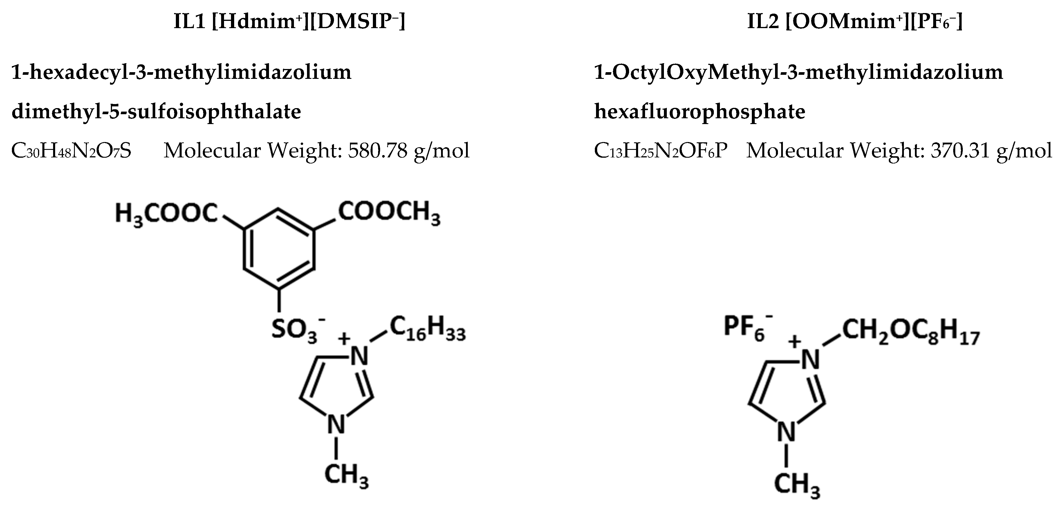
1-hexadecyl-3-methylimidazolium dimethyl-5-sulfoisophthalate ([Hdmim][DMSIP], IL1) synthesis (according to Colonna et al., slightly modified [10]).
1-octyloxymethyl-3-methylimidazolium hexafluorophosphate ([OOMim][PF6], IL2) synthesis (according to Pernak et al. [21]).
2.2.2. Membrane Preparation
2.3. Characterization
2.3.1. NMR Spectroscopy
2.3.2. Matrix Assisted Laser Desorption Time of Flight Mass Spectrometry (MALDI TOF MS) Analysis
2.3.3. Calorimetric Analyses
2.3.4. Thermogravimetric Analyses (TGA)
2.3.5. Fourier Transform Infrared (FTIR)
2.3.6. Scanning Electron Microscopy (SEM)
2.3.7. Contact Angle (CA)
2.3.8. Gas Permeation Tests
2.3.9. Antimicrobial Activity
2.3.10. IL Release
2.3.11. Cell Culture and Cytotoxicity Assay
3. Results
3.1. ILs Synthesis and Characterization and Membrane Preparation
3.2. Thermal Analysis
3.2.1. DSC Measurements
3.2.2. TGA
3.2.3. FTIR
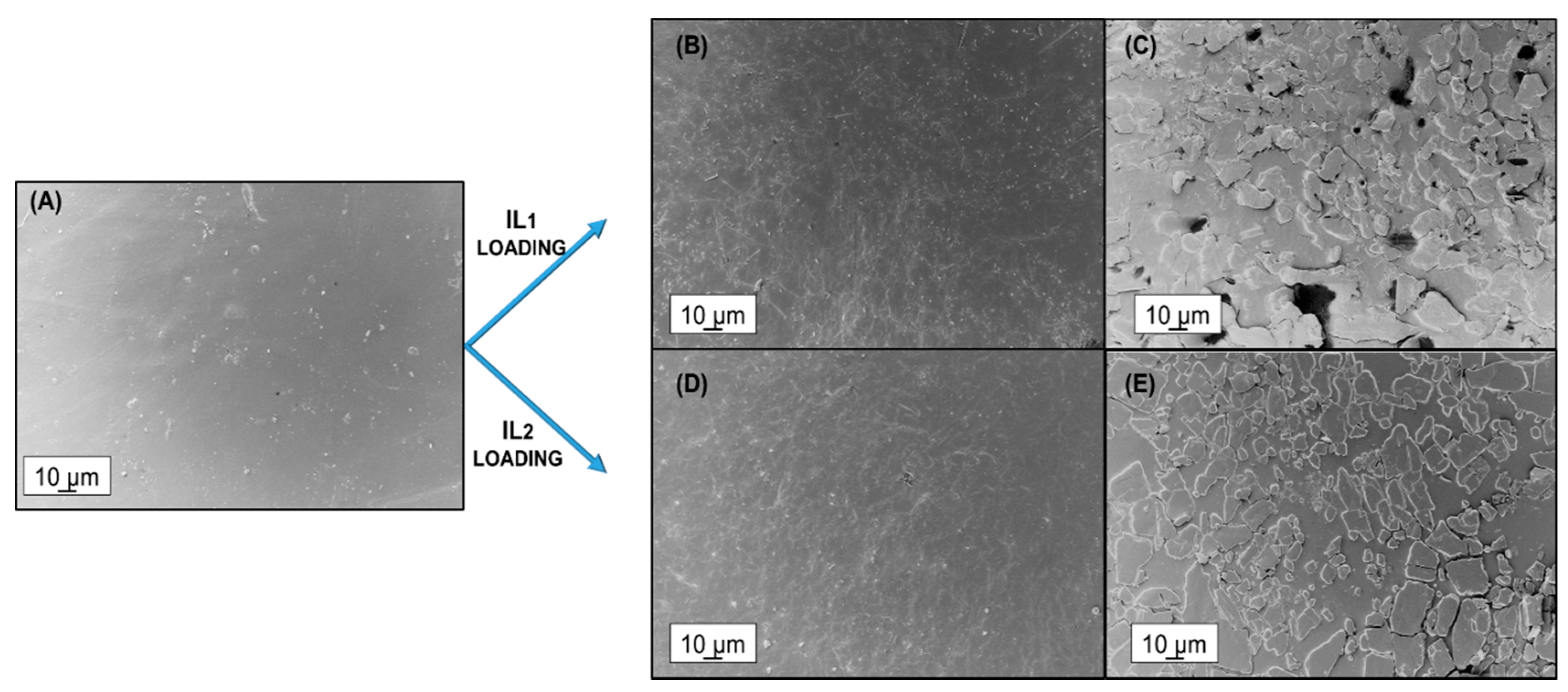
3.2.4. SEM
3.2.5. Contact Angle
3.2.6. Gas Permeation
3.2.7. Antimicrobial Activity
3.2.8. IL Release
3.2.9. Cytotoxicity of Pebax®Rnew/ILs Blends
4. Conclusions
Supplementary Materials
Author Contributions
Funding
Acknowledgments
Conflicts of Interest
References
- World Health Organization. World Health Organization Estimates of the Global Burden of Foodborne Diseases; WHO: Geneva, Switzerland, 2015. [Google Scholar]
- Muñoz-Bonilla, A.; Echeverria, C.; Sonseca, Á.; Arrieta, M.P.; Fernández-García, M. Bio-Based Polymers with Antimicrobial Properties towards Sustainable Development. Materials 2019, 12, 641. [Google Scholar] [CrossRef] [PubMed] [Green Version]
- Welton, T. Ionic Liquids: A Brief History. Biophys. Rev. 2018, 10, 691–706. [Google Scholar] [CrossRef] [PubMed] [Green Version]
- Huang, R.T.W.; Peng, K.C.; Shih, H.N.; Lin, G.H.; Chang, T.F.; Hsu, S.J.; Hsu, T.S.T.; Lin, I.J.B. Antimicrobial properties of ethoxyether-functionalized imidazolium salts. Soft Matter 2011, 7, 8392–8400. [Google Scholar] [CrossRef]
- Garcia, M.T.; Ribosa, I.; Perez, L.; Manresa, A.; Comelles, F. Aggregation Behavior and Antimicrobial Activity of Ester-Functionalized Imidazolium- and Pyridinium-Based Ionic Liquids in Aqueous Solution. Langmuir 2013, 29, 2536–2545. [Google Scholar] [CrossRef]
- Raucci, M.G.; Fasolino, I.; Pastore, S.G.; Soriente, A.; Capeletti, L.B.; Dessuy, M.B.; Giannini, C.; Schrekker, H.S.; Ambrosio, L. Antimicrobial Imidazolium Ionic Liquids for the Development of Minimal Invasive Calcium Phosphate-Based Bionanocomposites. ACS Appl. Mater. Interfaces 2018, 10, 42766–42776. [Google Scholar] [CrossRef]
- Yoo, B.; Jing, B.; Jones, S.E.; Lamberti, G.A.; Zhu, Y.; Shah, J.K.; Maginn, E.J. Molecular mechanisms of ionic liquid cytotoxicity probed by an integrated experimental and computational approach. Sci. Rep. 2016, 6, 19889. [Google Scholar] [CrossRef]
- Egorova, K.S.; Gordeev, E.G.; Ananikov, V.P. Biological Activity of Ionic Liquids and their Application in Pharmaceutics and Medicine. Chem. Rev. 2017, 117, 7132–7189. [Google Scholar] [CrossRef]
- Suchodolski, J.; Feder-Kubis, J.; Krasowska, A. Antifungal Activity of Ionic Liquids Based on (−)-Menthol: A Mechanism Study. Microbiol. Res. 2017, 197, 56–64. [Google Scholar] [CrossRef]
- Colonna, M.; Berti, C.; Binassi, E.; Fiorini, M.; Sullalti, S.; Acquasanta, F.; Vannini, M.; Di Gioia, D.; Aloisio, I. Imidazolium poly(butylene terephthalate) ionomers with long-term antimicrobial activity. Polymer 2012, 53, 1823–1830. [Google Scholar] [CrossRef]
- Pendleton, N.; Gilmore, B.F. The antimicrobial potential of ionic liquids: A source of chemical diversity for infection and biofilm control. Int. J. Antimicrob. Agents 2015, 46, 131–139. [Google Scholar] [CrossRef] [PubMed]
- Montalbán, M.G.; Hidalgo, J.M.; Collado-González, M.; Díaz Baños, F.G.; Víllora, G. Assessing Chemical Toxicity of Ionic Liquids on Vibrio fischeri: Correlation with Structure and Composition. Chemosphere 2016, 155, 405–414. [Google Scholar] [CrossRef]
- Bugatti, V.; Viscusi, G.; Di Bartolomeo, A.; Iemmo, L.; Zampino, D.C.; Vittoria, V.; Gorrasi, G. Ionic Liquid as dispersing agent of LDH-Carbon nanotubes into a biodegradable Vynil Alcohol polymer. Polymers 2020, 12, 495. [Google Scholar] [CrossRef] [Green Version]
- Gilmore, B.F. Antimicrobial Ionic Liquids. In Ionic Liquids. Applications and Perspectives; Kokorin, A., Ed.; InTechOpen: London, UK, 2011. [Google Scholar]
- Malik, M.A.; Hashim, M.A.; Nabi, F. Ionic liquids in supported liquid membrane technology. Chem. Eng. J. 2011, 171, 242–254. [Google Scholar] [CrossRef]
- Bernardo, P.; Jansen, J.C.; Bazzarelli, F.; Tasselli, F.; Fuoco, A.; Friess, K.; Izák, P.; Jarmarová, V.; Kačírková, M.; Clarizia, G. Gas transport properties of PEBAX®/Room Temperature Ionic Liquid gel membranes. Sep. Purif. Tech. 2012, 97, 73–82. [Google Scholar] [CrossRef]
- Muñoz-Bonilla, A.; Fernández-García, M. Poly(ionic liquid)s as antimicrobial materials. Eur. Polym. J. 2018, 105, 135–149. [Google Scholar] [CrossRef]
- Cai, J.; Jiang, J.; Zhou, Z.; Ding, Y.; Zhang, Y.; Wang, F.; Han, C.; Guo, J.; Shao, Q.; Du, H.; et al. Toughening Poly(lactic acid) by Melt Blending with Poly(ether-block-amide) Copolymer. Sci. Adv. Mater. 2017, 9, 1683–1692. [Google Scholar] [CrossRef] [Green Version]
- Yang, O.; Kim, H.L.; Weon, J.-I.; Seo1, Y.R. Endocrine-disrupting Chemicals: Review of Toxicological Mechanisms Using Molecular Pathway Analysis. J. Cancer Prev. 2015, 20, 12–24. [Google Scholar] [CrossRef]
- Fanelli, R.; Zuccato, E. Risks and benefits of PVC in medical applications. Boll. Chim. Farm. 2002, 141, 282–289. [Google Scholar]
- Pernak, J.; Czepukowicz, A.; Pozniak, R. New Ionic Liquids and Their Antielectrostatic Properties. Ind. Eng. Chem. Res. 2001, 40, 2379–2383. [Google Scholar] [CrossRef]
- Montando, G.; Carroccio, S.; Montaudo, M.S.; Puglisi, C.; Samperi, F. Recent advances in MALDI mass spectrometry of polymers. Macromol. Symp. 2004, 218, 101–112. [Google Scholar]
- Clarizia, G.; Bernardo, P.; Gorrasi, G.; Zampino, D.; Carroccio, S.C. Influence of the preparation method and photo-oxidation treatment on the thermal and gas transport properties of dense films based on a poly(ether-block-amide) copolymer. Materials 2018, 11, 1326. [Google Scholar] [CrossRef] [Green Version]
- Crank, J. The Mathematics of Diffusion, 2nd ed.; Clarendon Press: Oxford, UK, 1975. [Google Scholar]
- Conte, R.; Valentino, A.; Di Cristo, F.; Peluso, G.; Cerruti, P.; Di Salle, A.; Calarco, A. Cationic Polymer Nanoparticles-Mediated Delivery of miR-124 Impairs Tumorigenicity of Prostate Cancer Cells. Int. J. Mol. Sci. 2020, 21, 869. [Google Scholar] [CrossRef] [PubMed] [Green Version]
- Gómez, E.; Calvar, N.; Domínguez, Á. Thermal behavior of pure ionic liquids. Chapter 8. In Ionic Liquids—Current State of Art; Handy, S., Ed.; INTECH Open Science: London, UK, 2015; Available online: http://www.intechopen.com/ionic-liquids-current-state-of-the-art (accessed on 3 April 2020). [CrossRef] [Green Version]
- Crosthwaite, J.M.; Muldoon, M.J.; Dixon, J.K.; Anderson, J.L.; Brennecke, J.F. Phase transition and decomposition tempeatures, heat capacities and viscosities of pyridinium ionic liquids. J. Chem. Thermodyn. 2005, 37, 559–568. [Google Scholar] [CrossRef]
- Huddleston, J.G.; Visser, W.; Reichert, W.M.; Willauer, H.D.; Broker, G.A.; Rogers, R.D. Characterization and comparison of hydrophilic and hydrophobic room temperature ionic liquids incorporating the imidazolium cation. Green Chem. 2001, 3, 156–164. [Google Scholar] [CrossRef]
- Rogalsky, S.; Fatyeyeva, K.; Lyoshina, L.; Tarasyuk, O.; Bulko, O.; Lobok, S. Antimicrobial Properties and Thermal Stability of Polycarbonate Modified with 1-Alkyl-3-methylimidazolium Tetrafluoroborate Ionic Liquids. J. Appl. Polym. Sci. 2014, 131, 40050–40056. [Google Scholar] [CrossRef]
- Xi, T.; Tang, L.; Hao, W.; Yao, L.; Cui, P. Morphology and pervaporation performance of ionic liquid and waterborne polyurethane composite membranes. RSC Adv. 2018, 8, 7792–7799. [Google Scholar] [CrossRef] [Green Version]
- Berg, J.M.; Eriksson, L.G.T.; Claesson, P.M.; Børve, K.G.N. Three-Component Langmuir-Blodgett Films with a Controllable Degree of Polarity. Langmuir 1994, 10, 1225–1234. [Google Scholar] [CrossRef]
- Utrata-Wesolek, A. Antifouling surfaces in medical application. Polymery 2013, 58, 685–695. [Google Scholar] [CrossRef]
- Bugatti, V.; Bernardo, P.; Clarizia, G.; Viscusi, G.; Vertuccio, L.; Gorrasi, G. Ball Milling to produce composites based of natural clinoptilolite as carrier of salicylate in bio-based PA11. Polymers 2019, 11, 634. [Google Scholar] [CrossRef] [PubMed] [Green Version]
- Bondar, V.; Freeman, B.D.; Pinnau, I. Gas transport properties of poly(ether-b-amide) segmented block copolymers. J. Polym. Sci. Part B: Polym. Phys. 2000, 38, 2051–2062. [Google Scholar] [CrossRef]
- Docherty, K.; Kulpa, C.F. Toxicity and antimicrobial activity of imidazolium and pyridinium ionic liquids. Green Chem. 2005, 7, 185–189. [Google Scholar] [CrossRef]
- Gilmore, B.F.; Earle, M.J. Development of ionic liquid biocides against microbial biofilms: Designer microbicides for infection control. Chim. Oggi 2011, 29, 50–53. [Google Scholar]
- Cvjetko, M.; Radošević, K.; Tomica, A.; Slivac, I.; Vorkapić-Furač, J.; Srček, V. Cytotoxic Effects of Imidazolium Ionic Liquids on Fish and Human Cell Lines. Arch. Ind. Hyg. Toxicol. 2012, 63, 15–20. [Google Scholar] [CrossRef]
- Bakshi, K.; Mitra, S.; Sharma, V.K.; Jayadev, M.S.K.; Sakai, V.G.; Mukhopadhyay, R.; Gupta, A.; Ghosh, S.K. Imidazolium-based ionic liquids cause mammalian cell death due to modulated structures and dynamics of cellular membrane. Biochim Biophys Acta Biomembr. 2020, 1862, 183103. [Google Scholar] [CrossRef] [PubMed]
- Petkovic, M.; Seddon, K.R.; Rebelo, L.P.N.; Pereira, C.S. Ionic liquids: A pathway to environmental acceptability. Chem. Soc. Rev. 2011, 40, 1383–1403. [Google Scholar] [CrossRef] [PubMed]
- Domanska, U.; Bogel-Lukasik, E.; Bogel-Lukasik, R. 1-Octanol/Water Partition Coefficients of 1-Alkyl-3-methylimidazolium Chloride. Chem. Eur. J. 2003, 9, 3033–3041. [Google Scholar] [CrossRef]
- Ventura, S.P.M.; Gardas, R.L.; Goncalves, F.; Coutinho, J.A.P. Ecotoxicological risk profile of ionic liquids: Octanol-water distribution coefficients and toxicological data. J. Chem. Technol. Biotechnol. 2011, 86, 957–963. [Google Scholar] [CrossRef]
- Capello, C.; Fischer, U.; Hungerbühler, K. What is a green solvent? A comprehensive framework for the environmental assessment of solvents. Green Chem. 2007, 9, 927–934. [Google Scholar] [CrossRef]
- Taylor, M.S.; Daniels, A.U.; Andriano, K.P.; Heller, J. Six bioabsorbable polymers: In vitro acute toxicity of accumulated degradation products. J. Appl. Biomater. 1994, 5, 151–157. [Google Scholar] [CrossRef]


| Sample | PTMO | PA11 | ||
|---|---|---|---|---|
| Tm (°C) | ΔHm (J g−1) | Tm (°C) | ΔHm (J g−1) | |
| Neat Pebax®Rnew 25R53 | 12 | 27.8 | 142 | 4.0 |
| Pebax®Rnew—1% IL1 | 14 | 15.9 | 137 | 4.2 |
| Pebax®Rnew—5% IL1 | (−25) 14 | (6.4) 4.1 | 137 | 3.5 |
| Pebax®Rnew—1% IL2 | 13 | 14.3 | 134 | 3.6 |
| Pebax®Rnew—5% IL2 | 17 | 17.2 | 129 | 3.4 |
| Samples | TΔm= 5% (°C) a | Td1 (°C) b | % R c |
|---|---|---|---|
| Neat Pebax®Rnew 25R53 | 366 | 418 | 0.0 |
| Pebax®Rnew—1% IL1 | 337 | 413 | 0.2 |
| Pebax®Rnew—5% IL1 | 333 | 402 | 0.1 |
| Pebax®Rnew—1% IL2 | 354 | 422 | 0.2 |
| Pebax®Rnew—5% IL2 | 222 | 415 | 0.0 |
| IL Type (wt.%)_ | Permeability (Barrer) | Selectivity (–) | ||||
|---|---|---|---|---|---|---|
| Thickness | CO2 | O2 | N2 | He | CO2/N2 | O2/N2 |
| Neat Polymer _120 micron | 247 | 23.6 | 8.9 | 25.7 | 27.9 | 2.66 |
| IL1 (1%) 115 micron | 217 | 21.4 | 8.5 | 23.7 | 25.6 | 2.53 |
| IL1 (5%)_ 80 micron | 195 | 19.2 | 7.2 | 24.1 | 27.2 | 2.68 |
| IL2 (1%) 90 micron | 232 | 22.4 | 8.6 | 25.7 | 27.0 | 2.60 |
| IL2 (5%) 100 micron | 226 | 21.6 | 8.1 | 25.8 | 28.0 | 2.61 |
| Bacteria Strain | Inhibition Halos (mm) Induced by Pebax®Rnew/ILs Films | ||||
|---|---|---|---|---|---|
| Pebax®Rnew/IL1 | Pebax®Rnew/IL2 | ||||
| 1 wt.% | 5 wt.% | 1 wt.% | 5 wt.% | ||
| Gram− | E. coli | - | 1 ± 0.0 | 1 ± 0.0 | 4 ± 0.5 |
| Ps. fluorescens | 3 ± 0.2 | 3 ± 0.5 | - | 5 ± 0.5 | |
| S. enterica | - | - | - | - | |
| Gram+ | L. monocytogenes | 3 ± 0.5 | 3 ± 0.2 | - | - |
| B. subtilis | - | - | - | - | |
© 2020 by the authors. Licensee MDPI, Basel, Switzerland. This article is an open access article distributed under the terms and conditions of the Creative Commons Attribution (CC BY) license (http://creativecommons.org/licenses/by/4.0/).
Share and Cite
Clarizia, G.; Bernardo, P.; Carroccio, S.C.; Ussia, M.; Restuccia, C.; Parafati, L.; Calarco, A.; Zampino, D. Heterogenized Imidazolium-Based Ionic Liquids in Pebax®Rnew. Thermal, Gas Transport and Antimicrobial Properties. Polymers 2020, 12, 1419. https://doi.org/10.3390/polym12061419
Clarizia G, Bernardo P, Carroccio SC, Ussia M, Restuccia C, Parafati L, Calarco A, Zampino D. Heterogenized Imidazolium-Based Ionic Liquids in Pebax®Rnew. Thermal, Gas Transport and Antimicrobial Properties. Polymers. 2020; 12(6):1419. https://doi.org/10.3390/polym12061419
Chicago/Turabian StyleClarizia, Gabriele, Paola Bernardo, Sabrina C. Carroccio, Martina Ussia, Cristina Restuccia, Lucia Parafati, Anna Calarco, and Daniela Zampino. 2020. "Heterogenized Imidazolium-Based Ionic Liquids in Pebax®Rnew. Thermal, Gas Transport and Antimicrobial Properties" Polymers 12, no. 6: 1419. https://doi.org/10.3390/polym12061419