Synthesis and Characterization of a New Aluminum-Doped Bismuth Subcarbonate

 and
and
Abstract
:1. Introduction
2. Materials and Methods
2.1. Synthesis of Al-Doped Bi2O2CO3
2.2. Characterization of Bi2O2CO3:Al
3. Results
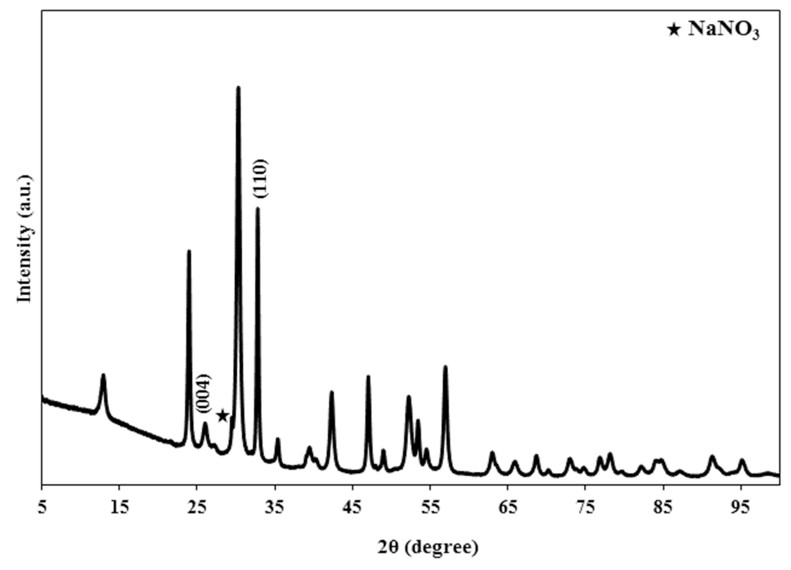
3.1. X-rays Diffraction
3.2. Fourier Transform Infrared Spectroscopy
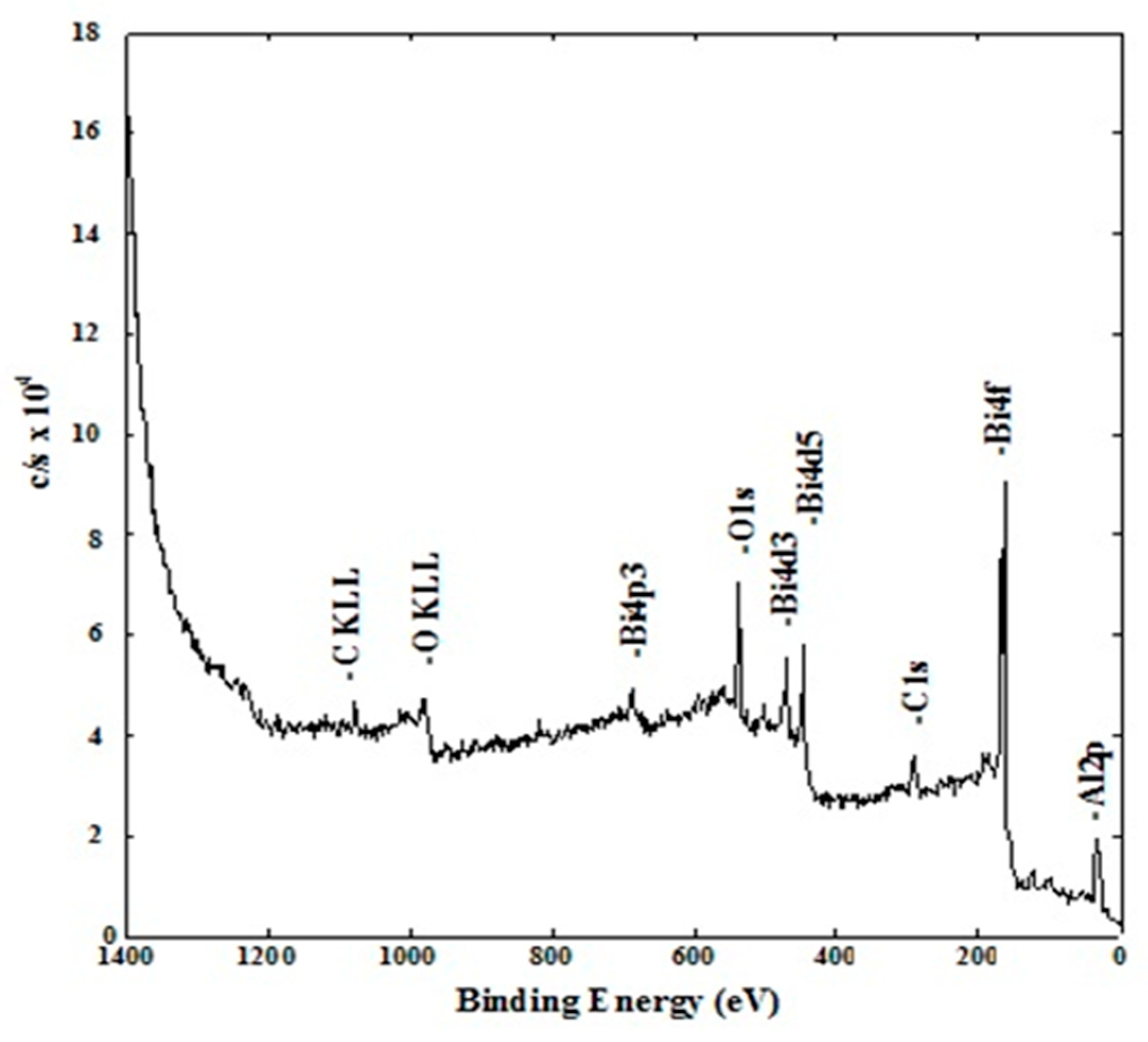
3.3. X-ray Photoelectron Spectroscopy, XPS
3.4. High Resolution XPS Analysis Per Element
3.4.1. C1s Region
3.4.2. O1s Region
3.4.3. Al2p Region
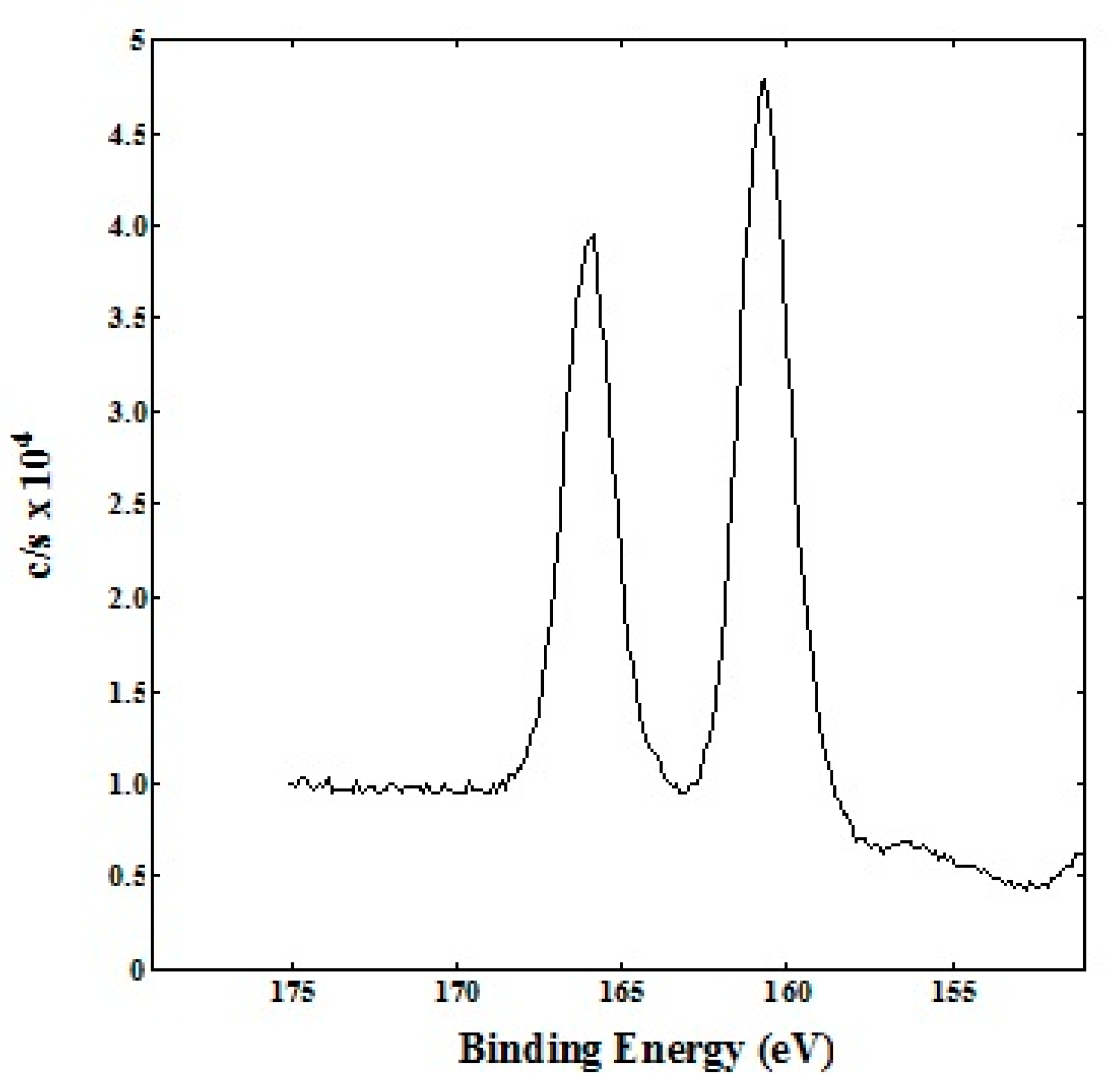
3.4.4. Bi4f Region
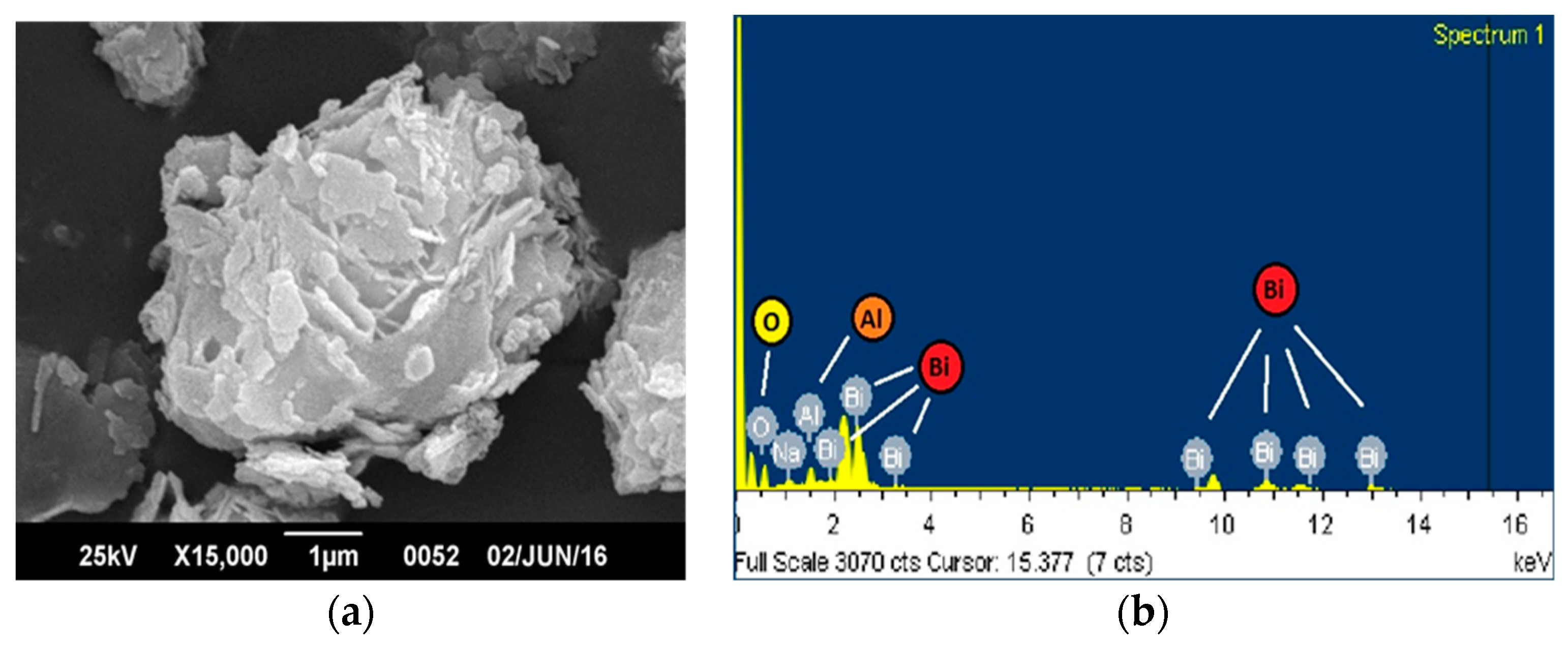
3.5. Scanning Electron Microscopy (SEM) and Energy Dispersive X-ray Spectroscopy (EDX)
4. Conclusions
Author Contributions
Funding
Acknowledgments
Conflicts of Interest
References
- Huang, H.; Tian, N.; Jin, S.; Zhang, Y.; Wang, S. Syntheses, characterization and nonlinear optical properties of a bismuth subcarbonate Bi2O2CO3. Solid State Sci. 2014, 30, 1–5. [Google Scholar] [CrossRef]
- Liu, Y.; Wang, Z.; Huang, B.; Yang, K.; Zhang, X.; Qin, X.; Dai, Y. Preparation, electronic structure, and photocatalytic properties of Bi2O2CO3 nanosheet. Appl. Surf. Sci. 2010, 257, 172–175. [Google Scholar] [CrossRef]
- Zhao, T.; Zai, J.; Xu, M.; Zou, Q.; Su, Y.; Wang, K.; Qian, X. Hierarchical Bi2O2CO3 microspheres with improved visible-light-driven photocatalytic activity. CrystEngComm 2011, 13, 4010–4017. [Google Scholar] [CrossRef]
- Chen, R.; Cheng, G.; So, M.H.; Wu, J.; Lu, Z.; Che, C.M.; Sun, H.; Wang, C.; Zhao, Z.; Luo, B.; et al. Hierarchical Bi2O2CO3 microspheres with improved visible-light-driven photocatalytic activity. CrystEngComm 2010, 13, 1–10. [Google Scholar]
- Zhao, H.; Tang, J.; Lai, Q.; Cheng, G.; Liu, Y.; Chen, R. Enhanced visible light photocatalytic performance of Sb-doped (BiO)2CO3 nanoplates. Catal. Commun. 2015, 58, 190–194. [Google Scholar] [CrossRef]
- Wang, C.; Zhao, Z.; Luo, B.; Fu, M.; Dong, F. Tuning the Morphological Structure and Photocatalytic Activity of Nitrogen-Doped (BiO)2CO3 by the Hydrothermal Temperature. J. Nanomater. 2014, 2014, 1–10. [Google Scholar]
- Tang, J.; Cheng, G.; Zhou, H.; Yang, H.; Lu, Z.; Chen, R. Shape-Dependent Photocatalytic Activities of Bismuth Subcarbonate Nanostructures. J. Nanosci. Nanotechnol. 2012, 12, 4028–4034. [Google Scholar] [CrossRef]
- Chen, R.; Cheng, G.; So, M.H.; Wu, J.; Lu, Z.; Che, C.M.; Sun, H. Bismuth subcarbonate nanoparticles fabricated by water-in-oil microemulsion-assisted hydrothermal process exhibit anti-Helicobacter pylori properties. Mater. Res. Bull. 2010, 45, 654–658. [Google Scholar] [CrossRef]
- Chen, R.; So, M.H.; Yang, J.; Deng, F.; Che, C.M.; Sun, H. Fabrication of bismuth subcarbonate nanotube arrays from bismuth citrate. Chem. Commun. 2006, 21, 2265–2267. [Google Scholar] [CrossRef]
- Cheng, G.; Yang, H.; Rong, K.; Lu, Z.; Yu, X.; Chen, R. Shape-controlled solvothermal synthesis of bismuth subcarbonate nanomaterials. J. Solid State Chem. 2010, 183, 1878–1883. [Google Scholar] [CrossRef]
- Selvamani, T.; Asiri, A.M.; Al-Youbi, A.O.; Anandan, S. Emergent Synthesis of Bismuth Subcarbonate Nanomaterials with Various Morphologies towards Photocatalytic Activities—An Overview. Mater. Sci. Forum 2013, 764, 169–193. [Google Scholar] [CrossRef]
- Dong, F.; Lee, S.C.; Wu, Z.; Huang, Y.; Fu, M.; Ho, W.K.; Zou, S.; Wang, B. Rose-like monodisperse bismuth subcarbonate hierarchical hollow microspheres: One-pot template-free fabrication and excellent visible light photocatalytic activity and photochemical stability for NO removal in indoor air. J. Hazard. Mater. 2011, 195, 346–354. [Google Scholar] [CrossRef] [PubMed]
- Chen, L.; Huang, R.; Yin, S.F.; Luo, S.L.; Au, C.T. Flower-like Bi2O2CO3: Facile synthesis and their photocatalytic application in treatment of dye-containing wastewater. Chem. Eng. J. 2012, 193, 123–130. [Google Scholar] [CrossRef]
- Cao, X.F.; Zhang, L.; Chen, X.T.; Xue, Z.L. Persimmon-like (BiO)2CO3 microstructures: Hydrothermal preparation, photocatalytic properties and their conversion into Bi2S3. CrystEngComm 2011, 13, 1939–1945. [Google Scholar] [CrossRef]
- Cheng, G.; Wu, J.; Xiao, F.; Yu, H.; Lu, Z.; Yu, X.; Chen, R. Synthesis of bismuth micro- and nanospheres by a simple refluxing method. Mater. Lett. 2009, 63, 2239–2242. [Google Scholar] [CrossRef]
- Nan, Z.; Chen, X.; Yang, Q.; Wang, X.; Shi, Z.; Hou, W. Structure transition from aragonite to vaterite and calcite by the assistance of SDBS. J. Colloid Interface Sci. 2008, 325, 331–336. [Google Scholar] [CrossRef] [PubMed]
- Ni, Z.; Sun, Y.; Zhang, Y.; Dong, F. Fabrication, modification and application of (BiO)2CO3 -based photocatalysts: A review. Appl. Surf. Sci. 2016, 365, 314–335. [Google Scholar] [CrossRef]
- Madhusudan, P.; Zhang, J.; Cheng, B.; Liu, G. Photocatalytic degradation of organic dyes with hierarchical Bi2O2CO3 microstructures under visible-light. CrystEngComm 2013, 15, 231–240. [Google Scholar] [CrossRef]
- Dong, F.; Sun, Y.; Fu, M.; Ho, W.K.; Lee, S.C.; Wu, Z. Novel in situ N-doped (BiO)2CO3 hierarchical microspheres self-assembled by nanosheets as efficient and durable visible light driven photocatalyst. Langmuir 2012, 28, 766–773. [Google Scholar] [CrossRef]
- Dong, F.; Sun, Y.; Ho, W.K.; Wu, Z. Controlled synthesis, growth mechanism and highly efficient solar photocatalysis of nitrogen-doped bismuth subcarbonate hierarchical nanosheets architectures. Dalton Trans. 2012, 41, 8270–8284. [Google Scholar] [CrossRef]
- Xiong, T.; Huang, H.; Sun, Y.; Dong, F. In situ synthesis of a C-doped (BiO)2CO3 hierarchical self-assembly effectively promoting visible light photocatalysis. J. Mater. Chem. A 2015, 3, 6118–6127. [Google Scholar] [CrossRef]
- Li, S.; Zhang, J.; Chen, X.; Feng, Z.; Li, C. Effect of surface loading and bulk doping of La3+ on the thermal stability and photocatalytic activity of Bi2O2CO3. Mater. Res. Bull. 2017, 94, 127–133. [Google Scholar] [CrossRef]
- Li, J.; Liu, Y.; Zhou, Y.; Liu, S.; Liang, Y.; Luo, T.; Dai, G. Enhanced visible-light photocatalytic activity of Bi2O2CO3 nanoplates by Fe-doping in the degradation of rhodamine B. Mater. Res. Bull. 2018, 107, 438–445. [Google Scholar] [CrossRef]
- Zhang, G.Y.; Wang, J.J.; Shen, X.Q.; Wang, J.J.; Wang, B.Y.; Gao, D.Z. Br-doped Bi2O2CO3 nanosheets with improved electronic structure and accelerated charge migration for outstanding photocatalytic behavior. Appl. Surf. Sci. 2019, 470, 63–73. [Google Scholar] [CrossRef]
- Егорышева, А.В.; Краев, А.С.; Гайтко, О.М.; Герасимова, Т.В.; Голодухина, С.В.; Агафонов, А.В. Электрореологические свойства α-Bi2O3 и Bi2O2CO3. Неорганические Материалы 2019, 55, 374–384. [Google Scholar]
- Liang, L.; Cao, J.; Lin, H.; Guo, X.; Zhang, M.; Chen, S. Enhancing visible light photocatalytic and photocharge separation of (BiO)2CO3 plate via dramatic I- ions doping effect. Mater. Res. Bull. 2016, 80, 329–336. [Google Scholar] [CrossRef]
- Shen, Z.; Han, Q.; Wang, X.; Zhu, J. Green synthesis of I− ions doped (BiO)2CO3 with enhanced visible-light photocatalytic activity. Mater. Lett. 2018, 214, 103–106. [Google Scholar] [CrossRef]
- Álvarez, L.; Rosas, F.; Márquez, E.; Velazco, E.J. Caracterización y estabilización de la fase metaestable del carbonato de calcio obtenida mediante la aplicación de una capa de Bi2O2CO3:Al a temperatura ambiente. Avances Quím. 2017, 12, 13–21. [Google Scholar]
- Tobon-Zapata, G.E.; Etcheverry, S.B.; Baran, E.J. Vibrational spectrum of bismuth subcarbonate. J. Mater. Sci. Lett. 1997, 16, 656–657. [Google Scholar] [CrossRef]
- Cen, W.; Xiong, T.; Tang, C.; Yuan, S.; Dong, F. Effects of morphology and crystallinity on the photocatalytic activity of (BiO)2CO3 nano/microstructures. Ind. Eng. Chem. Res. 2014, 53, 15002–15011. [Google Scholar] [CrossRef]
- Madhusudan, P.; Yu, J.; Wang, W.; Cheng, B.; Liu, G. Facile synthesis of novel hierarchical graphene- Bi2O2CO3 composites with enhanced photocatalytic performance under visible light. Dalton Trans. 2012, 41, 14345–14353. [Google Scholar] [CrossRef] [PubMed]
- Mohamed, M.; Al, M.; Moamen, S. A novel method for preparation of bismuth(III) carbonate basic (BiO)2CO3.3H2O upon the reaction of urea with various bismuth(III) salts at high temperature. Adv. Appl. Sci. Res. 2012, 3, 183–187. [Google Scholar]
- Shamaila, S.; Sajjad, A.K.L.; Chen, F.; Zhang, J. Study on highly visible light active Bi2O3 loaded ordered mesoporous titania. Appl. Catal. B Environ. 2010, 94, 272–280. [Google Scholar] [CrossRef]


| Frequency (cm−1) | Assignment |
|---|---|
| 3292 | Stretching vibration of H2O |
| 1477, 1385 | Anti-symmetric vibration v3 of CO32− |
| 1066 | Symmetric stretching vibration v1 of CO32− |
| 846 | Out-of-plane bending v2 of CO32− |
| 690 | In-plane deformation v4 of CO32− |
| 546 | Stretching vibration of Bi=O |
| Element | % Atomic |
|---|---|
| C1s | 30.44 |
| O1s | 50.21 |
| Al2p | 6.49 |
| Bi4f | 12.85 |
| Bond Energy for C1s (eV) | % by Type of C1s | Assignation |
|---|---|---|
| 285.0 | 10.72 | C-C, C-H Carbon from environmental contamination. |
| 287.0 | 50.52 | Some kind of C different to C-C, C-H |
| 290.2 | 38.76 | CO32− |
| Bond Energy for O1s (eV) | % by Type of O1s | Assignation |
|---|---|---|
| 531.8 | 38.66 | AlO(OH) |
| 533.2 | 41.22 | CO32– |
| 534.8 | 20.12 | H2O |
| Bond Energy for Al2p (eV) | % by Type of Al2p | Assignation |
|---|---|---|
| 75.8 | 92.11 | AlO(OH) |
| 77.3 | 07.89 | Al2O3 |
| Bond Energy for Bi4f (eV) | %Bi4f | Assignation |
|---|---|---|
| 160.2 | 12.85 | Bond energy greater than that of Bi2O3 (159.3 eV). It can be assigned to a chemical environment with oxygen and aluminum. This means that this chemical environment for bismuth is of greater ionic character. |
© 2019 by the authors. Licensee MDPI, Basel, Switzerland. This article is an open access article distributed under the terms and conditions of the Creative Commons Attribution (CC BY) license (http://creativecommons.org/licenses/by/4.0/).
Share and Cite
Álvarez, L.; Rojas de Gascue, B.; Tremont, R.J.; Márquez, E.; Velazco, E.J. Synthesis and Characterization of a New Aluminum-Doped Bismuth Subcarbonate. Crystals 2019, 9, 466. https://doi.org/10.3390/cryst9090466
Álvarez L, Rojas de Gascue B, Tremont RJ, Márquez E, Velazco EJ. Synthesis and Characterization of a New Aluminum-Doped Bismuth Subcarbonate. Crystals. 2019; 9(9):466. https://doi.org/10.3390/cryst9090466
Chicago/Turabian StyleÁlvarez, Loisangela, Blanca Rojas de Gascue, Rolando J. Tremont, Edgar Márquez, and Euclides J. Velazco. 2019. "Synthesis and Characterization of a New Aluminum-Doped Bismuth Subcarbonate" Crystals 9, no. 9: 466. https://doi.org/10.3390/cryst9090466