Article

A Novel Na(I) Coordination Complex with s-Triazine Pincer Ligand: Synthesis, X-ray Structure, Hirshfeld Analysis, and Antimicrobial Activity

1
Department of Chemistry, Faculty of Science, Alexandria University, Ibrahimia, P.O. Box 426, Alexandria 21321, Egypt
2
Department of Chemistry, University of Jyväskylä, P.O. Box 35, FI-40014 Jyväskylä, Finland
3
Department of Pharmaceutical Medicinal Chemistry, Faculty of Pharmacy (Girls), Al-Azhar University, Cairo 11754, Egypt
4
Department of Microbiology and Immunology, Faculty of Pharmacy (Girls), Al-Azhar University, Cairo 11754, Egypt
5
Department of Chemistry and Chemical Engineering, Chalmers University of Technology, SE-41296 Gothenburg, Sweden
6
Department of Chemistry, College of Science, King Saud University, P.O. Box 2455, Riyadh 11451, Saudi Arabia
*
Authors to whom correspondence should be addressed.
Crystals 2023, 13(6), 890; https://doi.org/10.3390/cryst13060890
Submission received: 5 May 2023 / Revised: 19 May 2023 / Accepted: 25 May 2023 / Published: 29 May 2023

Abstract

:
The pincer ligand 2,4-bis(3,5-dimethyl-1H-pyrazol-1-yl)-6-methoxy-1,3,5-triazine (bpmt) was used to synthesize the novel [Na(bpmt)2][AuCl4] complex through the self-assembly method. In this complex, the Na(I) ion is hexa-coordinated with two tridentate N-pincer ligands (bpmt). The two bpmt ligand units are meridionally coordinated to Na(I) via one short Na-N(s-triazine) and two slightly longer Na-N(pyrazole) bonds, resulting in a distorted octahedral geometry around the Na(I) ion. In the coordinated bpmt ligand, the s-triazine core is not found to be coplanar with the two pyrazole moieties. Additionally, the two bpmt units are strongly twisted from one another by 64.94°. Based on Hirshfeld investigations, the H···H (53.4%) interactions have a significant role in controlling the supramolecular arrangement of the [Na(bpmt)2][AuCl4] complex. In addition, the Cl···H (12.2%), C···H (11.5%), N···H (9.3%), and O···H (4.9%) interactions are significant. Antimicrobial investigations revealed that the [Na(bpmt)2][AuCl4] complex has promising antibacterial and antifungal activities. The [Na(bpmt)2][AuCl4] complex showed enhanced antibacterial activity for the majority of the studied gram-positive and gram-negative bacteria compared to the free bpmt (MIC = 62.5–125 µg/mL vs. MIC = 62.5–500 µg/mL, respectively) and Amoxicillin (MIC > 500 µg/mL) as a positive control. Additionally, the [Na(bpmt)2][AuCl4] complex had better antifungal efficacy (MIC = 125 µg/mL) against C. albicans compared to bpmt (MIC = 500 µg/mL).

1. Introduction

One of the most important pharmaceutical concerns in medicine is antibiotic-resistant microorganisms [1,2]. Efforts to address the serious problems arising from microbial infections include the design of novel antimicrobial agents with minimal side effects and enhanced chemical and pharmacological characteristics [3,4]. Generally, metal ions play a crucial role in biological processes due to their interactions with numerous biomolecules, including amino acids, enzymes, and serum albumin [5,6,7]. Thus, the self-assembly of individual molecules via non-covalent interactions, particularly metal–organic complexes, is one of the most promising research areas in this field [8,9,10,11,12]. The need to create molecular structures that can mimic the naturally existing molecules has driven researchers’ interest in the coordination chemistry of alkali metal ions in recent decades. The best example of alkali metal ions is Na(I), which is necessary for biological systems [13] and plays a crucial role in cancer therapy as it can make tumor cells more susceptible to death [14,15,16,17,18]. Recently, many Na(I) complexes have been reported and analyzed to determine their supramolecular structures [19,20,21,22]. It has been found that Na(I) can form homodinuclear, heterodinuclear, and trimetallic complexes [23,24,25,26]. Additionally, Na(I) can create coordination complexes with unexpected coordination geometries and various coordination numbers (CN). The coordination geometry of Na(I) can be tetrahedral (CN = 4), trigonal bipyramidal (CN = 5), octahedral (CN = 6), pentagonal pyramidal (CN = 7), square antiprism (CN = 8), dodecahedral (CN = 8), and trigonal-dodecahedral (CN = 8) [27,28,29,30,31].
In the literature, sodium complexes with octahedral coordination geometry are well known. For example, a 2D polymeric sodium complex with bis-tetrazole type ligand was reported by Sobral et. al. In this complex, the sodium metal ion has a distorted octahedral environment with a NaNO5 coordination sphere [32]. Another example is the mononuclear octahedral [Prolinate2-Na(MeOH)4]H+ complex, which has a NaO6 coordination sphere [33]. In this case, the sodium ion is coordinated with four methanol molecules and two prolinate anions as monodentate ligands. Furthermore, a new distorted octahedral sodium complex, [Na(H2O)5(DMF)]·(L) (L=1,5-naphthalenedisulfonate), was obtained, and its single crystal structure confirmed the NaO6 coordination sphere via one DMF and five H2O molecules [22]. A hepta-coordinated sodium complex was reported by Gourdon’s research group with dipicolinic acid as a ligand, and its structure was confirmed using single crystal X-ray structure, also showing a NaNO6 coordination environment [34]. To the best of our knowledge, there are no well-known pincer sodium complexes with s-triazine N-donor ligands and confirmed structures. However, Beer et al. reported a new sodium complex with bis-triazacyclononane tris-pyridyl N9-azacryptand as ligand [35]. This is considered the only pincer sodium complex that has been structurally characterized using single crystal X-ray crystallography (SCXRD) with nitrogen macrocyclic ligands, and it exhibits a NaN9 coordination environment. The most frequent ligands that form coordination compounds with alkali metal ions are Schiff bases, tridentate ligands (such as o-vanillin and benzhydryl ligands), crown ethers, etc. [23,36,37,38]. Generally, multidentate ligands play a significant role in coordination chemistry as they can form extra-stable metal complexes due to the chelate effect. Among these multidentate ligands, symmetrical triazine (s-triazine) derivatives are promising due to their diverse biological effects and powerful pharmacological properties [39,40,41,42,43]. Moreover, substances containing pyrazoles are considered effective biological agents [44,45,46]. Among s-triazine derivatives, the 2,4-bis(3,5-dimethyl-1H-pyrazol-1-yl)-6-methoxy-1,3,5-triazine (bpmt) ligand has been extensively studied by our research team for many reasons [4,47,48,49,50,51,52,53,54,55,56,57]. The bpmt ligand can be easily prepared with a high yield, and it is classified as a tridentate N-pincer ligand with different coordination behaviors. In addition, its metal complexes exhibit remarkable biological activities compared to the free bpmt ligand [4,48,51,52,53,55,56,58]. The pincer complexes of bpmt are stable and can be simply synthesized via the self-assembly of the functional ligand (bpmt) with a metal salt [54,57].
The goal of the current work was to synthesize a new pincer complex of the bpmt ligand through self-assembly with Na[AuCl4].2H2O. The molecular and supramolecular structures of the complex were explored using single crystal X-ray diffraction in combination with Hirshfeld calculations. In addition, the antimicrobial activity of the complex was evaluated.

2. Materials and Methods

2.1. Chemicals and Instrumentations

All chemicals were obtained from Sigma-Aldrich Company. Perkin-Elmer 2400 equipment (PerkinElmer, Waltham, MA, USA) was used to perform the elemental analyses (CHN). The FTIR spectra in KBr pellets were recorded in the range 4000–400 cm−1 using a Bruker Tensor 37 instrument (Waltham, MA, USA). NMR spectra were recorded in DMSO-d6 as a solvent using a JEOL-400 MHz spectrometer (JEOL, Ltd., Tokyo, Japan) at 298 K. Na content was determined using a Shimadzu atomic absorption spectrophotometer (AA-7000 series, Shimadzu, Ltd., Tokyo, Japan).

2.2. Syntheses

The bpmt pincer ligand was prepared using the procedure previously reported by our research group [57,59].

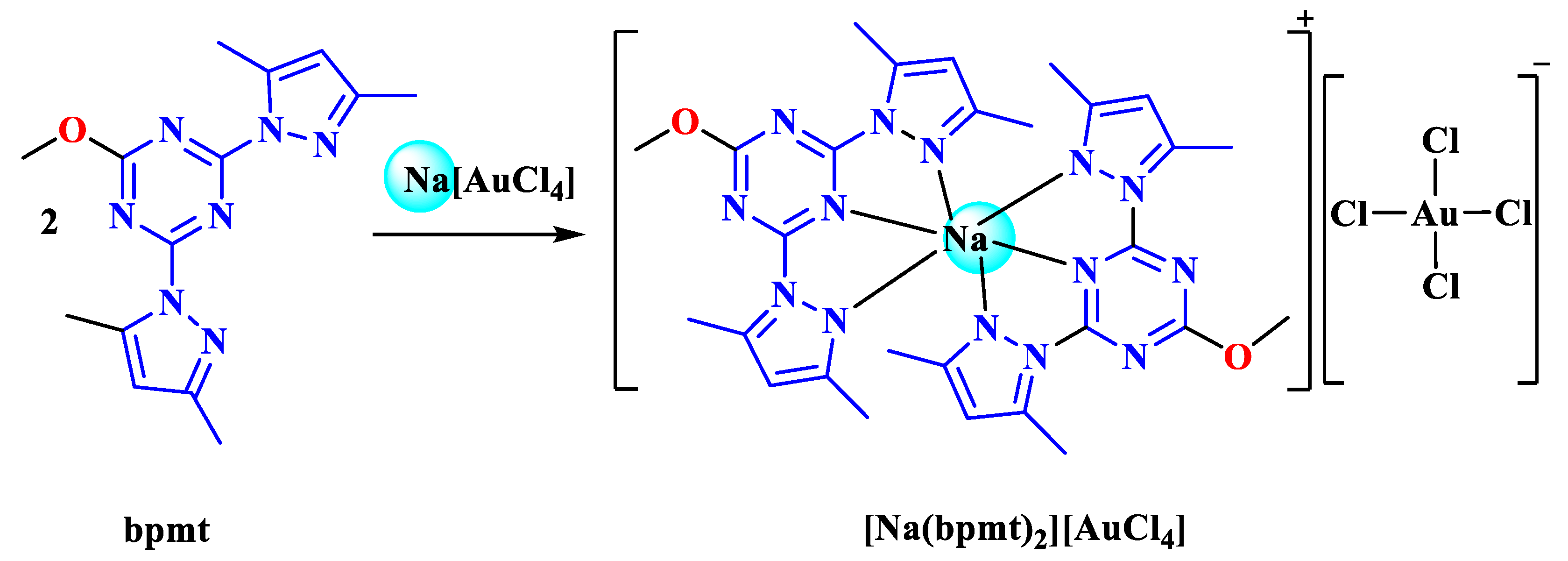
Synthesis of the [Na(bpmt)2][AuCl4] Complex

A 10 mL ethanolic solution of Na[AuCl4]·2H2O (39.8 mg, 0.1 mmol) was mixed with 10 mL of a hot ethanolic solution of bpmt (59.8 mg, 0.2 mmol). The resulting clear mixture was left at room-temperature for slow evaporation. After three days, yellow crystals were formed, which were collected through filtration and found suitable for single crystal X-ray diffraction measurements.
The yield of the reaction was 89%. The analytical calculation for C28H34AuCl4N14NaO2 yielded the following percentages: C, 35.02; H, 3.57; N, 20.42; Na, 22.99%. Upon analysis, the found percentages were as follows: C, 35.19; H, 3.45; N, 20.28%; Na, 22.78%.
The FTIR spectra of [Na(bpmt)2][AuCl4] exhibited peaks at the following wavenumbers (cm−1): 3421, 3107, 2956, 2930, 1601, 1548, 1417, 1367, 1221, 1123, 1046, 971, 813, and 750 (Figure S1, Supplementary Data). The FTIR spectra of the ligand (bpmt) displayed peaks at the following wavenumbers (cm−1): 3392, 3313, 2986, 2930, 1597, 1558, 1408, 1373, 1224, 1128, 1041, 973, 812, and 751 (Figure S2, Supplementary Data). The 1H NMR spectrum of [Na(bpmt)2][AuCl4] (500 MHz, DMSO-d6) revealed the following chemical shifts: δH 6.23 (s, 2H, Ar-H), 4.02 (s, 3H, OCH3), 2.64 (s, 6H, 2CH3), 2.19 (s, 6H, 2CH3) ppm. The 13C NMR spectrum (125 MHz, DMSO-d6) displayed the following chemical shifts: δC 172.4, 164.8, 152.6, 144.7, 112.2, 56.3, 15.9, 14.1 ppm (Figure S3, Supplementary Data).
The 1H NMR spectrum of the ligand (bpmt) (500 MHz, DMSO-d6) exhibited the following chemical shifts: δH 6.22 (s, 2H, Ar-H), 4.01 (s, 3H, OCH3), 2.64 (s, 6H, 2CH3), and 2.16 (s, 6H, 2CH3) ppm. The 13C NMR spectrum (125 MHz, DMSO-d6) displayed the following chemical shifts: δC 172.3, 164.8, 152.4, 144.6, 112.2, 56.3, 15.9, and 14.1 ppm (Figure S4, Supplementary Data).

2.3. Crystal Structure Determination

The crystal of [Na(bpmt)2][AuCl4] was immersed in cryo-oil, mounted in a loop, and measured at a temperature of 170 K. The X-ray diffraction data were collected using a Bruker Kappa Apex II diffractometer with Mo Kα radiation. The Denzo-Scalepack [60] software package was used for cell refinement and data reduction. A multi-scan absorption correction based on equivalent reflections (SADABS [61]) was applied to the intensities before the structure solution. The structure was solved using the intrinsic phasing method with SHELXT [62] software. Structural refinement was carried out using SHELXL [63] software with the SHELXLE [64] graphical user interface. All the hydrogen atoms were positioned geometrically and constrained to ride on their parent atoms, with C-H = 0.95–0.98 Å and Uiso = 1.2–1.5 Ueq (parent atom). The crystal data and structure refinement details are shown in Table 1.

2.4. Hirshfeld Surface Analysis

The topology analyses were performed using the Crystal Explorer 17.5 program [65] to generate 2D fingerprint plots and Hirshfeld surfaces [66].

2.5. Antimicrobial Studies

The antimicrobial activity [67] of the investigated ligand and its Na(I) complex were determined, as described in Method S1 (Supplementary Data).

3. Results and Discussion

3.1. Synthesis and Characterizations

The novel [Na(bpmt)2][AuCl4] complex was synthesized using a self-assembly technique by reacting Na[AuCl4].2H2O with the bpmt ligand (Scheme 1). Several spectroscopic techniques were used to confirm the structure of the resulting complex. Figures S1 and S2 (Supplementary Data) show the FTIR spectra of the [Na(bpmt)2][AuCl4] complex compared to the free bpmt ligand. The FTIR spectra of the [Na(bpmt)2][AuCl4] complex revealed the main vibrational characteristics of the functional organic ligand (bpmt) with slight variations. The stretching modes υ(C=N) and υ(C=C) in the free bpmt appeared at 1597 and 1558 cm−1, respectively. In the [Na(bpmt)2][AuCl4] complex, these stretching modes were observed at 1601 and 1548 cm−1, respectively. It was observed that the coordination of Na(I) with bpmt resulted in a blue shift for the υ(C=N) mode and a red shift in the υ(C=C) mode [55,57]. Thus, the complexation had an impact on both vibrational bands compared with the free ligand. Moreover, single crystal X-ray diffraction combined with Hirshfeld surface analysis was used to specifically determine the molecular and supramolecular structures of the [Na(bpmt)2][AuCl4] complex.

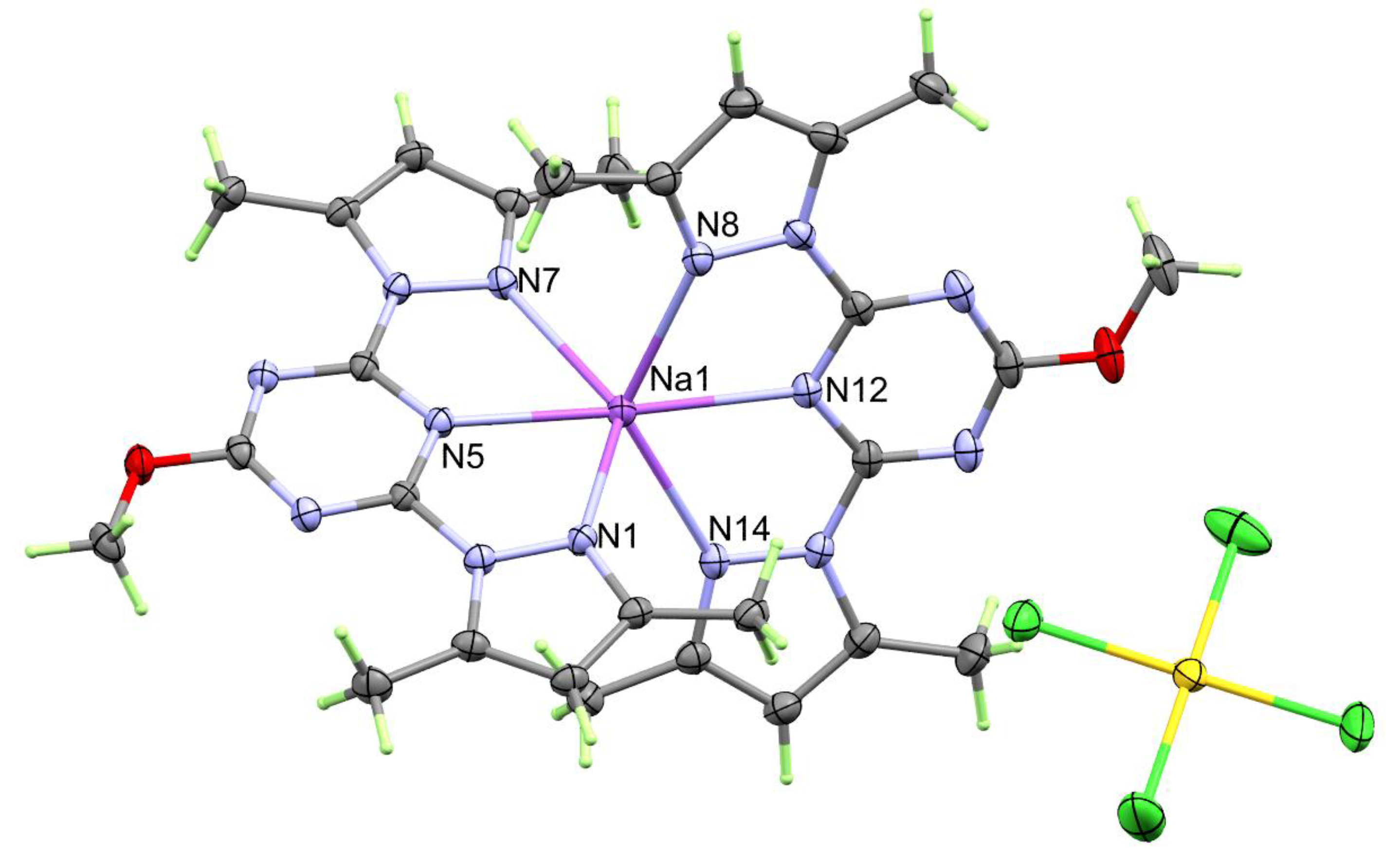
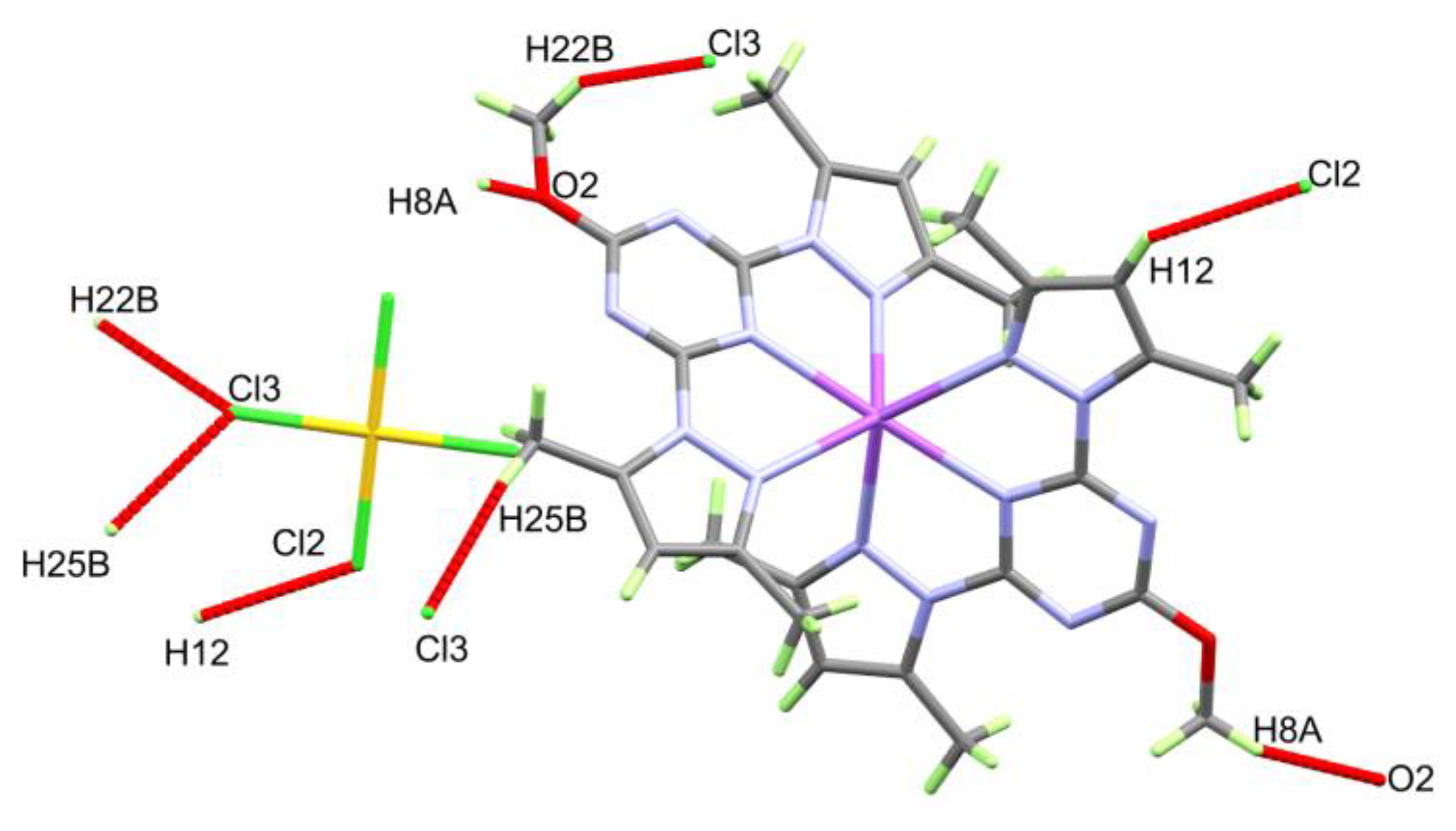
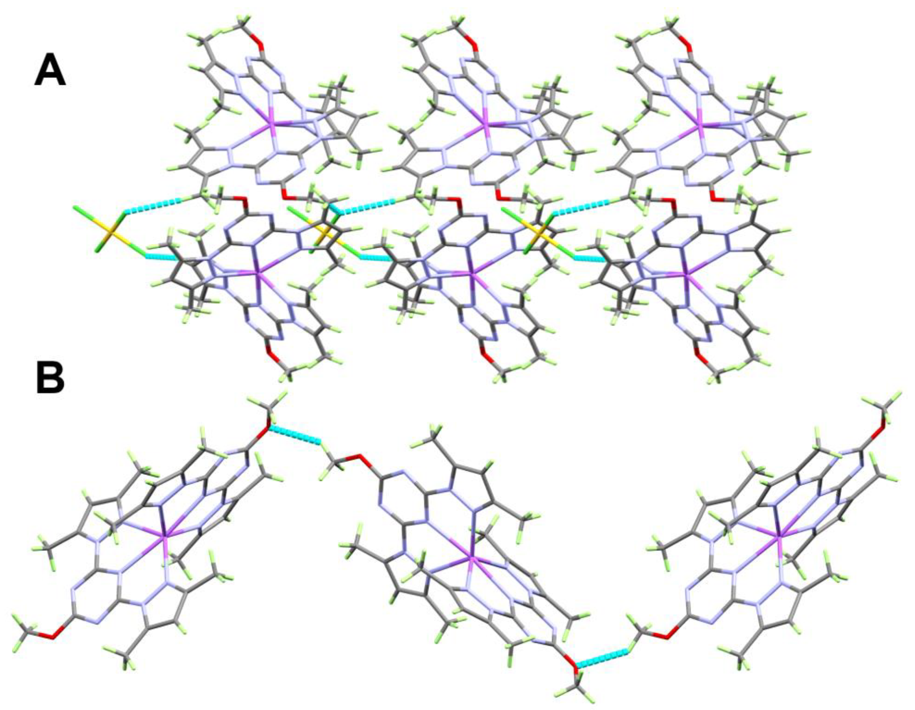
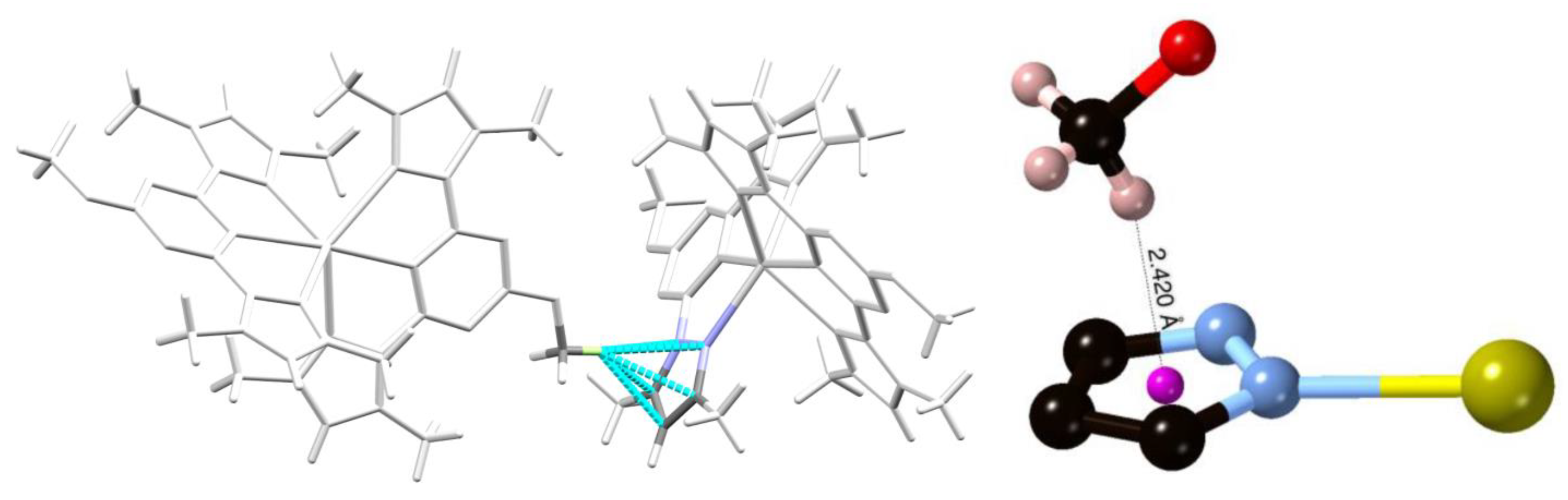
3.2. Crystal Structure Description

The X-ray structure of the [Na(bpmt)2][AuCl4] complex unequivocally confirmed the coordination between Na(I) and the bpmt ligand. The asymmetric unit of the resulting complex is shown in Figure 1, consisting of one formula unit of the cationic complex [Na(bpmt)2][AuCl4]. In this complex, the Na(I) ion coordinates with two units of the pincer ligand bpmt via the N-atoms of the two pyrazolyl moieties and one N-atom from the s-triazine core, while the tetrachloroaurate serves as the counter anion. The Na-N bond distances with the s-triazine core were found to be 2.443(3) Å (Na1-N5) and 2.429(3) Å (Na1-N12). It is worth noting that the two s-triazine cores of the two bpmt ligand units were found to be coordinated with the Na(I) ion in a trans configuration. The N5-Na-N12 angle was found to be 171.65(10)°, which was slightly deviated from the ideal value of 180°. Hence, the two bpmt ligand units were meridionally coordinated to the Na(I) ion. The Na-N bonds with the pyrazolyl moieties are generally slightly longer (2.438(3)–2.538(3) Å) than the Na-N(s-triazine) bond. The bite angles of the bpmt pincer chelate were in the range of 63.96(9)–64.98(9)°. In addition, the trans N(pyrazole)-Na-N(pyrazole) angles significantly deviated from the ideal value of 180°. The N(1)-Na(1)-N(7) and N(8)-Na(1)-N(14) angles were determined to be 128.40(9) and 128.18(9)°, respectively. The Na-N and N-Na-N angles are listed in Table 2.
It is worth noting that the s-triazine core and the two pyrazole moieties were not coplanar in the two bpmt ligand units. The N7N6C10C12C13 and N1N2C4C3C1 mean planes of the two pyrazole moieties formed angles of 5.32(7) and 4.81(8)°, respectively, with the N5C6N3C7N4C9 s-triazine core mean. The angles between the mean plane of the other s-triazine core N12C23N11C21N10C20 and the two pyrazole rings N13C24C26C27N14 and N9N8C15C17C18 were 11.18(9) and 8.42(8)°, respectively. Hence, the two pyrazole arms were twisted from the s-triazine core in both ligand units. In addition, the two bpmt ligand units were significantly twisted from each other, with a twist angle between their mean planes of 64.94(8)°, which significantly deviated from 90°. This deviation was possibly due to the steric preferences of the two bulky ligand units. The molecular packing of the [Na(bpmt)2][AuCl4] complex was controlled by the important C-H···Cl and C-H···O interactions shown in Figure 2. A list of these interactions along with their corresponding geometric parameters are given in Table 3.
The molecular packing of the [Na(bpmt)2][AuCl4] complex was controlled by three important interactions: C12-H12···Cl2, C22-H22B···Cl3, and C25-H25B···Cl3. These interactions exhibited donor to acceptor distances of 3.776(3), 3.750(5), and 3.839(4) Å, respectively. Additionally, the molecular packing was controlled by weak C8-H8A···O2 interaction. The donor C8 to acceptor O2 distance was 3.214(4) Å, while the C8-H8A···O2 angle was 132.5°. A view of the molecular packing is shown in Figure 3.
Furthermore, there were different levels of C-H···π interactions between H22C from the methoxy group and the pyrazole ring C24C26C27N14N13. The distance between H22C and the respective pyrazole atoms were 2.762, 2.673, 2.608, 2.665, and 2.718 Å. The H22C···centroid distance was calculated to be 2.420 Å, indicating significant C-H···π interactions. A view of the possible C-H···π interactions in the [Na(bpmt)2][AuCl4] complex is shown in Figure 4.

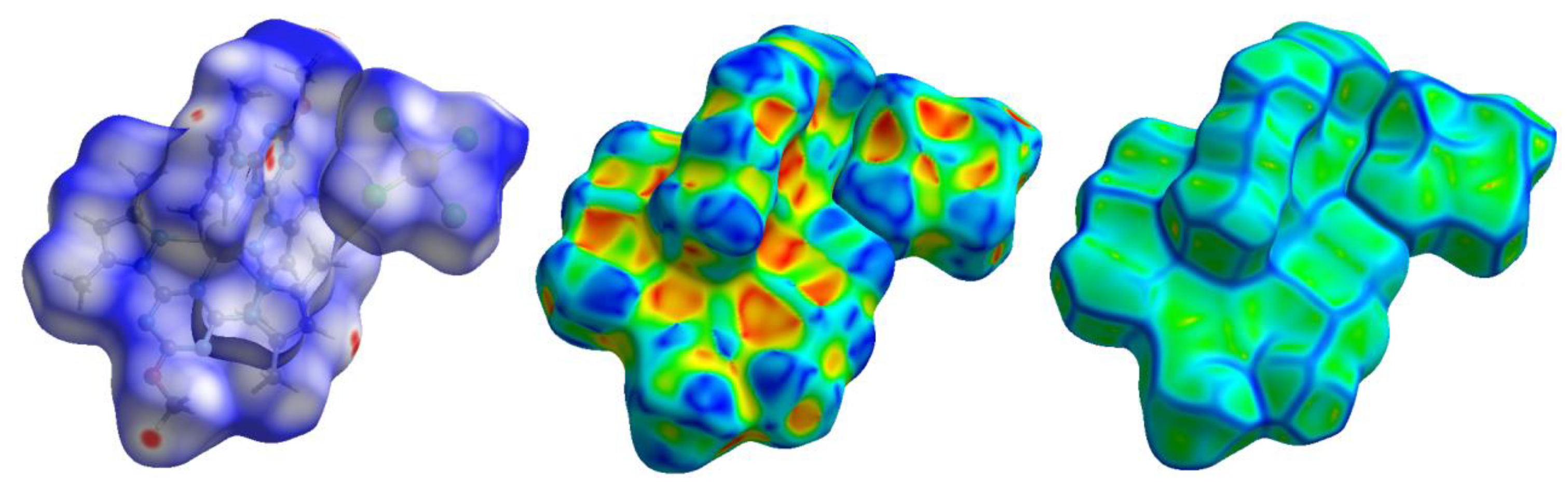
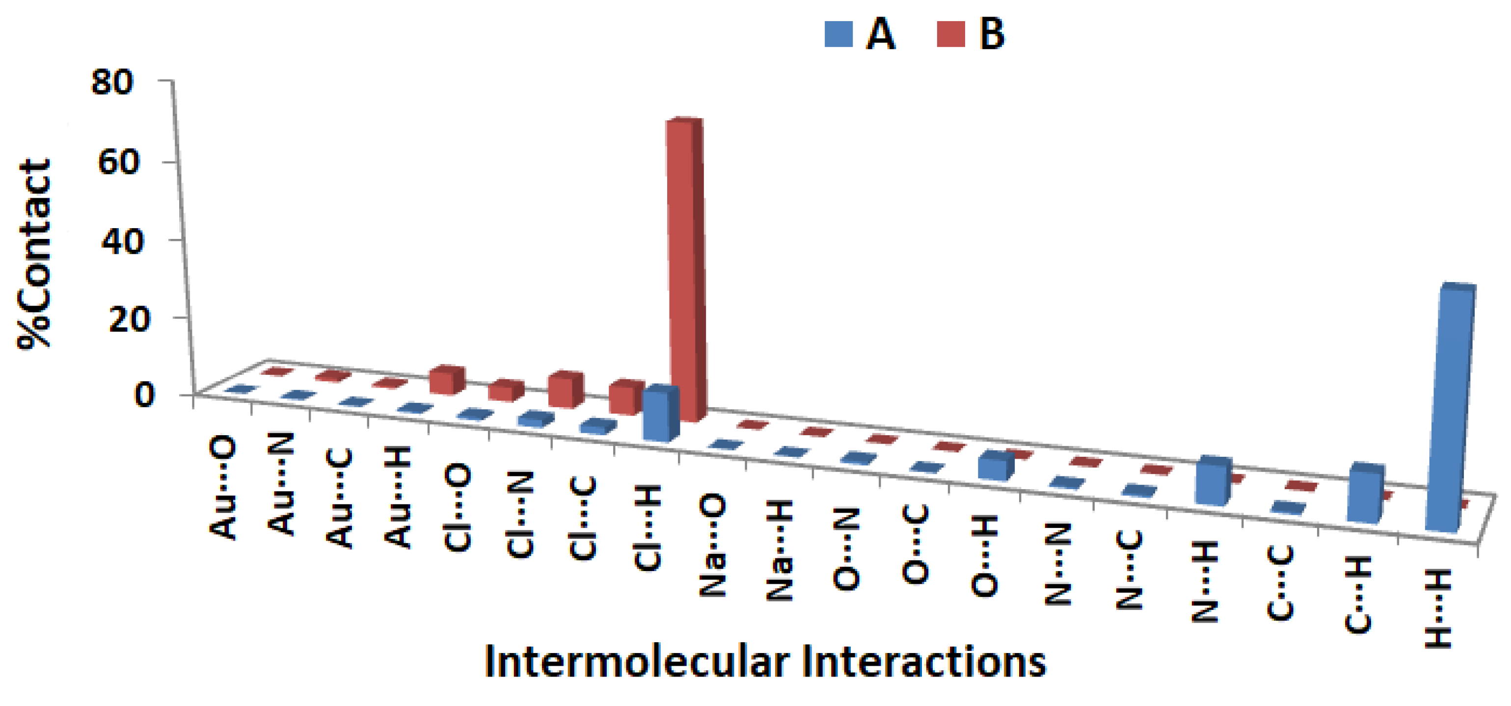
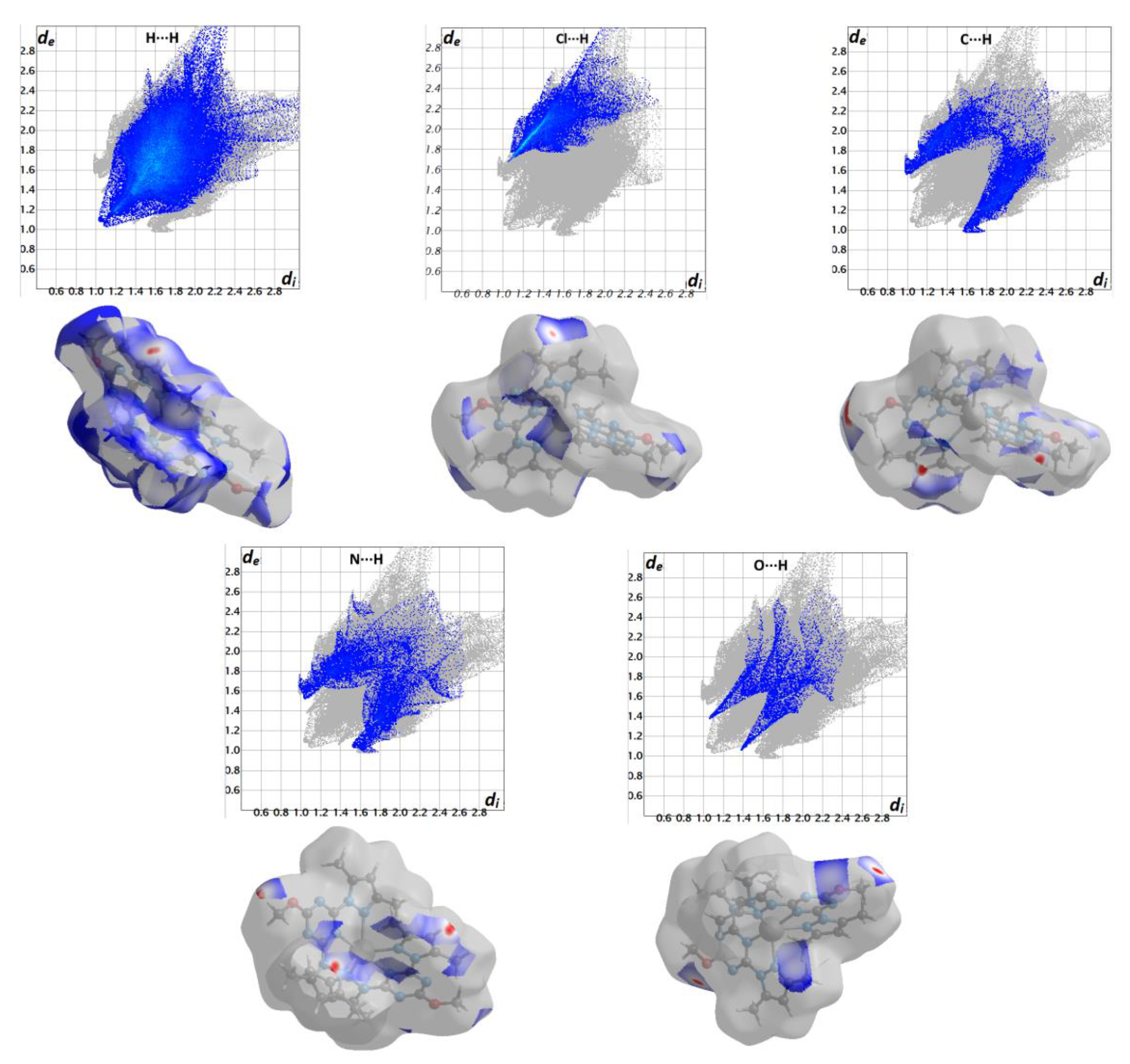
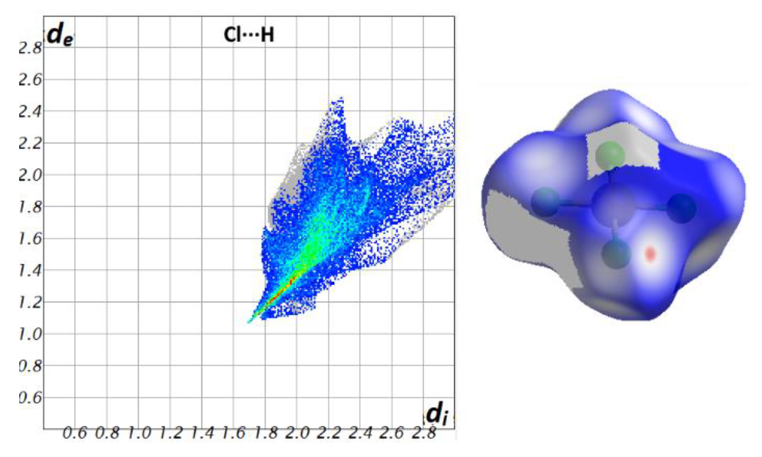
3.3. Hirshfeld Surfaces Analyses

An analysis of the various intermolecular interactions that occurred in the crystal structure of [Na(bpmt)2][AuCl4] was done using Hirshfeld surface calculations (Figure 5).
In this ionic complex, the Hirshfeld analysis was performed on the cationic (A) and anionic (B) units separately. The percentages of all possible contacts in the cationic complex [Na(bpmt)2]+ (A) and anion [AuCl4] (B) are presented in Table 4, and are presented graphically in Figure 6.
The Hirshfeld surfaces of the [Na(bpmt)2][AuCl4] complex, mapped with dnorm, shape index, and curvedness, are shown in Figure 5. Short contacts are apparent as deep red areas, while the white areas are related to contacts near the van der Waals radii sum of the interacting atoms, which were considered the most important in the molecular packing. Conversely, the blue areas represent long-distance interactions. The bright red spots on the dnorm maps represent the areas where H···H, Cl···H, C···H, N···H, and O···H interactions occurred. The decomposition of the fingerprint plot indicates their percentages in the cationic [Na(bpmt)2]+ (A) unit were 53.4, 12.2, 11.5, 9.3, and 4.9%, respectively. These contacts also appeared as sharp spikes in the fingerprint plots (Figure 7). The other intermolecular contacts, which appeared as blue areas in the dnorm maps, were less significant, and their intermolecular contacts were typically weak. The absence of red/blue triangles in the shape index map revealed that no aromatic π-π stacking contacts were present. Furthermore, the curved green regions in the curvedness map confirmed the absence of π-π stacking interactions.
For the anionic unit [AuCl4] (B), the significant contact observed was the Cl···H interaction, while other contacts had less significance. In this unit, the percentage of the Cl···H contacts was 74.1%. This type of non-covalent interaction exhibits the characteristics of short interactions, as clearly depicted in Figure 8.

3.4. Antimicrobial Studies

The antimicrobial activity of the free bpmt and its [Na(bpmt)2][AuCl4] pincer complex against a number of specified pathogenic microorganisms was evaluated. The minimum concentration that showed no observable microbial growth was determined as the minimum inhibitory concentration (MIC), and the results are listed in Table 5. The MIC data demonstrated that the free bpmt had no antibacterial activity against any of the studied strains except for K. pneumonia (50) and A. baumannii (8). On the other hand, the [Na(bpmt)2][AuCl4] complex showed improved antimicrobial activity against most bacterial strains compared to bpmt. The only exceptions were K. pneumonia (50), K. pneumonia (R124), and A. baumannii (8), where both compounds exhibited similar actions. The corresponding MIC values were 62.5, 500, and 125 µg/mL, respectively.
Interestingly, the MIC values of the [Na(bpmt)2][AuCl4] complex were in the range of 62.5–125 µg/mL, indicating higher antibacterial activity against most of the investigated bacteria compared to Amoxicillin (MIC > 500) as a standard antibiotic. It is clear that the [Na(bpmt)2][AuCl4] complex had higher MIC values (125 µg/mL) than Amoxicillin only for S. aureus and E. coli (≤7.8 and 62.5 µg/mL, respectively), and exhibited similar MIC values to Amoxicillin for P. aeruginosa, P. miabilis, and K. pneumonia (R124) (125, 125, and 500 µg/mL, respectively). In contrast, the free ligand bpmt demonstrated good antibacterial activity against only two bacterial strains, K. pneumonia (50) and A. baumannii (8), with MIC values of 62.5 and 125 µg/mL, respectively, compared to Amoxicillin (MIC > 500).
The [Na(bpmt)2][AuCl4] complex exhibited better antifungal activity than the free ligand bpmt. The MIC values were determined to be 125 and 500 µg/mL, respectively. Compared with Nystatin (MIC = 15.6 µg/mL), a standard antifungal medication, the [Na(bpmt)2][AuCl4] complex and its free bpmt ligand had lower antifungal activity against the studied fungus, C. albicans. Thus, the results revealed that the [Na(bpmt)2][AuCl4] complex had significant antibacterial properties against most of the gram-positive and gram-negative bacteria. Additionally, the [Na(bpmt)2][AuCl4] complex showed promising antifungal activity against C. albicans when compared to bpmt.

4. Conclusions

The novel [Na(bpmt)2][AuCl4] complex was obtained as highly crystalline product by reacting the bpmt pincer ligand with Na[AuCl4].2H2O using the self-assembly technique. Its structure was analyzed using single crystal X-ray diffraction, FTIR, and NMR spectroscopic techniques, in addition to elemental analysis and Hirshfeld topology analysis. The mononuclear homoleptic [Na(bpmt)2][AuCl4] complex had hexa-coordinated Na(I) with a distorted octahedral geometry. Hirshfeld studies were used to explore the importance of the H···H, Cl···H, C···H, N···H, and O···H contacts in the supramolecular structure of the [Na(bpmt)2][AuCl4] complex. Antimicrobial evaluation of the studied Na(I) complex revealed promising antibacterial and antifungal activities compared to the free bpmt ligand.

Supplementary Materials

The following supporting information can be downloaded at: https://www.mdpi.com/article/10.3390/cryst13060890/s1, Figure S1: FTIR spectra of the free ligand bpmt; Figure S2: FTIR spectra of the [Na(bpmt)2][AuCl4] complex; Figure S3: 1H and 13C NMR spectra of the free ligand (bpmt); Figure S4: 1H and 13C NMR spectra of [Na(bpmt)2][AuCl4]; Method S1: Antimicrobial activity assay.

Author Contributions

Conceptualization, S.M.S., M.S.A., M.A.M.A.-Y. and L.Ö.; methodology, A.Y.; software, S.M.S., M.H. and L.Ö.; validation, M.M.F.I., N.G.E.M., M.H. and L.Ö.; formal analysis, A.Y., M.M.F.I. and N.G.E.M.; investigation, A.Y., A.E.-F. and M.S.A.; resources, A.E.-F., M.A.M.A.-Y. and A.B.; data curation, L.Ö. and A.Y.; writing—original draft preparation, A.Y., A.E.-F., S.M.S., M.H., M.M.F.I., N.G.E.M. and L.Ö.; writing—review and editing, A.Y., M.A.M.A.-Y., S.M.S., M.S.A., M.M.F.I., N.G.E.M. and L.Ö.; visualization, A.Y., S.M.S. and L.Ö.; supervision, M.A.M.A.-Y., M.S.A., S.M.S. and L.Ö.; project administration, M.A.M.A.-Y. and A.B.; funding acquisition, A.B. All authors have read and agreed to the published version of the manuscript.

Funding

The authors would like to extend their sincere appreciation to the Researchers Supporting Project (RSP2023R64), King Saud University, Riyadh, Saudi Arabia.

Data Availability Statement

Not applicable.

Acknowledgments

The authors would like to extend their sincere appreciation to the Researchers Supporting Project (RSP2023R64), King Saud University, Riyadh, Saudi Arabia.

Conflicts of Interest

The authors declare no conflict of interest.

References

  1. Chu, D.T.; Plattner, J.J.; Katz, L. New directions in antibacterial research. J. Med. Chem. 1996, 39, 3853–3874. [Google Scholar] [CrossRef] [PubMed]
  2. Evans, A.; Kavanagh, K.A. Evaluation of metal-based antimicrobial compounds for the treatment of bacterial pathogens. Int. J. Med. Microbiol. 2021, 70, 001363. [Google Scholar] [CrossRef] [PubMed]
  3. Dalla Via, L.; Marciani Magno, S.; Gia, O.; Marini, A.M.; Da Settimo, F.; Salerno, S.; La Motta, C.; Simorini, F.; Taliani, S.; Lavecchia, A. Benzothiopyranoindole-based antiproliferative agents: Synthesis, cytotoxicity, nucleic acids interaction, and topoisomerases inhibition properties. J. Med. Chem. 2009, 52, 5429–5441. [Google Scholar] [CrossRef] [PubMed]
  4. Soliman, S.M.; El-Faham, A.; El Silk, S.E. Novel one-dimensional polymeric Cu(II) complexes via Cu(II)-assisted hydrolysis of the 2,4-bis(3,5-dimethyl-1H-pyrazol-1-yl)-6-methoxy-1,3,5-triazine pincer ligand: Synthesis, structure, and antimicrobial activities. Appl. Organomet. Chem. 2020, 34, e5941. [Google Scholar] [CrossRef]
  5. Fujimoto, S.; Yasui, H.; Yoshikawa, Y. Development of a novel antidiabetic zinc complex with an organoselenium ligand at the lowest dosage in KK-Ay mice. J. Inorg. Biochem. 2013, 121, 10–15. [Google Scholar] [CrossRef]
  6. Huang, Q.; Pan, Z.; Wang, P.; Chen, Z.; Zhang, X.; Xu, H. Zinc(II) and copper(II) complexes of β-substituted hydroxylporphyrins as tumor photosensitizers. Bioorg. Med. Chem. Lett. 2006, 16, 3030–3033. [Google Scholar] [CrossRef]
  7. Paesa, M.; de Ganuza, C.R.; Alejo, T.; Yus, C.; Irusta, S.; Arruebo, M.; Sebastian, V.; Mendoza, G. Elucidating the mechanisms of action of antibiotic-like ionic gold and biogenic gold nanoparticles against bacteria. J. Colloid Interface Sci. 2023, 633, 786–799. [Google Scholar] [CrossRef]
  8. Blake, A.J.; Champness, N.R.; Hubberstey, P.; Li, W.-S.; Withersby, M.A.; Schröder, M. Inorganic crystal engineering using self-assembly of tailored building-blocks. Coord. Chem. Rev. 1999, 183, 117–138. [Google Scholar] [CrossRef]
  9. Braga, D.; Maini, L.; Polito, M.; Scaccianoce, L.; Cojazzi, G.; Grepioni, F. Design of organometallic molecular and ionic materials. Coord. Chem. Rev. 2001, 216, 225–248. [Google Scholar] [CrossRef]
  10. Moulton, B.; Zaworotko, M.J. From molecules to crystal engineering: Supramolecular isomerism and polymorphism in network solids. Chem. Rev. 2001, 101, 1629–1658. [Google Scholar] [CrossRef]
  11. McManus, G.J.; Perry, J.J., IV; Perry, M.; Wagner, B.D.; Zaworotko, M.J. Exciplex fluorescence as a diagnostic probe of structure in coordination polymers of Zn2+ and 4,4′-bipyridine containing intercalated pyrene and enclathrated aromatic solvent guests. J. Am. Chem. Soc. 2007, 129, 9094–9101. [Google Scholar] [CrossRef]
  12. Macgillivray, L.R.; Papaefstathiou, G.S.; Friščić, T.; Hamilton, T.D.; Bučar, D.-K.; Chu, Q.; Varshney, D.B.; Georgiev, I.G. Supramolecular control of reactivity in the solid state: From templates to ladderanes to metal−organic frameworks. Acc. Chem. Res. 2008, 41, 280–291. [Google Scholar] [CrossRef]
  13. Verbalis, J.G. Disorders of body water homeostasis. Best Pract. Res. Clin. Endocrinol. Metab. 2003, 17, 471–503. [Google Scholar] [CrossRef]
  14. Fraser, S.P.; Ozerlat-Gunduz, I.; Brackenbury, W.J.; Fitzgerald, E.M.; Campbell, T.M.; Coombes, R.C.; Djamgoz, M.B. Regulation of voltage-gated sodium channel expression in cancer: Hormones, growth factors and auto-regulation. Philos. Trans. R. Soc. Lond. B Biol. Sci. 2014, 369, 20130105. [Google Scholar] [CrossRef]
  15. Li, M.; Xiong, Z.-G. Ion channels as targets for cancer therapy. Int. J. Physiol. Pathophysiol. Pharmacol. 2011, 3, 156–166. [Google Scholar]
  16. Arcangeli, A.; Crociani, O.; Lastraioli, E.; Masi, A.; Pillozzi, S.; Becchetti, A. Targeting ion channels in cancer: A novel frontier in antineoplastic therapy. Curr. Med. Chem. 2009, 16, 66–93. [Google Scholar] [CrossRef]
  17. Prevarskaya, N.; Skryma, R.; Shuba, Y. Ion channels and the hallmarks of cancer. Trends Mol. Med. 2010, 16, 107–121. [Google Scholar] [CrossRef]
  18. McCarthy, J.V.; Cotter, T.G. Cell shrinkage and apoptosis: A role for potassium and sodium ion efflux. Cell Death Differ. 1997, 4, 756–770. [Google Scholar] [CrossRef]
  19. Temel, H.; Hoşgören, H.; Çakır, Ü.; Boybay, M. Synthesis and Characterization of Na+ and Ba2+ Complexes with Some Lipophilic Diaza-18-Crown-6 Derivatives. In Proceedings of the Ninth International Symposium on Molecular Recognition and Inclusion, Lyon, France, 7–12 September 1996; Coleman, A.W., Ed.; Springer: Berlin/Heidelberg, Germany, 1998; pp. 527–530. [Google Scholar]
  20. Dou, J.; Li, D.; Sun, D.; Liu, Y.; Xu, L.; Bi, W.; Zheng, P. One-dimensional chain crown ether complex synthesis and crystal structure of novel complex:{[Na(18C6)][Na(18C6)(H2O)]}[Cu(mnt)2]. Indian J. Chem. 2001, 40A, 878–879. [Google Scholar]
  21. Xi-Shi, T.; Guang-Li, W.; Yuan-Yuan, L. Synthesis and crystal structure of a Na(I) complex with 4,4′-bipyridine and 2-formylbenzenesulfonate-hydrazine. St. Cerc. St. CICBIA 2015, 16, 173–177. [Google Scholar]
  22. Wang, L.H.; Ji, Z.X. Crystal structure of Na(I) complex with 1,5-naphthalenedisulfonate. In Advanced Materials Research; Trans Tech Publications Ltd.: Zurich, Switzerland, 2014; pp. 185–188. [Google Scholar]
  23. Jia, R.; Gao, T.; Chen, R.; Yang, Y.; Gao, P.; Wang, Y.; Yan, P. Syntheses and structures of homodinuclear (Na–Na) and heterodinuclear (Cu–Na, Cu–K) metal complexes. Aust. J. Chem. 2015, 69, 20–26. [Google Scholar] [CrossRef]
  24. Li, N.; Wang, M.; Ma, C.-B.; Hu, M.-Q.; Zhou, R.-W.; Chen, H.; Chen, C.-N. Synthesis and characterization of a new 2D trimetallic Mn/Ca/Na complex. Inorg. Chem. Commun. 2010, 13, 730–732. [Google Scholar] [CrossRef]
  25. Wang, R.-M.; Duan, Z.-F.; He, Y.-F.; Lei, Z.-Q. Heterogeneous catalytic aerobic oxidation behavior of Co–Na heterodinuclear polymeric complex of salen-crown ether. J. Mol. Catal. 2006, 260, 280–287. [Google Scholar] [CrossRef]
  26. Liu, X.; Xie, C.; Wang, X.; Shen, G.; Shen, D. A novel 2D polymeric structure of Cu–Na complex, in which phthalate gives seven coordination sites. Inorg. Chem. Commun. 2003, 6, 1433–1435. [Google Scholar] [CrossRef]
  27. Hogerheide, M.P.; Ringelberg, S.N.; Janssen, M.D.; Boersma, J.; Spek, A.L.; van Koten, G. Influence of intramolecular coordination on the aggregation of sodium phenolate complexes. X-ray structures of [NaOC6H4(CH2NMe2)-2]6 and [Na(OC6H2(CH2NMe2)2-2,6-Me-4)(HOC6H2(CH2NMe2)2-2,6-Me-4)]2. Inorg. Chem. 1996, 35, 1195–1200. [Google Scholar] [CrossRef]
  28. Wang, H.-S.; Zhang, K.; Wang, J.; Hu, Z.; Song, Y.; Zhang, Z.; Pan, Z.-Q. Regulating the distortion degree of the square antiprism coordination geometry in Dy–Na single ion magnets. CrystEngComm 2021, 23, 3175–3184. [Google Scholar] [CrossRef]
  29. Xu, X.; Ma, M.; Yao, Y.; Zhang, Y.; Shen, Q. Synthesis, characterisation of carbon-bridged (diphenolato) lanthanide complexes and their catalytic activity for diels–alder reactions. Eur. J. Inorg. Chem. 2005, 2005, 676–684. [Google Scholar] [CrossRef]
  30. Cunningham, D.; McArdle, P.; Mitchell, M.; Ní Chonchubhair, N.; O’Gara, M.; Franceschi, F.; Floriani, C. Adduct formation between alkali metal ions and divalent metal salicylaldimine complexes having methoxy substituents. A structural investigation. Inorg. Chem. 2000, 39, 1639–1649. [Google Scholar] [CrossRef]
  31. Goudarzi, A.; Saeidifar, M.; Aghapoor, K.; Mohsenzadeh, F.; Fenske, D.; Fuhr, O.; Ghassemzadeh, M. Unprecedented bi-and trinuclear palladium(II)-sodium complexes from a salophen-type schiff base: Synthesis, characterization, thermal behavior, and in vitro biological activities. J. Mol. Struct. 2023, 1272, 134224. [Google Scholar] [CrossRef]
  32. Wani, M.Y.; Silva, M.R.; Krishnakumar, B.; Kumar, S.; Al-Bogami, A.S.; Aqlan, F.M.; Sobral, A.J. Catalytic synthesis of 5-substituted tetrazoles: Unexpected reactions and products. J. Heterocycl. Chem. 2019, 56, 1613–1621. [Google Scholar] [CrossRef]
  33. Belveren, S.; Poyraz, S.; Ülger, M.; Pask, C.M.; Döndaş, H.A.; Sansano, J.M. Synthesis, structure and bioactivity of a mononuclear octahedral [prolinate2-Na(MeOH)4] H+ complex. Inorganica Chim. Acta 2020, 504, 119456. [Google Scholar] [CrossRef]
  34. Laid, P.; Gourdon, A.; Launay, J.-P. Chemistry of Iron with Dipicolinic Acid. 1. Mononuclear Complexes of Iron(I1) or Iron(1II). Inorg. Chem. 1995, 34, 5129–5137. [Google Scholar]
  35. Brown, A.; Bunchuay, T.; Crane, C.G.; White, N.G.; Thompson, A.L.; Beer, P.D. A Bis-Triazacyclononane Tris-Pyridyl N9-Azacryptand “Beer Can” Receptor for Complexation of Alkali Metal and Lead(II) Cations. Chem. Eur. J. 2018, 24, 10434–10442. [Google Scholar] [CrossRef] [PubMed]
  36. Jeon, Y.-M.; Kim, J.; Whang, D.; Kim, K. Molecular container assembly capable of controlling binding and release of its guest molecules: Reversible encapsulation of organic molecules in sodium ion complexed cucurbituril. J. Am. Chem. Soc. 1996, 118, 9790–9791. [Google Scholar] [CrossRef]
  37. Piotrowski, H.; Polborn, K.; Hilt, G.; Severin, K. A self-assembled metallomacrocyclic ionophore with high affinity and selectivity for Li+ and Na+. J. Am. Chem. Soc. 2001, 123, 2699–2700. [Google Scholar] [CrossRef] [PubMed]
  38. Khristolyubov, D.O.; Lyubov, D.M.; Cherkasov, A.V.; Fukin, G.K.; Shavyrin, A.S.; Trifonov, A.A. Alkali-metal alkyl complexes with the tridentate benzhydryl ligand [2,2′-(4-MeC6H4NMe2)2CH]. Organometallics 2018, 37, 1627–1634. [Google Scholar] [CrossRef]
  39. Menicagli, R.; Samaritani, S.; Signore, G.; Vaglini, F.; Dalla Via, L. In vitro cytotoxic activities of 2-alkyl-4,6-diheteroalkyl-1,3,5-triazines: New molecules in anticancer research. J. Med. Chem. 2004, 47, 4649–4652. [Google Scholar] [CrossRef]
  40. Pandey, V.K.; Tusi, S.; Tusi, Z.; Joshi, M.; Bajpai, S. Synthesis and biological activity of substituted 2,4,6-s-triazines. Acta Pharm. 2004, 54, 1–12. [Google Scholar]
  41. Lübbers, T.; Angehrn, P.; Gmünder, H.; Herzig, S.; Kulhanek, J. Design, synthesis, and structure–activity relationship studies of ATP analogues as DNA gyrase inhibitors. Bioorg. Med. Chem. Lett. 2000, 10, 821–826. [Google Scholar] [CrossRef]
  42. Al-Khodir, F.A.; Al-Warhi, T.; Abumelha, H.M.; Al-Issa, S. Synthesis, chemical and biological investigations of new Ru(III) and Se(IV) complexes containing 1,3,5-triazine chelating derivatives. J. Mol. Struct. 2019, 1179, 795–808. [Google Scholar] [CrossRef]
  43. Klenke, B.; Stewart, M.; Barrett, M.P.; Brun, R.; Gilbert, I.H. Synthesis and biological evaluation of s-triazine substituted polyamines as potential new anti-trypanosomal drugs. J. Med. Chem. 2001, 44, 3440–3452. [Google Scholar] [CrossRef]
  44. Fadaly, W.A.; Elshaier, Y.A.; Nemr, M.T.; Abdellatif, K.R. Design, synthesis, modeling studies and biological evaluation of pyrazole derivatives linked to oxime and nitrate moieties as nitric oxide donor selective COX-2 and aromatase inhibitors with dual anti-inflammatory and anti-neoplastic activities. Bioorg. Chem. 2023, 134, 106428. [Google Scholar] [CrossRef]
  45. Rabah, R.R.A.; Sebastian, A.; Vunnam, S.; Sultan, S.; Tarazi, H.; Anbar, H.S.; Shehata, M.K.; Zaraei, S.-O.; Elgendy, S.M.; Al Shamma, S.A. Design, synthesis, and biological evaluation of a new series of pyrazole derivatives: Discovery of potent and selective JNK3 kinase inhibitors. Bioorg. Med. Chem. 2022, 69, 116894. [Google Scholar] [CrossRef]
  46. Ali, S.A.; Awad, S.M.; Said, A.M.; Mahgoub, S.; Taha, H.; Ahmed, N.M. Design, synthesis, molecular modelling and biological evaluation of novel 3-(2-naphthyl)-1-phenyl-1H-pyrazole derivatives as potent antioxidants and 15-lipoxygenase inhibitors. J. Enzyme Inhib. Med. Chem. 2020, 35, 847–863. [Google Scholar] [CrossRef]
  47. Dahlous, K.A.; Soliman, S.M.; El-Faham, A.; Massoud, R.A. Synthesis, molecular and supramolecular structures of symmetric dinuclear Cd(II) azido complex with bis-pyrazolyl s-triazine pincer ligand. Symmetry 2022, 14, 2409. [Google Scholar] [CrossRef]
  48. Soliman, S.M.; Almarhoon, Z.; Sholkamy, E.N.; El-Faham, A. Bis-pyrazolyl-s-triazine Ni(II) pincer complexes as selective gram positive antibacterial agents; synthesis, structural and antimicrobial studies. J. Mol. Struct. 2019, 1195, 315–322. [Google Scholar] [CrossRef]
  49. Soliman, S.M.; Almarhoon, Z.; El-Faham, A. Synthesis, molecular and supramolecular structures of new Cd(II) pincer-type complexes with s-triazine core ligand. Crystals 2019, 9, 226. [Google Scholar] [CrossRef]
  50. Soliman, S.M.; El-Faham, A. Synthesis, X-ray structure, and DFT studies of five-and eight-coordinated Cd(II) complexes with s-triazine N-pincer chelate. J. Coord. Chem. 2019, 72, 1621–1636. [Google Scholar] [CrossRef]
  51. Refaat, H.M.; Alotaibi, A.A.; Dege, N.; El-Faham, A.; Soliman, S.M. Synthesis, structure and biological evaluations of Zn(II) pincer complexes based on s-triazine type chelator. Molecules 2022, 27, 3625. [Google Scholar] [CrossRef]
  52. Refaat, H.M.; Alotaibi, A.A.; Dege, N.; El-Faham, A.; Soliman, S.M. Co(II) complexes based on the bis-pyrazol-s-triazine pincer ligand: Synthesis, X-ray structure studies, and cytotoxic evaluation. Crystals 2022, 12, 741–750. [Google Scholar] [CrossRef]
  53. Soliman, S.M.; Elsilk, S.E.; El-Faham, A. Syntheses, structure, Hirshfeld analysis and antimicrobial activity of four new Co(II) complexes with s-triazine-based pincer ligand. Inorg. Chim. Acta 2020, 510, 119753. [Google Scholar] [CrossRef]
  54. Dahlous, K.A.; Soliman, S.M.; El-Faham, A.; Massoud, R.A. Synthesis and X-ray structure combined with Hirshfeld and aim studies on a new trinuclear Zn(II)-azido complex with s-triazine pincer ligand. Crystals 2022, 12, 1786–1799. [Google Scholar] [CrossRef]
  55. Lasri, J.; Al-Rasheed, H.H.; El-Faham, A.; Haukka, M.; Abutaha, N.; Soliman, S.M. Synthesis, structure and in vitro anticancer activity of Pd(II) complexes of mono-and bis-pyrazolyl-s-triazine ligands. Polyhedron 2020, 187, 114665. [Google Scholar] [CrossRef]
  56. Soliman, S.M.; Elsilk, S.E.; El-Faham, A. Synthesis, structure and biological activity of Zinc(II) pincer complexes with 2,4-bis(3,5-dimethyl-1H-pyrazol-1-yl)-6-methoxy-1,3,5-triazine. Inorg. Chim. Acta 2020, 508, 119627. [Google Scholar] [CrossRef]
  57. Soliman, S.M.; El-Faham, A. Synthesis, characterization, and structural studies of two heteroleptic Mn(II) complexes with tridentate N,N,N-pincer type ligand. J. Coord. Chem. 2018, 71, 2373–2388. [Google Scholar] [CrossRef]
  58. Soliman, S.M.; Massoud, R.A.; Al-Rasheed, H.H.; El-Faham, A. Syntheses and structural investigations of penta-coordinated Co(II) complexes with bis-pyrazolo-s-triazine pincer ligands, and evaluation of their antimicrobial and antioxidant activities. Molecules 2021, 26, 3633. [Google Scholar] [CrossRef]
  59. Soliman, S.M.; El-Faham, A. One pot synthesis of two Mn(II) perchlorate complexes with s-triazine NNN-pincer ligand; molecular structure, Hirshfeld analysis and DFT studies. J. Mol. Struct. 2018, 1164, 344–353. [Google Scholar] [CrossRef]
  60. Otwinowski, Z.; Minor, W. Processing of X-ray Diffraction Data Collected in Oscillation Mode. Methods Enzymol. 1997, 276, 307–326. [Google Scholar]
  61. Sheldrick, G.M. SADABS-Bruker Nonius Scaling and Absorption Correction; Bruker AXS, Inc.: Madison, WI, USA, 2012. [Google Scholar]
  62. Sheldrick, G.M. Shelxt–integrated space-group and crystal-structure determination. Acta Cryst. 2015, A71, 3–8. [Google Scholar] [CrossRef]
  63. Sheldrick, G.M. Crystal structure refinement with SHELXL. Acta Cryst. 2015, C71, 3–8. [Google Scholar]
  64. Hübschle, C.B.; Sheldrick, G.M.; Dittrich, B. A Qt Graphical User Interface for SHELXL. J. Appl. Cryst. 2011, 44, 1281–1284. [Google Scholar] [CrossRef]
  65. Mackenzie, C.F.; Spackman, P.R.; Jayatilaka, D.; Spackman, M.A. Crystalexplorer model energies and energy frameworks: Extension to metal coordination compounds, organic salts, solvates and open-shell systems. IUCrJ 2017, 4, 575–587. [Google Scholar] [CrossRef]
  66. Hirshfeld, F.L. Bonded-atom fragments for describing molecular charge densities. Theor. Chim. Acta 1977, 44, 129–138. [Google Scholar] [CrossRef]
  67. Wayne, P. Clinical and laboratory standards institute: Performance standards for antimicrobial susceptibility testing: 20th informational supplement. In CLSI Document M100-S20; Clinical and Laboratory Standards Institute: Wayne, NY, USA, 2010. [Google Scholar]
Scheme 1. Synthesis of the [Na(bpmt)2][AuCl4] complex.
Crystals 13 00890 sch001
Figure 1. Structure with atom numbering for [Na(bpmt)2][AuCl4]. Atom color codes are: N: blue; O: red; C: grey; Au: yellow; Cl: green; H: light green; Na: purple.
Crystals 13 00890 g001
Figure 2. The important C-H···Cl and C-H···O contacts in the crystal structure of [Na(bpmt)2][AuCl4].
Crystals 13 00890 g002
Figure 3. Packing structure for the [Na(bpmt)2][AuCl4] complex via C-H···Cl (A) and C-H···O (B) interactions along ac and bc planes, respectively.
Crystals 13 00890 g003
Figure 4. The possible C-H···π interactions in the [Na(bpmt)2][AuCl4] complex.
Crystals 13 00890 g004
Figure 5. Hirshfeld surfaces mapped with dnorm (left), shape index (middle), and curvedness (right) for the [Na(bpmt)2] [AuCl4] complex.
Crystals 13 00890 g005
Figure 6. All of the possible intermolecular contacts in the [Na(bpmt)2][AuCl4] complex. A and B refer to the cationic and anionic parts of the [Na(bpmt)2][AuCl4] complex, respectively.
Crystals 13 00890 g006
Figure 7. The dnorm maps and fingerprint plots of the most important interactions in the cationic complex unit [[Na(bpmt)2]+ (A).
Crystals 13 00890 g007
Figure 8. The dnorm map and fingerprint plot of Cl···H contact in the [AuCl4] anion.
Crystals 13 00890 g008
Table 1. Crystal data for [Na(bpmt)2][AuCl4].
CCDC2252757
empirical formulaC28H34AuCl4N14NaO2
fw960.45
temp (K)170(2)
λ(Å)0.71073
cryst systMonoclinic
space groupP21/c
a (Å)10.24060(10)
b (Å)14.12690(10)
c (Å)26.0992(3)
β (deg)96.2770(10)
V3)3753.08(6)
Z4
ρcalc (Mg/m3)1.700
μ(Mo Kα) (mm−1)4.264
No. reflns.63394
Unique reflns.9264
Completeness to θ = 25.242°99.2%
GOOF (F2)1.038
Rint0.0365
R1 a (I ≥ 2σ)0.0335
wR2 b (I ≥ 2σ)0.644
a R1 = Σ||Fo| − |Fc||/Σ|Fo|. b wR2 = {Σ[w(Fo2Fc2)2]/Σ[w(Fo2)2]}1/2.
Table 2. Bond lengths (Å) and angles (°) for [Na(bpmt)2][AuCl4] complex.
BondLength/ÅBondLength/Å
Bond distances
Na(1)-N(5)2.443(3)Na(1)-N(12)2.429(3)
Na(1)-N(1)2.438(3)Na(1)-N(8)2.469(3)
Na(1)-N(7)2.497(3)Na(1)-N(14)2.538(3)
Bond angles
N(12)-Na(1)-N(1)112.14(9)N(5)-Na(1)-N(7)64.02(8)
N(12)-Na(1)-N(5)171.65(10)N(8)-Na(1)-N(7)84.75(9)
N(1)-Na(1)-N(5)64.48(8)N(12)-Na(1)-N(14)63.96(9)
N(12)-Na(1)-N(8)64.98(9)N(1)-Na(1)-N(14)83.42(9)
N(1)-Na(1)-N(8)108.21(9)N(5)-Na(1)-N(14)121.75(9)
N(5)-Na(1)-N(8)108.19(9)N(8)-Na(1)-N(14)128.18(9)
N(12)-Na(1)-N(7)118.36(9)N(7)-Na(1)-N(14)127.72(9)
N(1)-Na(1)-N(7)128.40(9)
Table 3. The geometric parameters of the C-H···Cl and C-H···O interactions in [Na(bpmt)2][AuCl4] complex.
D-H···AD-H/ÅH···A/ÅD···A/ÅD-H···A/˚
C8-H8A···O20.982.4703.214(4)132
C12-H12···Cl20.952.9483.776(3)146.5
C22-H22B···Cl30.982.9333.750(5)141.5
C25-H25B···Cl30.982.8653.839(4)172.7
Table 4. The percentages of all possible contacts in [Na(bpmt)2][AuCl4] complex.
Contact%ContactContact%ContactContact%Contact
[Na(bpmt)2]+ (A)[AuCl4] (B)
Au···N0.3O···N0.8Au···O0.1
Au···C0.2O···C0.2Au···N1.0
Au···H0.5O···H4.9Au···C0.6
Cl···O0.8N···N0.5Au···H5.7
Cl···N2.0N···C0.6Cl···O3.7
Cl···C1.9N···H9.3Cl···N7.6
Cl···H12.2C···C0.6Cl···C7.1
Na···O0.1C···H11.5Cl···H74.1
Na···H0.2H···H53.4
Table 5. MIC values (µg/mL) of the [Na(bpmt)2][AuCl4] complex and its free ligand bpmt.
Tested Compoundbpmt[Na(bpmt)2][AuCl4]Control
Gram-positive bacteria
S. aureus (ATCC 25923)500125≤7.8 a
MRSA (ATCC43300)500125>500 a
MRSA (1)500125>500 a
E. fecium (31)500125>500 a
Gram-negative bacteria
E. coli (ATCC 25922)50012562.5 a
K. pneumonia (ATCC 700603)500125>500 a
P. aeruginosa (ATCC 29853)500125125 a
A. baumannii (ATCC 19606)500125>500 a
P. miabilis500125125 a
K. pneumonia (50)62.562.5>500 a
K. pneumonia isolates (R124)500500>500 a
P. aeruginosa (5)500125500 a
A. baumannii (8)125125>500 a
Fungi
C. albicans50012515.6 b
a Amoxicillin; b Nystatin.
Disclaimer/Publisher’s Note: The statements, opinions and data contained in all publications are solely those of the individual author(s) and contributor(s) and not of MDPI and/or the editor(s). MDPI and/or the editor(s) disclaim responsibility for any injury to people or property resulting from any ideas, methods, instructions or products referred to in the content.

