Article

Influence of Polymeric Blends on Bioceramics of Hydroxyapatite

by
Eduardo da Silva Gomes
1,
Antônia Millena de Oliveira Lima
1,2,
Sílvia Rodrigues Gavinho
3,
Manuel Pedro Fernandes Graça
3,*,
Susana Devesa
4,* and
Ana Angélica Mathias Macêdo
1
1
Federal Institute of Maranhão (IFMA)—Campus Imperatriz, Research Laboratory, Imperatriz 65919-050, Brazil
2
Foundation for Research and Scientific and Technological Development of Maranhão, (FAPEMA), São Luís 65075-340, Maranhão, Brazil
3
I3N and Physics Department, University of Aveiro, 3810-193 Aveiro, Portugal
4
CEMMPRE, Centre for Mechanical Engineering, Materials and Processes, Department of Mechanical Engineering, University of Coimbra, Rua Luís Reis Santos, 3030-788 Coimbra, Portugal
*
Authors to whom correspondence should be addressed.
Crystals 2023, 13(10), 1429; https://doi.org/10.3390/cryst13101429
Submission received: 1 August 2023 / Revised: 13 September 2023 / Accepted: 23 September 2023 / Published: 26 September 2023
(This article belongs to the Special Issue Advances in New Functional Biomaterials for Medical Applications)

Abstract

:
Bioceramics are used to repair, rebuild, and replace parts of the human body, e.g., bones, joints and teeth, in the form of powder, coatings or prostheses. The synthetic hydroxyapatite [Ca10(PO4)6(OH)2 (HAP)] based on calcium phosphate has been widely used in the medical and dental areas due to the chemical similarity with the inorganic component of human bone tissue. In this work, hydroxyapatite nanocrystalline powders were synthesized by the solid-state reaction method and sintered with a galactomannan and chitosan blend. The bioceramics studied were prepared from 70%, 80% and 90% of hydroxyapatite with 30%, 20% and 10% of galactomannan and chitosan blends, respectively. The influence of the blend content on the bioceramics was investigated through structural, vibrational, thermal, morphological and dielectric characterizations. It was observed that the increase in the blend percentage promoted an increase in the grain size, which was followed by a decrease in the density and hardness of the samples. The sample with a higher amount of polymeric blend also presented a higher dielectric constant and higher losses.

1. Introduction

The various pathologies that affect the bone structure, such as osteoporosis and loss of bone mass, encouraged the search for synthetic materials that facilitate bone repair, aiming for the rapid restoration of physiological functions [1,2]. Among these materials, bioceramics are used to repair, rebuild and replace parts of the human body, e.g., bones, joints, teeth and others, in the form of powder or coatings for prostheses [3,4].
The biomineral phase, based on calcium phosphates, is the main constituent of calcified tissues (bone, enamel and dentin), which is present in human bone in a composition range of about 65–70%, whereas the water content is in the range of 5–8% and the organic phase constitutes the remaining content [5].
Calcium phosphate salts, such as the synthetic hydroxyapatite Ca10(PO4)6(OH)2 (HAP), have been widely used in the orthopedic and dental fields since they have chemical similarity with the inorganic component of human bone tissue and high stability in the presence of biological fluids. As a consequence, HAP has osteoconductivity, biocompatibility and bioactivity properties, i.e., the ability to form chemical bonds with neighboring hard tissues after implantation [5,6,7,8,9,10,11].
The HAP has been studied in the form of powder and coating, such as nano or microparticles [12,13], with its synthesis being reported by different wet and dry methods, e.g., precipitation in aqueous solutions, sol–gel processes, high-energy grinding, etc. [14,15,16,17,18,19]. The various techniques used for the synthesis of HAP infer different characteristics that influence, for example, its orthopedic and dental application [4,9].
In the wet synthesis processes, since aqueous solutions are used, the by-product is mainly water, producing, generally, nano-sized and homogeneous powders. Moreover, there is a very low probability of contamination. However, disadvantages, such as the inherent difficulty in controlling the exact stoichiometric composition of the product and the long time required to obtain HAP powders, culminate in poor reproducibility and high processing costs.
In the dry synthesis processes, i.e., processes that do not require solvents, there is no need for specific controllable conditions, leading to high reproducibility and low cost. However, the risk of contamination can be increased during the milling process, with most of the dry processes having difficulty producing nano-sized HAP powders [20,21,22].
The HAP obtained at low temperatures presents low crystallinity being a fragile material. At high temperatures (above 900 °C), HAP presents good crystallinity and can produce a more resistant material and is also easy to handle [23].
HAP can be used as a biomaterial and as ion exchangers, adsorbents and catalysts; however, one of the most effective uses of HAP is as a hybrid material with biopolymers [24,25], having, as an example, its joint use with collagen, chitosan and chitin, synthesized by various methods, including precipitation, electrochemical deposition and blending, i.e., a biomimetic process with simulation of the biological fluid [26,27,28,29,30,31,32,33]. These biopolymers end up inferring changes in the properties of HAP, particularly the dielectric, vibrational, morphological and optical properties [34,35,36].
In this context, chitosan, a polycationic linear biopolymer, amino derived from the partial or total deacetylation process of the chitin [37], with a structure formed by the repetition of N-acetyl-D-glucosamine or 2-acetamido-2-deoxy-D-glucose units in diluted β(1→4) connections [38], contains numerous advantages, e.g., displaying high biodegradability, high biocompatibility, chemical inertia and good film-forming properties [39,40].
Galactomannan, a polysaccharide present in the seed endosperm of a variety of leguminous plants, consists of linear chains of (1–4) linked β-d-mannopyranosyl residues, most of which are substituted with (1–6) linked α-d-galactopyranosyl side-chain residues, with the ratio of mannose to galactose depending on the plant source and the method of extraction. These compounds have been studied in binary mixtures with other polysaccharides due to their gel formation ability, rheological properties and applicability in several systems [41,42,43,44].
The biopolymers mentioned, once added to the synthesis of HAP, can lead to changes in its properties. Therefore, in this study, HAP nanocrystalline powders were synthesized and sintered with a galactomannan and chitosan blend. The influence of the blend on the bioceramic was investigated through structural, vibrational, thermal, dielectric and morphological characterizations.

2. Materials and Methods

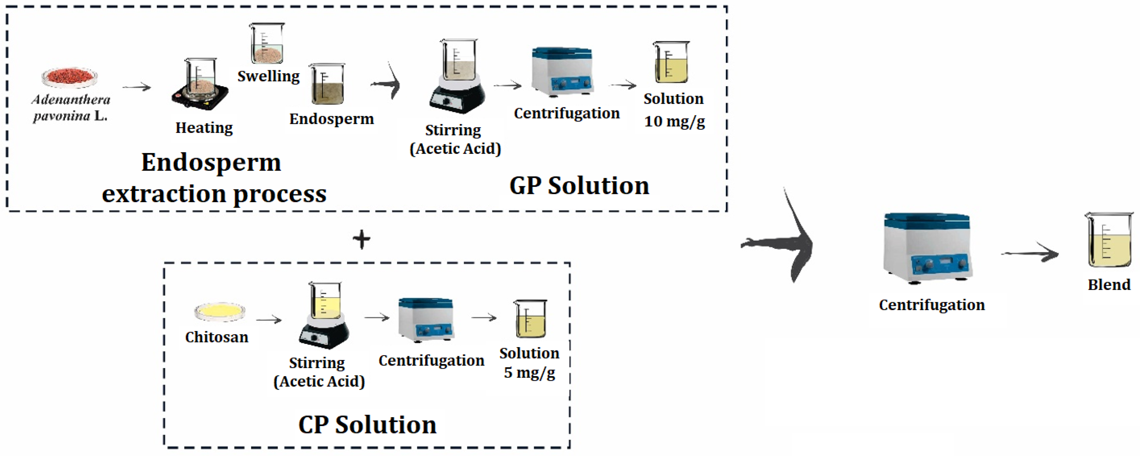
2.1. Bioceramics Preparation

The sample preparation consisted of six steps: synthesis of hydroxyapatite, galactomannan extraction and solution preparation, chitosan solution preparation, polymeric blend preparation and, finally, the production of the bioceramics composed of hydroxyapatite and the polymeric blend.
The synthesis of HAP was performed by the high-energy method, using stoichiometric amounts of calcium hydroxide, Ca(OH)2 (Vetec, 97%), and calcium hydrogen phosphate, CaHPO4 (Aldrich, St. Louis, MO, USA, 99%), according to the chemical reaction presented in Equation (1):
4Ca(OH)2 + 6CaHPO4 → Ca10(PO4)6(OH)2 + 6H2O
The starting materials were ground in a Fritsch Pulverisette 6 planetary ball mill. During grinding, air-sealed stainless steel bowls and balls were used, with a rotation speed of 370 rpm. To avoid excessive heat, the synthesis was performed in 30 min stages with 10 min pauses, with a total duration of 20 h.
The crude galactomannan was extracted from the seeds of Adenanthera pavonina L. The endosperms were obtained after heating the seeds in boiling distilled water for 20 min with enzymatic inactivation, followed by swelling for 12 h. The seed coats were removed and then the endosperms were separated from the embryo and stored under refrigeration [34].
The galactomannan solution (GP) was obtained by the solubilization of the endosperms at room temperature in a 0.1% acetic acid solution (pH = 3.0) for 1 h under mechanical agitation. The solution was centrifuged at 14,560× g (10,000 rpm) for 1 h, and the dry matter of the suspension was obtained by heating it at 100 °C until a constant weight was reached. The resulting solution was taken to a final concentration of 10 mg/g.
The chitosan solution (CP) was obtained by the solubilization of the chitosan powder (Sigma/Aldrich, St. Louis, MO, USA,) at room temperature in 0.1% acetic acid solution (pH = 3.0) for 24 h under mechanical agitation. The solution was centrifuged at 14,560× g for 1 h, with the resulting solution taken to a final concentration of 5 mg/g.
The polymeric blend of galactomannan and chitosan (GC) was prepared from the homogenization of GP (10 mg/g) and CP (5 mg/g) solutions, obtained by agitation at room temperature, centrifuged at 582 g for 20 min and stored under refrigeration until use.
Figure 1 shows the representative flowchart of GC blend preparation and the processes that preceded it.
Finally, the bioceramics were prepared from 70%, 80% and 90% of HAP nanocrystalline powder with 30%, 20% and 10% of GC blends, respectively, and categorized as follows: H70GC30, H80GC20 and H90GC10. The resulting powders were molded into cylindrical pellets that were 10 mm in diameter and 1 mm in height by cold uniaxial pressing (260 MPa) in a hydraulic press. The samples were then sintered at 900 °C for 5 h at a heating rate of 5 °C/min.
Figure 2 shows the representative flowchart of the bioceramics preparation process.

2.2. Bioceramics Characterization

X-ray diffraction (XRD) patterns were determined by a Rigaku DMAXB diffractometer configured in a Bragg–Brentano geometry with Cu Kα radiation (40 kV and 25 mA). The analyses were performed at room temperature (27 °C) with an angular interval of 10–70° (2θ) at a rate of 1°/min. The XRD patterns were analyzed by the Rietveld refinement method [45]. The quantitative analysis of the Rietveld refinement was performed using the BGMN software with the Profex interface [46]. This method consists of minimizing the sum of squares of the difference between the intensities observed and calculated for each point of the powder diffraction pattern (Equation (2)).
M = i w i I O B S i I C A L C i 2
where M is the minimum residue, I O B S i is the intensity of the experimental diffraction pattern in the i-point, I C A L C i is the intensity of the theoretical diffraction pattern in the i-point and w i is the weight for each measured point [18]. To evaluate the quality of the adjustment, quantitative factors that require reliable data are used, and among them, we can define the standard weight residue, R W P , and the expected residue, R e x p , presented in Equations (3) and (4).
R W P = i w i I O B S i I C A L C i 2 i w i I O B S i 2 1 2
R e x p = N P i w i I O B S i 2 1 2
where N is the total number of observed points and P is the number of fitted parameters. From the mathematical point of view, the factor with the greatest statistical significance to be evaluated is the R W P because it depends on the minimum residue M. Another important factor is S, also called the goodness of fit (Equation (5)), and should be close to 1.0 at the end of the refinement process.
S = R W P R e x p
The size of the crystallite, LC, can be estimated from the Scherrer formula, presented in Equation (6), and using the peaks obtained from X-ray diffraction.
L C = k λ β cos θ
where k is the constant that depends on the morphology and direction of the material network (and is equal to 0.9), λ is the wavelength of X-rays, β is the width at half height of the diffracted peak (FWHM) and θ is the Bragg angle of the corresponding diffracted peak [47]. The percentage degree of crystallinity (XC) was determined using Equation (7) [48].
X C = 100 × I e x p I b a c k I e x p
where I e x p refers to the total integrated area of the experimental pattern obtained by X-ray diffraction and I b a c k is the integral area of the baseline that corresponds to the amorphous region.
The Fourier transform infrared (FTIR) spectra of hydroxyapatite and hydroxyapatite samples with polymeric blends were obtained using KBr, measured in the region between 400 and 4000 cm−1, using the SHIMATZU FTIR-283B spectrometer.
The experimental density was measured by Archimedes’ principle using a pycnometer.
The thermogravimetry (TG) analyses were performed using the SHIMADZU TGA 50H thermal analyzer. The samples were heated in an inert atmosphere of nitrogen gas at a heating rate of 10 °C/min. The differential scanning calorimetry (DSC) studies were performed in a Shimadzu DSC50 thermal analyzer, with utilization of the same heating rate.
To perform scanning electron microscopy (SEM), the samples were coated with gold and analyzed in a Phillips XL-30 operating with primary electron groups limited between 12 and 20 KeV. The average grain size was estimated from the obtained micrographs.
To evaluate the hardness of the samples, a SHIMADZU HMV2 microdurometer was used, and was equipped with a standard Vickers microhardness tester, upon which a load of 920.7 mN was applied for 20 s. We then performed 10 indentations in each sample, with 10 mm in diameter and 1 mm in thickness. The hardness is the mechanical property used to know the strength of the material, and it is related to the material’s density and morphology. The Vickers microhardness tester consists of a pyramidal diamond penetrator that is forced against the specimen. The Vickers hardness, HV, of each sample was determined according to Equation (8).
H V = L 2 d 2
where d is the average length of the indentation diagonal, expressed in meters, and L the indentation load, in Newton [49,50].
The dielectric measurements of the GC blend and the bioceramics H70GC30, H80GC20 and H90GC10 were performed using the Solartron 1260 Impedance Analyzer in the frequency range from 1 Hz to 30 MHz at room temperature (27 °C). The real part of the dielectric permittivity, ε , was calculated from the measured capacitance, C, of the samples, and is given by Equation (9).
ε = C d ε 0 A
where d is sample thickness, A is the surface area of the electrode and ε 0 is the vacuum permittivity (8,851,014 × 10−12 F/m).
The complex part of the dielectric permittivity, ε , was obtained from the resistance, R, of the samples by applying Expression (10):
ε = d R ω ε 0 A
The real and complex parts of the dielectric permittivity can be related by the tangent loss, tanδ, which is given by Equation (11).
t a n δ = ε ε
The ε measures the ability of a dielectric to store energy and relates to the polarization that occurs in the material subject to an electric field. The ability of a material to convert electromagnetic energy into heat is measured by the ε [51,52,53,54,55,56].

3. Results

3.1. XRD and Rietveld Refinement

The structure of the HAP (sintered at 900 °C for 5 h with a heating rate of 5 °C/min) was determined by XRD. Figure 3a shows the XRD patterns and the ICSD 429,746 profile [57]. It is observed that the HAP assumes all the characteristic peaks of the hexagonal phase of hydroxyapatite, not presenting peaks that could be assigned to secondary phases. The crystallite size, LC, and the percentage degree of crystallinity, XC, of the sintered HAP, obtained using Equations (6) and (7), are (30.45 ± 0.7) nm and 37.52%, respectively, and are depicted in Table 1. Silva et al. [58], when synthesizing hydroxyapatite samples by high-energy mechanical grinding, obtained an average crystallite size between 22 and 39 nm, showing good agreement with this work.
With the Rietveld refinement, a quantitative analysis of the crystalline phase of HAP was performed. Figure 3b confirms that the sample presents 100% of the Ca10(PO4)6(OH)2 phase, with a hexagonal structure characteristic of the space group P63/m, and the network parameters and density presented in Table 1. The results found are aligned with the values previously reported [57,58]. The RWP and S values, obtained using Equations (3)–(5), are satisfactory since they are within their typical range [19,59].

3.2. Infrared Spectroscopy

The FTIR spectrum for the HAP sample, measured in the region ranging from 2000 cm−1 to 400 cm−1, is displayed in Figure 4. The characteristic bands, assigned to the functional groups phosphate (PO43−), carbonate (CO32−) and hydroxyl (OH) can be identified: the bands at 569 cm−1, 603 cm−1, 968 cm−1, 1043 cm−1 and 1093 cm−1 can be attributed to the PO43− ion; the bands at 1419 cm−1 and 1469 cm−1 arise from vibrations of the CO32− ions; and vibrations associated to OH are observed at 630 cm−1 and 1649 cm−1. The band at 1649 cm−1 corresponds to free OH, attributed to symmetric deformation in the molecule of H2O adsorbed in the synthesis process. The bands at 1469 cm−1 and 1419 cm−1 are assigned to the asymmetric stretching vibration of carbonate ions. The bands at 1093 cm−1 and 1043 cm−1 may be ascribed to the triply degenerated ν3 anti-symmetric stretching of the P–O band, and the 968 cm−1 band can be due to the ν1 non-degenerated symmetric stretching of the P–O bond. The band refers to the oscillation mode of the OH- ions, and the band of the P-OH bond corresponding to the water adsorbed on the surface is visible at 630 cm−1. Finally, the bands at 603 cm−1 and 569 cm−1 can be attributed to the triply degenerated ν4 vibration of the O–P–O bond, and the band at 478 cm−1 may be related to the doubly degenerated ν2 O–P–O bending.
The results obtained are in agreement with the literature [15,16,47,60,61,62,63,64] and the observed bands and their corresponding assignments can be consulted in Table 2.
The FTIR spectrum of the GC blend, measured in the region from 2000 cm−1 to 400 cm−1, is also depicted in Figure 4. The bands at 808 cm−1 and 879 cm−1 indicate the presence of α–D–galactopyranose and β–D–mannopyranose units, respectively, and, at 1014 cm−1, a common band of polysaccharides is also present. It is noteworthy that the band between 1000 and 1110 cm−1 is attributed to C-O-C stretching vibration and, in the region from 1350 to 1450 cm−1, the bands correspond to the symmetrical deformations of the CH2 and COH groups [65,66,67,68].
According to [69], these spectra can be influenced by parameters such as the deacetylation percentage or crystallinity. The IR spectra of chitosan, which is essentially produced from chitin by a deacetylation reaction, corresponds to a convolution of specific signals for carbohydrates and absorption due to amine and amide functions. Therefore, the band at 1575 cm−1 arises from the peptide bond vibration amide II and can be ascribed to the N–H stretching vibrations, while the 1660 cm−1 band is assigned to the stretching vibrations of C=O due to the peptide bond vibration amide I [69,70,71,72,73].
The analysis of the IR of the bioceramics H70GC30, H80GC20 and H90GC10 showed the same absorptions of HAP.

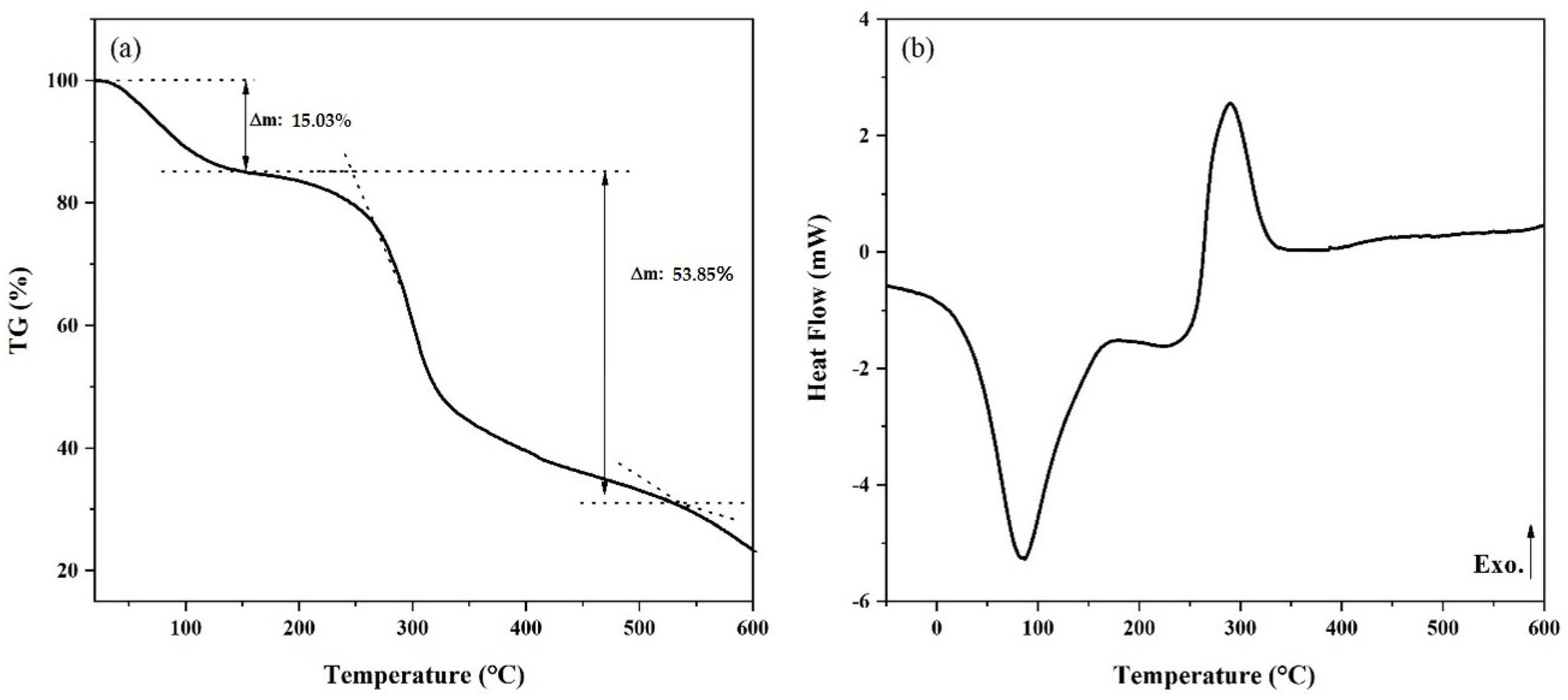
3.3. Thermal Analysis

To study the thermal behavior of HAP and the GC blend, thermogravimetry and differential scanning calorimetry measurements were performed and are depicted in Figure 5 and Figure 6.
The TG curve shows a three-stage weight loss at the temperature ranges of 20–100, 100–300 and 300–500 °C, approximately.
In the first stage, the weight loss is due to the evaporation of adsorbed water [72,73]. The presence of water, physically adsorbed in the synthesis process of HAP, was already discussed in the infrared spectrum analysis. The second stage shows weight loss due to the release of adsorbed and lattice water, which is in accordance with the findings of Tõnsuaadu et al. [74], which stated that the lattice water is irreversibly lost between 200 and 400 °C. The third stage may represent the loss of water from decomposition, which means that in the present case, the removal of water molecules starts at lower temperatures when compared to the literature. According to Tõnsuaadu et al. [75], the dehydroxylation of HAP with the removal of water molecules in the air atmosphere starts at 900 °C. However, Bulina et al. [73] reported that this process probably starts at a lower temperature, specifically 600 °C, and Mandal et al. [72] considered the temperature of 780 °C to be the beginning point of the dihydroxylation process. Such a temperature discrepancy should be attributed to the differences between samples and the experimental method [75]. Above 500 °C, HAP is thermally stable since stoichiometric hydroxyapatite with a Ca/P ratio of 1.67 is stable up to 1200 °C [76].
In Figure 5b, the endothermic event present in the range 20 to 100 °C, more precisely at 52.74 °C, corroborates the information given by the TG curve and can be attributed to the evaporation of adsorbed water [77]. In the range of 100–300 °C, a broad exothermic peak can be considered. This peak is due to crystal growth and lattice strain release during the heating process. In the grinding process, a large amount of strain and defects are incorporated into the powder particles and, when the milled sample is heated, grain growth takes place and strain is released, although no phase transformation occurs up to 1200 °C [72].
The thermogram of the GC blend, shown in Figure 6a, indicates that the thermal decomposition occurs in two stages. In the first, in the range of 25 to 150 °C, there is a loss of 15.03% of the initial mass of the sample resulting from the water release. In the second, between 200 and 350 °C, a pronounced decline in the TG curve is observed, referring to the disintegration of macromolecules related to the polymeric groups present in the blend as well as the decomposition of the organic material, which results in the loss of another 53.85% of the initial mass.
In Figure 6b, the thermal transitions present in the GC blend were verified through the DSC. The two events present in the thermogram are represented by two peaks, one endothermic at 85 °C and one exothermic at 290 °C, respectively. The endothermic peak is related to the loss of water in the structure and the exothermic peak with the degradation of the material. The GC blend can be considered thermally stable due to the presence of two well-defined thermal transition regions [68,72].
In the thermal analyses of the bioceramics H70GC30, H80GC20 and H90GC10, no divergences were observed with the results presented by HAP. This occurred since the biopolymers degraded at a temperature of around 290 °C; therefore, the bioceramics only show the presence of HAP, an outcome that can relate to the results obtained in the FTIR.

3.4. Morphological Analysis

The sample morphology was investigated using SEM. Figure 7 presents the micrographs of H70GC30, H80GC20 and H90GC10 bioceramics, with a magnification factor of 25,000×. The grains can be described as small spherical particles, being in consonance with the morphology presented by pure hydroxyapatite.
Table 3 shows the average grain size, the experimental density and the Vickers hardness obtained for each sample. For the HAP, the average estimated grain size, 0.5 μm, is aligned with the values reported by Macêdo et al. [34]. It can be observed that the increase in the CG blend content promoted an increase in the average grain size. This grain growth has an impact on the porosity of the samples since a decrease in the experimental density can be observed.
The Vickers hardness of HAP, H70GC30, H80GC20 and H90GC10 bioceramics was calculated with Equation (8). It is observed that the hardness of the bioceramics decreased with the biopolymer addition. This behavior was expected since the main contribution of the decrease of the microhardness is the decrease in the ceramic density [50].

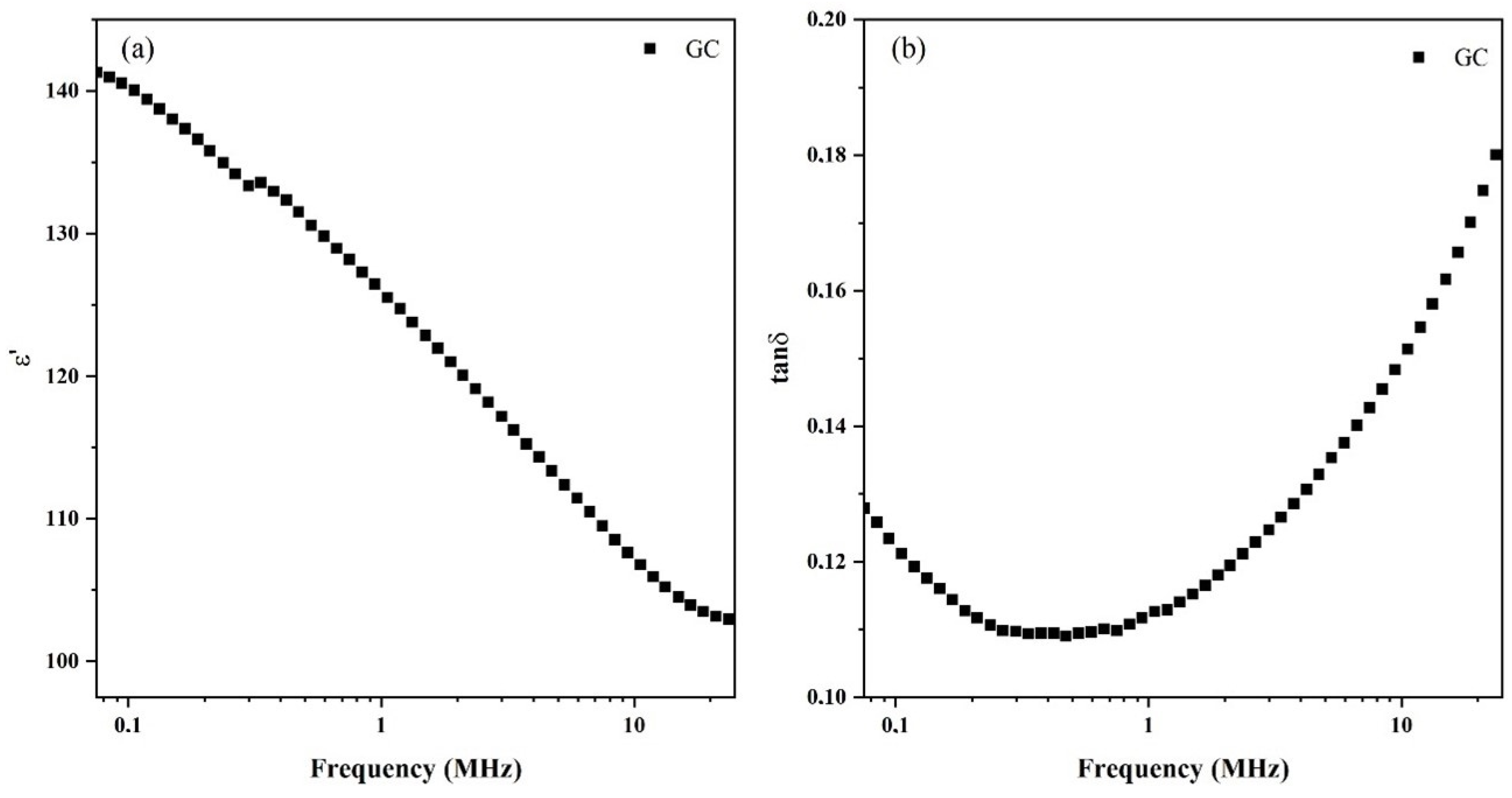
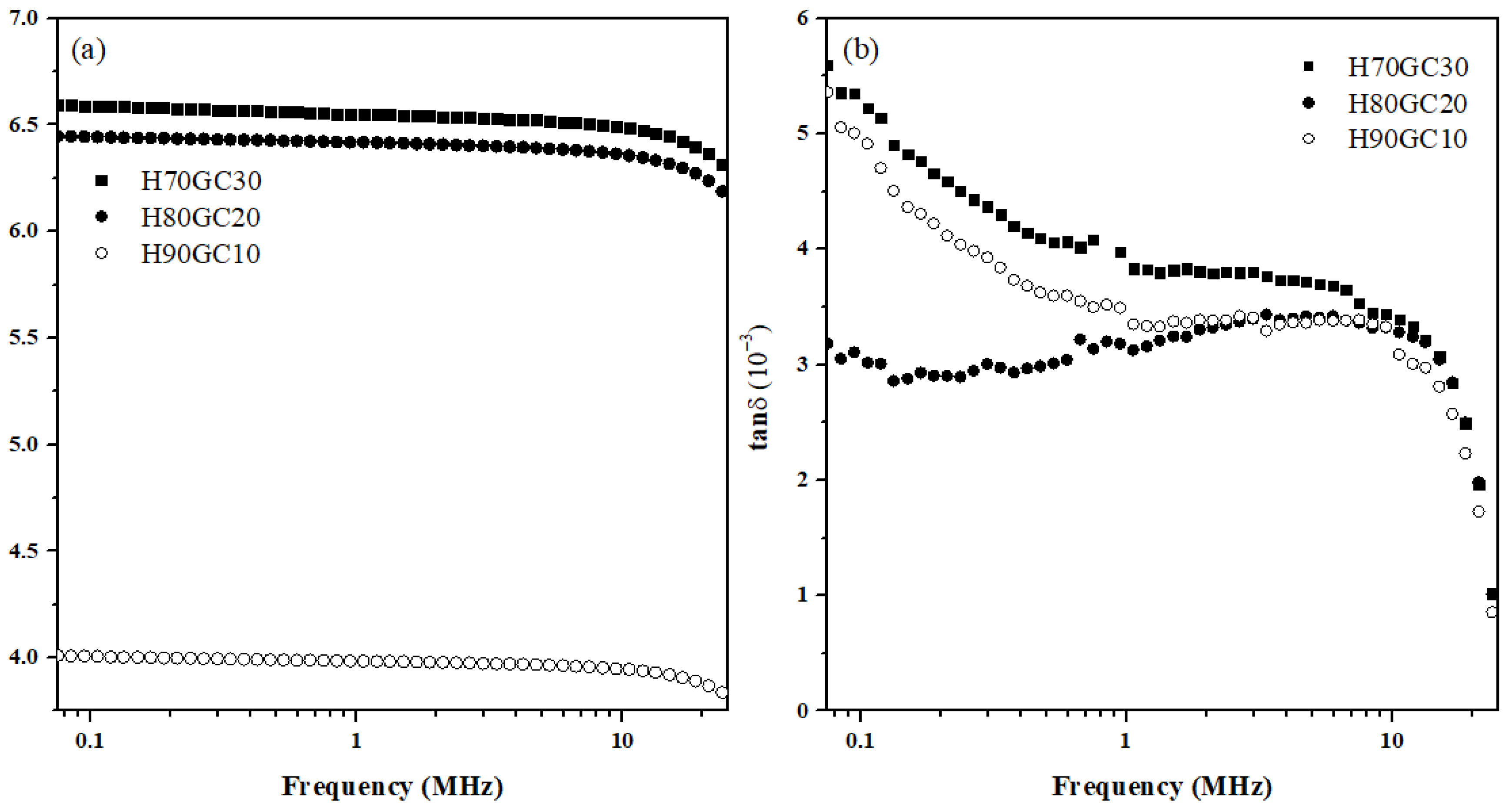
3.5. Dielectric Spectroscopy

The dielectric behavior of the GC blend as a function of frequency is depicted in Figure 8. Figure 8a shows that the dielectric constant, ε , at room temperature and in the frequency range considered. This constant decreases monotonically with increasing frequency. The continuous decrease of the dielectric constant with increasing frequency is a common behavior for all dielectric materials [78] and is due to the fact that when the frequency of the applied electric field increases, the mechanism of polarization will not be able to follow the change in the electric field and, therefore, the contribution of polarization to the dielectric constant will diminish [51,78,79]. At higher frequencies, when already in the limit of the measurement window, it is possible to infer that the ε values tend to stabilize, a prediction supported by the dielectric behavior observed in several polymers [79]. Figure 8b presents the tangent loss, tanδ, as a function of the frequency. The inexistence of peaks shows that, at room temperature and in the frequency range of the measurements, the CG blend does not present any relaxation phenomenon and, since the values of tanδ are inferior to one, based on Equation (11), it is possible to conclude that the dielectric losses are inferior to the dielectric constant.
The dielectric properties of the bioceramics H70GC30, H80GC20 and H90GC10, depicted in Figure 9, are related to the different types of polarization present in the sample. The interfaces of bioceramics with biopolymers have a large number of defects that result in an unequal distribution of charges [80]. It can be verified in Figure 9a that the increase in the amount of GC promoted the increase of ε . This enhancement in the dielectric constant values shows the increased energy storage capacity of the studied bioceramics [78,79,80,81,82,83]. With the addition of GC, cations are the dominant charges and may be responsible for the increase in the dielectric constant [34]. Until ≈ 10 MHz, the samples show a quasi-frequency independent behavior, indicating that the materials do not exhibit intense polarization processes and that the stored charge remains practically constant in the frequency range considered. At higher frequencies (>10 MHz), the charge carriers are unable to follow the rapid changes in the applied electric field and, as a consequence, the dielectric constant values start to decrease [81,82].
Figure 9b presents the tanδ as a function of the frequency for the studied bioceramics. Since the tanδ values are inferior to one, the energy dissipated is inferior to the stored energy [84]. It is also noted that the highest dissipated energy is from bioceramics containing higher concentrations of biopolymer (H70GC30).
The dielectric properties of inorganic compounds containing galactomannan and chitosan have direct relationships with the concentration of the biopolymer [61,65,69,85].

4. Conclusions

Overall, the XRD pattern, Rietveld refinement and FTIR collectively indicated that the methodology of synthesis of HAP nanocrystalline powders was efficient, allowing us to obtain a single HAP phase. The FTIR spectrum showed that the GC blending process did not promote an alteration of the vibrational properties, presenting characteristic bands of galactomannan and chitosan.
Dielectric, morphological, hardness and density analyses confirmed that the GC blend, as well as its concentration, influenced the properties of the HAP. The dielectric analysis also showed a relationship between the concentration of the GC blend and the dielectric constant of the bioceramic since the blend favored the increase of ε without compromising the tanδ, which presented values in the order of 10−3.

Author Contributions

Conceptualization, E.d.S.G., M.P.F.G. and A.A.M.M.; methodology, E.d.S.G. and A.A.M.M.; software, S.R.G. and S.D.; validation, S.D., M.P.F.G. and A.A.M.M.; formal analysis, M.P.F.G. and A.A.M.M.; investigation, E.d.S.G., A.M.d.O.L., S.R.G. and S.D.; resources, S.D., M.P.F.G. and A.A.M.M.; data curation, A.A.M.M.; writing—original draft preparation, E.d.S.G.; writing—review and editing, S.R.G., S.D. and M.P.F.G.; visualization, A.M.d.O.L.; supervision, A.A.M.M.; project administration, A.A.M.M.; funding acquisition, M.P.F.G. All authors have read and agreed to the published version of the manuscript.

Funding

This research was funded by FEDER funds through the COMPETE 2020 Program and National Funds through FCT—Portuguese Foundation for Science and Technology—under the project LISBOA-01-0247-FEDER-039985/POCI-01-0247-FEDER-039985, LA/P/0037/2020, UIDP/50025/2020, and UIDB/50025/2020 of the Associate Laboratory Institute of Nanostructures, Nanomodelling and Nanofabrication—i3N. S.R. Gavinho acknowledges FCT—Portuguese Foundation for Science and Technology—for the PhD grant (SFRH/BD/148233/2019).

Institutional Review Board Statement

Not applicable.

Informed Consent Statement

Not applicable.

Data Availability Statement

The data presented in this study are available on request from the corresponding author.

Conflicts of Interest

The authors declare no conflict of interest.

References

  1. Ragunathan, S.; Govindasamy, G.; Raghul, D.R.; Karuppaswamy, M.; Vijayachandra Togo, R.K. Hydroxyapatite reinforced natural polymer scaffold for bone tissue regeneration. Mater. Today Proc. 2020, 23, 111–118. [Google Scholar] [CrossRef]
  2. Arvidson, K.; Abdallah, B.M.; Applegate, L.A.; Baldini, N.; Cenni, E.; Gomez-Barrena, E.; Granchi, D.; Kassem, M.; Konttinen, Y.T.; Mustafa, K.; et al. Bone regeneration and stem cells. J. Cell. Mol. Med. 2011, 15, 718–746. [Google Scholar] [CrossRef] [PubMed]
  3. Kalita, S.J.; Bhardwaj, A.; Bhatt, H.A. Nanocrystalline calcium phosphate ceramics in biomedical engineering. Mater. Sci. Eng. C 2007, 27, 441–449. [Google Scholar] [CrossRef]
  4. Daglilar, S.; Erkan, M.E. A study on bioceramic reinforced bone cements. Mater. Lett. 2007, 61, 1456–1459. [Google Scholar] [CrossRef]
  5. Sadat-Shojai, M.; Khorasani, M.T.; Dinpanah-Khoshdargi, E.; Jamshidi, A. Synthesis methods for nanosized hydroxyapatite with diverse structures. Acta Biomater. 2013, 9, 7591–7621. [Google Scholar] [CrossRef]
  6. Hwang, K.; Lim, Y. Chemical and structural changes of hydroxyapatite films by using a sol–gel method. Surf. Coat. Technol. 1999, 115, 172–175. [Google Scholar] [CrossRef]
  7. Suchanek, W.L.; Byrappa, K.; Shuk, P.; Riman, R.E.; Janas, V.F.; Ten Huisen, K.S. Preparation of magnesium-substituted hydroxyapatite powders by the mechanochemical–hydrothermal method. Biomaterials 2004, 25, 4647–4657. [Google Scholar] [CrossRef]
  8. Rigo, E.C.D.S.; Oliveira, L.C.; Santos, L.A.; Boschi, A.O.; Carrodeguas, R.G. Implantes metálicos recobertos com hidroxiapatita. Res. Biomed. Eng. 1999, 15, 21–29. [Google Scholar]
  9. Assis, C.M.D.; Vercik, L.C.D.O.; Santos, M.L.D.; Fook, M.V.L.; Guastaldi, A.C. Comparison of crystallinity between natural hydroxyapatite and synthetic cp-Ti/HA coatings. Mater. Res. 2005, 8, 207–211. [Google Scholar] [CrossRef]
  10. Nikpour, M.R.; Rabiee, S.M.; Jahanshahi, M.J.C.P.B.E. Synthesis and characterization of hydroxyapatite/chitosan nanocomposite materials for medical engineering applications. Compos. Part B 2012, 43, 1881–1886. [Google Scholar] [CrossRef]
  11. Silva, C.C.; Valente, M.A.; Graça, M.P.F.; Sombra, A.S.B. Preparation and optical characterization of hydroxyapatite and ceramic systems with titanium and zirconium formed by dry high-energy mechanical alloying. Solid State Sci. 2004, 6, 1365–1374. [Google Scholar] [CrossRef]
  12. Motskin, M.; Wright, D.M.; Muller, K.; Kyle, N.; Gard, T.G.; Porter, A.E.; Skepper, J.N. Hydroxyapatite nano and microparticles: Correlation of particle properties with cytotoxicity and biostability. Biomaterials 2009, 30, 3307–3317. [Google Scholar] [CrossRef] [PubMed]
  13. Wang, S.; Kempen, D.H.; Yaszemski, M.J.; Lu, L. The roles of matrix polymer crystallinity and hydroxyapatite nanoparticles in modulating material properties of photo-crosslinked composites and bone marrow stromal cell responses. Biomaterials 2009, 30, 3359–3370. [Google Scholar] [CrossRef] [PubMed]
  14. Mohandes, F.; Salavati-Niasari, M.; Fathi, M.; Fereshteh, Z. Hydroxyapatite nanocrystals: Simple preparation, characterization and formation mechanism. Mater. Sci. Eng. C 2014, 45, 29–36. [Google Scholar] [CrossRef]
  15. Yasukawa, A.; Kandori, K.; Tanaka, H.; Gotoh, K. Preparation and structure of carbonated calcium hydroxyapatite substituted with heavy rare earth ions. Mater. Res. Bull. 2012, 47, 1257–1263. [Google Scholar] [CrossRef]
  16. Yasukawa, A.; Gotoh, K.; Tanaka, H.; Kandori, K. Preparation and structure of calcium hydroxyapatite substituted with light rare earth ions. Colloids Surf. A 2012, 393, 53–59. [Google Scholar] [CrossRef]
  17. Kaygili, O.; Dorozhkin, S.V.; Ates, T.; Al-Ghamdi, A.A.; Yakuphanoglu, F. Dielectric properties of Fe doped hydroxyapatite prepared by sol–gel method. Ceram. Int. 2014, 40, 9395–9402. [Google Scholar] [CrossRef]
  18. Zhang, W.; Cao, N.; Chai, Y.; Xu, X.; Wang, Y. Synthesis of nanosize single-crystal strontium hydroxyapatite via a simple sol–gel method. Ceram. Int. 2014, 40, 16061–16064. [Google Scholar] [CrossRef]
  19. Silva, C.C.; Graça, M.P.F.; Valente, M.A.; Sombra, A.S.B. Structural study of Fe2O3-doped calcium phosphates obtained by the mechanical milling method. Phys. Scr. 2009, 79, 055601. [Google Scholar] [CrossRef]
  20. Nasiri-Tabrizi, B.; Fahami, A.; Ebrahimi-Kahrizsangi, R. Effect of milling parameters on the formation of nanocrystalline hydroxyapatite using different raw materials. Ceram. Inter. 2013, 39, 5751–5763. [Google Scholar] [CrossRef]
  21. Rhee, S.H. Synthesis of hydroxyapatite via mechanochemical treatment. Biomaterials 2002, 23, 1147–1152. [Google Scholar] [CrossRef] [PubMed]
  22. Cho, J.S.; Rhee, S.H. Formation mechanism of nano-sized hydroxyapatite powders through spray pyrolysis of a calcium phosphate solution containing polyethylene glycol. J. Eur. Ceram. Soc. 2013, 33, 233–241. [Google Scholar] [CrossRef]
  23. Liu, H.S.; Chin, T.S.; Lai, L.S.; Chiu, S.Y.; Chung, K.H.; Chang, C.S.; Lui, M.T. Hydroxyapatite synthesized by a simplified hydrothermal method. Ceram. Inter. 1997, 23, 19–25. [Google Scholar] [CrossRef]
  24. Lala, S.; Satpati, B.; Kar, T.; Pradhan, S.K. Structural and microstructural characterizations of nanocrystalline hydroxyapatite synthesized by mechanical alloying. Mater. Sci. Eng. C 2013, 33, 2891–2898. [Google Scholar] [CrossRef] [PubMed]
  25. Macuvele, D.L.P.; Nones, J.; Matsinhe, J.V.; Lima, M.M.; Soares, C.; Fiori, M.A.; Riella, H.G. Advances in ultra high molecular weight polyethylene/hydroxyapatite composites for biomedical applications: A brief review. Mater. Sci. Eng. C 2017, 76, 1248–1262. [Google Scholar] [CrossRef] [PubMed]
  26. Chen, F.; Wang, Z.C.; Lin, C.J. Preparation and characterization of nano-sized hydroxyapatite particles and hydroxyapatite/chitosan nano-composite for use in biomedical materials. Mater. Lett. 2002, 57, 858–861. [Google Scholar] [CrossRef]
  27. Zhang, L.J.; Feng, X.S.; Liu, H.G.; Qian, D.J.; Zhang, L.; Yu, X.L.; Cui, F.Z. Hydroxyapatite/collagen composite materials formation in simulated body fluid environment. Mater. Lett. 2004, 58, 719–722. [Google Scholar] [CrossRef]
  28. Maeda, Y.; Jayakumar, R.; Nagahama, H.; Furuike, T.; Tamura, H. Synthesis, characterization and bioactivity studies of novel β-chitin scaffolds for tissue-engineering applications. Int. J. Biol. Macromol. 2008, 42, 463–467. [Google Scholar] [CrossRef]
  29. Manara, S.; Paolucci, F.; Palazzo, B.; Marcaccio, M.; Foresti, E.; Tosi, G.; Sabbatini, S.; Sabatino, P.; Altankov, G.; Roveri, N. Electrochemically-assisted deposition of biomimetic hydroxyapatite–collagen coatings on titanium plate. Inorg. Chim. Acta 2008, 361, 1634–1645. [Google Scholar] [CrossRef]
  30. Madhumathi, K.; Shalumon, K.T.; Rani, V.D.; Tamura, H.; Furuike, T.; Selvamurugan, N.; Nair, S.V.; Jayakumar, R. Wet chemical synthesis of chitosan hydrogel–hydroxyapatite composite membranes for tissue engineering applications. Int. J. Biol. Macromol. 2009, 45, 12–15. [Google Scholar] [CrossRef]
  31. Pang, X.; Casagrande, T.; Zhitomirsky, I. Electrophoretic deposition of hydroxyapatite–CaSiO3–chitosan composite coatings. J. Colloid Interface Sci. 2009, 330, 323–329. [Google Scholar] [CrossRef] [PubMed]
  32. Trakoolwannachai, V.; Kheolamai, P.; Ummartyotin, S. Development of hydroxyapatite from eggshell waste and a chitosan-based composite: In vitro behavior of human osteoblast-like cell (Saos-2) cultures. Int. J. Biol. Macromol. 2019, 134, 557–564. [Google Scholar] [CrossRef] [PubMed]
  33. Pereira, M.B.B.; França, D.B.; Araújo, R.C.; Silva Filho, E.C.; Rigaud, B.; Fonseca, M.G.; Jaber, M. Amino hydroxyapatite/chitosan hybrids reticulated with glutaraldehyde at different pH values and their use for diclofenac removal. Carbohydr. Polym. 2020, 236, 116036. [Google Scholar] [CrossRef] [PubMed]
  34. Macêdo, A.A.M.; Sombra, A.S.B.; Mazzetto, S.E.; Silva, C.C. Influence of the polysaccharide galactomannan on the dielectrical characterization of hydroxyapatite ceramic. Compos. Part B 2013, 44, 95–99. [Google Scholar] [CrossRef]
  35. Pawlik, A.; Rehman, M.A.U.; Nawaz, Q.; Bastan, F.E.; Sulka, G.D.; Boccaccini, A.R. Fabrication and characterization of electrophoretically deposited chitosan-hydroxyapatite composite coatings on anodic titanium dioxide layers. Electrochim. Acta 2019, 307, 465–473. [Google Scholar] [CrossRef]
  36. Cunha, C.S.; Castro, P.J.; Sousa, S.C.; Pullar, R.C.; Tobaldi, D.M.; Piccirillo, C.; Pintado, M.M. Films of chitosan and natural modified hydroxyapatite as effective UV-protecting, biocompatible and antibacterial wound dressings. Int. J. Biol. Macromol. 2020, 159, 1177–1185. [Google Scholar] [CrossRef]
  37. Olivera, S.; Muralidhara, H.B.; Venkatesh, K.; Guna, V.K.; Gopalakrishna, K.; Kumar, Y. Potential applications of cellulose and chitosan nanoparticles/composites in wastewater treatment: A review. Carbohydr. Polym. 2016, 153, 600–618. [Google Scholar] [CrossRef]
  38. Croisier, F.; Jérôme, C. Chitosan-based biomaterials for tissue engineering. Eur. Polym. J. 2013, 49, 780–792. [Google Scholar] [CrossRef]
  39. Thanyacharoen, T.; Chuysinuan, P.; Techasakul, S.; Noenplab, A.N.L.; Ummartyotin, S. The chemical composition and antioxidant and release properties of a black rice (Oryza sativa L.)-loaded chitosan and polyvinyl alcohol composite. J. Mol. Liq. 2017, 248, 1065–1070. [Google Scholar] [CrossRef]
  40. Thanyacharoen, T.; Chuysinuan, P.; Techasakul, S.; Nooeaid, P.; Ummartyotin, S. Development of a gallic acid-loaded chitosan and polyvinyl alcohol hydrogel composite: Release characteristics and antioxidant activity. Int. J. Biol. Macromol. 2018, 107, 363–370. [Google Scholar] [CrossRef]
  41. Kök, M.S.; Hill, S.E.; Mitchell, J.R. Viscosity of galactomannans during high temperature processing: Influence of degradation and solubilisation. Food Hydrocoll. 1999, 13, 535–542. [Google Scholar] [CrossRef]
  42. Fernandes, P.B.; Gonçalves, M.P.; Doublier, J.L. A rheological characterization of kappa-carrageenan/galactomannan mixed gels: A comparison of locust bean gum samples. Carbohydr. Polym. 1991, 16, 253–274. [Google Scholar] [CrossRef]
  43. Bresolin, T.M.B.; Milas, M.; Rinaudo, M.; Reicher, F.; Ganter, J.L.M.S. Role of galactomannan composition on the binary gel formation with xanthan. Int. J. Biol. Macromol. 1999, 26, 225–231. [Google Scholar] [CrossRef] [PubMed]
  44. Vendruscolo, C.W.; Andreazza, I.F.; Ganter, J.L.M.S.; Ferrero, C.; Bresolin, T.M.B. Xanthan and galactomannan (from M. scabrella) matrix tablets for oral controlled delivery of theophylline. Int. J. Pharm. 2005, 296, 1–11. [Google Scholar] [CrossRef]
  45. Rietveld, H.M. Line profiles of neutron powder-diffraction peaks for structure refinement. Acta Crystallogr. 1967, 22, 151–152. [Google Scholar] [CrossRef]
  46. Doebelin, N.; Kleeberg, R. Profex: A graphical user interface for the Rietveld refinement program BGMN. J. Appl. Crystallogr. 2015, 48, 1573–1580. [Google Scholar] [CrossRef]
  47. Hayakawa, S.; Li, Y.; Tsuru, K.; Osaka, A.; Fujii, E.; Kawabata, K. Preparation of nanometer-scale rod array of hydroxyapatite crystal. Acta Biomater. 2009, 5, 2152–2160. [Google Scholar] [CrossRef]
  48. Vinodhini, P.A.; Sangeetha, K.; Thandapani, G.; Sudha, P.N.; Jayachandran, V.; Sukumaran, A. FTIR, XRD and DSC studies of nanochitosan, cellulose acetate and polyethylene glycol blend ultrafiltration membranes. Int. J. Biol. Macromol. 2017, 104, 1721–1729. [Google Scholar] [CrossRef]
  49. Hoepfner, T.P.; Case, E.D. The influence of the microstructure on the hardness of sintered hydroxyapatite. Ceram. Int. 2003, 29, 699–706. [Google Scholar] [CrossRef]
  50. Pinto Filho, F.; Nogueira, R.E.F.Q.; Graça, M.P.F.; Valente, M.A.; Sombra, A.S.B.; Silva, C.C.D. Structural and mechanical study of the sintering effect in hydroxyapatite doped with iron oxide. Physica B 2008, 403, 3826–3829. [Google Scholar] [CrossRef]
  51. Figueiró, S.D.; Macêdo, A.A.M.; Melo, M.R.S.; Freitas, A.L.P.; Moreira, R.A.; De Oliveira, R.S.; Goés, J.C.; Sombra, A.S.B. On the dielectric behaviour of collagen–algal sulfated polysaccharide blends: Effect of glutaraldehyde crosslinking. Biophys. Chem. 2006, 120, 154–159. [Google Scholar] [CrossRef] [PubMed]
  52. Peixoto, M.V.; Costa, F.M.; Devesa, S.; Graça, M.P.F. Structural, Morphological and Dielectric Characterization of BiFeO3 Fibers Grown by the LFZ Technique. Crystals 2023, 13, 960. [Google Scholar] [CrossRef]
  53. Ashoorirad, M.; Saviz, M.; Fallah, A. On the electrical properties of collagen macromolecule solutions: Role of collagen-water interactions. J. Mol. Liq. 2020, 300, 112344. [Google Scholar] [CrossRef]
  54. Salema, A.A.; Yeow, Y.K.; Ishaque, K.; Ani, F.N.; Afzal, M.T.; Hassan, A. Dielectric properties and microwave heating of oil palm biomass and biochar. Ind. Crops Prod. 2013, 50, 366–374. [Google Scholar] [CrossRef]
  55. Dorey, R. Microstructure–property relationships: How the microstructure of the film affects its properties. In Ceramic Thick Films for MEMS and Microdevices; Dorey, R., Ed.; William Andrew Publishing: Norwich, NY, USA, 2012; pp. 85–112. [Google Scholar] [CrossRef]
  56. Graça, M.P.F.; da Silva, M.F.; Valente, M.A. NaNbO3 crystals dispersed in a B2O3 glass matrix–Structural characteristics versus electrical and dielectrical properties. Solid State Sci. 2009, 11, 570–577. [Google Scholar] [CrossRef]
  57. Kazin, P.E.; Pogosova, M.A.; Trusov, L.A.; Kolesnik, I.V.; Magdysyuk, O.V.; Dinnebier, R.E. Crystal structure details of La-and Bi-substituted hydroxyapatites: Evidence for LaO+ and BiO+ with a very short metal–oxygen bond. J. Solid State Chem. 2016, 237, 349–357. [Google Scholar] [CrossRef]
  58. Silva, C.C.; Pinheiro, A.G.; Miranda, M.A.R.; Góes, J.C.; Sombra, A.S.B. Structural properties of hydroxyapatite obtained by mechanosynthesis. Solid State Sci. 2013, 5, 553–558. [Google Scholar] [CrossRef]
  59. Sarkar, A.; Kannan, S. In situ synthesis, fabrication and Rietveld refinement of the hydroxyapatite/titania composite coatings on 316 L SS. Ceram. Int. 2014, 40, 6453–6463. [Google Scholar] [CrossRef]
  60. Bhadang, K.A.; Gross, K.A. Influence of fluorapatite on the properties of thermally sprayed hydroxyapatite coatings. Biomaterials 2004, 25, 4935–4945. [Google Scholar] [CrossRef]
  61. Silva, C.C.D.; Rocha, H.H.B.; Freire, F.N.A.; Santos, M.R.P.; Sabóia, K.D.A.; Góes, J.C.; Sombra, A.S.B. Hydroxyapatite screen-printed thick films: Optical and electrical properties. Mater. Chem. Phys. 2005, 92, 260–268. [Google Scholar] [CrossRef]
  62. Sanosh, K.P.; Chu, M.C.; Balakrishnan, A.; Lee, Y.J.; Kim, T.N.; Cho, S.J. Synthesis of nano hydroxyapatite powder that simulate teeth particle morphology and composition. Curr. Appl. Phys. 2009, 9, 1459–1462. [Google Scholar] [CrossRef]
  63. Xia, Z.; Liao, L.; Zhao, S. Synthesis of mesoporous hydroxyapatite using a modified hard-templating route. Mater. Res. Bull. 2009, 44, 1626–1629. [Google Scholar] [CrossRef]
  64. Yang, P.; Quan, Z.; Li, C.; Kang, X.; Lian, H.; Lin, J. Bioactive, luminescent and mesoporous europium-doped hydroxyapatite as a drug carrier. Biomaterials 2008, 29, 4341–4347. [Google Scholar] [CrossRef]
  65. Wang, J.; Somasundaran, P. Study of galactomannose interaction with solids using AFM, IR and allied techniques. J. Colloid Interface Sci. 2007, 309, 373–383. [Google Scholar] [CrossRef] [PubMed]
  66. Tang, E.S.K.; Huang, M.; Lim, L.Y. Ultrasonication of chitosan and chitosan nanoparticles. Int. J. Pharm 2003, 265, 103–114. [Google Scholar] [CrossRef]
  67. Ding, W.; Lian, Q.; Samuels, R.J.; Polk, M.B. Synthesis and characterization of a novel derivative of chitosan. Polymer 2003, 44, 547–556. [Google Scholar] [CrossRef]
  68. Govindan, S.; Nivethaa, E.A.K.; Saravanan, R.; Narayanan, V.; Stephen, A. Synthesis and characterization of chitosan–silver nanocomposite. Appl. Nanosci. 2012, 2, 299–303. [Google Scholar] [CrossRef]
  69. Lima, C.G.A.; De Oliveira, R.S.; Figueiró, S.D.; Wehmann, C.F.; Góes, J.C.; Sombra, A.S.B. DC conductivity and dielectric permittivity of collagen–chitosan films. Mater. Chem. Phys. 2016, 99, 284–288. [Google Scholar] [CrossRef]
  70. Figueiró, S.D.; Góes, J.C.; Moreira, R.A.; Sombra, A.S.B. On the physico-chemical and dielectric properties of glutaraldehyde crosslinked galactomannan–collagen films. Carbohydr. Polym. 2004, 56, 313–320. [Google Scholar] [CrossRef]
  71. Park, S.H.; Chun, M.K.; Choi, H.K. Preparation of an extended-release matrix tablet using chitosan/Carbopol interpolymer complex. Int. J. Pharm. 2018, 347, 39–44. [Google Scholar] [CrossRef]
  72. Mandal, T.; Mishra, B.K.; Garg, A.; Chaira, D. Optimization of milling parameters for the mechanosynthesis of nanocrystalline hydroxyapatite. Powder Technol. 2014, 253, 650–656. [Google Scholar] [CrossRef]
  73. Bulina, N.V.; Makarova, S.V.; Baev, S.G.; Matvienko, A.A.; Gerasimov, K.B.; Logutenko, O.A.; Bystrov, V.S. A study of thermal stability of hydroxyapatite. Minerals 2021, 11, 1310. [Google Scholar] [CrossRef]
  74. Tonsuaadu, K.; Gross, K.A.; Plūduma, L.; Veiderma, M. A review on the thermal stability of calcium apatites. J. Therm. Anal. Calorim. 2012, 110, 647–659. [Google Scholar] [CrossRef]
  75. Wang, T.; Dorner-Reisel, A.; Müller, E. Thermogravimetric and thermokinetic investigation of the dehydroxylation of a hydroxyapatite powder. J. Eur. Ceram. Soc. 2004, 24, 693–698. [Google Scholar] [CrossRef]
  76. Mostafa, N.Y. Characterization, thermal stability and sintering of hydroxyapatite powders prepared by different routes. Mater. Chem. Phys. 2005, 94, 333–341. [Google Scholar] [CrossRef]
  77. Zima, A. Hydroxyapatite-chitosan based bioactive hybrid biomaterials with improved mechanical strength. Spectrochim. Acta Part A 2018, 193, 175–184. [Google Scholar] [CrossRef]
  78. Rahman, P.M.; Mujeeb, V.A.; Muraleedharan, K.; Thomas, S.K. Chitosan/nano ZnO composite films: Enhanced mechanical, antimicrobial and dielectric properties. Arabian J. Chem. 2018, 11, 120–127. [Google Scholar] [CrossRef]
  79. Raja, V.; Sharma, A.K.; Rao, V.N. Impedance spectroscopic and dielectric analysis of PMMA-CO-P4VPNO polymer films. Mater. Lett. 2004, 58, 3242–3247. [Google Scholar] [CrossRef]
  80. Bonardd, S.; Robles, E.; Barandiaran, I.; Saldías, C.; Leiva, Á.; Kortaberria, G. Biocomposites with increased dielectric constant based on chitosan and nitrile-modified cellulose nanocrystals. Carbohydr. Polym. 2018, 199, 20–30. [Google Scholar] [CrossRef]
  81. Al-Muntaser, A.A.; Pashameah, R.A.; Alzahrani, E.; AlSubhi, S.A.; Hameed, S.T.; Morsi, M.A. Graphene nanoplatelets/TiO2 hybrid nanofiller boosted PVA/CMC blend based high performance nanocomposites for flexible energy storage applications. J. Polym. Environ. 2023, 31, 2534–2548. [Google Scholar] [CrossRef]
  82. El-Naggar, A.M.; Heiba, Z.K.; Kamal, A.M.; Alzahrani, K.E.; Abd-Elkader, O.H.; Mohamed, M.B. Impact of natural melanin doping on the structural, optical and dielectric characteristics of the PVP/CMC blend. J. Taibah Univ. Sci. 2023, 17, 2190731. [Google Scholar] [CrossRef]
  83. Abdullah, A.Q.; Ali, N.A.; Hussein, S.I.; Hakamy, A.; Abd-Elnaiem, A.M. Improving the Dielectric, Thermal, and Electrical Properties of Poly (Methyl Methacrylate)/Hydroxyapatite Blends by Incorporating Graphene Nanoplatelets. J. Inorg. Organomet. Polym. Mater. 2023, 1–12. [Google Scholar] [CrossRef]
  84. Bhatt, A.S.; Bhat, D.K.; Santosh, M.S. Electrical and magnetic properties of chitosan-magnetite nanocomposites. Physica B 2010, 405, 2078–2082. [Google Scholar] [CrossRef]
  85. Costa, M.M.; Sohn, R.S.T.M.; Macêdo, A.A.M.; Mazzetto, S.E.; Graça, M.P.F.; Sombra, A.S.B. Study of the temperature and organic bindings effects in the dielectric and structural properties of the lithium ferrite ceramic matrix (LiFe5O8). J. Alloys Compd. 2011, 509, 9466–9471. [Google Scholar] [CrossRef]
Figure 1. Representative flowchart of the extraction of galactomannan and preparation processes of the GP and CP solutions as well as the synthesis of the GC blend.
Crystals 13 01429 g001
Figure 2. Representative flowchart of the H70GC30, H80GC20 and H90GC10 bioceramics preparation.
Crystals 13 01429 g002
Figure 3. (a) XRD pattern of the HAP sintered and ICSD 429746. (b) Rietveld refinement of the HAP spectrum.
Crystals 13 01429 g003
Figure 4. Infrared spectra of HAP and GC blend measured in the range of 400 to 2000 cm−1.
Crystals 13 01429 g004
Figure 5. Thermal stability curves of HAP: (a) TG and (b) DSC.
Crystals 13 01429 g005
Figure 6. Thermal stability curves of GC blend: (a) TG and (b) DSC.
Crystals 13 01429 g006
Figure 7. Micrographs of the sintered bioceramics: (a) H70GC30, (b) H80GC20 and (c) H90GC10.
Crystals 13 01429 g007
Figure 8. (a) ε and (b) tanδ as a function of the frequency of the GC blend.
Crystals 13 01429 g008
Figure 9. (a) ε and (b) tanδ as a function of the frequency of H70GC30, H80GC20 and H90GC10 bioceramics.
Crystals 13 01429 g009
Table 1. Structural parameters obtained through the XRD pattern and Rietveld refinement.
SampleLattice
Parameter
RWP (%)Sρ (g/cm3)LC (nm)XC (%)
a = b (Å)c (Å)V (Å3)
HAP9.4204 (7)6.8823 (5)528.9358 (5)15.011.083.14730.45 ± 0.737.52
Table 2. FTIR bands with corresponding vibrations for HAP and GC.
BandsWavenumber (cm−1)
HAPGC
υ2 (O-P-O)478-
υ4 (O-P-O)569-
υ4 (O-P-O)603-
OH630-
α–D–galactopyranose-808
β–D–mannopyranose-879
υ1 (P-O)968-
CH3-1139
υ3 (P-O)1043-
υ3 (P-O)1093-
C-O-C stretching vibration 1000 to 1110
Symmetric deformations of groups CH2 and COH-1350 to 1450
υ3 (CO32−)1419-
υ3 (CO32−)1469-
N−H-1575
OH1649-
C=O-1660
Table 3. Average grain size, Vickers hardness and density of HAP, H70GC30, H80GC20 and H90GC10 bioceramics.
BioceramicsAverage Grain Size (μm)Density (g/cm3)Vickers Hardness (GPa)
HAP0.52.611.9
H70GC300.752.200.73
H80GC200.692.320.79
H90GC100.602.670.97
Disclaimer/Publisher’s Note: The statements, opinions and data contained in all publications are solely those of the individual author(s) and contributor(s) and not of MDPI and/or the editor(s). MDPI and/or the editor(s) disclaim responsibility for any injury to people or property resulting from any ideas, methods, instructions or products referred to in the content.

