
Reappraisal of The Optical Textures of Columnar Phases in Terms of Developable Domain Structures with Relaxed Constraints and a Rationale for The Striated Texture

Abstract
:1. Preface

2. Introduction
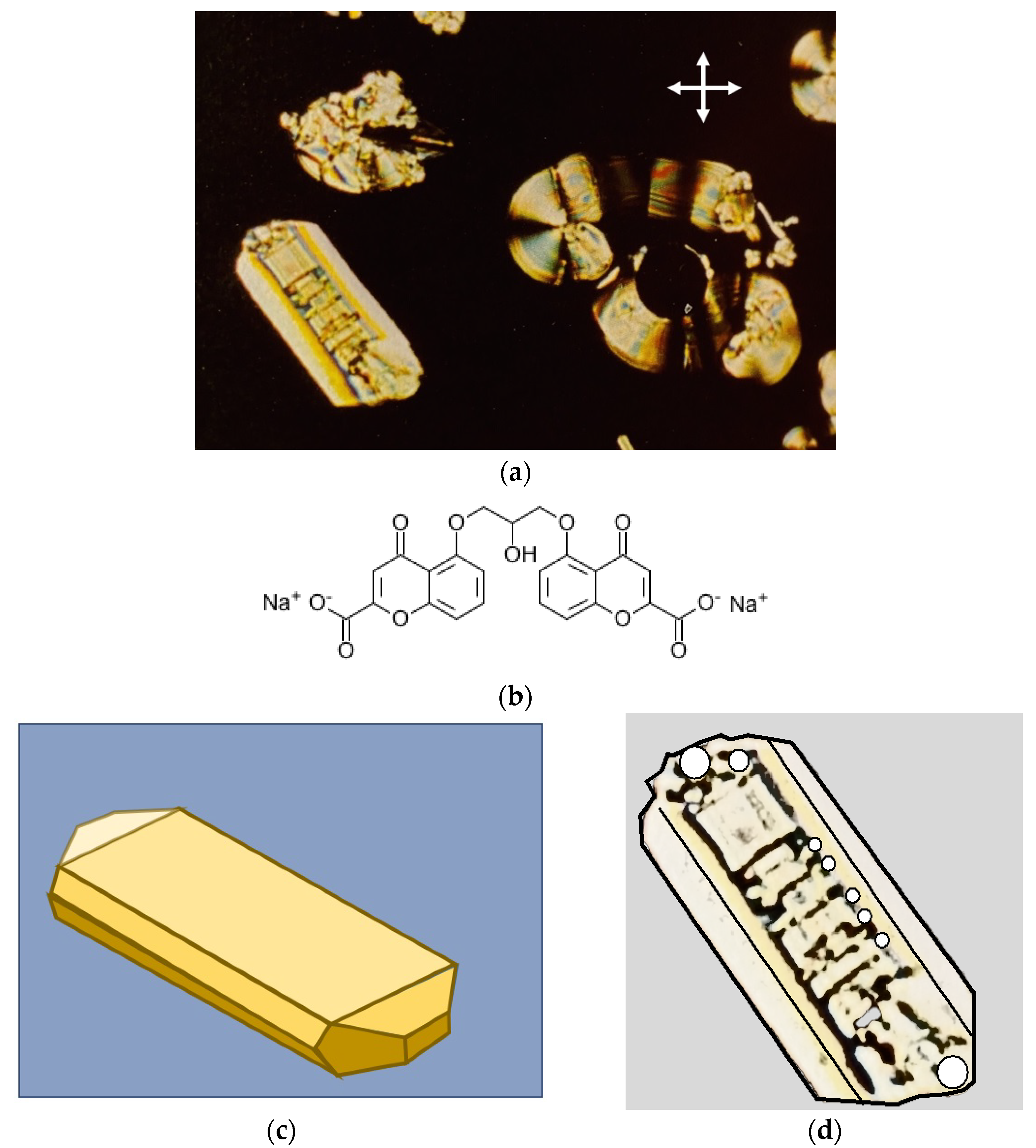
2.1. Diisobutylsilane Diol and Intal

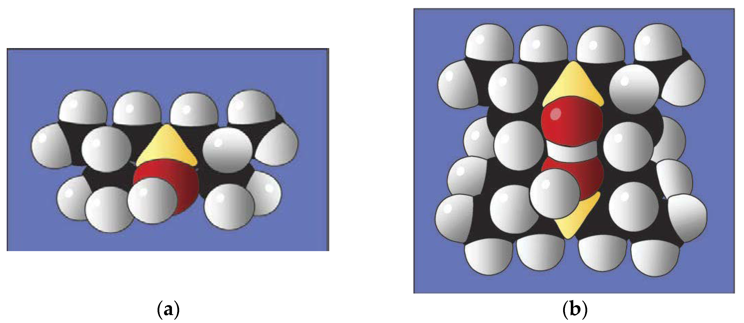
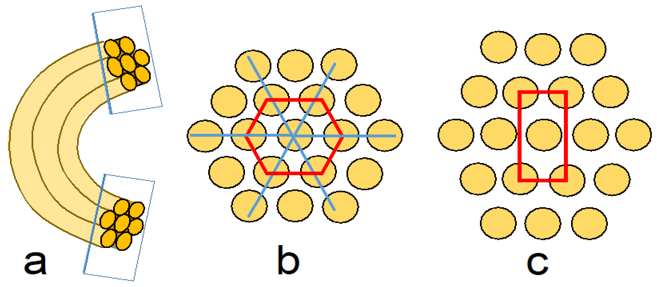
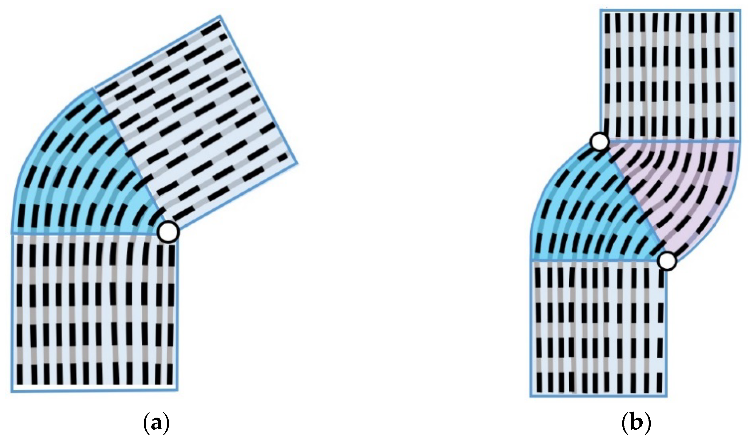
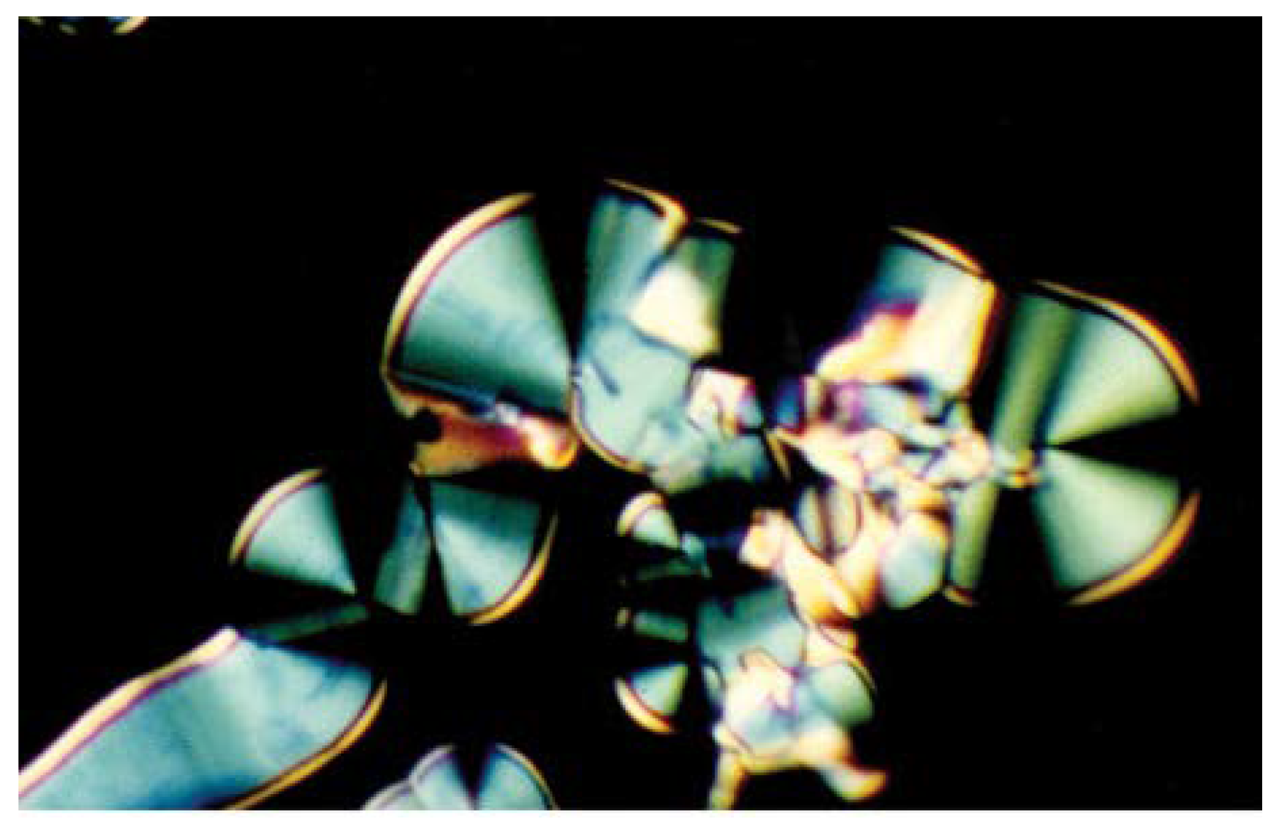
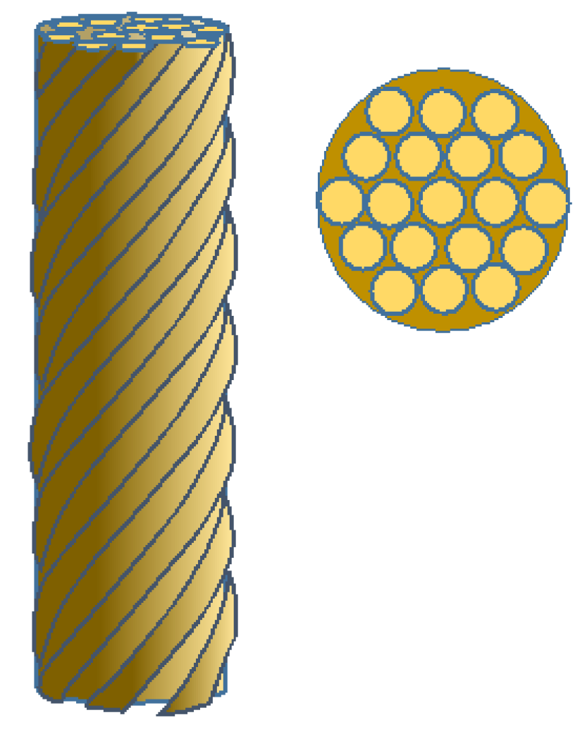
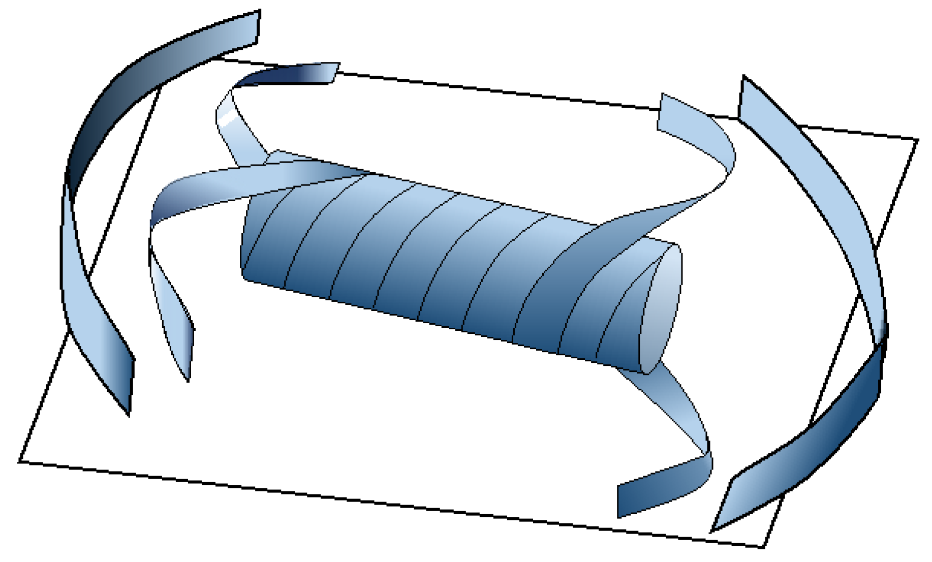
2.2. Developable Domains
2.3. Compression Structures


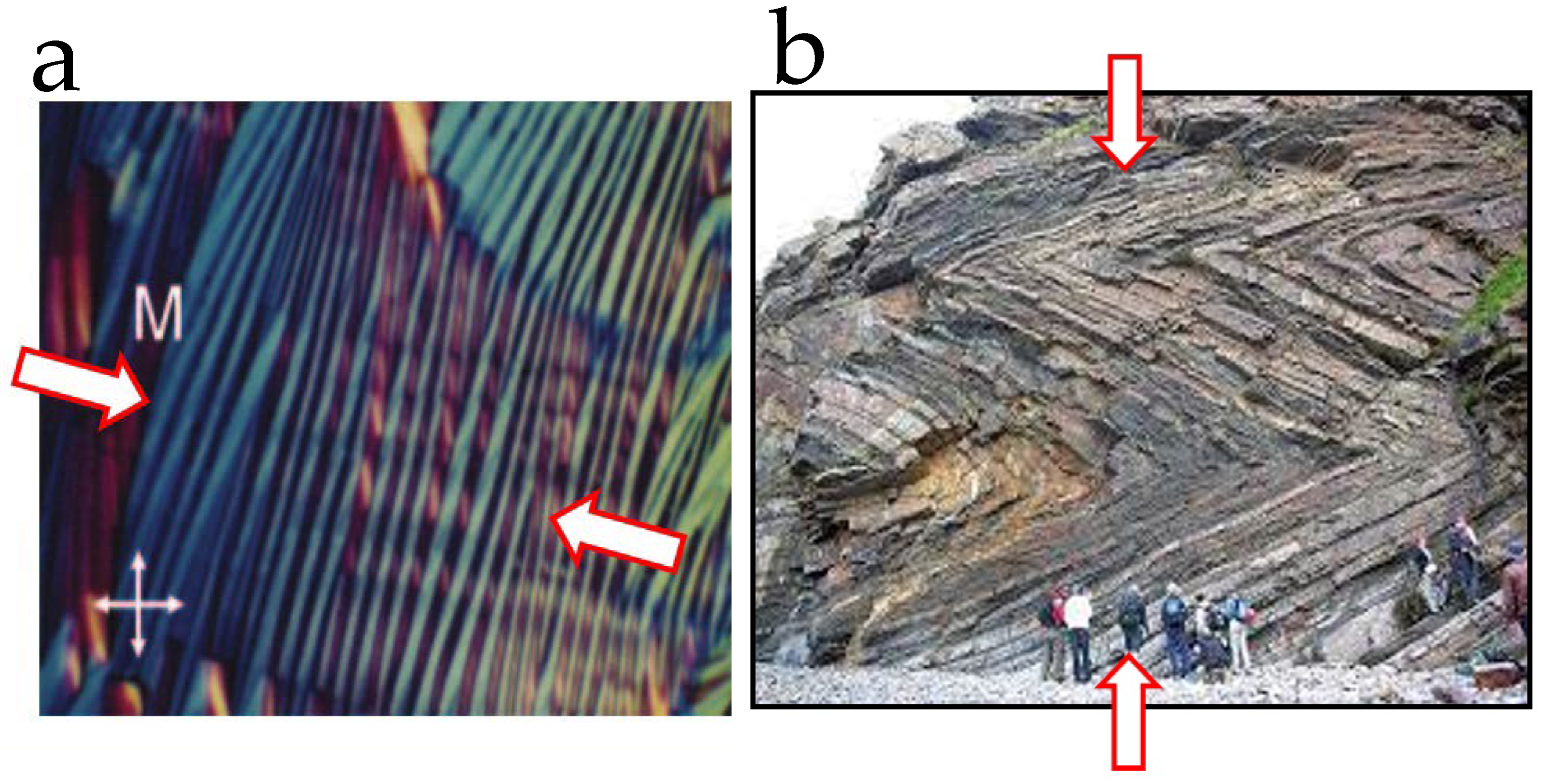
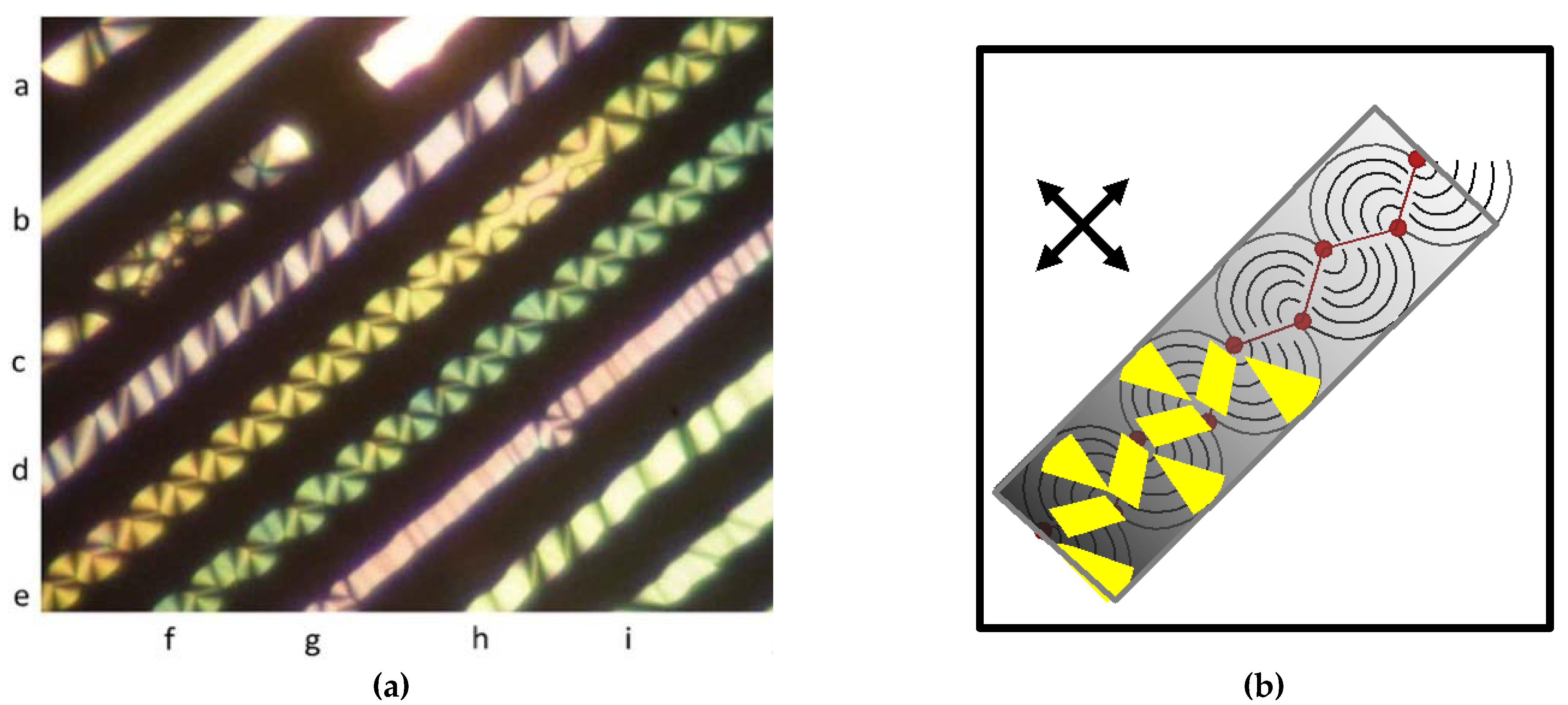
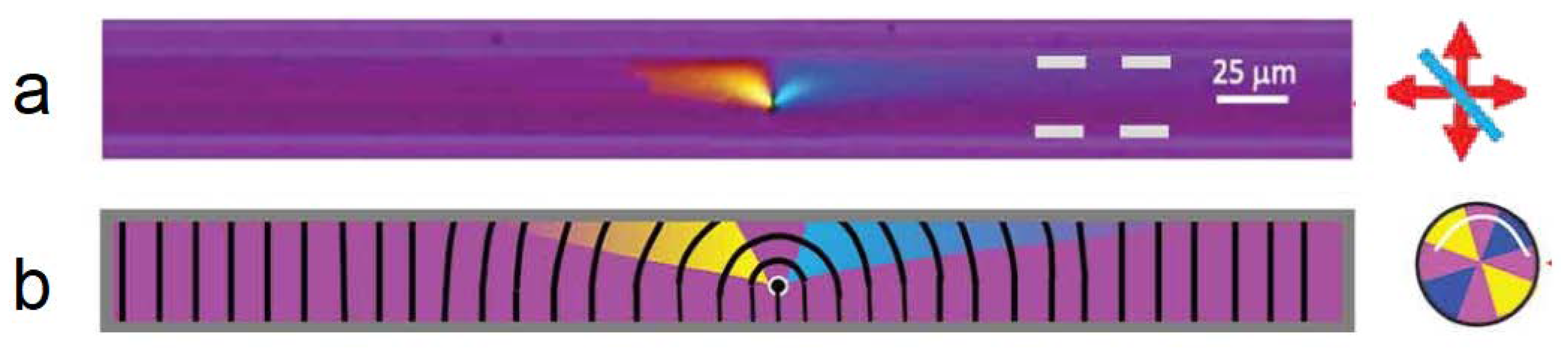
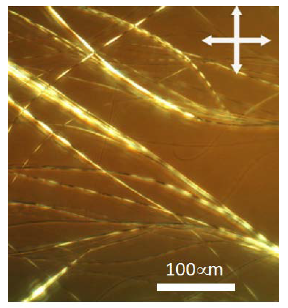
2.4. Optical Textures of Columnar Mesophase Stripes
3. Discussion
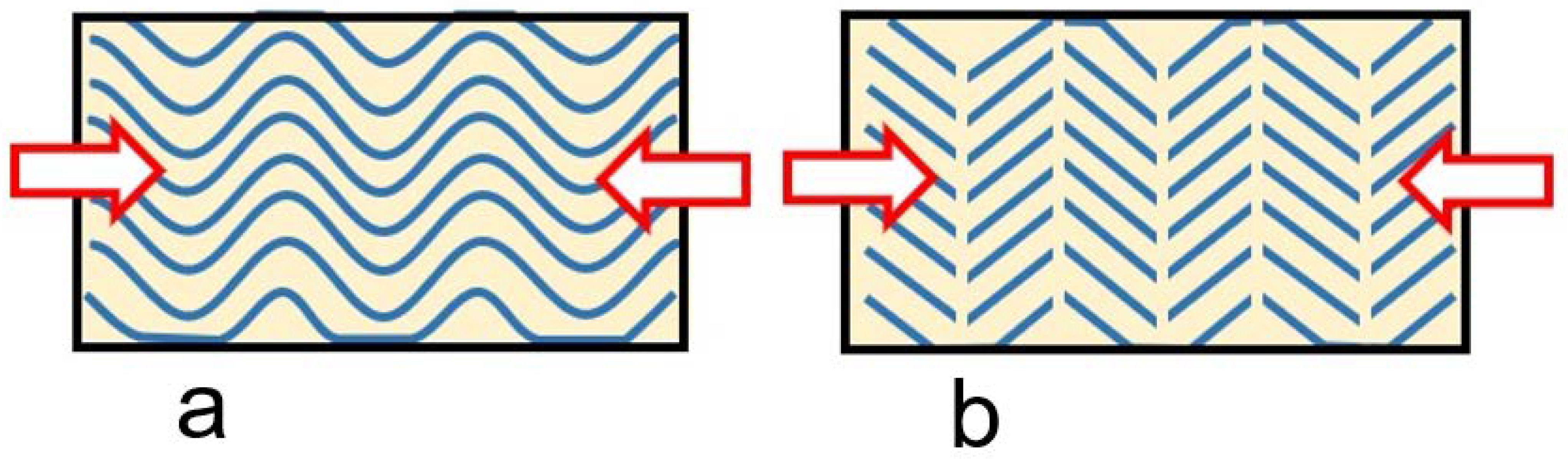
3.1. Slippage
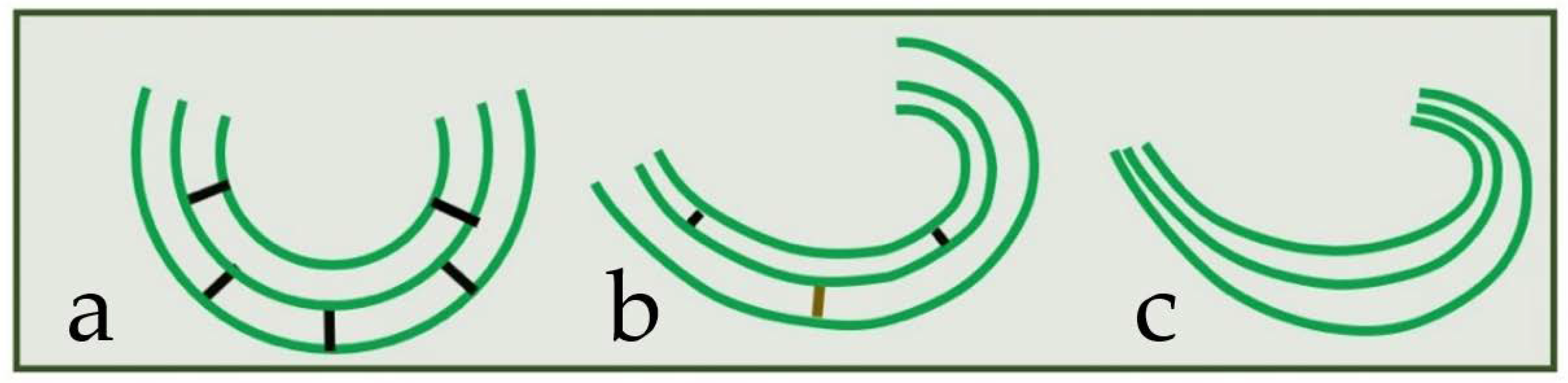
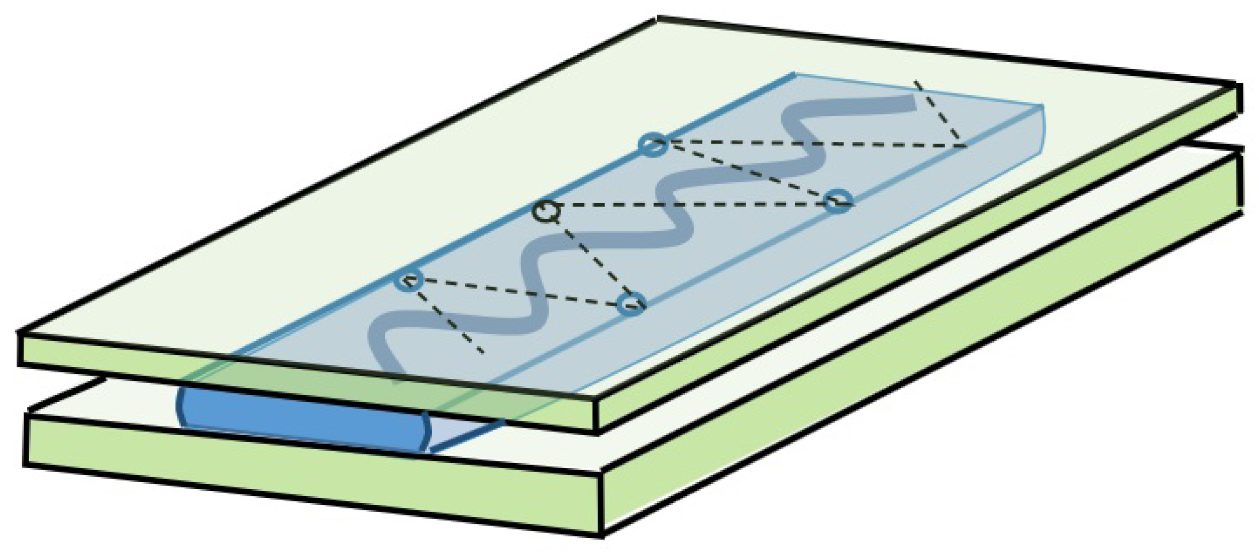
3.2. Striations
“The well-developed striated textures shown by M phases are frequently displayed by M phases between crossed polars, particularly when they have formed slowly or have aged somewhat”.[1] page 114
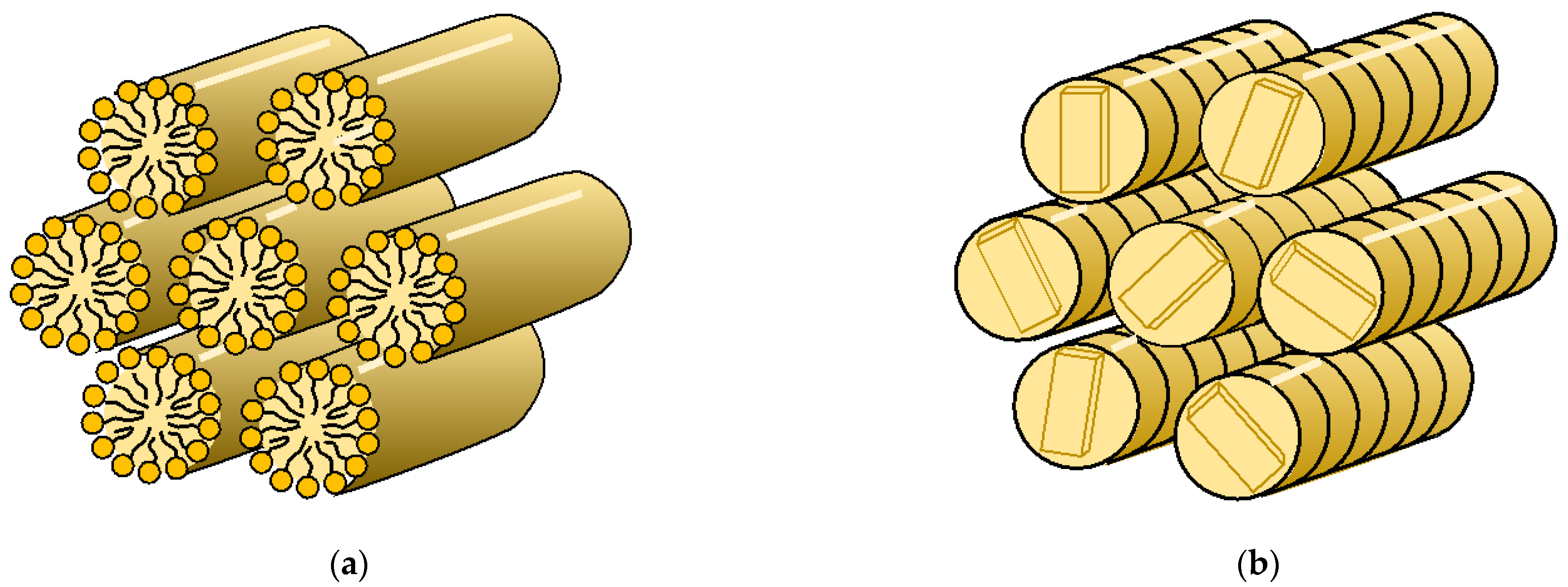
3.3. Helical Bundles
“The relatively restrictive geometry of equidistant fields raises interesting questions about the relationship between the problem of packing finite versus infinite equidistant curves …. The structure of finite equidistant bundles may be much less constrained than equidistant fields. Discrete equidistant bundles of this sort have ready applications to physical systems, from collagen triple helices and other dense packed biological systems”.[19]

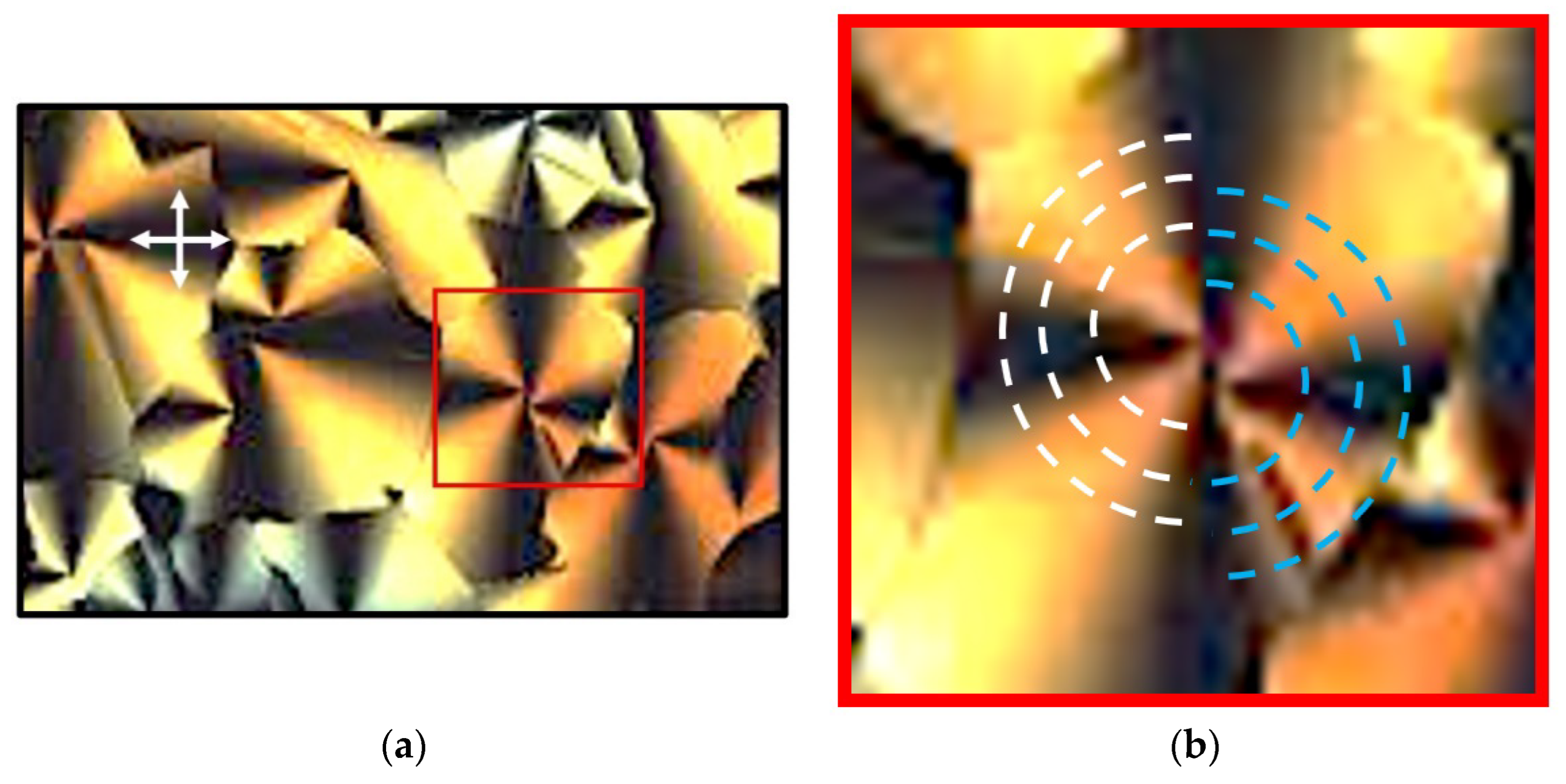
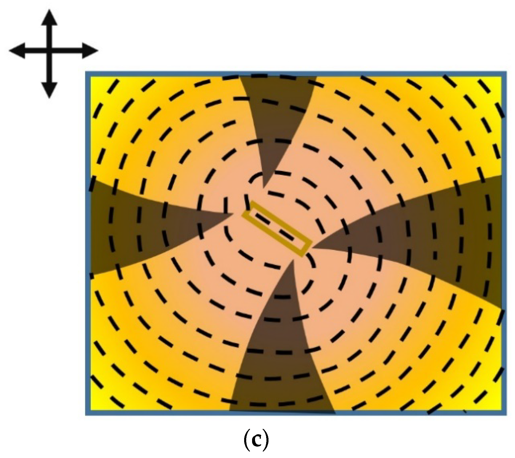
3.4. Multi-Pole Nucleation


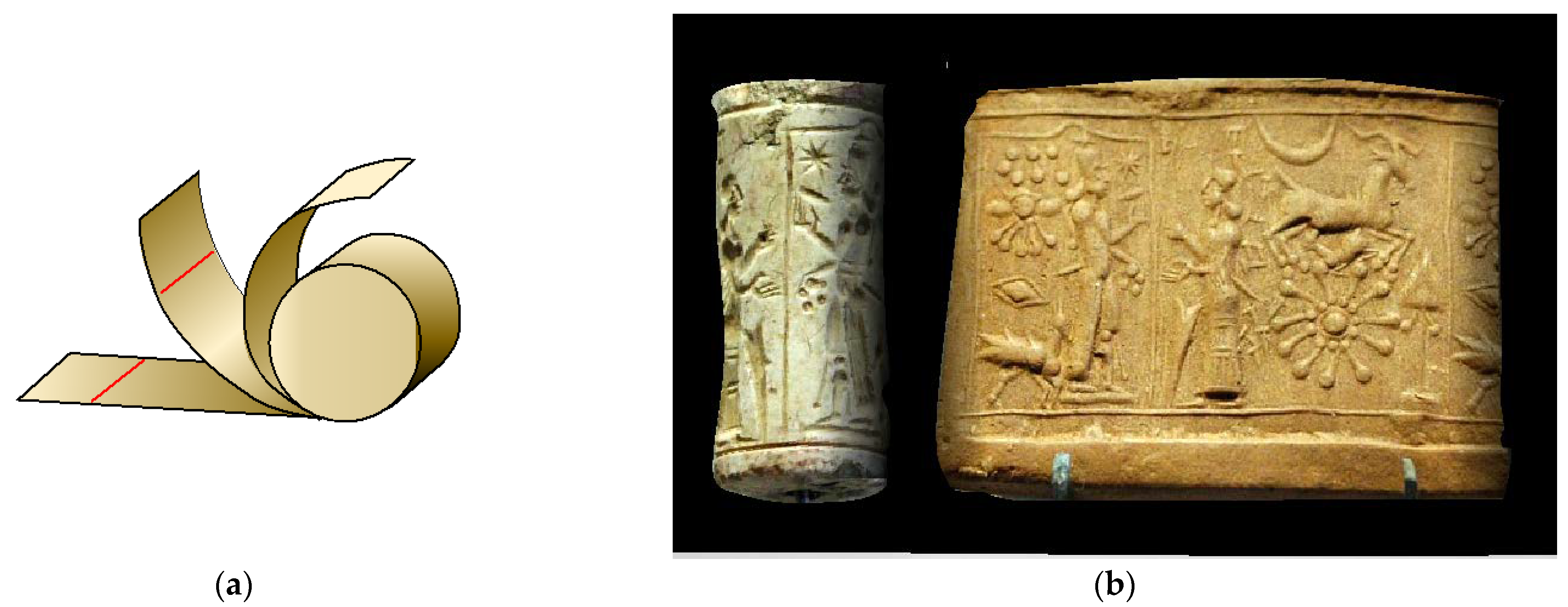
3.5. Helical Fibrils
4. Retrospect
Funding
Acknowledgments
Conflicts of Interest
Dedication
References
- Hartshorne, N.H. (Ed.) The Microscopy of Liquid Crystals; Volume 48 of the Microscope Series; Microscope Publications the University of California Publications: Chicago, IL, USA, 2009; ISBN1 0904962032. ISBN2 9780904962031. [Google Scholar]
- Bouligand, Y. Defects and textures of hexagonal discotics. J. Phys. 1980, 41, 1307–1315. [Google Scholar] [CrossRef]
- Kleman, M. Developable domains in hexagonal liquid crystals. J. Phys. 1980, 41, 737–745. [Google Scholar] [CrossRef]
- Hartshorne, N.H.; Stuart, A. (Eds.) Crystals and the Polarising Microscope, 4th ed.; First published 1934; Hodder & Stoughton Educational: London, UK, 1970; ISBN-10: 0713122560; ISBN-13: 978-713122565. [Google Scholar]
- Dunmur, D.; Slukin, T. (Eds.) Soap, Science, and Flat-Screen TVs: A History of Liquid Crystals; OUP Oxford: Oxford, UK, 2014; ISBN 10-198700830; ISBN 13-0198700838. [Google Scholar]
- Barrall, E.M., II; Porter, R.S.; Johnson, J.F. Temperatures of Liquid Crystal Transitions in Cholesteryl Esters by Differential Thermal Analysis. J. Phys. Chem. 1966, 70, 385–390. [Google Scholar] [CrossRef] [PubMed]
- Bernal, J.D.; Fankuchen, I. Crystallographic studies of Plant Virus Preparations. J. Gen. Physiol. 1941, 25, 111–146. [Google Scholar] [CrossRef] [PubMed] [Green Version]
- Lydon, J.E. A personal history of the early days of chromonics. Liq. Cryst. Today 2007, 16, 13–27. [Google Scholar] [CrossRef]
- Lydon, J.E. New models for the mesophases of disodium cromoglycate (INTAL). Mol. Cryst. Liq. Cryst. 1980, 64, 19–24. [Google Scholar] [CrossRef]
- Lydon, J.E. Chromonic Liquid Crystals. Curr. Opin. Colloid Interface Sci. 1998, 3, 458–466. [Google Scholar] [CrossRef]
- Bunning, J.D.; Lydon, J.E.; Eaborn, C.M.; Jackson, P.; Goodby, J.W.; Gray, G.W. Classification of the mesophase of diisobutylsilanediol. J Chem. Soc. Faraday Trans. 1982, 1, 713–724. [Google Scholar] [CrossRef]
- Lydon, J.E. The Pre-history of discotic mesophases—A personal account of the study of the mesophase of diisobutylsilane diol. Liquid Cryst. 2015, 425, 666–677. [Google Scholar] [CrossRef]
- Chandrasekhar, S.; Sadashiva, B.K.; Suresh, K.A. Liquid crystals of disc-like molecules. Pramana 1977, 9, 471–480. [Google Scholar] [CrossRef]
- Bushby, R.J. The Prehistory of discotic liquid crystal. Liq. Cryst. Today 2014, 42, 14–17. [Google Scholar] [CrossRef]
- Demus, D.; Richter, L. (Eds.) Textures of Liquid Crystals; VEB Deutscher Verlag für Grundstoffindustrie: Leipzig, Germany, 1978; ISBN1 10: 3527257969. ISBN2 13: 9783527257966. [Google Scholar]
- Gray, G.W.; Goodby, J.W.; Hill, L. (Eds.) Smectic Liquid Crystals: Textures and Structures; The University of California: Downtown Oakland, CA, USA, 1984; ISBN1 0863440258. ISBN2 9780863440250. [Google Scholar]
- Dierking, I. (Ed.) Textures of Liquid Crystals; Wiley-VCH Verlag GmbH & Co. KGaA: Weinheim, Germany, 2003; ISBN1 9783527307258. ISBN2 9783527602056. [Google Scholar] [CrossRef]
- Collon, D. First Impressions: Cylinder Seals in the Ancient Near East, Their History and Significance; British Museum Publication: London, UK, 1987. [Google Scholar]
- Atkinson, D.W.; Santangelo, C.D.; Grayson, G.M. Constant spacing of filament bundles. New J. Phys. 2019, 21, 062001. [Google Scholar] [CrossRef]
- Pinterest. Available online: https://www.geologypage.com/2018/12/millook-haven-beach-england.html (accessed on 5 May 2022).
- Bramble, J.P.; Tate, D.J.; Evans, S.D.; Lydon, J.E.; Bushby, R.J. Alternating defects and egg and dart textures in de-wetted stripes of discotic liquid crystal. Liq. Cryst. 2021, 49, 543–558. [Google Scholar] [CrossRef]
- Nakata, M.; Zanchetta, G.; Chapman, B.D.; Jones, C.D.; Cross, J.O.; Pindak, R.; Bellini, T.; Clarke, N.A. DNA duplexes end-to-end stacking and liquid crystal condensation of 6– to 20–Base Pairs. Science 2007, 318, 1276–1279. [Google Scholar] [CrossRef] [PubMed]
- Atkins, E.D.T.; Parker, K.D. The helical structure of a β-D-1,3-xylan. J. Polym. Sci. C Polym. Symp. 2007, 28, 69–81. [Google Scholar] [CrossRef]
- Khan, A.A.; Kamarudin, M.A.; Qasim, M.M.; Wilkinson, T.D. Formation of physical-gel redox electrolytes through self-assembly of discotic liquid crystals: Applications in dye sensitized solar cell. Electrochim. Acta 2017, 244, 162–171. [Google Scholar] [CrossRef] [Green Version]
- Doyle, A.C. A case of identity. In The Adventures of Sherlock Holmes, Adventure III Sherlock Holmes; The Strand Magazine; The Strand: London, UK, 1891. [Google Scholar]


Publisher’s Note: MDPI stays neutral with regard to jurisdictional claims in published maps and institutional affiliations. |
© 2022 by the author. Licensee MDPI, Basel, Switzerland. This article is an open access article distributed under the terms and conditions of the Creative Commons Attribution (CC BY) license (https://creativecommons.org/licenses/by/4.0/).
Share and Cite
Lydon, J.E. Reappraisal of The Optical Textures of Columnar Phases in Terms of Developable Domain Structures with Relaxed Constraints and a Rationale for The Striated Texture. Crystals 2022, 12, 1180. https://doi.org/10.3390/cryst12081180
Lydon JE. Reappraisal of The Optical Textures of Columnar Phases in Terms of Developable Domain Structures with Relaxed Constraints and a Rationale for The Striated Texture. Crystals. 2022; 12(8):1180. https://doi.org/10.3390/cryst12081180
Chicago/Turabian StyleLydon, John E. 2022. "Reappraisal of The Optical Textures of Columnar Phases in Terms of Developable Domain Structures with Relaxed Constraints and a Rationale for The Striated Texture" Crystals 12, no. 8: 1180. https://doi.org/10.3390/cryst12081180