Article

Enhanced Photocatalytic Performance of Ag3PO4/Mn-ZnO Nanocomposite for the Degradation of Tetracycline Hydrochloride

1
Department of Physics, College of Science, King Faisal University, Al Ahsa 31982, Saudi Arabia
2
Department of Physics, University of Gujrat, H.H. Campus, Gujrat 50700, Pakistan
3
Department of Physics, Institute of Physics and Material Science, University of Okara, Okara 56300, Pakistan
4
Department of Chemistry, University of Gujrat, H.H. Campus, Gujrat 50700, Pakistan
5
Department of Chemistry, College of Science, Princess Nourah Bint Abdulrahman University, Riyadh 11671, Saudi Arabia
6
Department of Basic Sciences, Preparatory Year Deanship, King Faisal University, Al Ahsa 31982, Saudi Arabia
7
Department of Civil and Environmental Engineering, College of Engineering, King Faisal University, Al Ahsa 31982, Saudi Arabia
*
Authors to whom correspondence should be addressed.
Crystals 2022, 12(8), 1156; https://doi.org/10.3390/cryst12081156
Submission received: 1 August 2022 / Revised: 11 August 2022 / Accepted: 12 August 2022 / Published: 17 August 2022
(This article belongs to the Section Materials for Energy Applications)

Abstract

:
Using sustainable photocatalysts, photocatalytic degradation has emerged as one of the viable strategies to combat water pollution through eco-friendly and cost-effective means. Visible-light-active Ag3PO4/Mn-ZnO nanocomposite photocatalysts were produced in this study using a simple hydrothermal method and varied concentrations of Ag3PO4 to Mn-ZnO ranging from 0 to 5 wt percent. X-ray diffraction, scanning electron microcopy, energy-dispersive X-ray, transmission electron microscopy, UV–visible spectroscopy, Fourier transform infra-red spectrophotometer, and photoluminescence spectroscopy were used to examine the structural, morphological, and optical properties of synthesized materials. Visible light was used to test the photocatalytic activity of produced Ag3PO4/Mn-ZnO photocatalysts for the breakdown of tetracycline (TC) hydrochloride. In comparison to the other samples, the 3% Ag3PO4/Mn-ZnO nanocomposite exhibited superior activity as a result of improved visible light absorption and suppressed charge carrier recombination. In addition, this sample demonstrated good stability of TC in an aqueous environment after five consecutive cycles. This research will enhance the scope of photocatalysis for environmental applications.

1. Introduction

The presence of toxic organic dyes, disinfectants, medications, preservatives, and additives in the environment has negative consequences on humans, aquatic ecosystems, and environmental biodiversity. As a result of researchers’ heightened focus on this critical issue [1,2], eco-friendly solutions for effluents and enhanced control over environmental contaminants are being developed to mitigate the adverse impacts of these contaminations on humans. Urgent provision must be adopted to provide cleaned and sanitized water to suit the needs of humans, aquatic life, and irrigation [3]. For the treatment of water originating from various industries, a large array of new approaches is employed. These tactics include oxidation, filtration, precipitation, coagulation, adsorption, and flocculation. They are physical, chemical, and biological in nature. However, it should be noted that these approaches are not very effective because they generate secondary pollutants that are still harmful to the environment [4]. There is an urgent need for creative, eco-friendly, and cost-effective techniques to improve the efficacy of water treatment in order to generate clean water for the benefit of human and aquatic life.
Photocatalytic degradation is a good technique for removing contaminants from waste water as compared to other methods [5]. In photocatalysis, it is necessary to employ a semiconductor material that is non-toxic, cost-effective, has a broad absorption spectrum, etc. In the past few decades, a variety of semiconductors such as metal oxides and metal sulfides, e.g., TiO2, ZnO, WO3, MoS2, and CdS, have emerged as promising candidates for photocatalysis [6]. ZnO has become a leading and successful material in the field of photodegradation due to its unique qualities, which include cost-effectiveness, excellent photo-stability, easy preparation, non-toxicity, and exceptional chemical stability [7]. However, pure ZnO is a semiconducting material with a wide band gap (3.37 eV) that only absorbs light in the ultraviolet part of the solar spectrum [7,8]. Consequently, only 3–5% of the solar spectrum is utilized for photocatalytic activity, limiting its practical applicability [9,10,11]. Diverse procedures, such as regulating crystal phase, oxygen vacancy production, active facet and morphology development, doping, noble metal deposition, and preparation of composites with other semiconductors are applied to address this issue[12,13,14,15,16,17,18]. These methodologies have effectively increased the photocatalytic efficiency of ZnO for the breakdown of contaminants. Doping ZnO with Mn is an efficient strategy to reduce the band gap of ZnO via introducing intermediate states between the conduction and valence bands of ZnO [19,20,21,22]. Moreover, it is known that Mn in ZnO adopts divalent state, thus Mn2+ ions have the possibility to substitute Zn2+ in ZnO lattice [21]. Another useful feature that enhances photocatalytic activity is to increase the capturing of electrons on the doping center, thus good electron transportation and charge carrier’s separation can be achieved [22]. Mn2+ ions with half-filled configuration have the ability to trap electrons effectively to promote the charge carrier’s separation process for higher photocatalytic activity [21].
The coupling of a semiconductor with a semiconducting material with a large energy band gap to form a composite is also a useful strategy for increasing the photocatalytic efficiency of a material [23]. Therefore, the coupling of Ag3PO4 with Mn-doped ZnO (Ag3PO4/Mn-ZnO) is an effective method for enhancing the photocatalytic activity of the resulting photocatalyst [24]. This was carried out owing to the fact that Ag3PO4 is a visible-light-active semiconductor with 2.45 eV band gap energy, having conduction band potential at 0.329 eV energy and valance band potential at 2.589 eV energy. No literature was identified on this nanocomposite-based substance, as far as we are aware.
In this study, Ag3PO4/Mn-ZnO nanocomposite was fabricated using a simple hydrothermal process and evaluated by XRD, SEM, EDX, TEM, UV–Vis spectroscopy, FTIR, and PL spectroscopy. In addition, the photodegradation activity against the antibiotic tetracycline hydrochloride was investigated utilizing the produced Ag3PO4/Mn-ZnO nanocomposite under visible light.

2. Experimental Method

2.1. Chemical Reagents

Zinc acetate (ZnC₄H₆O₄), manganese acetate (C4H6MnO4), silver nitrate (AgNO3), disodium phosphate (Na2HPO4), methanol (CH3OH), sodium hydroxide (NaOH), and distilled water (H2O) were purchased directly from Sigma-Aldrich (Sigma Aldrich, St. Louis, MI, USA) and utilized as obtained.

2.2. Fabrication of Materials

2.2.1. Fabrication of Mn-ZnO

Mn-doped ZnO was synthesized using hydrothermal method. Solution ‘A’ was synthesized by adding 3 g of zinc acetate into 50 mL of methanol. Then, solution ‘B’ was prepared by the addition of 0.06 g manganese acetate into 25 mL of methanol with constant stirring for 15 min. Next, both solutions A and B were mixed to obtain solution ‘C’. After that, sodium hydroxide was also poured dropwise into this solution ‘C’ in order to maintain the pH level at 11 under constant stirring for 15 min to obtain a homogeneous solution. For hydrothermal processing, the final solution was poured into a Teflon-lined stainless-steel autoclave and maintained at 150 °C for 3 h in a Memmert oven. The final product was purified using methanol and distilled water three times. After filtration, the product was maintained at 70 °C in a laboratory oven for drying purpose, and finally the dried Mn-ZnO product was collected. Pure ZnO was prepared by following the same route except adding dopant precursor [25,26]. The as-synthesized samples were labeled as Z (pure ZnO) and MZ (Mn-doped ZnO).

2.2.2. Fabrication of Ag3PO4/Mn-ZnO

To synthesize Ag3PO4/Mn-ZnO nanocomposite, 2 g of Mn-ZnO was dissolved into 50 mL of distilled water and magnetically stirred for 20 min to obtain solution ‘A’. In the next step, 0.02 g of AgNO3 was added into 30 mL of distilled water to make solution ‘B’, and stirred magnetically for 20 min. Next, the solutions ‘A’ and ‘B’ were mixed and continuously stirred to form the homogenous solution ‘C’. Then, 0.1 M ammonia solution (20 mL) was added dropwise into the solution ‘C’. This final solution was again kept for magnetic stirring till the formation of transparent solution. Afterward, 0.02 g of Na2HPO4 was also added into the above solution under continuous stirring of 20 min, and precipitates were obtained finally. After this, centrifuging and washing was performed before drying at 70 °C for 10 h. By changing the weight percentages of Ag3PO4 in the above-mentioned synthesis route, the other remaining samples with different concentrations of Ag3PO4 were also collected. Pure Ag3PO4 was synthesized by following the same route except adding Mn-ZnO [25]. All these samples were labeled as AP (pure Ag3PO4) 1APMZ (1 wt.%Ag3PO4/Mn-ZnO), 3APMZ (3 wt.% Ag3PO4/Mn-ZnO), and 5APMZ (5 wt.% Ag3PO4/Mn-ZnO).

2.3. Characterization

After synthesis, nano-materials were analyzed with the help of X-ray powder diffraction using an X-ray diffractometer (Shimadzu XRD 7000) equipped with radiation source of Cu Kα radiation (λ = 1.5418 Å) to study structural properties, including crystallite size and crystal phase. The morphological study, such as shape, size, and arrangement of particles of the synthesized nano-materials, was performed via scanning electron microscope (ZIESS Germany Gemini SEM 500) furnished with an energy-dispersive X-ray spectrometer, EDX (High Wycombe, UK), for the analysis of chemical composition of nano-materials. Furthermore, transmission electron microscopy (TEM; JEOL JEM-200EX electron microscope) was used to study the morphology of prepared samples. To examine the optical properties of as-synthesized nanomaterials, a UV–visible spectrophotometer (UV-1800, Shimadzu, Koyota, Japan) was used and absorption spectrums were recorded over the range of 300–800 nm. The specific functional groups were determined by Fourier transform infrared spectrophotometer (FTIR, Perkin-Elmer System 2000). A fluorescence spectrophotometer was used to study the photoluminescence (PL) spectra (F-7000 DC-0506, HTACHI, Japan).

2.4. Photodegradation Activity

The photodegradation efficiency of as-synthesized nanomaterials was estimated against tetracycline hydrochloride, an antibiotic. Photocatalytic reaction was conducted in a photocatalytic reactor (BL-GHX-V, Shanghai, China) equipped with a xenon lamp of 250 watt emitting visible light (290–700 nm). The visible light (λ ≥ 400 nm) used in all photocatalytic experiments was obtained using filter with cut-off wavelength of 400 nm. To examine the degradation activity of as-synthesized photocatalysts, a 40 mg quantity of the photocatalyst (0.4 g/L) was added into 100 mL of tetracycline hydrochloride possessing concentration of 25 mg/100 mL during each experiment. The solution was kept under constant stirring for 60 min under dark in order to achieve adsorption–desorption equilibrium. After this step, the solution was illuminated by visible light and 5 mL solution was taken out after every 30 min time duration. For the purpose of antibiotic’s degradation efficiency measurement, a UV–Vis spectrophotometer (UV-1800, Shimadzu, Japan) was employed.
For reusability experiment (1 cycle), the photocatalyst was separated from the TC solution using centrifugation. Then, the obtained photocatalyst was washed seven times to remove the remnants of TC attached to the surface. After this, the photocatalyst was dried in an oven at 100 °C for 4 h. The same process was repeated for another four cycles. The time span between each cycle was about 6 h.

3. Results and Discussion
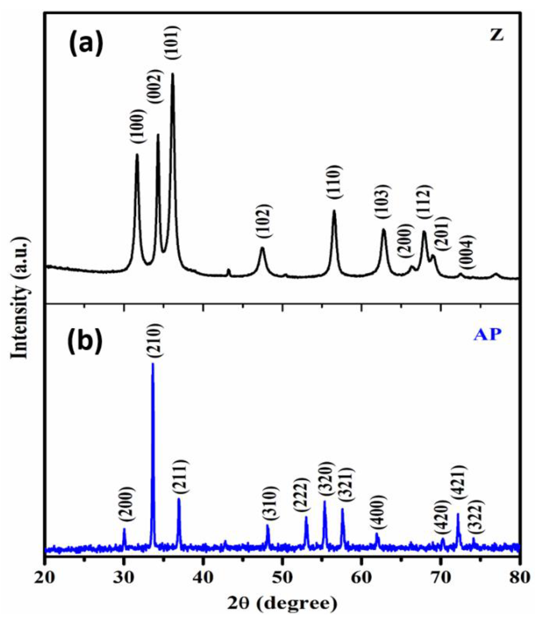
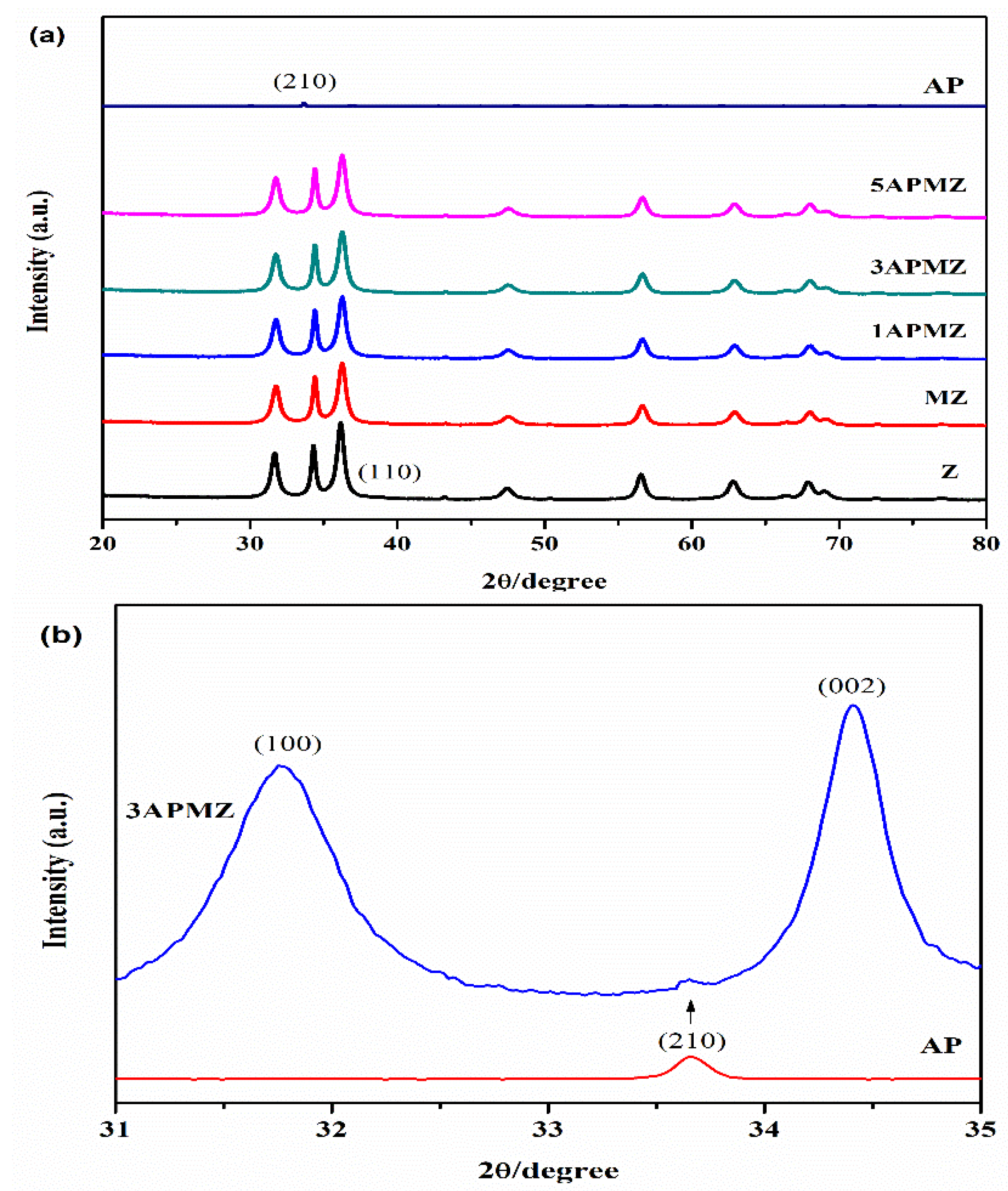
3.1. XRD Analysis

The fabricated samples were characterized to study crystalline structure using X-ray diffraction (XRD). The XRD results for pure ZnO and Ag3PO4 samples are depicted in Figure 1. The diffraction peaks with hkl values (100), (002), (101), (102), (110), (103), (200), (112), and (201) were observed at 31.5°, 34.2°, 36.2°, 35.6°, 47.4°, 56.6°, 62.4°, 68.2°, and 69.1°, respectively (see Figure 1a), and a characteristic peak was also observed at 36.2°. These values were in good agreement with standard data (JCPDS No. 79-0205) and also confirmed the hexagonal phase wurtzite structure of zinc oxide. The presence of these intense and sharp diffraction peaks prove the high crystallinity of zinc oxide. The diffraction peaks for Ag3PO4 nano-material were observed at planes (200), (210), (211), (310), (320), (321), (400), (420), and (421), corresponding to 2θ values at 30°, 33.6°, 36.7°, 48.2°, 53.2°, 55.3°, 57.8°, 62.1°, 70.1°, and 72.7°, respectively, as revealed in Figure 1b, and a characteristic peak was also detected at 2θ = 33.6° value. All these planes were well indexed with standardized values (JCPDS No. 06-0505). The crystalline structure of as-prepared Ag3PO4 was confirmed as cubic body centered.
Figure 2a presents a comparison of XRD patterns of Z, MZ, 1APMZ, 3APMZ, 5APMZ, and AP samples. It can be observed that by doping Mn into ZnO, the intensities of diffraction peaks were reduced due to the fact that Mn was incorporated into the ZnO crystal lattice. No extra diffraction peak was observed in the XRD graph of the MZ sample, which confirmed absence of impurity traces [27]. Intensities of diffraction peaks related to pure ZnO are very high in comparison to Ag3PO4, so its diffraction peaks were suppressed in the resulting pattern. An enlarged view of the XRD pattern related to the 3APMZ sample, as shown in Figure 2b, shows that there was a presence of (210) diffraction peak which was due to Ag3PO4 material. This confirms the formation of Ag3PO4/Mn-ZnO nanocomposite. Moreover, the crystallite sizes of all synthesized samples were calculated based on Scherrer equation. The calculated crystallite sizes for Ag3PO4, ZnO, Mn-ZnO, 1APMZ, 5APMZ, and 3APMZ were 20 nm, 34 nm, 32 nm, 28 nm, 21.5 nm, and 20.3 nm, respectively. This indicates a decrease in crystallite size due to doping of Mn and loading of Ag3PO4.

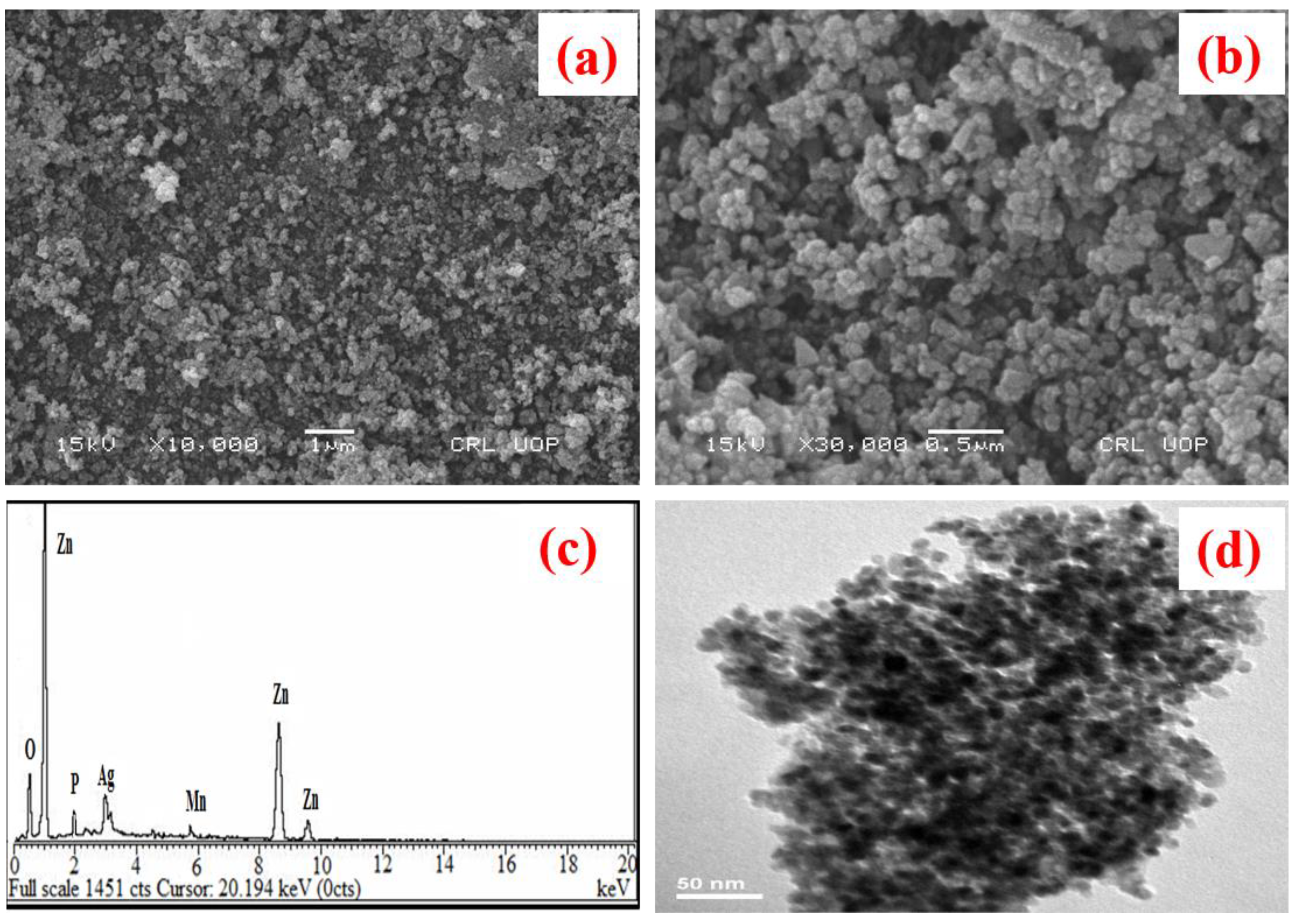
3.2. SEM, EDX, and TEM Analysis

SEM images of as-synthesized 3APMZ nanocomposites with different magnification are shown in Figure 3a,b, respectively. This image (Figure 3b) revealed that 3APMZ nanocomposite was based on nano-clusters-based morphology. Furthermore, these nano-clusters are found to possess irregular shapes with different sizes. Furthermore, the energy-dispersive X-ray (EDX) spectroscopy was operated for the analysis of chemical composition and purity of synthesized 3APMZ nanocomposite. EDX pattern confirmed the presence of O, P, Ag, Mn, and Zn, as is obvious from Figure 3c. In the indexed table (see Figure 3c), the atomic and weight percentages of elements present in as-synthesized 3APMZ nanocomposite were also presented. The highly pure nature of the as-synthesized sample was confirmed by this pattern as there was no other element presence in the material. Figure 3d exhibits the TEM nanoparticles-based structure of Ag3PO4/Mn-ZnO composite which has average size of both types of nanoparticles in the range of 20–34 nm. It can be seen that there are two types of nanoparticles, which might be Ag3PO4 nanoparticles (with smaller size) deposited on larger-sized Mn-ZnO nanoparticles.

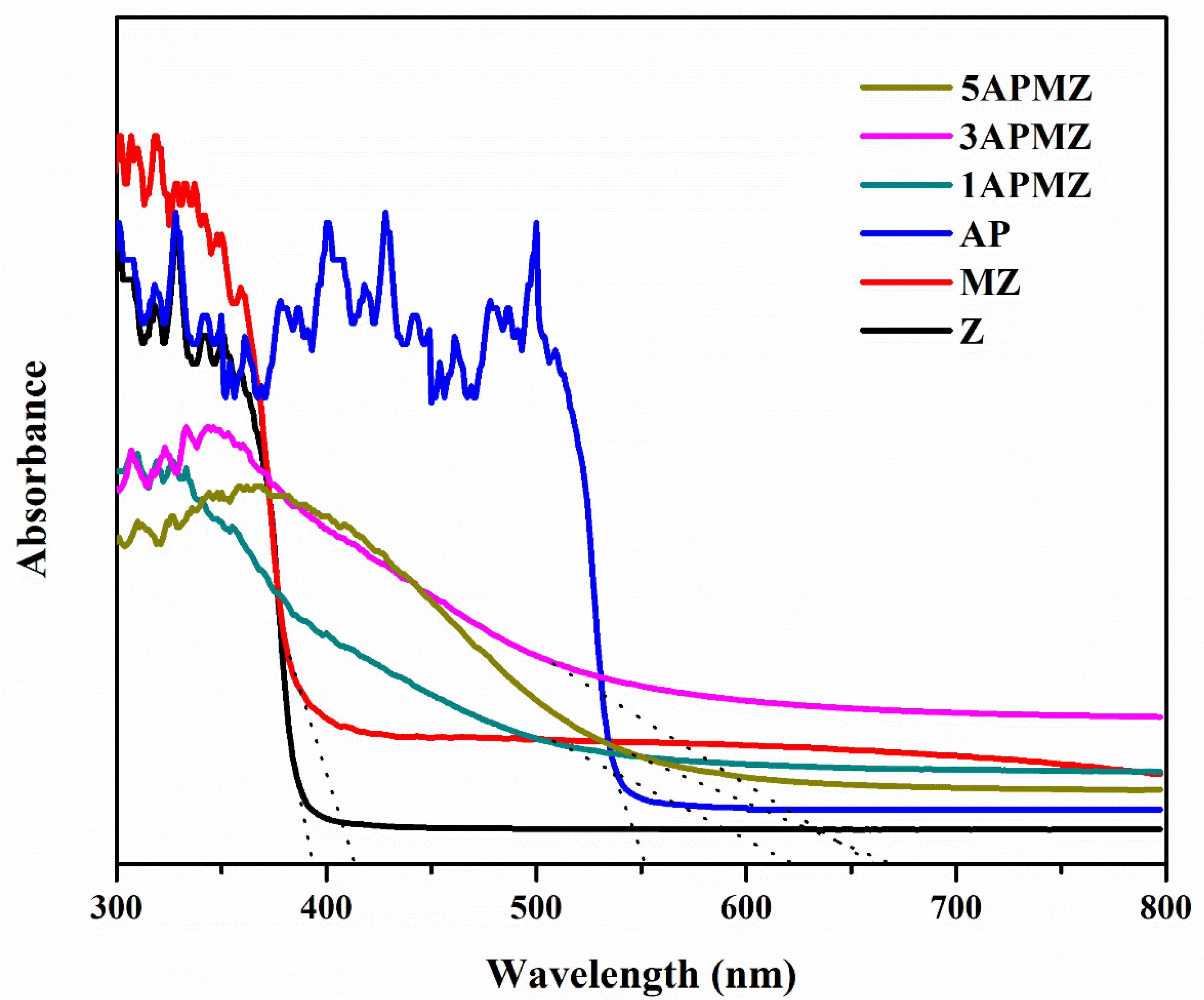
3.3. UV–Visible Spectroscopy
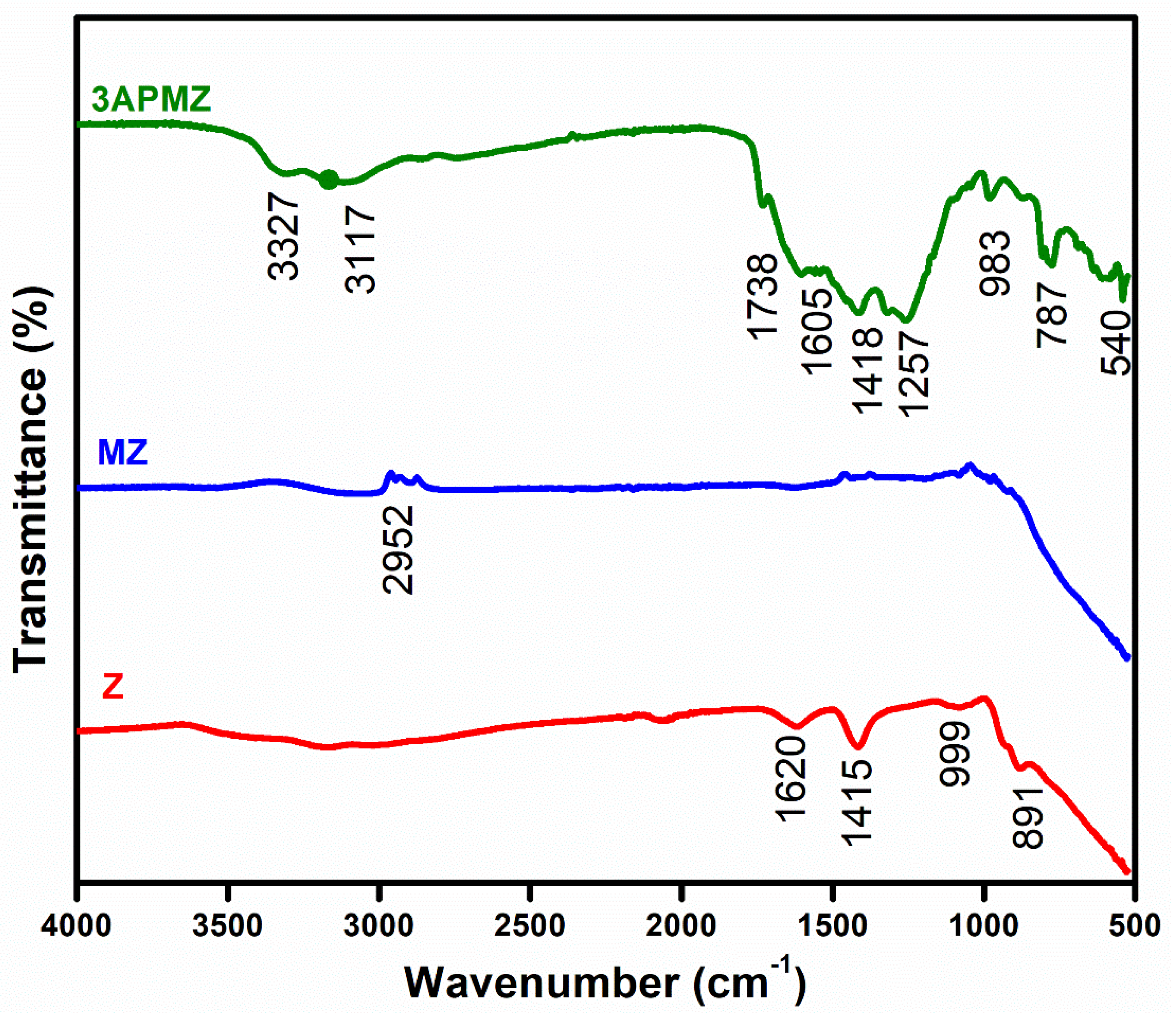
By using a UV–Vis spectrophotometer, the absorption spectra were recorded over the range of 300–800 nm and are shown in Figure 4. One can see that pure ZnO fell in the range of the UV region with 393.53 nm wavelength, so it can only harvest 3–5% of the electromagnetic spectrum, while Mn doping into ZnO caused a red shift in absorption range towards higher wavelength corresponding to visible light region. The reason behind this red shift is the formation of impurity levels within the band gap of the ZnO nanostructure [19]. From the absorption spectrum, it can be concluded that incorporation of Ag3PO4 into Mn-ZnO resulted in further shifting towards longer wavelength in the visible region. It is proposed that this happened due to the fact that Ag3PO4 facilitated transportation of photo-excited electron–hole pairs. The band gap energies were also calculated and are given in Table 1. The decrease in band gap energy due to increasing concentration of Ag3PO4 in a synthesized nanocomposite shows that the material will be effective for higher absorption of visible light and enhanced photocatalytic performance.

3.4. FTIR Analysis

FTIR spectroscopic study gives quantitative and qualitative information about differential groups and bonds in chemical compounds. Figure 5 shows the FTIR spectra of ZnO (Z), Mn-ZnO (MZ), and Ag3PO4/Mn-ZnO (3APMZ) at room temperature in the range of 500 to 4000 cm−1. Stretching vibrations of the ZnO show the bands at 891 and 999 cm−1 in the tetrahedral coordination. The minor shift was observed towards the higher wavenumber in the characteristic peak of the ZnO because of Mn doping into the ZnO structure, which shows the bond strengths. Moreover, other absorption peaks are detected at 2952, 3117, and 3327 cm−1 for MZ and 3APMZ samples, respectively. The bending mode origin of H–OH is observed at 1257, 1418, 1605, and 1738 cm−1 for the 3APMZ sample. The stretching bands are found at 540, 787, and 983 cm−1 for the respective 3APMZ samples [28].

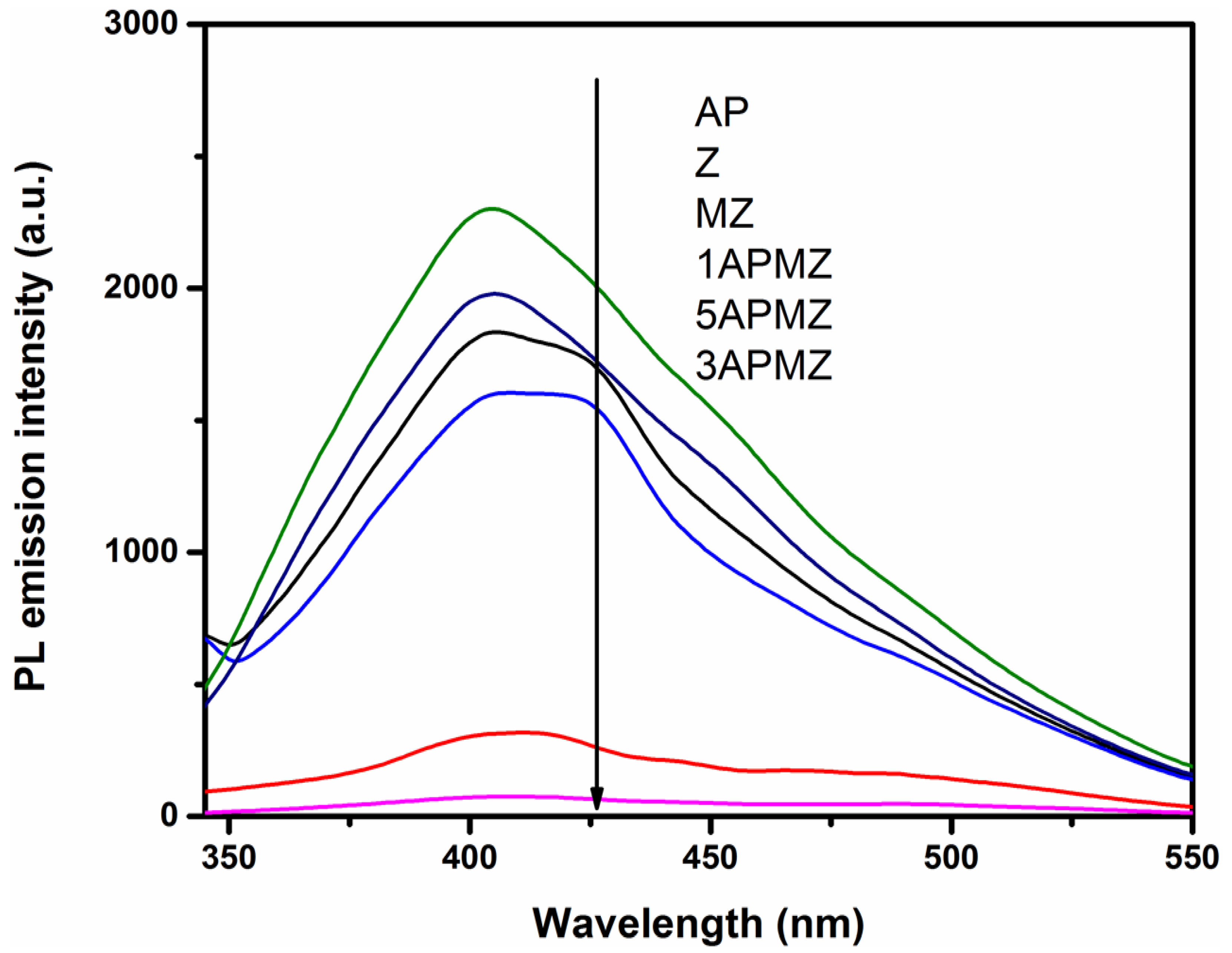
3.5. Photoluminescence Analysis

Generation, transmission, and separation proficiency of charge carriers can be studied via photoluminescence (PL) analysis. If the PL emission intensity is higher, then it shows recombination of charge carriers that is high and fast [29]. PL spectra of different photocatalysts at the excitation wavelength of 325 nm are depicted in Figure 6. As shown in the results, the Ag3PO4 (AP) spectrum reveals the higher recombination of electrons and holes as compared to all other photocatalysts. PL emission intensity of ZnO decreases due to combination of Mn and Ag3PO4, as shown in Figure 6. Moreover, the 3APMZ composite photocatalyst exhibited minimum PL emission intensity, which is due to the reduced recombination of electrons/holes as compared to all other composite samples. In summary, the results demonstrated that the loading of Mn and Ag3PO4 into ZnO is a suitable approach to diminish the recombination of charges. Therefore, the 3APMZ sample is expected to perform the best for enhanced photocatalytic activity in the degradation of tetracycline hydrochloride.

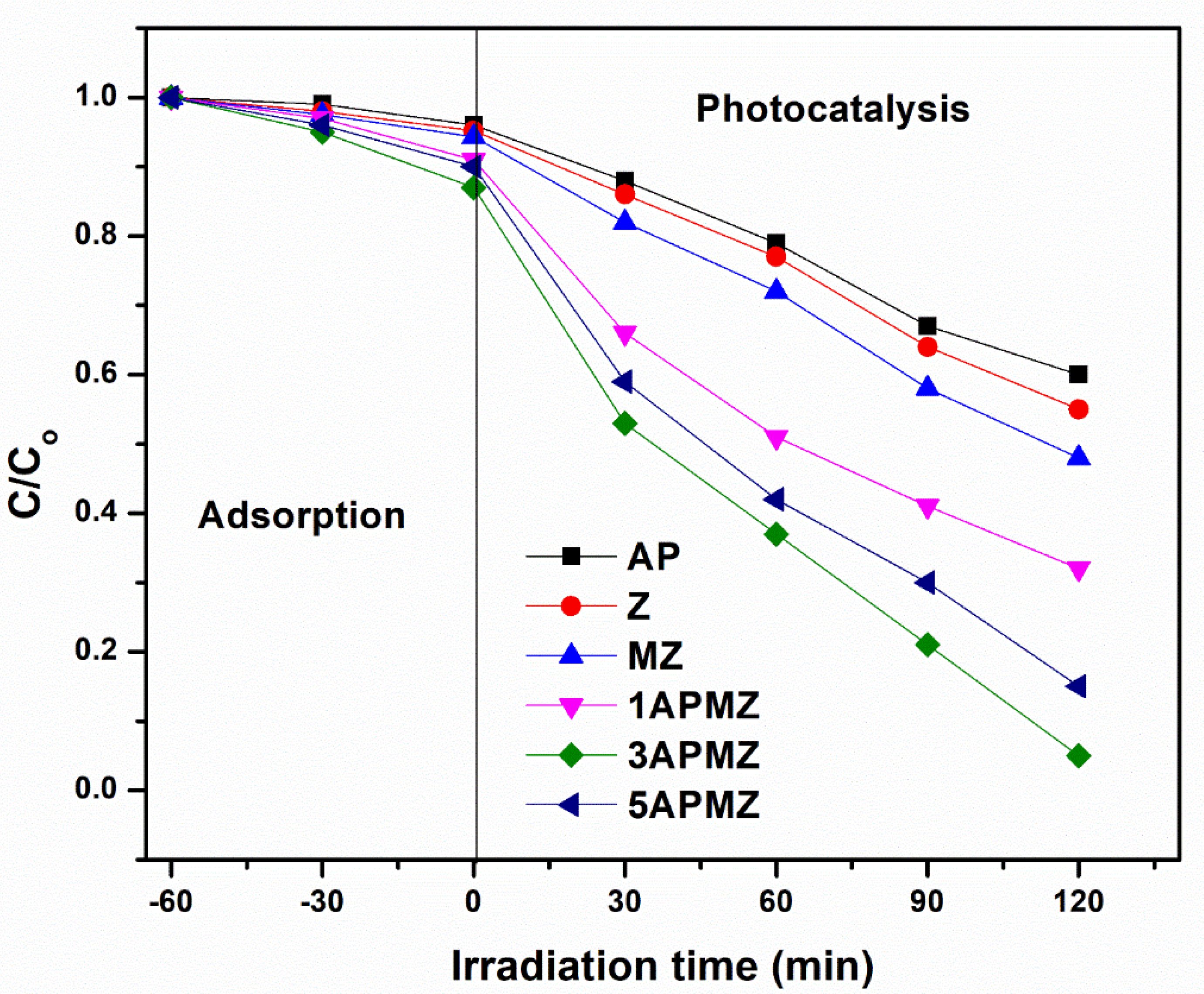
3.6. Photodegradation Activity

To evaluate the efficiency of the prepared composites, the photocatalytic properties of the synthesized photocatalysts, including pure Ag3PO4 (AP), ZnO(Z), Mn-ZnO (MZ), 1 wt.% Ag3PO4/Mn-ZnO (1APMZ), 3 wt.% Ag3PO4/Mn-ZnO (3APMZ), and 5 wt.% Ag3PO4/Mn-ZnO (5APMZ), were examined by the photodecomposition of tetracycline (TC) under visible light irradiation. As depicted in Figure 7, the adsorption–desorption equilibrium of TC using different photocatalysts was investigated for 60 min reaction in the dark. Almost 13% of TC was adsorbed via 3APMZ composite, which is more than other synthesized composites, showing that 3APMZ composites have higher adsorption ability. Furthermore, the degradation of TC for different samples was found to be AP (57 %), Z (64 %), MZ (69 %), 1APMZ (81 %), 3APMZ (95 %), and 5APMZ (85 %), respectively. The pseudo-first-order reaction rate constant is found via the following formula [29]:
K app = 1 t   ln Co C
where Co is the TC concentration before irradiation and C is the TC concentration after irradiation of time. We calculated the pseudo-first-order reaction rate constants as 0.0037 min−1 for AP, 0.0045 min−1 for Z, 0.0064 min−1 for MZ, 0.0097 min−1 for 1APMZ, 0.01164 min−1 for 5APMZ, and 0.01263 min−1 for 3APMZ.
As compared to all other samples, the 3APMZ photocatalysts showed 95% degradation of TC under visible light illumination. This shows that optimum loading of Ag3PO4 into Mn-ZnO (3APMZ) enhanced the photodegradation performance of the photocatalyst. However, excessive loading of Ag3PO4 (5APMZ) above optimum level decreases the efficiency of material due to higher recombination of photoexcited charges, as was confirmed by PL results. Moreover, the photocatalytic performance of the prepared photocatalyst (3APMZ) was compared with the literature, as shown in Table 2. The results show that the prepared photocatalyst in this study has good photocatalytic activity compared to most of the studies.

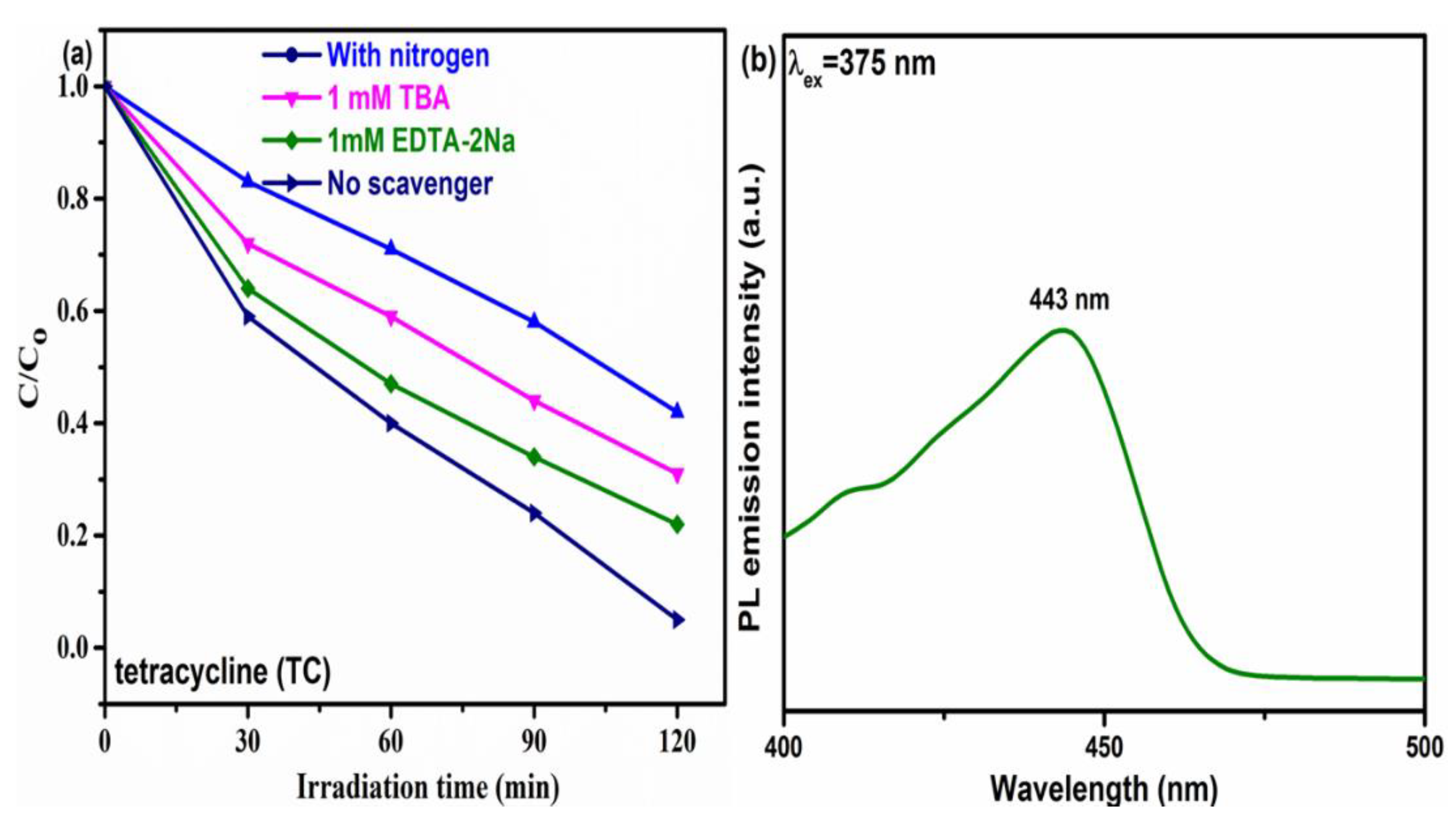
3.7. Effect of Scavengers

The photocatalytic oxidative species could be known by using ethylenediamine tetra acetic acid disodium (EDTA-2Na) and tert-butanol (TBA) as scavengers of holes (h+) and hydroxyl radicals (•OH), respectively. Simultaneously, nitrogen gas was purged to study the outcome of dissolved oxygen for photodegradation. Figure 8a shows the decrease of photocatalytic activity in TC degradation by the introduction of nitrogen gas, EDTA-2Na, and TBA, respectively, when the 3APMZ sample was used as photocatalyst. The results show that dissolved oxygen, hydroxyl radical, and holes were actively engaged in photodegradation of TC. However, dissolved oxygen played a crucial role in the photocatalytic process as compared to other radicals. Furthermore, in order to observe generation of hydroxyl radicals (•OH), coumarin was used, which generated 7-hydroxycoumarin by reacting with •OH, as can be seen in the PL spectrum of Figure 8b [33].

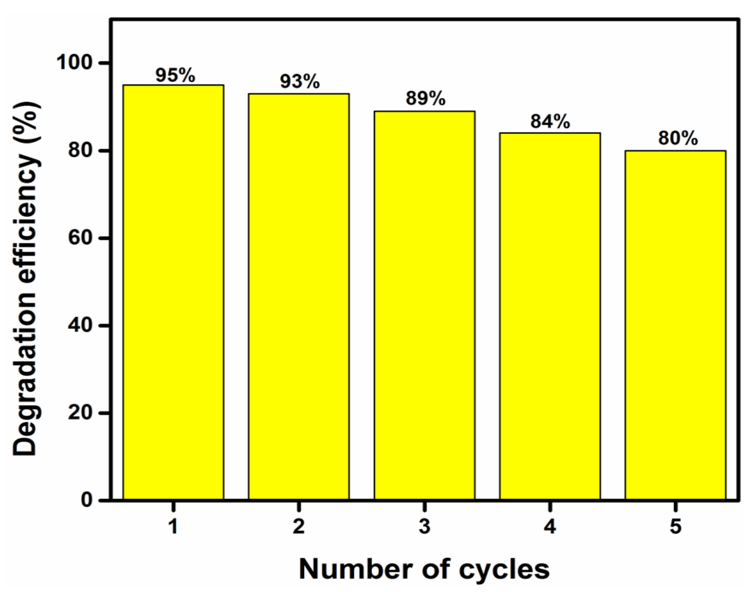
3.8. Photocatalytic Stability

The photocatalytic stability is very significant in addition to the photocatalytic efficiency for practical applications. Five circulating runs were carried out for the degradation of TC under the irradiance of visible light, as displayed in Figure 9. The decrease in degradation efficiency of the photocatalyst (3APMZ) for five consecutive cycles was observed from 95% to 80%, as shown in Figure 9. This decrease could be attributed to the decrease in photocatalyst dose of approximately 8% during each reuse cycle. More briefly, the initial concentration of photocatalyst was 0.4 g (1 cycle) and it was decreased to 0.391 g (2 cycles), 0.372 g (3 cycles), 0.347 g (4 cycles), and 0.327 g (5 cycles). These results are consistent with previous studies [34,35]. It can be concluded that the 3APMZ composite has good stability and reusability in TC degradation.

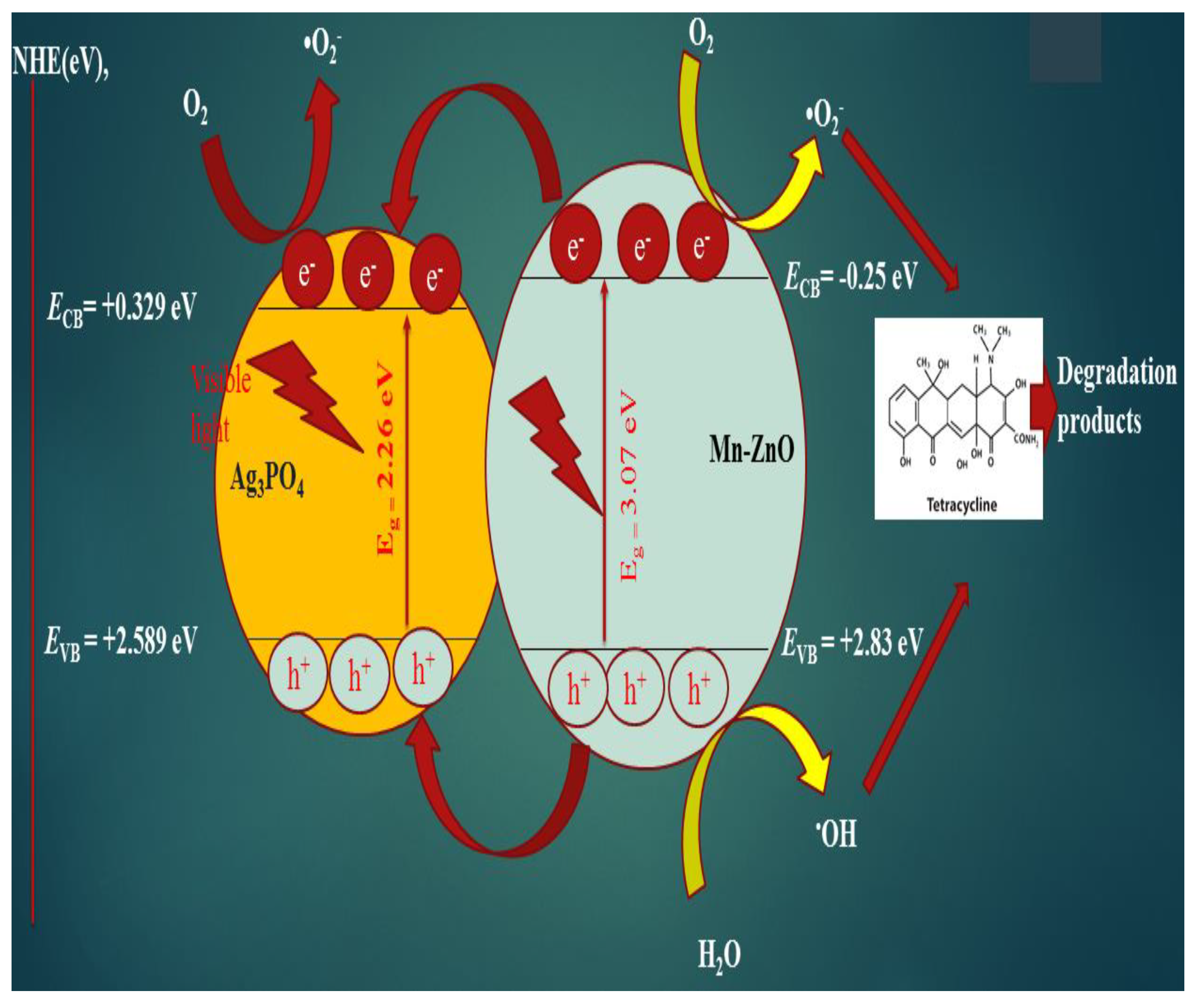
3.9. Proposed Mechanism

The proposed photocatalytic reaction mechanism for enhanced performance of 3APMZ in the degradation of TC is shown in Figure 10. During normal photocatalytic reaction in Ag3PO4 and Mn-ZnO semiconductors, the excitation of electrons and holes occurred due to incident visible light exposure. It was already found that doping of Mn2+ could change the band structure of ZnO, which results in the formation of impurity levels in the band gap of ZnO [21]. In the Mn-doped ZnO, photoexcited electrons move to the conduction band via impurity states of Mn2+. Therefore, Mn2+ ions played a vital role in reducing the recombination of charge carriers during photocatalysis. Upon light irradiation, the electrons were shifted towards the conduction bands (CB) of both semiconducting materials from their valence bands, where these electrons interacted with oxygen (O2) to create superoxide radicals (•O2). Simultaneously, hydroxyl radical (•OH) was produced due to reaction of photo-excited holes in the valence bands of both Ag3PO4 and Mn-ZnO. The above produced radicals contributed to degrading the TC due to photocatalytic oxidation and reduction reactions. Moreover, for the determination of actual charge transfer mechanism, the valence band potential (EVB) and conduction band potential (ECB) of Ag3PO4 and Mn-ZnO were calculated using the below equations.
E VB = X E o + 0.5 E g
E CB = E VB E g
where X is the electronegativity of the atoms, which is 5.96 eV for Ag3PO4 [36] and 5.79 eV for Mn-ZnO [37], E o is the hydrogen scale free electron energy which has ∼4.5 eV value, and Eg is the energy band gap. As Mn-ZnO (−0.25 eV vs. NHE) has more conduction band potential as compared to Ag3PO4 (+0.329 eV vs. NHE), electrons were shifted from the conduction band of Mn-ZnO toward the conduction band of Ag3PO4 [38,39]. Moreover, holes were shifted from the valence band of Mn-ZnO (+2.83 eV vs. NHE) to the valence band of Ag3PO4 (+2.589 eV vs. NHE) due to p–n heterojunctions [40]. In this way, the formed Type-II p–n junction between Ag3PO4 and Mn-ZnO facilitated the charge carrier transportation efficiently, and photocatalytic activity was enhanced.

4. Conclusions

In this investigation, nanocomposites based on ZnO were shown to be effective as visible-light photocatalysts for the breakdown of the antibiotic tetracycline hydrochloride. The integration of Mn and Ag3PO4 into ZnO resulted in improvements to the material’s structural, optical, and morphological properties, respectively, in the samples that were produced. A modified photocatalyst demonstrated improved photocatalytic performance for the breakdown of tetracycline, and a nanocomposite containing three percent Ag3PO4/Mn-ZnO was discovered to be a very efficient photocatalyst material in comparison to all of the other samples. On the basis of these findings, Ag3PO4/Mn-ZnO has the potential to be an excellent future commercial photocatalytic material for the treatment of wastewater when exposed to visible light.

Author Contributions

Conceptualization, M.W.A. and K.K.; data curation, H.A. and B.S.; formal analysis, H.A., A.B. and N.Z.; funding acquisition, M.W.A. and M.F.; investigation, H.A., N.R.K., S.N. and M.K.H.; methodology, H.A., N.R.K., S.N., M.K.H. and M.F.; project administration, M.W.A.; resources, M.W.A., A.B. and K.K.; software, M.K.H., A.B. and B.S.; supervision, M.W.A., A.B., B.S. and N.Z.; validation, H.A., N.R.K., S.N., M.K.H. and B.S.; visualization, M.K.H. and B.S.; writing—original draft, M.W.A., N.R.K., M.F. and N.Z.; writing—review and editing, M.W.A., S.N., M.F., K.K. and N.Z. All authors have read and agreed to the published version of the manuscript.

Funding

This work was supported by the Deanship of Scientific Research, Vice Presidency for Graduate Studies and Scientific Research, King Faisal University, Saudi Arabia [Project No. GRANT314]. The authors would like to acknowledge Princess Nourah bint Abdulrahman University Researchers Supporting Project number (PNURSP2022R230), Princess Nourah bint Abdulrahman University, Riyadh, Saudi Arabia.

Institutional Review Board Statement

Not applicable.

Informed Consent Statement

Not applicable.

Data Availability Statement

Not applicable.

Acknowledgments

This work was supported by the Deanship of Scientific Research, Vice Presidency for Graduate Studies and Scientific Research, King Faisal University, Saudi Arabia [Project No. GRANT314]. The authors would like to acknowledge Princess Nourah bint Abdulrahman University Researchers Supporting Project number (PNURSP2022R230), Princess Nourah bint Abdulrahman University, Riyadh, Saudi Arabia.

Conflicts of Interest

The authors declare no conflict of interest.

References

  1. Tahir, M.B.; Sagir, M. Carbon nanodots and rare metals (RM = La, Gd, Er) doped tungsten oxide nanostructures for photocatalytic dyes degradation and hydrogen production. Sep. Purif. Technol. 2019, 209, 94–102. [Google Scholar] [CrossRef]
  2. Tahir, M.B.; Nabi, G.; Rafique, M.; Khalid, N.R. Nanostructured-based WO3 photocatalysts: Recent development, activity enhancement, perspectives and applications for wastewater treatment. Int. J. Environ. Sci. Technol. 2017, 14, 2519–2542. [Google Scholar] [CrossRef]
  3. Pickett, M.T.; Roberson, L.B.; Calabria, J.L.; Bullard, T.J.; Turner, G.; Yeh, D.H. Regenerative water purification for space applications: Needs, challenges, and technologies towards’ closing the loop. Life Sci. Space Res. 2020, 24, 64–82. [Google Scholar] [CrossRef] [PubMed]
  4. Tahir, M.B.; Kiran, H.; Iqbal, T. The detoxification of heavy metals from aqueous environment using nano-photocatalysis approach: A review. Environ. Sci. Pollut. Res. 2019, 26, 10515–10528. [Google Scholar] [CrossRef]
  5. Xie, W.; Li, R.; Xu, Q. Enhanced photocatalytic activity of Se- doped TiO2 under visible light irradiation. Sci. Rep. 2018, 8, 8752. [Google Scholar] [CrossRef]
  6. Santangelo, S. Electrospun nanomaterials for energy applications: Recent advances. Appl. Sci. 2019, 9, 1049. [Google Scholar] [CrossRef]
  7. Khalid, N.; Hammad, A.; Tahir, M.; Rafique, M.; Iqbal, T.; Nabi, G.; Hussain, M. Enhanced photocatalytic activity of Al and Fe co-doped ZnO nanorods for methylene blue degradation. Ceram. Int. 2019, 45, 21430–21435. [Google Scholar] [CrossRef]
  8. Chen, J.; Ouyang, W.; Yang, W.; He, J.H.; Fang, X. Recent progress of heterojunction ultraviolet photodetectors: Materials, integrations, and applications. Adv. Funct. Mater. 2020, 30, 1909909. [Google Scholar] [CrossRef]
  9. Polat, K. Thin film photocatalyst made from Fe2O3/2D graphene/Cu working in the visible region of the solar spectrum. Solid State Commun. 2020, 319, 113993. [Google Scholar] [CrossRef]
  10. Abebe, B.; Murthy, H.A.; Amare, E. Enhancing the photocatalytic efficiency of ZnO: Defects, heterojunction, and optimization. Environ. Nanotechnol. Monit. Manag. 2020, 14, 100336. [Google Scholar] [CrossRef]
  11. Wu, G.; Xing, W. Fabrication of ternary visible-light-driven semiconductor photocatalyst and its effective photocatalytic performance. Mater. Technol. 2019, 34, 292–300. [Google Scholar] [CrossRef]
  12. Tahir, M.; Tasleem, S.; Tahir, B. Recent development in band engineering of binary semiconductor materials for solar driven photocatalytic hydrogen production. Int. J. Hydrogen Energy 2020, 45, 15985–16038. [Google Scholar] [CrossRef]
  13. Li, Y.; Liu, F.T.; Chang, Y.; Wang, J.; Wang, C.W. High efficient photocatalytic activity from nanostructuralized photonic crystal-like pn coaxial hetero-junction film photocatalyst of Cu3SnS4/TiO2 nanotube arrays. Appl. Surf. Sci. 2017, 426, 770–780. [Google Scholar] [CrossRef]
  14. Hasija, V.; Raizada, P.; Sudhaik, A.; Sharma, K.; Kumar, A.; Singh, P.; Jonnalagadda, S.B.; Thakur, V.K. Recent advances in noble metal free doped graphitic carbon nitride based nanohybrids for photocatalysis of organic contaminants in water: A review. Appl. Mater. Today 2019, 15, 494–524. [Google Scholar] [CrossRef]
  15. Tasleem, S.; Tahir, M. Current trends in strategies to improve photocatalytic performance of perovskites materials for solar to hydrogen production. Renew. Sustain. Energy Rev. 2020, 132, 110073. [Google Scholar] [CrossRef]
  16. Nasr, M.; Eid, C.; Habchi, R.; Miele, P.; Bechelany, M. Recent progress on titanium dioxide nanomaterials for photocatalytic applications. ChemSusChem 2018, 11, 3023–3047. [Google Scholar] [CrossRef]
  17. Khalid, N.R.; Arshad, A.; Tahir, M.B.; Hussain, M.K. Fabrication of p–n heterojunction Ag2O@Ce2O nanocomposites make enables to improve photocatalytic activity under visible light. Appl. Nanosci. 2021, 11, 199–206. [Google Scholar] [CrossRef]
  18. Montero-Muñoz, M.; Ibarra, J.E.R.; Rodríguez-Páez, J.E.; Ramirez, A.; A Huamaní-Coaquira, J. Shape-control of Zinc Oxide nanoparticles: Enhancing photocatalytic activity under UV irradiation. J. Phys. Conf. Ser. 2017, 792, 012068. [Google Scholar] [CrossRef]
  19. Achouri, F.; Corbel, S.; Balan, L.; Mozet, K.; Girot, E.; Medjahdi, G.; Said, M.B.; Ghrabi, A.; Schneider, R. Porous Mn-doped ZnO nanoparticles for enhanced solar and visible light photocatalysis. Mater. Des. 2016, 101, 309–316. [Google Scholar] [CrossRef]
  20. Thakur, D.; Sharma, A.; Awasthi, A.; Rana, D.S.; Singh, D.; Pandey, S.; Thakur, S. Manganese-Doped Zinc Oxide Nanostructures as Potential Scaffold for Photocatalytic and Fluorescence Sensing Applications. Chemosensors 2020, 8, 120. [Google Scholar] [CrossRef]
  21. Wang, Y.; Hao, X.; Wang, Z.; Dong, M.; Cui, L. Facile fabrication of Mn2+-doped ZnO photocatalysts by electrospinning. R. Soc. Open Sci. 2020, 7, 191050. [Google Scholar] [CrossRef]
  22. Wang, Y.; Cheng, J.; Yu, S.; Alcocer, E.J.; Shahid, M.; Wang, Z.; Pan, W. Synergistic effect of N-decorated and Mn2+ doped ZnO nanofibers with enhanced photocatalytic activity. Sci. Rep. 2016, 6, 32711. [Google Scholar] [CrossRef]
  23. Liu, H.Y.; Niu, C.G.; Guo, H.; Liang, C.; Huang, D.W.; Zhang, L.; Yang, Y.Y.; Li, L. In suit constructing 2D/1D MgIn2S4/CdS heterojunction system with enhanced photocatalytic activity towards treatment of wastewater and H2 production. J. Colloid Interface Sci. 2020, 576, 264–279. [Google Scholar] [CrossRef] [PubMed]
  24. Goswami, B.; Singha, R. Effect of Mn Doping on Optical Properties of ZnO Nanoparticles. Int. J. Innov. Res. Sci. Eng. Technol. 2015, 4, 2577–2582. [Google Scholar]
  25. Gusmão, L.A.; Peixoto, D.A.; Marinho, J.Z.; Romeiro, F.C.; Gonçalves, R.F.; Longo, E.; de Oliveira, C.A.; Lima, R.C. Alkali influence on ZnO and Ag-doped ZnO nanostructures formation using the microwave-assisted hydrothermal method for fungicidal inhibition. J. Phys. Chem. Solids 2021, 158, 110234. [Google Scholar] [CrossRef]
  26. Hussain, M.K.; Khalid, N.; Tanveer, M.; Kebaili, I.; Alrobei, H. Fabrication of CuO/MoO3 pn heterojunction for enhanced dyes degradation and hydrogen production from water splitting. Int. J. Hydrogen Energy 2022, 47, 15491–15504. [Google Scholar] [CrossRef]
  27. Al Farsi, B.; Al Marzouqi, F.; Al-Maashani, M.; Souier, M.T.; Myint, M.T.Z.; Al-Abri, M.Z. Rapid microwave-assisted fabrication of Al-doped zinc oxide nanorods on a glass substrate for photocatalytic degradation of phenol under visible light irradiation. Mater. Sci. Eng. B 2021, 264, 114977. [Google Scholar] [CrossRef]
  28. Xiong, G.; Pal, U.; Serrano, J.G. Correlations among size, defects, and photoluminescence in ZnO nanoparticles. J. Appl. Phys. 2007, 101, 024317. [Google Scholar] [CrossRef]
  29. Tanveer, M.; Nisa, I.; Nabi, G.; Hussain, M.K.; Khalid, S.; Qadeer, M. Sol-gel extended hydrothermal pathway for novel Cd-Zn co-doped Mg-ferrite nano-structures and a systematic study of structural, optical and magnetic properties. J. Magn. Magn. Mater. 2022, 553, 169245. [Google Scholar] [CrossRef]
  30. Zhang, A.; Zhang, J. Effects of europium doping on the photocatalytic behavior of BiVO4. J. Hazard. Mater. 2010, 173, 265–272. [Google Scholar] [CrossRef]
  31. Semeraro, P.; Bettini, S.; Sawalha, S.; Pal, S.; Licciulli, A.; Marzo, F.; Lovergine, N.; Valli, L.; Giancane, G. Photocatalytic degradation of tetracycline by ZnO/γ-Fe2O3 paramagnetic nanocomposite material. Nanomaterials 2020, 10, 1458. [Google Scholar] [CrossRef]
  32. Wetchakun, N.; Chainet, S.; Phanichphant, S.; Wetchakun, K. Efficient photocatalytic degradation of methylene blue over BiVO4/TiO2 nanocomposites. Ceram. Int. 2015, 41, 5999–6004. [Google Scholar] [CrossRef]
  33. Wafi, A.; Szabó-Bárdos, E.; Horváth, O.; Makó, É.; Jakab, M.; Zsirka, B. Coumarin-based quantification of hydroxyl radicals and other reactive species generated on excited nitrogen-doped TiO2. J. Photochem. Photobiol. A Chem. 2021, 404, 112913. [Google Scholar] [CrossRef]
  34. Siboni, M.S.; Samadi, M.T.; Yang, J.K.; Lee, S.M. Photocatalytic reduction of Cr (VI) and Ni (II) in aqueous solution by synthesized nanoparticle ZnO under ultraviolet light irradiation: A kinetic study. Environ. Technol. 2011, 32, 1573–1579. [Google Scholar] [CrossRef]
  35. Suharyadi, E.; Muzakki, A.; Istiqomah, N.I.; Puspitarum, D.L.; Purnama, B.; Djuhana, D. Reusability of photocatalytic CoFe2O4@ZnO core–shell nanoparticles for dye degradation. ECS J. Solid State Sci. Technol. 2022, 11, 023004. [Google Scholar] [CrossRef]
  36. Chen, X.; Yu, C.; Zhu, R.; Li, N.; Chen, J.; Li, S.; Xia, W.; Xu, S.; Wang, H.; Chen, X. Ag3PO4 deposited on CuBi2O4 to construct Z-scheme photocatalyst with excellent visible-light catalytic performance toward the degradation of diclofenac sodium. Nanomaterials 2019, 9, 959. [Google Scholar] [CrossRef]
  37. Yu, Z.; Moussa, H.; Liu, M.; Schneider, R.; Moliere, M.; Liao, H. Heterostructured metal oxides-ZnO nanorods films prepared by SPPS route for photodegradation applications. Surf. Coat. Technol. 2019, 375, 670–680. [Google Scholar] [CrossRef]
  38. Batool, M.; Daoush, W.M.; Hussain, M.K. Dye Sequestration Using Biosynthesized Silver Nanoparticles Adsorbent in Aqueous Solutions. Crystals 2022, 12, 662. [Google Scholar] [CrossRef]
  39. Nemiwal, M.; Zhang, T.C.; Kumar, D. Recent progress in g-C3N4, TiO2 and ZnO based photocatalysts for dye degradation: Strategies to improve photocatalytic activity. Sci. Total Environ. 2021, 767, 144896. [Google Scholar] [CrossRef]
  40. Qamar, M.A.; Shahid, S.; Javed, M.; Sher, M.; Iqbal, S.; Bahadur, A.; Li, D. Fabricated novel g-C3N4/Mn doped ZnO nanocomposite as highly active photocatalyst for the disinfection of pathogens and degradation of the organic pollutants from wastewater under sunlight radiations. Colloids Surf. A Physicochem. Eng. Asp. 2021, 611, 125863. [Google Scholar] [CrossRef]
Figure 1. (a,b) The XRD patterns for pure ZnO and Ag3PO4 samples, respectively.
Crystals 12 01156 g001
Figure 2. (a) XRD patterns of Z, MZ, 1APMZ, 3APMZ, 5APMZ, and AP samples. (b) Enlarged view of XRD for AP and 3APMZ samples.
Crystals 12 01156 g002
Figure 3. (a,b) SEM images of 3APMZ nanocomposites at different magnifications. (c) EDX graph of 3APMZsample. (d) TEM image of 3APMZsample.
Crystals 12 01156 g003
Figure 4. Absorption spectra of Z, MZ, 1APMZ, 3APMZ, 5APMZ, and AP samples.
Crystals 12 01156 g004
Figure 5. FTIR spectra of pure Z, MZ, and 3APMZ samples.
Crystals 12 01156 g005
Figure 6. PL analysis of all the samples in the range of wavelength 345 nm to 550 nm at 325 nm excitation wavelength.
Crystals 12 01156 g006
Figure 7. Photodegradation rate of tetracycline hydrochloride shown by Z, MZ, 1APMZ, 3APMZ, 5APMZ, and AP samples.
Crystals 12 01156 g007
Figure 8. (a) Photogenerated reactive species trapped by dissimilar scavengers through degradation of TC on 3APMZ. (b) The 7-hydroxycoumarin PL emission intensity was observed at 443 nm under the irradiance of visible light.
Crystals 12 01156 g008
Figure 9. Reusability of 3APMZ heterojunction in five consecutive runs.
Crystals 12 01156 g009
Figure 10. Photocatalytic mechanism of TC degradation under visible light illumination using 3APMZ.
Crystals 12 01156 g010
Table 1. The band gap energies comparison among Z, MZ, 1APMZ, 3APMZ, 5APMZ, and AP samples.
SamplesWavelengths (nm)Band Gap Energy (eV)
Z393.533.15
MZ403.283.07
AP547.802.26
1APMZ616.992.01
3 APMZ659.421.88
5 APMZ631.031.96
Table 2. Comparative photocatalytic study for the degradation of TC with other studies.
PhotocatalystDyeLight SourceTime (min)Degradation (%)Ref.
CuO-MoO3Rhodamine BVisible light12099[26]
Ag2O/Ce2OMethyl orangeVisible light150100[17]
Eu/BiVO4Methyl orangeVisible light18052[30]
ZnO/γ-Fe2O3TetracyclineVisible light15089[31]
TiO2/BiVO4Methylene blueVisible light12071[32]
APMZTetracyclineVisible light12095Present work
Publisher’s Note: MDPI stays neutral with regard to jurisdictional claims in published maps and institutional affiliations.

