
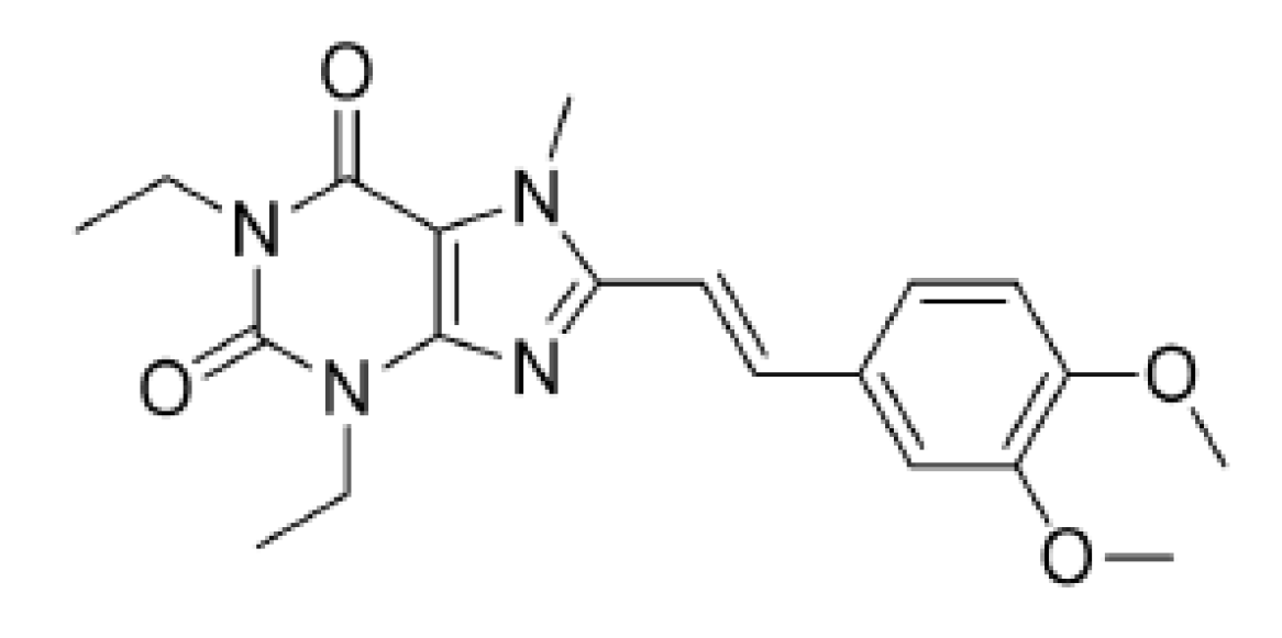
Studies on the Crystal Forms of Istradefylline: Structure, Solubility, and Dissolution Profile

Abstract
:1. Introduction
2. Materials and Methods
2.1. Materials
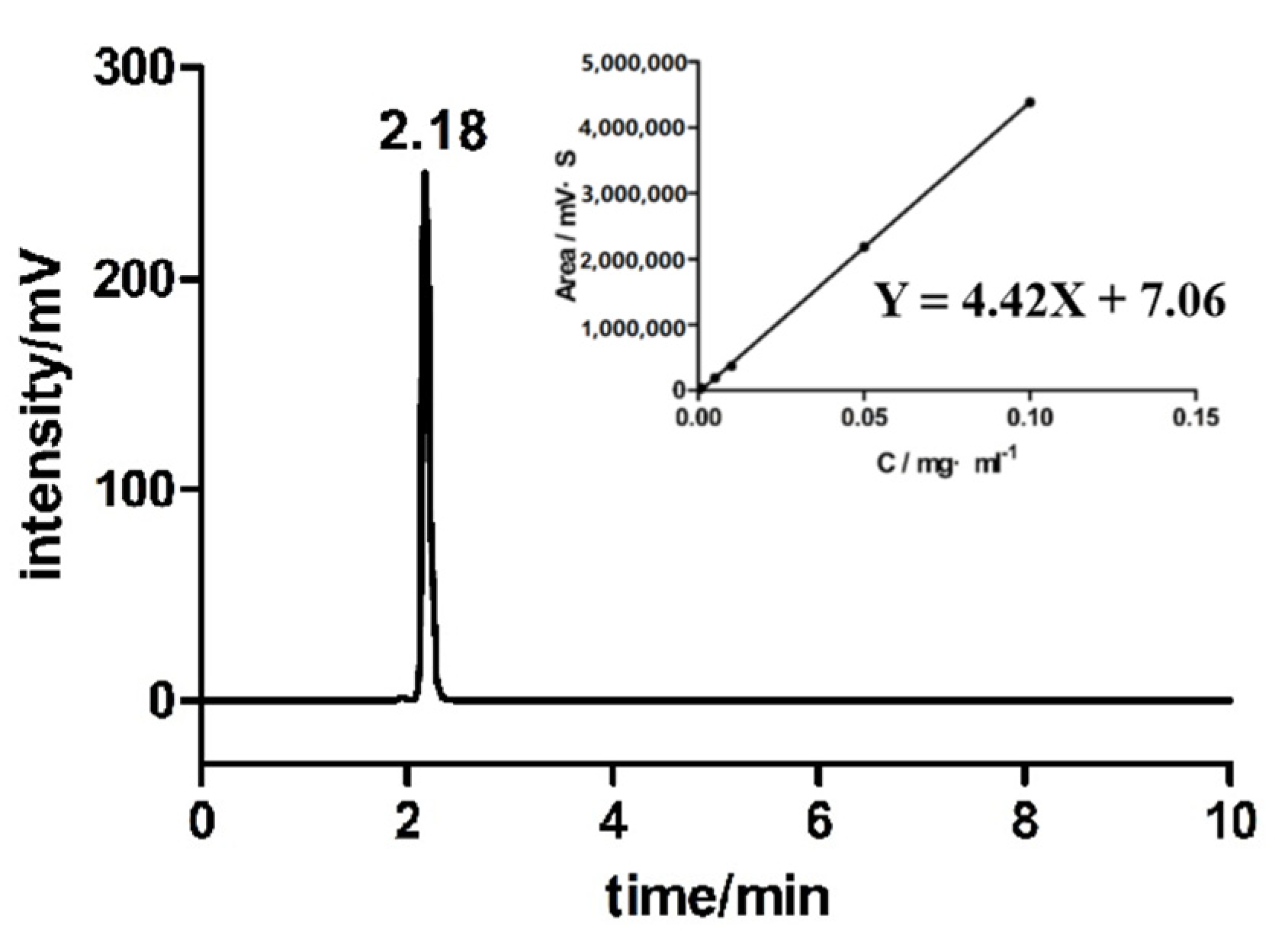
2.2. HPLC analysis
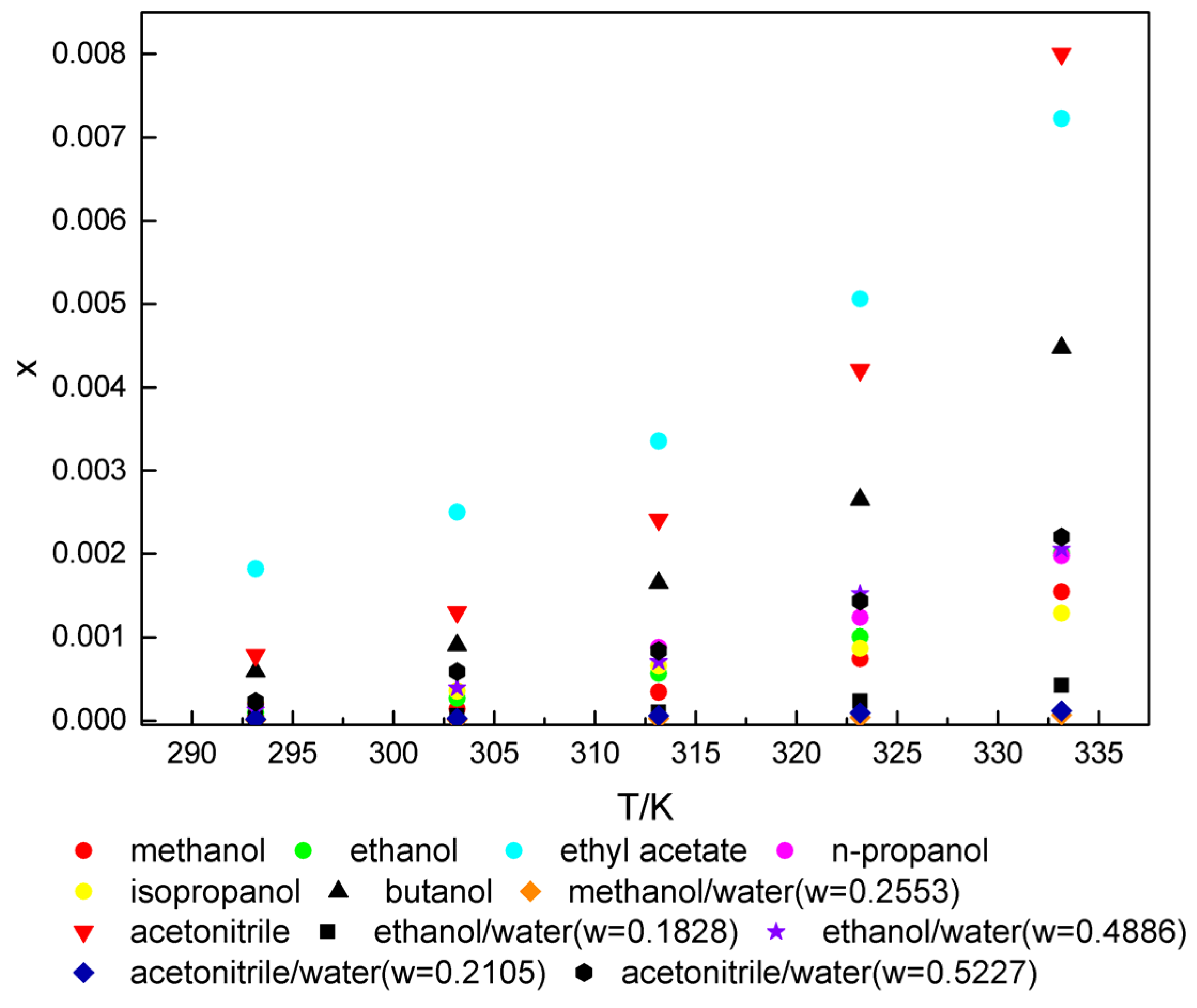
2.3. Solubility of Istradefylline in Diverse Organic Solvents and Solvent Mixtures with Water
2.4. Preparation of Single Crystal
2.5. X-ray Diffraction
2.6. Differential Scanning Calorimetry (DSC) and Thermogravimetric Analysis (TGA)
2.7. Fourier-Transform Infrared Spectral Analysis (FT-IR)
2.8. Particle Size and Specific Surface Area Analysis (BET)
2.9. Dissolution Study
3. Results and Discussion
3.1. Solubility of Istradefylline
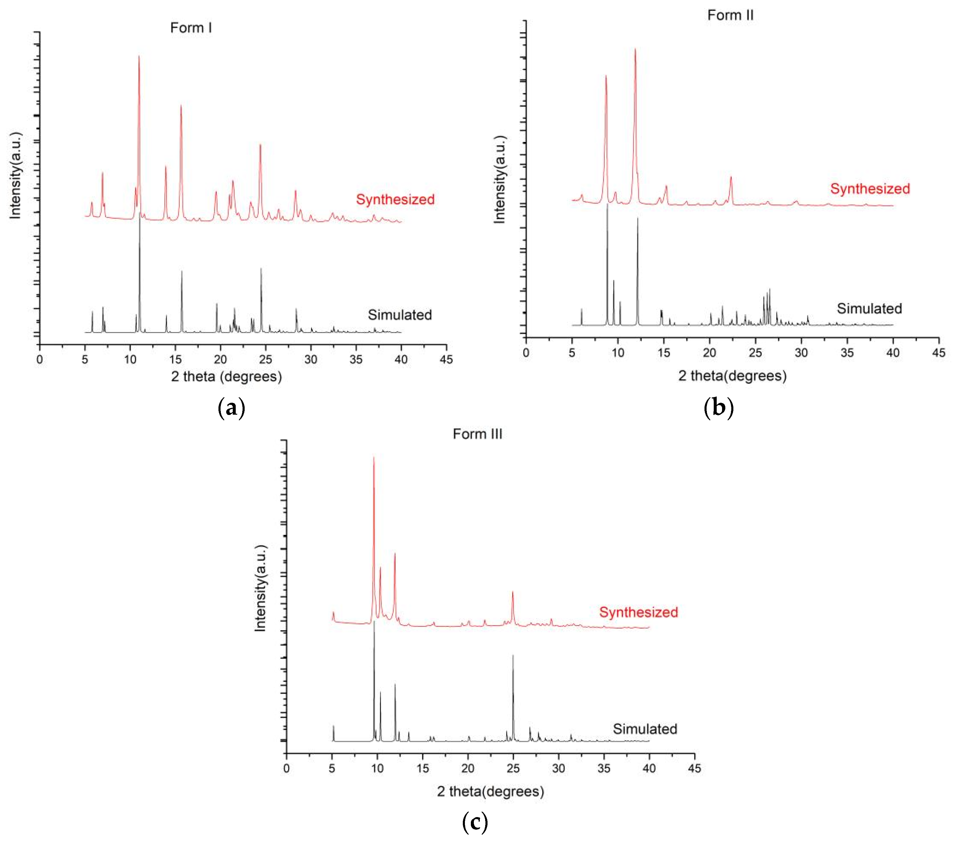
3.2. Powder X-ray Diffraction
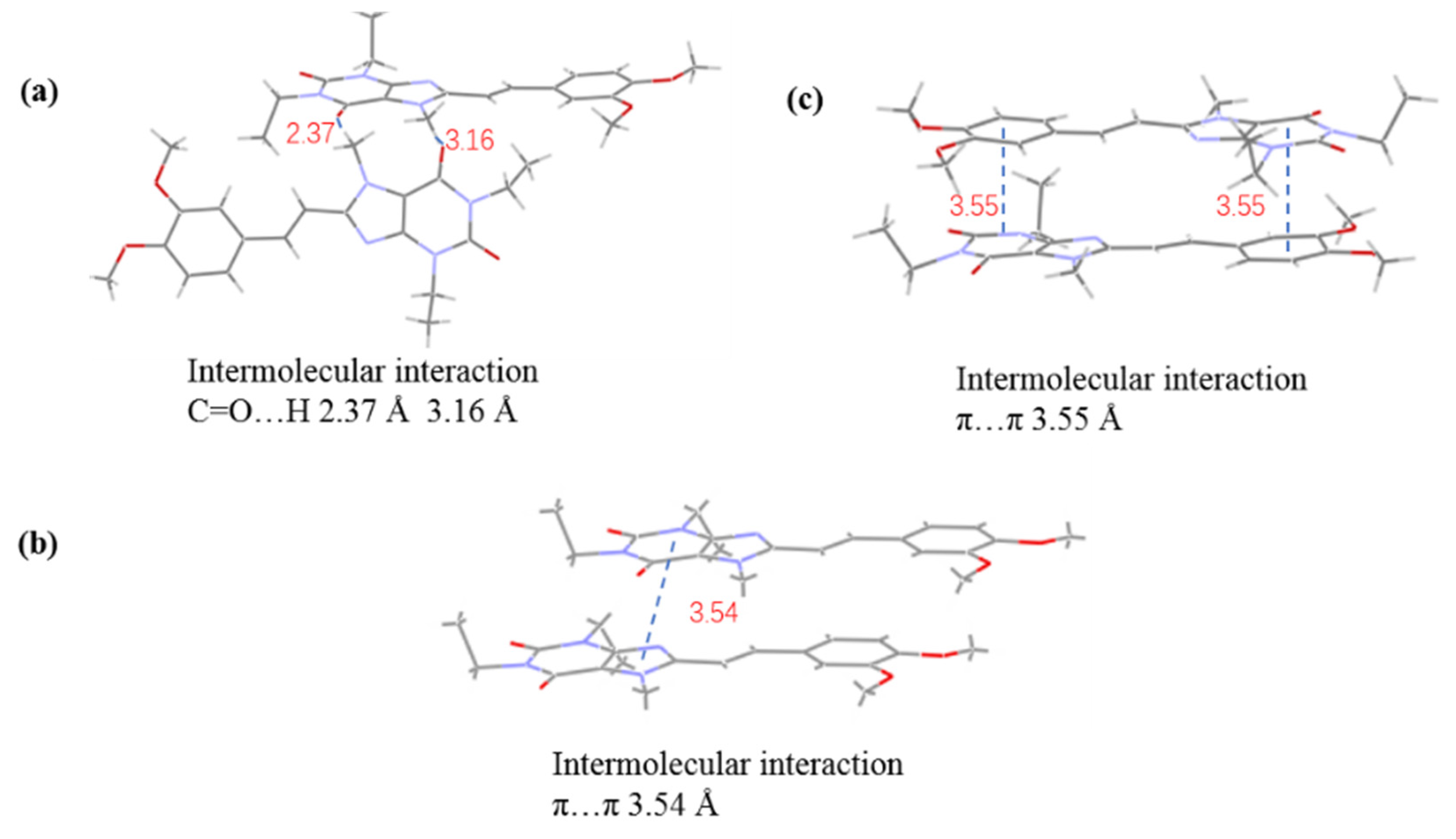
3.3. Single-Crystal X-ray Diffraction
3.4. TGA
3.5. FT-IR Analysis
3.6. Particle Size and BET Analysis
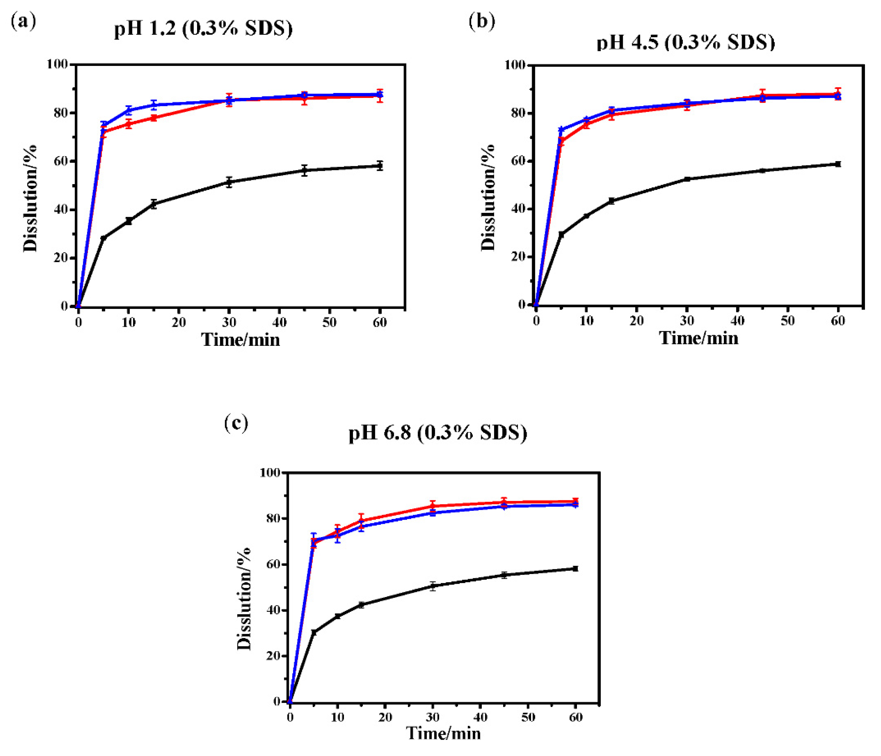
3.7. Dissolution Curve Test
4. Conclusions
5. Patents
Supplementary Materials
Author Contributions
Funding
Institutional Review Board Statement
Informed Consent Statement
Data Availability Statement
Acknowledgments
Conflicts of Interest
References
- Zheng, J.Y.; Zhang, X.H.; Zhen, X.C. Development of Adenosine A2A Receptor Antagonists for the Treatment of Parkinson’s Disease: A Recent Update and Challenge. ACS Chem. Neurosci. 2019, 10, 783–791. [Google Scholar] [CrossRef]
- Shook, B.C.; Jackson, P.F. Adenosine A2A Receptor Antagonists and Parkinson’s Disease. ACS Chem. Neurosci. 2011, 2, 555–567. [Google Scholar] [CrossRef] [Green Version]
- Hauser, R.A.; Shulman, L.M.; Trugman, J.M.; Roberts, J.W.; Sussman, N.M. Study of istradefylline in patients with Parkinson’s disease on levodopa with motor fluctuations. Mov. Disord. 2010, 23, 2177–2185. [Google Scholar] [CrossRef]
- Tian, S.; Wang, X.; Li, L.L.; Zhang, X.H.; Li, Y.Y.; Zhu, F.; Hou, T.J.; Zhen, X.C. Discovery of Novel and Selective Adenosine A2A Receptor Antagonists for Treating Parkinson’s Disease through Comparative Structure-Based Virtual Screening. J. Chem. Inf. Model. 2017, 57, 1474–1487. [Google Scholar] [CrossRef]
- Lu, J.; Cui, J.; Li, X.H.; Wang, X.; Zhou, Y.; Yang, W.J.; Chen, M.; Zhao, J.; Pei, G. An Anti-Parkinson’s Disease Drug via Targeting Adenosine A2A Receptor Enhances Amyloid-β Generation and γ-Secretase Activity. PLoS ONE 2016, 11, e0166415. [Google Scholar] [CrossRef] [Green Version]
- Uchida, S.I.; Soshiroda, K.; Okita, E.; Uchida, M.K.; Mori, A.; Jenner, P.; Kanda, T. The adenosine A2A receptor antagonist, istradefylline enhances the anti-parkinsonian activity of low doses of dopamine agonists in MPTP-treated common marmosets. Eur. J. Pharmacol. 2015, 747, 160–165. [Google Scholar] [CrossRef]
- Ko, W.K.D.; Sandrine, M.C.; Li, Q.; Yang, J.Z.; Steve, M.; Elsa, Y.P.; Erwan, B. An evaluation of istradefylline treatment on Parkinsonian motor and cognitive deficits in 1-methyl-4-phenyl-1,2,3,6-tetrahydropyridine (MPTP)-treated macaque models. Neuropharmacology. 2016, 110, 48–58. [Google Scholar] [CrossRef]
- Matsuura, K.; Kajikawa, H.; Tabei, K.I.; Satoh, M.; Kida, H.; Nakamura, N.; Tomimoto, H. The effectiveness of istradefylline for the treatment of gait deficits and sleepiness in patients with Parkinson’s Disease. Neurosci. Lett. 2018, 662, 158–161. [Google Scholar] [CrossRef]
- Kataoka, H.; Sugie, K. Does istradefylline really have a dystonic mechanism? J. Neurol. Sci. 2018, 388, 233–234. [Google Scholar] [CrossRef]
- Fumio, S.; Junichi, S.; Nobuaki, K.; Joji, N.; Shizuo, S.; Shunji, I.; Hiromi, N. Therapeutic Agents for Parkinson’s Disease. EP0590919, 29 December 1999. [Google Scholar]
- Li, J.; Han, Y.F.; Li, Y.X.; Chen, S.W.; Gong, P. Improved synthesis process of adenosine A2A receptor inhibitor istradefylline. Chin. J. Med. Chem. 2018, 2, 220–224. [Google Scholar]
- Uchida, S.I.; Soshiroda, K.; Okita, E.; Uchida, M.K.; Mori, A.; Jenner, P.; Kanda, T. The adenosine A2A receptor antagonist, istradefylline enhances anti-parkinsonian activity induced by combined treatment with low doses of L-DOPA and dopamine agonists in MPTP-treated common marmosets. Eur. J. Pharmacol. 2015, 766, 25–30. [Google Scholar] [CrossRef] [PubMed]
- Uchida, S.I.; Tashiro, T.; Kawai-Uchida, M.; Mori, A.; Jenner, P.; Kanda, T. The adenosine A2A-receptor antagonist istradefylline enhances the motor response of L-DOPA without worsening dyskinesia in MPTP-treated common marmosets. J. Pharmacol. Sci. 2014, 124, 480–485. [Google Scholar] [CrossRef] [PubMed] [Green Version]
- Huang, T.H.; Lu, D.Q.; Ling, X.Q.; Wang, X.X.; Liu, T.Q.; Shen, F.F.; He, K.F. Thermodynamic models for determination of the solid-liquid equilibrium of Istradefylline in ethyl acetate plus (isopropanol, tetrahydrofuran, acetone) binary solvent mixtures. J. Chem. Thermodyn. 2017, 111, 31–40. [Google Scholar] [CrossRef]
- Ge, Y.H.; Li, T.T.; Cheng, J.J. Crystal Type I of Azilsartan Polymorphs: Preparation and Analysis. J. Cryst. Process. Technol. 2016, 6, 1–10. [Google Scholar] [CrossRef] [Green Version]
- Yadav, M.R.; Shaikh, A.R.; Ganesan, V.; Giridhar, R.; Chadha, R. Studies on the crystal forms of pefloxacin: Preparation, characterization and dissolution profile. J. Pharm. Sci. 2008, 97, 2637–2648. [Google Scholar] [CrossRef]
- Ganesan, V. Studies on the crystal forms of moxifloxacin: Preparation, characterization and dissolution profile. Pharm. Anal. Acta 2013, 4, 135. [Google Scholar]
- Zhang, X.M.; Sun, F.X.; Zhang, T.T.; Jia, J.T.; Su, H.M.; Wang, C.H.; Zhu, G.S. Three pharmaceuticals cocrystals of adefovir: Syntheses, structures and dissolution study. J. Mol. Struct. 2015, 1100, 395–400. [Google Scholar] [CrossRef]
- Llinas, A.; Barbas, R.; Font-Bardia, M.; Quayle, M.J.; Velaga, S.; Prohens, R. Two New Polymorphic Cocrystals of Zafirlukast: Preparation, Crystal Structure, and Stability Relations. Cryst. Growth Des. 2015, 15, 4162–4169. [Google Scholar] [CrossRef]
- Hua, D.Y.; Chen, G.L.; Shen, W.J. Determination of two crystal forms of famotidine by differential scanning calorimetry. J. Chin. Med. Ind. 1991, 22, 78–79. [Google Scholar]
- Wang, J.; Zhang, R.H.; Sun, S.Y. Study on the polycrystalline form of nimodipine. Acta pharm. Sin. 1995, 30, 443–448. [Google Scholar]
- Jiao, L.T.; Zhang, L.; Yang, D.Z.; Yang, S.Y.; Du, G.H.; Lv, Y. Raman spectroscopic analysis and dissolution tests of nimodipine crystal forms. Her. Med. 2017, 36, 1175–1179. [Google Scholar]
- Bao, J.Y.; Huang, H.; Yu, D.J.; Wei, W.; Jiang, Y.W.; Zhang, X.Q. Polymorphs of Istradefylline. CN104744464A, 1 July 2015. [Google Scholar]
- Dong, D.D. The invention relates to a method for preparing Istradefylline crystal form III by ball milling. CN108117554A, 5 June 2018. [Google Scholar]
- Wang, C.H. A new crystal form of Istradefylline and its preparation method. CN105884776A, 24 August 2016. [Google Scholar]
- Gong, D.H.; Wang, J.; Gai, J.H.; Yang, J.; Sun, W.J.; Yang, M.; Yang, C.Q.; Ma, Y.X. A new crystal form of Istradefylline and its preparation method. CN106279169A, 4 January 2017. [Google Scholar]
- Dong, D.D. A preparation method of Istradefylline crystal form II for treating Parkinson’s disease. CN108101907A, 1 June 2018. [Google Scholar]
- Bourne, S.A.; Villiers, M.D.; Crider, A.M.; Caira, M.R. Polymorphism of the antitubercular Isoxyl. Cryst Growth Des. 2011, 11, 4950–4957. [Google Scholar]
- Cui, K.J.; Yang, Y.M.; Meng, Z.H.; Xu, G.R.; Xu, Z.B. Solubility of tetranitrodimerglycoluril (TNDGU) in different solvents at temperatures between 293.15 K and 313.15 K. J. Chem. Eng. Data 2014, 59, 2620–2622. [Google Scholar] [CrossRef]


| Process | Material Name | Function | Batch Size/g | Proportion/% |
|---|---|---|---|---|
| Tablet | Istradefylline | Drug | 20.00 | 14.29 |
| MCC PH102 | Fillers | 67.50 | 48.21 | |
| Lactose | Diluents | 42.00 | 30.00 | |
| PVPP | Disintegrant | 9.80 | 7.00 | |
| MS | Lubricants | 0.70 | 0.50 | |
| Total | 140.0 | 100.0 | ||
| Coating Single piece of content | Opadry | Materials | 5.2 | 3% |
| Purified water | Solvent | 59.8 | Final removal | |
| Actual use (1.2 times preparation) | Opadry | Materials | 54.4 | 3% |
| Purified water | Solvent | 626.0 | Final removal | |
| pH | Solubility of Istradefylline (µg/mL) |
|---|---|
| 1 | 0.41 |
| 2 | 0.39 |
| 3 | 0.32 |
| 4 | 0.31 |
| 7 | 0.27 |
| 8 | 0.18 |
| 10 | 0.11 |
| 12 | 0.10 |
| Compounds | Form I | Form II | Form III |
|---|---|---|---|
| Chemical formula | C20H24N4O4 | C20H26N4O5 | C22H29N5O5 |
| Formula weight | 384.43 | 402.45 | 443.50 |
| Crystal system | monoclinic | monoclinic | monoclinic |
| Space group | P21 | P21/c | P21/m |
| a/Å | 13.6762(17) | 4.5436(5) | 9.430(9) |
| b/Å | 4.7483(7) | 23.776(2) | 7.129(7) |
| c/Å | 16.464(2) | 18.6282(17) | 17.587(18) |
| α/° | 90 | 90 | 90 |
| β/° | 112.39(4) | 95.065(7) | 103.514(10) |
| γ/° | 90 | 90 | 90 |
| vol/Å3 | 988.5(2) | 2004.6(3) | 1149.5(19) |
| Z | 2 | 4 | 2 |
| ρcalcg/cm3 | 1.292 | 1.334 | 1.281 |
| Gof | 1.017 | 1.036 | 1.052 |
| R | R1= 0.0552, wR2 = 0.1107 | R1 = 0.0888, wR2 = 0.2892 | R1 = 0.083, wR2 = 0.27 |
Publisher’s Note: MDPI stays neutral with regard to jurisdictional claims in published maps and institutional affiliations. |
© 2022 by the authors. Licensee MDPI, Basel, Switzerland. This article is an open access article distributed under the terms and conditions of the Creative Commons Attribution (CC BY) license (https://creativecommons.org/licenses/by/4.0/).
Share and Cite
Wang, Y.; Xu, Y.; Zheng, Z.; Xue, M.; Meng, Z.; Xu, Z.; Li, J.; Lin, Q. Studies on the Crystal Forms of Istradefylline: Structure, Solubility, and Dissolution Profile. Crystals 2022, 12, 917. https://doi.org/10.3390/cryst12070917
Wang Y, Xu Y, Zheng Z, Xue M, Meng Z, Xu Z, Li J, Lin Q. Studies on the Crystal Forms of Istradefylline: Structure, Solubility, and Dissolution Profile. Crystals. 2022; 12(7):917. https://doi.org/10.3390/cryst12070917
Chicago/Turabian StyleWang, Yiyun, Youwei Xu, Zhonghui Zheng, Min Xue, Zihui Meng, Zhibin Xu, Jiarong Li, and Qing Lin. 2022. "Studies on the Crystal Forms of Istradefylline: Structure, Solubility, and Dissolution Profile" Crystals 12, no. 7: 917. https://doi.org/10.3390/cryst12070917