Article

Hyaluronic Acid-Protein Conjugate Modified Iron-Based MOFs (MIL-101 (Fe)) for Efficient Therapy of Neuroblastoma: Molecular Simulation, Stability and Toxicity Studies

1
Department of Pharmaceutics, Manipal College of Pharmaceutical Sciences, Manipal Academy of Higher Education, Manipal 576104, Karnataka, India
2
Department of Atomic and Molecular Physics, Manipal Academy of Higher Education, Manipal 576104, Karnataka, India
3
Department of Pharmaceutical Chemistry, Manipal College of Pharmaceutical Sciences, Manipal Academy of Higher Education, Manipal 576104, Karnataka, India
*
Author to whom correspondence should be addressed.
These authors contributed equally to this work.
Crystals 2022, 12(10), 1484; https://doi.org/10.3390/cryst12101484
Submission received: 1 September 2022 / Revised: 10 October 2022 / Accepted: 13 October 2022 / Published: 19 October 2022
(This article belongs to the Section Inorganic Crystalline Materials)

Abstract

:
Iron-based metal-organic frameworks (MIL (101)) have recently gained attention in materials science for biomedical applications. In the present work, Iron-based MOF (MIL-101(Fe)) were coated with lactoferrin (Lf) conjugated with hyaluronic acid (HA) and investigated its potential for delivering 5-fluorouracil (5-FU), along with assessing the toxicity profile. The synthesised nanoparticles were extensively characterised using spectroscopic, X-Ray, thermal and electron microscopic techniques. 5-FU was loaded into MOFs, and the drug-loading efficiency and drug release pattern were studied, along with stability testing in pH and serum protein. The toxicity of MIL-101(Fe) was assessed using both in vitro and in vivo techniques such as the haemolysis assay, cell viability assay and acute and subacute toxicity studies in animals. In silico molecular simulation was done to assess the Lf and Tf interaction. The molecular interaction of Lf with Transferrin (Tf) showed strong molecular interaction and negligible fluctuation in the RMSD (root mean square deviation) values. The MOFs were stable and demonstrated sustained drug release patterns. The in vitro cell studies demonstrated biocompatibility and enhanced cellular internalisation of MOFs. The in vivo toxicity studies supported the in vitro results. The synthesised MOFs demonstrated potential as a targeted delivery platform for cancer targeting.

1. Introduction

An integrative area, nanotechnology is concerned with designing and developing nano-scale devices using engineering, physics, biology and chemistry principles [1,2,3,4]. Due to advances in nanoscience, the biomedical field has achieved several benefits due to developments in the diagnosis, imaging and treatment of various diseases [5,6,7,8,9,10,11,12,13,14,15,16,17,18,19,20,21]. Nanomedicine is a vast subject field that encompasses a multitude of nano-sized components. A study on the rational targeting and delivery of diagnostic agents and therapeutics via interstitial or intravenous administration routes with the support of nanomaterials is currently at the forefront of nanomedicine ventures [22]. The latest developments in the coordinated structure and the desired surface modification of inorganic nanomaterials have brought new potential to this growing field of nano-research.
Metal organic frameworks (MOFs) are the centre of attention among inorganic nanocarriers because of their exceptional characteristics [5]. MOF nanomaterials are porous crystalline materials designed to form frameworks by bonding metal ions or clusters connected to organic ligands [5,23,24]. MOFs are renowned for their controllable pore size, large surface areas and versatile surface internal characteristics. In several applications, such as gas adsorption/separation [25,26], catalysis [27,28] and drug delivery [29,30], as these inherent characteristics make MOFs very appealing. MIL-101 (MIL, Material of Institute Lavoisier), among the most prominent MOFs, was first discovered by Férey and associates in 2005 [31]. MIL-101(Fe) is a mesoporous carrier consisting of nontoxic materials with a pore scale of 12 Å × 29 Å and 16 Å × 34 Å [32]. Iron, an inorganic bridge, and terephthalic acid (H2BDC), an organic linker, are used to develop this MOF. At about 22 μM, iron resides in blood plasma. Despite all promising applications, especially in biomedicine, they have posed substantial toxicological concerns.
It is imperative to examine their toxicity profile before any bio-application of MOFs [5,33]. The in vivo toxicity of MOFs has only been reported in a few papers [34,35]. The in vivo acute toxicity of Fe(III) carboxylate MOFs delivered intravenously to rats was tested by Baati et al. (2013) and reported to have low acute toxicity [34]. The toxicity of MIL-101(Fe) has never been recorded to date following these pioneering studies. It is a potential risk to human health that has not been understood yet and demands immediate investigation. Therefore, this research aimed to assess the consequence of orally administered MIL-101(Fe) on Wistar rats by assessing hemocompatibility, in vitro cytotoxicity, and in vivo acute and subacute toxicity. The study also aims to check the molecular interaction of lactoferrin (Lf) with the transferrin (Tf) receptor, utilising molecular docking and molecular dynamics simulation studies.

2. Materials and Methods

2.1. Materials

Iron chloride hexahydrate, acetic acid, 1,4-benzenedicarboxylic acid (BDC), N,N–dimethylformamide (DMF) and bovine serum albumin (BSA) were procured from Sigma-Aldrich, India(Bangalore, India). 5-Fluorouracil (5-FU) was procured from TCI, India (Tamil Nadu, India). Hyaluronic acid was obtained from Novozymes (Bagsværd, Denmark), and lactoferrin (Bioferrin™) was obtained from Glanbia, USA (Fitchburg, MA, USA) as a gift sample. All other reagents and all other solvents and chemicals were of analytical grade and utilised without additional purification.

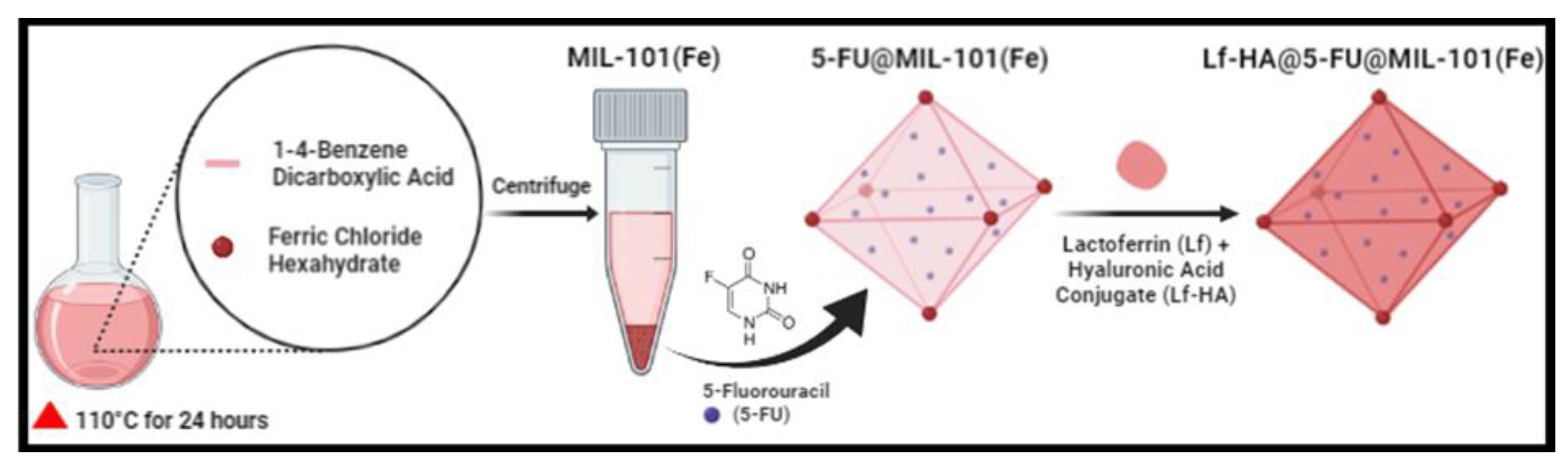
2.2. Synthesis of Fe-Based MOF (MIL-101(Fe))

With some modifications, the Fe-based MOF MIL-101(Fe) was developed using a conventional solvothermal technique utilising the reported procedures. In the traditional solvothermal approach, and a solution of iron salt was made by sonicating 80 mL of DMF with acetic acid (1 mL) and FeCl3⋅6H2O (10 mmol), accompanied by the introduction of BDC (5 mmol). The solution was thoroughly mixed before being moved to an autoclave (Teflon-lined 100 mL) and heated for 24 h at 110 °C. After 24 h, the autoclave temperature was reduced to the ambient temperature, and the precipitate was separated from the mixed solution by centrifuging the sample for 15 min at 15,000 rpm. The material obtained was refined by washing with DMF three times and eventually with methanol, centrifuged and vacuum dried at 60 °C overnight. The drug-loading capacity of MIL-101(Fe) was estimated using 5-FU as a model drug. The drug loading was performed post-synthetically by dispersing the MIL-101(Fe) MOFs in 5-FU solution (5 mg/mL) in the ratio of 1:1 for 24 h, and the synthesised material was referred to as 5-FU@MIL-101(Fe) [36,37].

2.3. Lactoferrin (Lf) Conjugation with Hyaluronic Acid (HA)

As previously reported by our group, the coupling of Lf to the HA was due to the free amino groups of Lf and the carboxyl group of HA [38]. In distilled water, HA (10% w/w), EDC (0.96 mg), and NHS (1.15 mg) were all dissolved. Using a magnetic stirrer, the aforementioned liquid was gently swirled for 10 min at ambient temperature. Lf (5 mg) was introduced to the above-mentioned mixture, which was then constantly swirled for 12 h at ambient temperature. To get rid of unreacted agents, the resultant mixture was thoroughly dialysed (MWCO 30000) under water for 48 h. The obtained Lf-modified HA (Lf-HA) was surface coated on 5-FU@MIL-101(Fe) to produce Lf-HA@5-FU@MIL-101(Fe), henceforth referred to as surface-modified MIL-101(Fe) (SM-MIL).

2.4. Molecular Docking and Molecular Dynamics (MD) Simulation

In the present study, Schrödinger’s (Schrödinger, LLC, New York, NY, USA) Maestro (version 11.8) was utilised to perform molecular docking and dynamics simulation investigations in Linux Ubuntu 18.04.1 LTS console, Intel® Core™ i5-10600K Processor (10th Generation), 16 GB Ram and NVIDIA® GeForce® 4GB graphics card on an HP workstation.

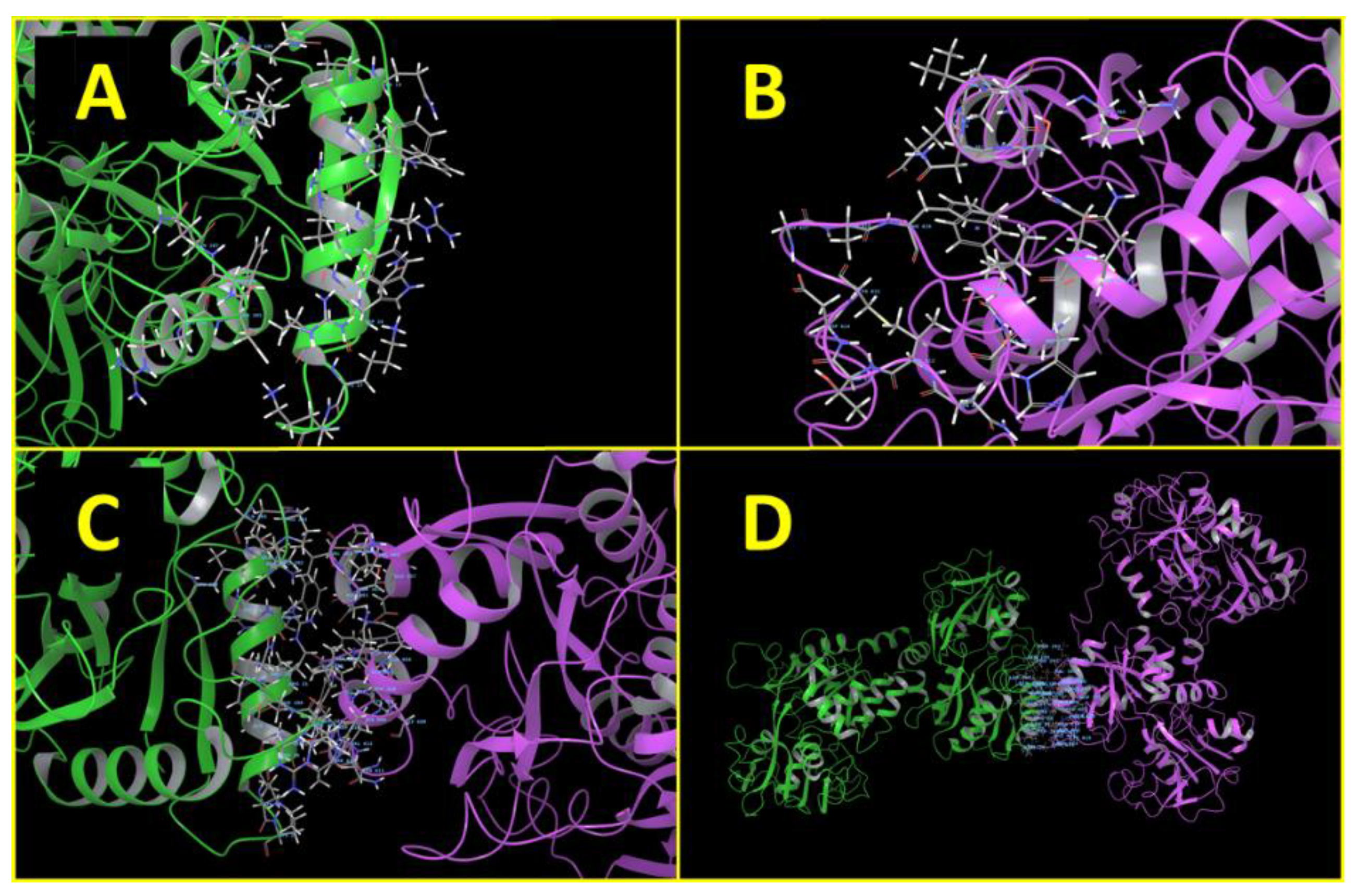
2.4.1. Molecular Docking Studies

Molecular docking studies were conducted to fully comprehend the Lf’s binding synergy with Tf receptors. There are more than 10 Lf protein structures accessible in the Protein Data Bank (PDB) of RCSB, so a structure with 1BLF PDB ID at a frequency of 2.8 Å was chosen and extracted for this experiment, along with a Tf structure reported to the PDB with PDB ID: 3V83 at a frequency of 2.10 Å. The extracted protein structure was organised using the Protein Preparation Wizard tool. The protein used for docking was introduced, then optimised and altered; minimisation throughout the protein generation process, which comprises sub-steps such as bridging absent chains and loops utilising the Prime tool, trailed by optimisation and minimisation using the OPLS3e force field. The essential residues and the dynamic site have been conserved in the structure of a protein. Beyond 5 Å, nonprotein water molecules were eliminated, and hetero atom stages were developed. The protein–protein docking analysis was performed, and the protein–protein complex, which was docked, was then utilised for the MD simulation analysis [39,40,41].

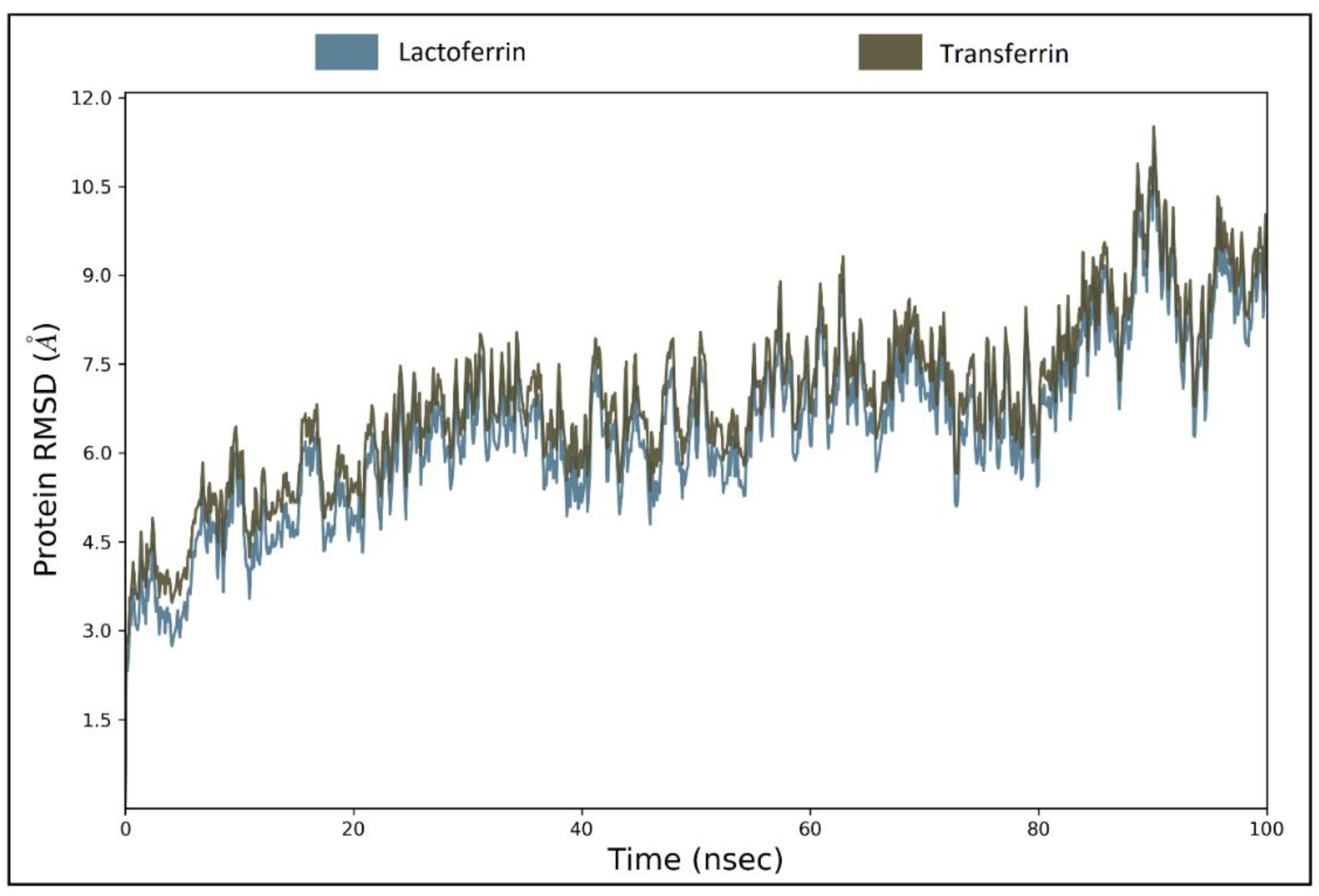
2.4.2. MD Simulation

MD simulation is extensively utilised to comprehend the dynamics and functionality of the protein–protein interaction. MD simulation was carried out employing Desmond coupled with Maestro to comprehend the protein’s dynamical behaviour in the existence of another protein. MD simulation is a three-step method: (1) system constructer, (2) minimisation and (3) dynamics. In the simulation, the OPLS3e force field was utilised. The Simple Point Charge (SPC) model was chosen for the solvent model in the system function, which was placed in an orthorhombic box, and the technique of measuring the box size was buffer. To balance the charges, sodium and chloride ions were utilised. The organised system was then equilibrated, and its energy was minimised. The NPT (particle number, pressure and temperature) ensembles were utilised to fix the temperature to 300 K and the pressure to 1.01325 bar. The number of atoms in this simulation experiment was about 1,68,765. MD simulation was performed for 100 nanoseconds (ns), and the frequency of trajectory monitoring was 100 picoseconds (ps). The Simulation Interaction Diagram (Desmond’s) was utilised to analyse the simulation experiment [39,40,41].

2.5. Characterisations of SM-MIL

2.5.1. Particle Size, Zeta Potential and Surface Morphology

The synthesised SM-MIL was characterised for the particle size, polydispersion index (PDI) and zeta potential using Malvern Zetasizer (Zetasizer Nano ZS, Malvern Instruments Inc., Malvern, UK). The morphological surface properties of synthesised nanoparticles (NPs) were examined with scanning electron microscopy (SEM). The surface area and pore volume of the SM-MIL NPs were further resolute using the Brunauer–Emmett–Teller (BET) analyser.

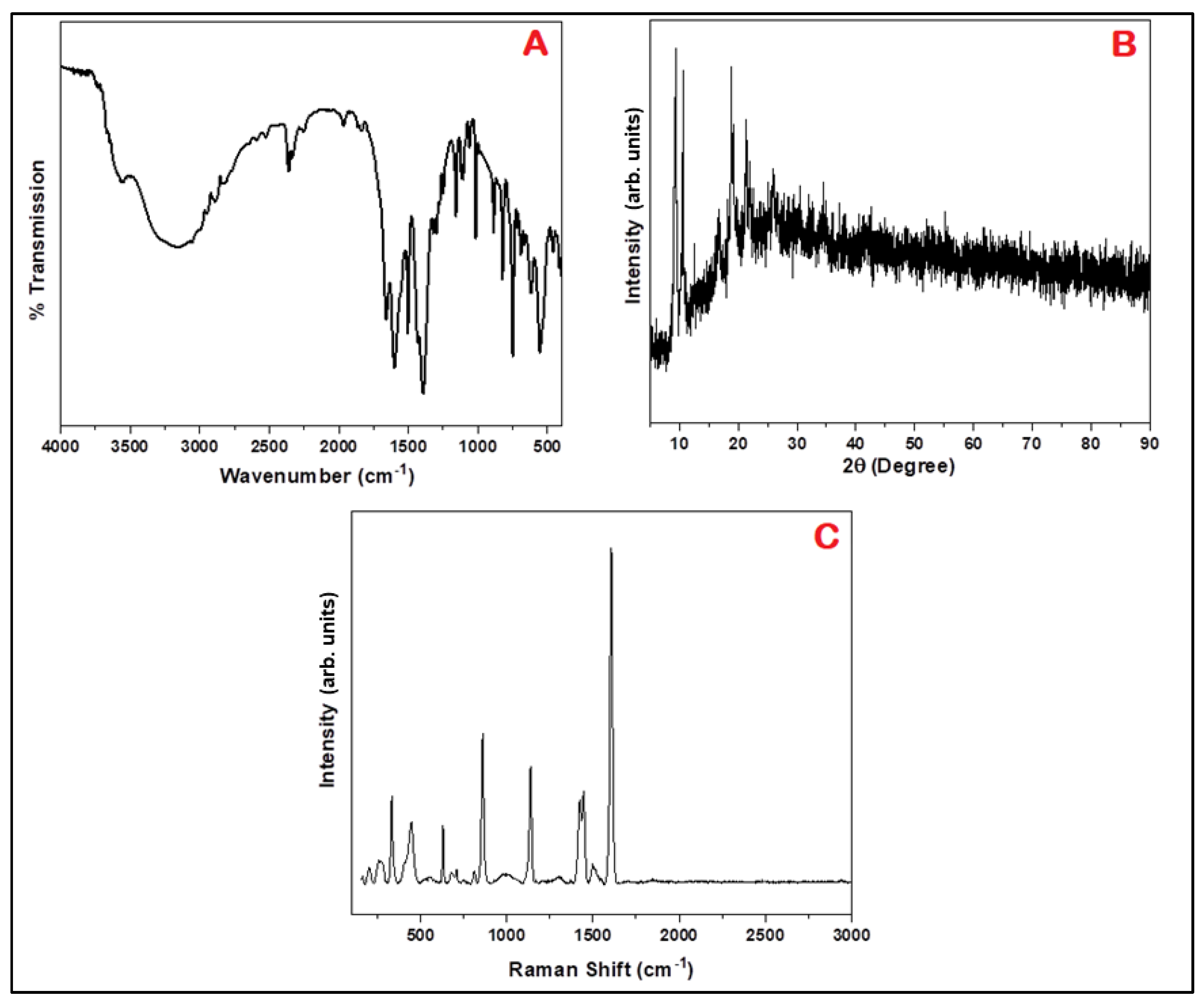
2.5.2. Spectroscopic Analysis

Several spectroscopic techniques have been used to study SM-MIL. To obtain the FTIR spectrum, the Bruker Alpha II ATR-FTIR instrument was used by scanning from a range of 500 cm−1 to 4000 cm−1. The obtained spectrum was baseline corrected and normalised for transmittance. The Ultima IV (Rigaku, Tokoyo, Japan) X-ray diffractometer was utilised to acquire information for PXRD to evaluate the crystalline structure of SM-MIL, along with Raman spectroscopy (B&W TEK, i-Raman Plus, Plainsboro, NJ, USA). Energy-dispersive spectroscopy was investigated to evaluate the elemental alignment of SM-MIL.

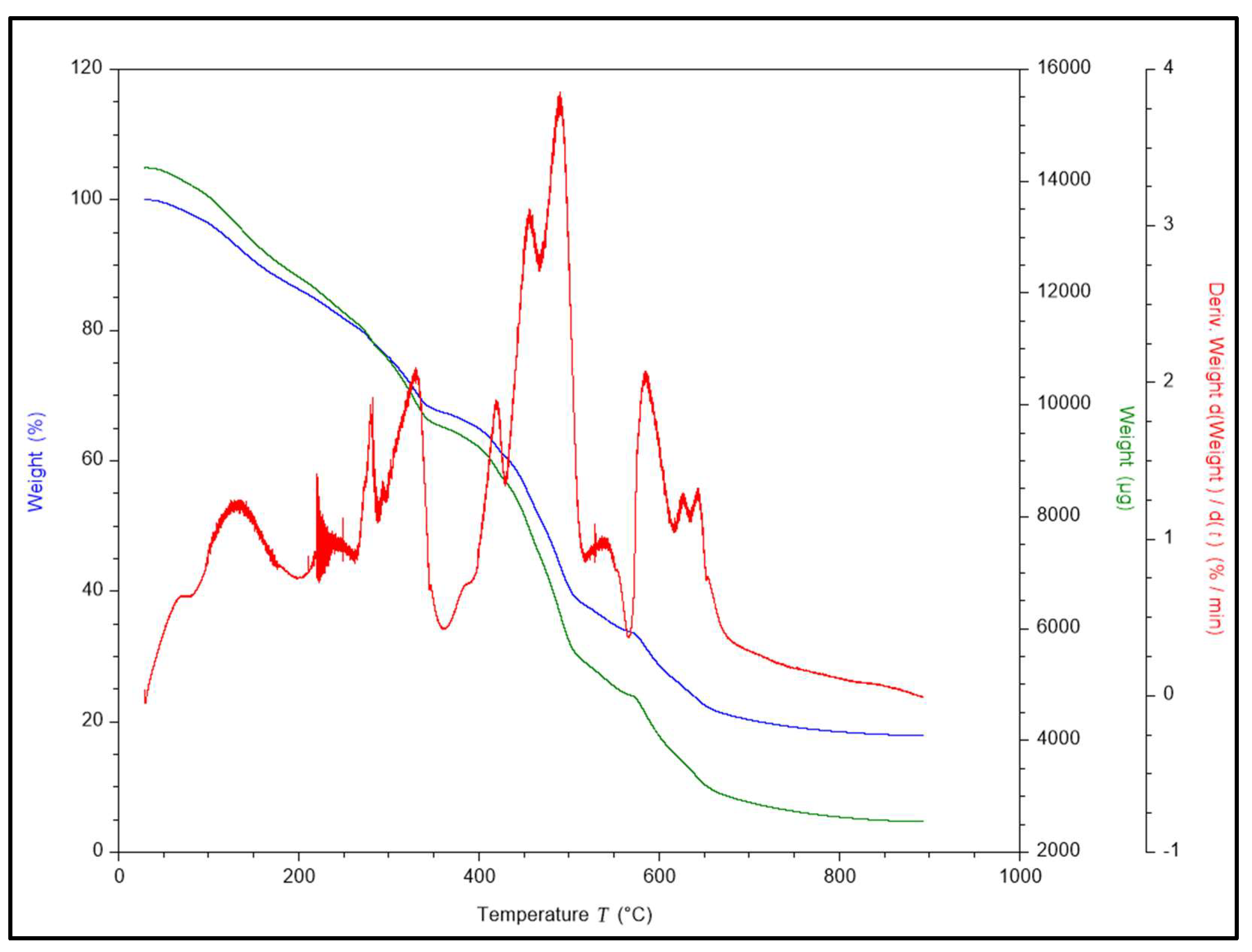
2.5.3. Thermal Stability

The thermogravimetric analysis (TGA; Discovery TGA 55, TA instruments, Milford, CT, USA) examined the material’s thermal stability. A sample of 10 mg was positioned in a platinum pan and heated up to 900 °C at 10 °C/min and recorded the weight change.

2.6. Drug Loading and In Vitro Drug Release Studies

The drug entrapment and loading efficacy were resolute using UV–Vis spectroscopy to measure the absorbance of the supernatant, as reported in a previous report [14]. The release of 5-FU was investigated using the dialysis bag technique at 37 ± 1 °C in two media (phosphate-buffered saline, pH 7.4 and pH 5.5). A dialysis bag (soaked overnight) was filled with a drug-loaded nanoparticle dispersion containing 5 mg equivalents of 5-FU and tightened at the ends. This bag was then submerged in 50 mL of receptor solution upheld at 37± 1 °C and swirled at 100 rpm. At various time intermissions ranging from 0 to 24 h, aliquots (1 mL) were removed from the receptor section and substituted with the same volume of the new medium. The quantity of drug released was assessed by evaluating the absorbance of aliquots with UV–Vis spectroscopy.

2.7. Drug Release Kinetics

The mathematical models are valuable tools for understanding and assessing drug release patterns and mechanisms from formulations and for analysing formulation design [42]. Kinetic models such as first order, zero order, Korsmeyer-Peppas and Higuchi’s models were used to investigate the in vitro release study data to discover the model best suited the data concerning drug release [43,44].

2.8. Chorioallantoic Membrane (CAM) Assay

The CAM assay was used to examine the impact of the proposed formulation on angiogenesis. Fertilised eggs of chickens were obtained and stored at 37 °C in an incubator (moistened). A small opening was cut into the eggshell on the 9th day, and the testing solutions were put into the chorioallantoic membrane. After the treatment, the opening was repacked with adhesive tape, and the chicken eggs were returned to the incubator. The alterations in blood vessels were noticed after 24 h [39].

2.9. In Vitro Viability and Cellular Internalisation

The in vitro cell viability study was done on IMR-32 neuroblastoma cells, as reported previously by our group. The cells were cultured at a density of 5 × 10⁴ cells in 96-well plates and incubated with different concentrations (0, 5, 10, 15, 20 and 25μg/mL) of SM-MIL for a duration of 24 and 48 h. For cellular internalisation, the SM-MIL were labelled with FITC for imaging. The sub-confluent cell cultures of IMR-32 were grown on coverslips (24 h, 37 °C) and then incubated with SM-MIL for 4 h. After incubation, the coverslips were visualised under a Confocal microscope (Zeiss LSM 510- Germany) at 60× to assess the uptake at the excitation wavelength and emission wavelength of 488 nm and 560 nm, respectively.

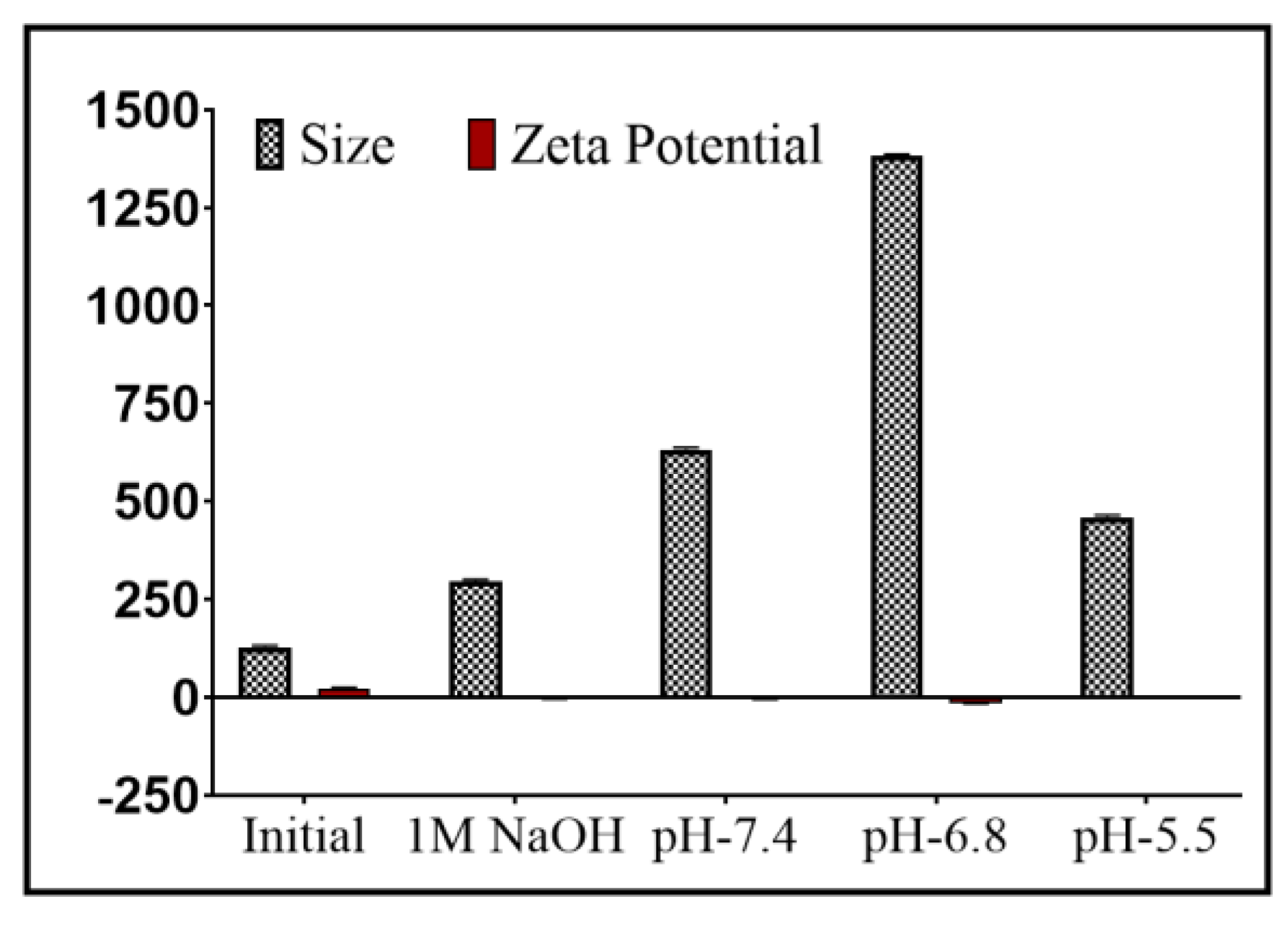
2.10. Stability Studies

The produced MIL-101(Fe) stability was evaluated for pH and in biological conditions. When evaluating the stability of nanoformulations, pH is a critical factor. The consequence of pH on the stability and size of MIL-101(Fe) was evaluated by dispersing 5 mg of MIL-101(Fe) in the media of different pH (pH: 5.5, 6.8, 7.4, 8.0 and 9.0) overnight and assessed with the Malvern Zeta sizer for particle Size, PDI and zeta potential.
Protein adsorption experiments helped analyse the serum protein’s impact on the stability of MIL-101(Fe). In this experiment, albumin was utilised as a typical protein. Five milligrams of MIL-101(Fe) were reconstituted in PBS (pH 7.4) and suspended in 2% and 4% w/v concentration solutions of bovine serum albumin (BSA). The dispersion was then set at 37 °C for 8 h and 100 rpm in an orbital shaker and further centrifuged at 10,000 rpm to eliminate the unadsorbed protein and examined for size, PDI and zeta potential. The NPs were incubated in serum for 24 h to assess the serum stability of the MIL-101(Fe).

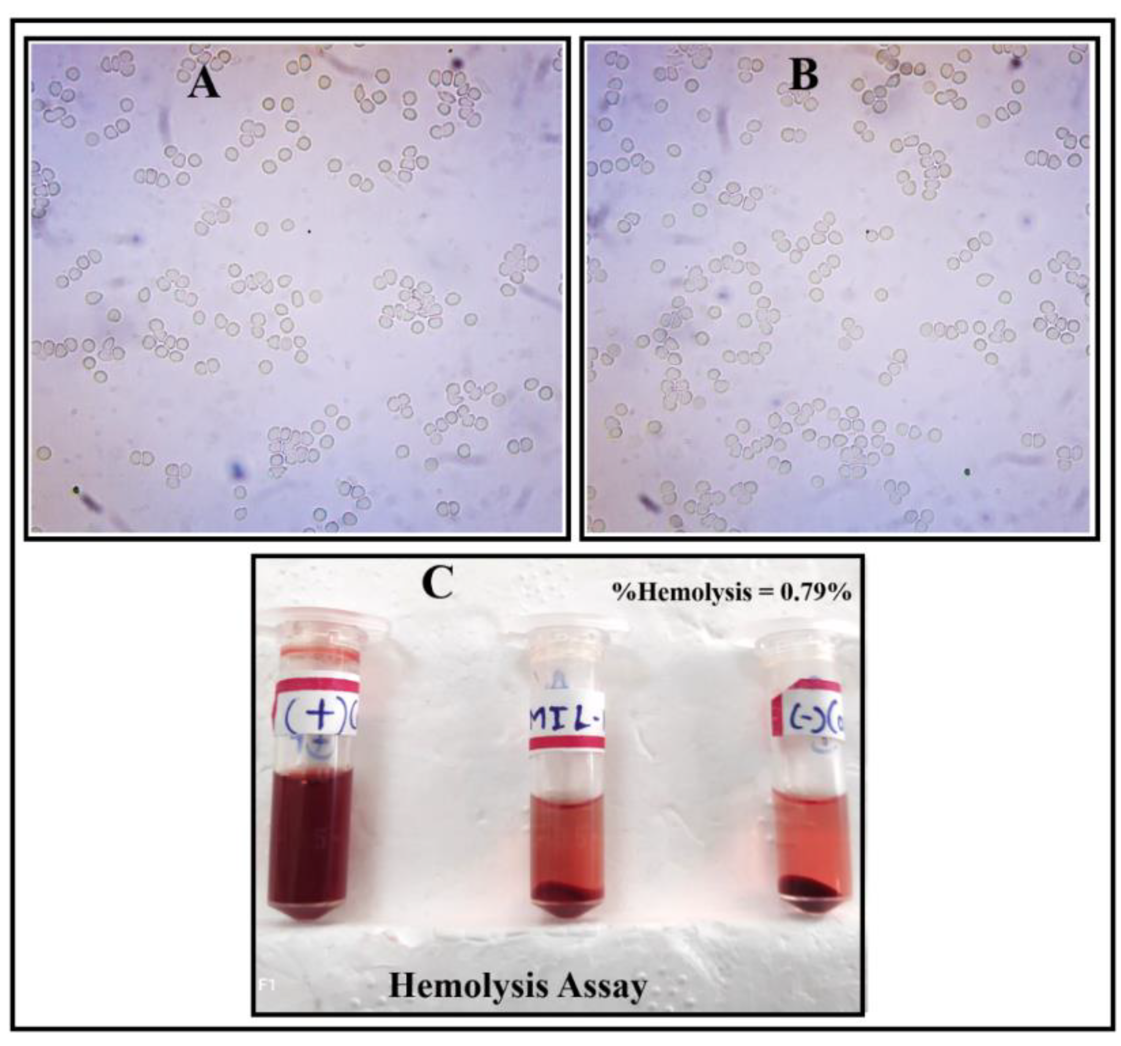
2.11. Interaction with Erythrocytes

To investigate the hemocompatibility of synthesised MOFs, an analysis of the interaction of SM-MIL with erythrocytes was performed following a previously reported method [29]. In particular, blood was obtained from rats by retro-orbital plexus perforation into the tube-containing EDTA solution and centrifuged for 15 min at 2500 rpm. The pellets of erythrocytes collected were treated three times, utilising PBS pH 7.4. SM-MIL (dispersed with 5 mg/mL PBS) was transferred to the erythrocyte suspension and incubated for 2 h at 37 °C. After incubation, the suspension was analysed under a Motic microscope to examine the haemolysis (if any) due to SM-MIL. As a control, erythrocytes combined with saline were used.
The hemocompatibility was assessed by diluting 800 mL of blood with 1 mL of PBS buffer [45,46]. This suspension (0.2 mL) was mixed with 0.8 mL of SM-MIL suspension (2 mg/mL in PBS) before being incubated at 37 °C for 60 min. After centrifuging the sample at 2500 rpm for 15 min, the intact erythrocytes were isolated from the incubated sample. To allow for haemoglobin oxidation, the collected supernatant was left at room temperature for 10 min. Utilising the given formula below, the optical density of oxyhaemoglobin was measured at 545 nm, and the percentage of haemolysis was calculated:
% Haemolysis = (OD of sample OD of negative control)/(OD of positive control − OD of negative control)

2.12. In Vivo Toxicity Studies

The Institutional Animal Ethics Committee (IAEC) from the Manipal Academy of Higher Education, Manipal, approved the experimental protocol (IAEC Reg No: 94/PO/ReBi/S/99/CPCSEA). Wistar rats (male) utilised for the experiment were provided by the Central Animal Research Centre Facility, Kasturba Medical College, Manipal. Wistar rats weighing 200–250 g each were individually housed per cage. Animals were allowed to acclimatise to research laboratory environments for 2 weeks before conducting the study. The animals were housed in a conditioned atmosphere at 25 ± 2 °C and supplemented with standard water and animal food from the laboratory. Acute and subacute toxicity of the synthesised SM-MIL MOFs were assessed using single- and multiple-dose studies as reported in the previous literature [47].

2.12.1. Acute Toxicity Study

The acute toxicity experiment for SM-MIL was conducted following a single I.V. injection at the given doses. Nine male Wistar rats were split into three groups for acute toxicity assessment (control, Test 1 and Test 2, comprising 3 animals each). All subjects, except for the control group of animals treated with saline, were injected with SM-MIL dispersed in 0.2 mL saline through the tail vein at doses of 15 and 20 mg/kg. The body weight change, dermal modifications, the existence of ascites and grooming or diminished motion were assessed daily. Utilising a body condition scoring (BCS) method, the nutritional status of the rats was evaluated. Around 100 μL of blood was obtained after 14 days from the retroorbital plexus into a dipotassium EDTA-containing tube and subjected to a complete blood count (CBC) examination.

2.12.2. Subacute Toxicity Study

The subacute toxicity experiment involved assessing the toxicity of SM-MIL NPs at a given dose for a short period after multiple injections. Healthy 6 male Wistar rats were selected and assigned into two groups of 3 each (test group and control group). The animals in the test group were given an I.V. injection thrice (1st, 4th and 7th days) in a week through the tail vein at a dose of 50 mg/kg of SM-MIL NPs dispersed in 0.2 mL saline. After 7 days of injection, a complete blood count (CBC), along with different biochemical parameters such as glucose, creatinine, alanine aminotransferase (ALT), total bilirubin, aspartate aminotransferase (AST), blood urea nitrogen (BUN), cholesterol, gamma-glutamyl transferase (GGT), albumin and total protein analysis, was done by collecting around 100 μL of the blood in a tube containing dipotassium EDTA solution from the retro-orbital plexus of the animal. The animal was then sacrificed, and various tissue organs such as the liver, kidney, brain, spleen and heart were collected in 10% formalin solution and stored (48 h) for histopathological analysis. The test and control specimens were compared via microscopic examination using a LX-500 LED trinocular Research microscope (Labomed, Los Angeles, CA, USA), and the images were captured with a MiaCam CMOS AR 6pro microscope camera (Noida, India).

3. Results and Discussion

3.1. Synthesis of Fe-Based MOF (SM-MIL)

The synthesis of SM-MIL is schematically depicted in Figure 1. The mechanism behind MIL-101(Fe) synthesis is a facile solvothermal approach. Initially, FeCl3 was dissolved in DMF solvent, and acetic acid was added as a structural modulator. To this solution, when the BDC linker, separately dissolved in DMF, was added and subjected to thermal treatment for 24 h, a brown solution was formed, indicating the formation of MIL-101(Fe)NPs. The washed and dried MIL-101(Fe) NPs showed an average particle size of 127.1 nm, PDI of 0.123 and zeta potential value of 21.8 mV with a practical yield of 96%. The size of Lf-HA conjugate-coated nanoparticles was found to be 141.7 nm, while the zeta potential was found to be 17.9 mV. This decrease in zeta potential value, although not considerably, for Lf-HA conjugate-coated MIL-101(Fe) suggests efficient coating of the Lf-HA conjugate on the surface of MIL-101(Fe).

3.2. Molecular Docking and Molecular Dynamics (MD) Simulation

3.2.1. Molecular Docking Studies

Computational docking studies were undertaken based on the experimental data to comprehend the location of the binding site and the optimum potential conformation for binding Lf to Tf utilising protein–protein docking. Molecular docking studies revealed that Lf potentially attaches to Tf and exhibits variable binding with various amino acids, as shown in Figure 2. The overall number of hydrogen bonds, salt bridges, Pi stacking, disulphides, vdW clash and the distance between two amino acids of two proteins, Lf and Tf, respectively, is shown in Table 1 [39,40,41].

3.2.2. MD Simulation

As illustrated in Figure 3, MD simulation experiments were conducted to investigate several factors, such as protein–protein complex stability, binding mode projection and relationships with the Lf-binding site and Tf. Throughout the simulation, a frame was captured every 100 ps and stored in a trajectory. The experiment of MD simulation generated around 1000 frames in total. The metric root means square deviation (RMSD) indicates protein stability and structural deviation. In the case of protein RMSD of Lf and Tf, it was discovered that they moved together throughout a 100 ns simulation period, as shown in Figure 4. While there was some drift, later, it remained steady up to 7.5 RMSD until 60 ns [39,40,41].

3.3. Spectroscopic Characterisation of MIL-101(Fe)

The FT-IR spectrum of MIL-101(Fe) is depicted in Figure 5A. MIL-101(Fe) have typical absorption peaks of 1598.69, 1394.28, 1016.30 and 750.17 cm−1, which may primarily be caused by vibrations of the carboxylate groups available in the MOFs and related to MIL-101, as previously published [42]. Moreover, at 1321.96 and 1297.85 cm−1, respectively, C–N vibrations of aromatic amines were detected.
MIL-101(Fe)’s XRD pattern, as shown in Figure 5B, was in close alignment with the previously published work [48], suggesting that MIL-101(Fe) was synthesised successfully. MIL-101(Fe) diffraction patterns have no impure diffraction peaks, indicating that the synthesised compound is pure. It showed that the stability of the MOFs is exceptional, which is beneficial for their use as a drug delivery system. These results are further verified by Raman spectra in Figure 5C. Several MIL-101(Fe) bands are mostly connected to organic ligands linked to C=O in the carboxylic group and the C-C bond of the benzene ring, respectively [49]. EDS spectroscopy (Figure 6A) was also done to determine the MIL-101(Fe) elemental composition. Figure 6B–D shows the abundance of Fe, C and O, which correspond to the primary elemental makeup of the MIL-101(Fe) skeleton. Nitrogen was also found (Figure 6E), indicating the existence of residual solvent (DMF).

3.4. Morphological Characteristics

Scanning electron microscopy (SEM) was utilised to examine the morphologies of MIL-101(Fe). A standard octahedral shape was observed for the study, as depicted in Figure 7A,B, which closely aligns with the previous studies [48]. The number of particles with a size diversity of ≈200 nm in the MIL-101(Fe) sample was observed. The MIL-101(Fe) BET surface area was determined to be 0.24 (sq.m/gm). Process optimisation and thermal induction can improve the sample’s smaller area and pore volume.

3.5. Thermal Stability

The MIL-101(Fe) thermal stability was inspected by TGA investigation. The TGA examination of MIL-101(Fe), as depicted in Figure 8, revealed that the TGA curves remained relatively stable up to 270 °C, which is similar to past findings [48]. The first weight loss (20%) amid 34 and 270 °C in the MIL-101(Fe) curves may be attributed to the evaporation of certain DMF and water-like solvent molecules used in the procedure. The second loss between 270 and 600 °C occurred from the gradual matrix breakdown of MIL-101(Fe). The findings of the thermal analysis led us to infer that the prepared MIL-101(Fe) sample exhibits similar thermal activity to the previously published literature [48].

3.6. Drug Loading and In Vitro Drug Release

Using 5-FU as a model drug, the drug loading efficiency of SM-MIL was investigated. The drug entrapment and loading efficiency were estimated to be 45% and 22.36%, respectively. The release of 5-FU from the SM-MIL was studied in two media (Figure 9) to replicate the extracellular (pH 7.4 and 5.5) and intracellular (pH 5.5: tumour cells) pH conditions. The pH of 5.5 mimics the low pH found in the tumour microenvironment, which is expected to be the target of the developed nanoformulation. The acidic pH is also found in endosomes following endocytosis [37]. Drug release in the pH 5.5 medium was substantially faster (94.54% after 12 h) than in the PBS medium 7.4 pH (27.63% after 12 h), implying the improved drug release in an acidic medium.

3.7. Release Model Kinetics

To determine the drug release constant and regression coefficient, the drug release data were fitted to various kinetic models. The drug release model does not follow zero-order kinetics, as shown by the low r2 values (<0.73) obtained with zero-order release plots, and the same is true for first-order release kinetics (r2 < 0.72). Higuchi’s model could best describe the release patterns of plain 5-FU and SM-MIL (pH 5.5), as the graphs demonstrated strong linearity with correlation coefficient values of 0.92 and 0.89, respectively. The Korsmeyer-Peppas plots, which demonstrated fair linearity with slope values less than 0.5 and r2 values of 0.93 and 0.93 for 5-FU and SM-MIL (pH 5.5), further supported the diffusion mechanism of drug release for these two samples. These findings show that diffusion control dominated the drug release mechanism from these two samples [50].
On the other hand, as the r2 values for all of the kinetics models were so low, SM-MIL (@pH 7.4) did not demonstrate any specific drug release mechanism. Even though the “n” value in the Korsmeyer-Peppas model was discovered to be 0.13, the graph’s linearity was also very low (r2 was about 0.71). Furthermore, the drug-loaded SM-MIL (pH 7.4) displayed biphasic release in the drug release profile. Therefore, the drug release from SM-MIL (pH 7.4) might follow mixed-order kinetics.

3.8. Chorioallantoic Membrane (CAM) Assay

The angiogenic and antiangiogenic potential of biomolecules and therapeutics are often studied using CAM, which comprises a dense network of capillaries. After 24 h of treatment with SM-MIL, the studies revealed antiangiogenic activity, decreased blood vessel development and a significant reduction in the branching vascular structure (Figure 10C). After 24 h of treatment with the negative control, lenalidomide, no blood vessel development was seen (Figure 10B). The growth of blood vessels was found in phosphate-buffered saline, 7.4 pH (PBS). However, the branching of vessels was not evident and was less mature (Figure 10A). The findings revealed that SM-MIL promoted antiangiogenic activity. This can be quite helpful for cancer-related therapies, where an optimised formulation with 5-FU as an active ingredient might be beneficial.

3.9. In Vitro Cell Viability and Cellular Internalization Study

The cell viability of SM-MIL was performed to assess the biocompatibility of synthesised nanoparticles and drug-loaded nanoparticles. The cell viability of placebo SM-MIL was found to be more than 90% (93.7 ± 3.2%) even at the maximum concentration of 25 µg/mL, suggesting the safety of SM-MIL, which can also be attributed to the fact that the coating of HA over MOFs has been reported to demonstrate enhanced biocompatibility. In the case of drug-loaded SM-MIL, the suppression of cell viability was more prominent. The decrease in cell viability in the case of SM-MIL was found to be 32.1 ± 3.8% and 41.3 ± 3.6% after 24 and 48 h, respectively.
The cellular Internalisation of nanoparticles has been reported to increase with HA coating, although it has not been reported to date for MIL(Fe) 101. The cellular uptake of SM-MIL is shown in Figure 11. The presence of nanoparticles can be seen as aggregates around the cell membrane, suggesting possible localisation of HA-coated nanoparticles on the membrane surface, followed by internalisation of the same inside cells.

3.10. Stability Studies

Before the NPs enter the tumour cells, they must pass through the physiological pH of the blood, so it is essential to analyse their physiological pH stability. The impact of different pH media on the size and stability of MIL-101(Fe) indicated the pH-dependent changes concerning size, PDI and zeta potential. The preliminary size, PDI and zeta potential of the framework were 127.1 nm, 0.123 and 21.8 mV, respectively. While the size, PDI and zeta potential of the structure are observed in Figure 12 after dispersing it overnight in 1M NaOH, 7.4 pH PBS, 6.8 pH PBS and 5.5 pH PBS. From the above data, we can derive that the size and stability of MIL-101(Fe) in various pH media can change significantly. Thus, it can be concluded that the framework could show pH-dependent drug release when administered through the oral, intravenous or intramuscular route.
The fate of NPs is decided based on the extent of the protein corona formation when the NPs interact with the serum proteins. We utilised bovine serum albumin (BSA) as a model protein to understand this interaction and incubated the MIL-101(Fe) NPs for 4 h with BSA at two distinct concentrations (2% w/v and 4% w/v). Concerning the particle size and zeta potential, the degree of interaction was examined. The particle sizes of MIL-101(Fe) NPs incubated with 2% and 4% BSA were raised to 151.2 nm and 155.5 nm, respectively; the zeta potential values decreased to 16.5 mV and 14.3 mV, respectively (Figure 13). Although albumin had a negative charge, it interacted weakly with the NPs due to the presence of amino acids such as Lys, His and Arg, suggesting the stabilisation of MIL-101(Fe) NPs in the serum protein environment. These findings confirmed the stability of MIL-101(Fe) NPs in the biological media.

3.11. Interaction with Erythrocytes

The study of the interaction of erythrocytes with nanoformulations is significant for the administration of nanoformulations through the parenteral route. RBCs act as osmometers (which swell or shrink as dictated by their osmotic environment), and the lysis of RBCs can be caused by some physical or osmotic changes in the blood, resulting in haemoglobin release. Haemoglobinuria, which is used as a diagnostic parameter for blood poisoning, is caused by haemoglobin excretion in the urine. Through a microscopic analysis using the Motic microscope and haemolysis assay, we studied the interaction of SM-MIL NPs with RBCs. The morphology of RBCs did not alter (Figure 14A,B) when the SM-MIL NPs were incubated with erythrocytes, suggesting the absence of any interaction among them. In addition, as shown in Figure 14C, the haemolysis assay shows that the % haemolysis caused by SM-MIL NPs was less than 5%, which is considered a critical safe value at a 2 mg/mL concentration [45]. This indicates the SM-MIL NP biosafety and compatibility.

3.12. In Vivo Toxicity Studies

The increasing interest in using nanomaterials to deliver therapeutics has facilitated in vivo experiments to determine the systemic toxicity of the NPs. The acute toxicity was carried out in Wistar rats to assess the effect of a single dose at different concentrations of SM-MIL NPs for 14 days. The body weight of the rats of the control group was compared with all the treated group rats. There was no significant difference in the body weight values (Table 2), even at the dose of 20 mg/kg, and the animals did not show impaired mobility, dermal changes or the presence of ascites. Further, the values of the CBC analysis, as tabulated in Table 3, corresponding to days 7 and 14, indicated that the haematological parameters of the treated group were similar to the control group and well within the reference range. Therefore, it can be inferred that the SM-MIL NPs did not exhibit acute toxicity in the rats up to the dose of 20 mg/kg.
Subacute toxicity studies reveal the effect of multiple doses on the body. We could observe no significant change in the body weight of rats compared to control animals, even at the dose of 50 mg/kg (Table 2), and there was no unusual response or impaired mobility or any signs of infection in any of the rats. The haematological parameters are tabulated in Table 3. Though few parameters differed from the control group, all were well within the reference array.
Further, the histopathological studies were performed using tissues from various organs, as shown in Figure 15. The spleen, liver, heart and brain were collected, and the excised tissue samples were stored in 10% formalin for 48 h. Macroscopic examination of the tissues of the control specimen was compared with the test specimen. The cardiac muscle and the myocardium of the heart tissue showed no inflammation, congestion or necrosis. The liver tissue section of both groups showed lobules with central vein and portal triads along the periphery of lobules. No decongestion and necrosis were observed; however, mildly congested sinusoids were observed in both groups. No histological changes were observed in the spleen tissue. The cerebrum and hippocampus of the crystal violet-stained brain tissue showed a varying number of healthy neurons (pale round well, defined nuclear boundary with prominent nuclei) and degenerated neurons that are darkly stained with fragmented and shrunken nuclei in both groups. In conclusion, no histopathological abnormalities or lesions were detected in main organs such as the liver, brain, heart and spleen, proving that the detected results for the SM-MIL MOF synthesised are safe as a drug carrier for multiple dosages up to 50 mg/kg per dose in Wistar rats.

4. Conclusions

In conclusion, the SM-MIL NPs were biocompatible and did not trigger the haemolysis of male Wistar rat RBCs. SM-MIL demonstrated no significant acute (visible or haematological) toxicity in Wistar rats with a single dose of up to 50 mg/kg. The findings of the subacute toxicity experiments demonstrated the stable and nontoxic aspects of SM-MIL at all analysed doses, as no substantial toxicity in rat or histopathological analysis of different organs, such as the liver, heart, spleen and brain, revealed any grade of disruption or pathological alterations in either the haematological or serological study. These results suggest that SM-MIL NPs are encouraging and safe novel carriers for drug delivery applications.

Author Contributions

Conceptualization, A.P. and S.M.; methodology, A.P.; software, A.N.N. and S.B.; validation, S.M., A.P. and A.N.N.; formal analysis, A.N.N., S.K. and A.P.; investigation, A.N.N. and S.H.N.; resources, S.M., G.G.S. and S.D.G.; data curation, S.K., G.F. and B.S.P.; writing—original draft preparation, A.N.N.; writing—review and editing, A.P., S.K., A.N.N.; visualization, A.P. and S.M.; supervision, S.M. and A.P.; project administration, S.M.; funding acquisition, S.M. All authors have read and agreed to the published version of the manuscript.

Funding

The authors are thankful to the (i) Manipal Academy of Higher Education (MAHE), Manipal, India for the TMA Pai Doctoral Fellowship to Ajinkya Nitin Nikam and Gasper Fernandes, (ii) Board of Research in Nuclear Science (BRNS), Department of Atomic Energy (DAE), Government of India, for the Junior Research Fellowship to Sanjay Kulkarni and (iii) All India Council for Technical Education (AICTE), Government of India, New Delhi, for the National Doctoral Fellowship to Bharath Singh Padya.

Data Availability Statement

Not applicable.

Acknowledgments

The authors are thankful to the Manipal College of Pharmaceutical Sciences, MAHE, Manipal, India, for providing the necessary facilities to carry out this research.

Conflicts of Interest

The authors declare no conflict of interest.

References

  1. Farokhzad, O.C.; Langer, R. Impact of Nanotechnology on Drug Delivery. ACS Nano 2009, 3, 16–20. [Google Scholar] [CrossRef]
  2. Ferrari, M. Cancer nanotechnology: Opportunities and challenges. Nat. Cancer 2005, 5, 161–171. [Google Scholar] [CrossRef]
  3. Park, K. Nanotechnology: What it can do for drug delivery. J. Control. Release 2007, 120, 1–3. [Google Scholar] [CrossRef] [PubMed] [Green Version]
  4. Park, K. Facing the Truth about Nanotechnology in Drug Delivery. ACS Nano 2013, 7, 7442–7447. [Google Scholar] [CrossRef] [Green Version]
  5. Pandey, A.; Dhas, N.; Deshmukh, P.; Caro, C.; Patil, P.; García-Martín, M.L.; Padya, B.; Nikam, A.; Mehta, T.; Mutalik, S. Heterogeneous surface architectured metal-organic frameworks for cancer therapy, imaging, and biosensing: A state-of-the-art review. Coord. Chem. Rev. 2020, 409, 213212. [Google Scholar] [CrossRef]
  6. Nikam, A.; More, M.P.; Pandey, A.P.; Patil, P.; Patil, A.G.; Deshmukh, P.K. Design and development of thiolated graphene oxide nanosheets for brain tumor targeting. Int. J. Polym. Mater. Polym. Biomater. 2019, 69, 611–621. [Google Scholar] [CrossRef]
  7. Jha, A.; Nikam, A.N.; Kulkarni, S.; Mutalik, S.P.; Pandey, A.; Hegde, M.; Rao, B.S.S.; Mutalik, S. Biomimetic nanoarchitecturing: A disguised attack on cancer cells. J. Control. Release 2020, 329, 413–433. [Google Scholar] [CrossRef]
  8. Kotha, R.; Fernandes, G.; Nikam, A.N.; Kulkarni, S.; Pandey, A.; Pandey, S.; Mutalik, S. Surface engineered bimetallic nanoparticles based therapeutic and imaging platform: Recent advancements and future perspective. Mater. Sci. Technol. 2020, 36, 1729–1748. [Google Scholar] [CrossRef]
  9. Nikam, A.N.; Pandey, A.; Fernandes, G.; Kulkarni, S.; Mutalik, S.P.; Padya, B.S.; George, S.D.; Mutalik, S. Copper sulphide based heterogeneous nanoplatforms for multimodal therapy and imaging of cancer: Recent advances and toxicological perspectives. Coord. Chem. Rev. 2020, 419, 213356. [Google Scholar] [CrossRef]
  10. Shreya, A.B.; Pandey, A.; Nikam, A.N.; Patil, P.O.; Sonawane, R.; Deshmukh, P.K.; Mutalik, S. One- pot development of spray dried cationic proliposomal dry powder insufflation: Optimization, characterization and bio-interactions. J. Drug Deliv. Sci. Technol. 2020, 61, 102298. [Google Scholar] [CrossRef]
  11. Pandey, A.; Nikam, A.N.; Fernandes, G.; Kulkarni, S.; Padya, B.S.; Prassl, R.; Das, S.; Joseph, A.; Deshmukh, P.K.; Patil, P.O.; et al. Black Phosphorus as Multifaceted Advanced Material Nanoplatforms for Potential Biomedical Applications. Nanomaterials 2020, 11, 13. [Google Scholar] [CrossRef]
  12. Padya, B.S.; Pandey, A.; Nikam, A.; Kulkarni, S.; Fernandes, G.; Mutalik, S. Chapter 33—Two-dimensional materials-based nanoplatforms for lung cancer management: Synthesis, properties, and tar-geted therapy. In Advanced Drug Delivery Systems in the Management of Cancer; Dua, K., Mehta, M., Pinto, T.d.A., Pont, L.G., Williams, K.A., Rathbone, M.J., Eds.; Academic Press: Cambridge, MA, USA, 2021; pp. 415–429. [Google Scholar] [CrossRef]
  13. Pandey, A.; Nikam, A.; Basavraj, S.; Mutalik, S.; Gopalan, D.; Kulkarni, S.; Padya, B.; Fernandes, G.; Mutalik, S. Chapter 25—Nose-to-brain drug delivery: Regulatory aspects, clinical trials, patents, and future perspectives. In Direct Nose-to-Brain Drug Delivery; Pardeshi, C.V., Souto, E.B., Eds.; Academic Press: Cambridge, MA, USA, 2021; pp. 495–522. [Google Scholar] [CrossRef]
  14. Kulkarni, S.; Pandey, A.; Nikam, A.N.; Nannuri, S.H.; George, S.D.; Fayaz, S.M.A.; Vincent, A.P.; Mutalik, S. ZIF-8 nano confined protein-titanocene complex core-shell MOFs for efficient therapy of Neuroblastoma: Optimization, molecular dynamics and toxicity studies. Int. J. Biol. Macromol. 2021, 178, 444–463. [Google Scholar] [CrossRef]
  15. Mehta, S.; Kulkarni, S.; Nikam, A.N.; Padya, B.S.; Pandey, A.; Mutalik, S. Liposomes as Versatile Platform for Cancer Theranostics: Therapy, Bio-imaging, and Toxicological Aspects. Curr. Pharm. Des. 2021, 27, 1977–1991. [Google Scholar] [CrossRef]
  16. Dhas, N.; Kudarha, R.; Pandey, A.; Nikam, A.N.; Sharma, S.; Singh, A.; Garkal, A.; Hariharan, K.; Singh, A.; Bangar, P.; et al. Stimuli responsive and receptor targeted iron oxide based nanoplatforms for multimodal therapy and imaging of cancer: Conjugation chemistry and alternative therapeutic strategies. J. Control. Release 2021, 333, 188–245. [Google Scholar] [CrossRef]
  17. Pandey, A.; Nikam, A.N.; Padya, B.S.; Kulkarni, S.; Fernandes, G.; Shreya, A.B.; García, M.C.; Caro, C.; Páez-Muñoz, J.M.; Dhas, N.; et al. Surface architectured black phosphorous nanoconstructs based smart and versatile platform for cancer theranostics. Coord. Chem. Rev. 2021, 435, 213826. [Google Scholar] [CrossRef]
  18. Fernandes, G.; Pandey, A.; Kulkarni, S.; Mutalik, S.P.; Nikam, A.N.; Seetharam, R.N.; Kulkarni, S.S.; Mutalik, S. Supramolecular Dendrimers Based Novel Platforms for Effective Oral Delivery of Therapeutic Moieties. J. Drug Deliv. Sci. Technol. 2021, 64, 102647. [Google Scholar] [CrossRef]
  19. Nannuri, S.H.; Nikam, A.N.; Pandey, A.; Mutalik, S.; George, S.D. Subcellular Imaging and Diagnosis of Cancer using Engineered Nanoparticles. Curr. Pharm. Des. 2022, 28, 690–710. [Google Scholar] [CrossRef]
  20. Dhas, N.; García, M.C.; Kudarha, R.; Pandey, A.; Nikam, A.N.; Gopalan, D.; Fernandes, G.; Soman, S.; Kulkarni, S.; Seetharam, R.N.; et al. Advancements in cell membrane camouflaged nanoparticles: A bioinspired platform for cancer therapy. J. Control. Release 2022, 346, 71–97. [Google Scholar] [CrossRef]
  21. Navti, P.D.; Pandey, A.; Nikam, A.N.; Padya, B.S.; Kalthur, G.; Koteshwara, K.B.; Mutalik, S. Ionic Liquids Assisted Topical Drug Delivery for Permeation Enhancement: Formulation Strategies, Biomedical Applications, and Toxicological Perspective. AAPS PharmSciTech 2022, 23, 161. [Google Scholar] [CrossRef]
  22. Moghimi, S.M.; Hunter, A.; Murray, J.C. Nanomedicine: Current status and future prospects. FASEB J. 2004, 19, 311–330. [Google Scholar] [CrossRef] [PubMed] [Green Version]
  23. Zhou, H.C.; Long, J.R.; Yaghi, O.M. Introduction to Metal–Organic Frameworks. Chem. Rev. 2012, 112, 673–674. [Google Scholar] [CrossRef]
  24. Yang, Q.; Xu, Q.; Jiang, H.-L. Metal–organic frameworks meet metal nanoparticles: Synergistic effect for enhanced catalysis. Chem. Soc. Rev. 2017, 46, 4774–4808. [Google Scholar] [CrossRef] [PubMed]
  25. Kim, J.; Yang, S.-T.; Choi, S.B.; Sim, J.; Kim, J.; Ahn, W.-S. Control of catenation in CuTATB-n metal–organic frameworks by sonochemical synthesis and its effect on CO2 adsorption. J. Mater. Chem. 2011, 21, 3070–3076. [Google Scholar] [CrossRef]
  26. Li, J.-R.; Ma, Y.; McCarthy, M.C.; Sculley, J.; Yu, J.; Jeong, H.-K.; Balbuena, P.B.; Zhou, H.-C. Carbon dioxide capture-related gas adsorption and separation in metal-organic frameworks. Coord. Chem. Rev. 2011, 255, 1791–1823. [Google Scholar] [CrossRef]
  27. Corma, A.; García, H.; Xamena, F.X.L. Engineering Metal Organic Frameworks for Heterogeneous Catalysis. Chem. Rev. 2010, 110, 4606–4655. [Google Scholar] [CrossRef]
  28. Zhu, L.; Liu, X.-Q.; Jiang, H.-L.; Sun, L.-B. Metal–Organic Frameworks for Heterogeneous Basic Catalysis. Chem. Rev. 2017, 117, 8129–8176. [Google Scholar] [CrossRef]
  29. Pandey, A.; Kulkarni, S.; Vincent, A.P.; Nannuri, S.H.; George, S.D.; Mutalik, S. Hyaluronic acid-drug conjugate modified core-shell MOFs as pH responsive nanoplatform for multimodal therapy of glioblastoma. Int. J. Pharm. 2020, 588, 119735. [Google Scholar] [CrossRef] [PubMed]
  30. Beg, S.; Rahman, M.; Jain, A.; Saini, S.; Midoux, P.; Pichon, C.; Ahmad, F.J.; Akhter, S. Nanoporous metal organic frameworks as hybrid polymer–metal composites for drug delivery and biomedical applications. Drug Discov. Today 2017, 22, 625–637. [Google Scholar] [CrossRef]
  31. Férey, G.; Mellot-Draznieks, C.; Serre, C.; Millange, F.; Dutour, J.; Surblé, S.; Margiolaki, I. A Chromium Terephthalate-Based Solid with Unusually Large Pore Volumes and Surface Area. Science 2005, 309, 2040–2042. [Google Scholar] [CrossRef]
  32. Almáši, M.; Zeleňák, V.; Palotai, P.; Beňová, E.; Zeleňáková, A. Metal-organic framework MIL-101(Fe)-NH2 functionalized with different long-chain polyamines as drug delivery system. Inorg. Chem. Commun. 2018, 93, 115–120. [Google Scholar] [CrossRef]
  33. Sajid, M. Toxicity of nanoscale metal organic frameworks: A perspective. Environ. Sci. Pollut. Res. 2016, 23, 14805–14807. [Google Scholar] [CrossRef] [Green Version]
  34. Baati, T.; Njim, L.; Neffati, F.; Kerkeni, A.; Bouttemi, M.; Gref, R.; Najjar, M.F.; Zakhama, A.; Couvreur, P.; Serre, C.; et al. In depth analysis of the in vivo toxicity of nanoparticles of porous iron(iii) metal–organic frameworks. Chem. Sci. 2013, 4, 1597–1607. [Google Scholar] [CrossRef]
  35. Liu, C.-H.; Chiu, H.-C.; Sung, H.-L.; Yeh, J.-Y.; Wu, K.C.-W.; Liu, S.-H. Acute oral toxicity and repeated dose 28-day oral toxicity studies of MIL-101 nanoparticles. Regul. Toxicol. Pharmacol. 2019, 107, 104426. [Google Scholar] [CrossRef]
  36. Wang, Y.; Lin, W.; Yu, S.; Huang, X.; Lang, X.; He, Q.; Gao, L.; Zhu, H.; Chen, J. A biocompatible Zr-based metal-organic framework UiO-66-PDC as an oral drug carrier for pH-response release. J. Solid State Chem. 2021, 293, 121805. [Google Scholar] [CrossRef]
  37. Jarai, B.M.; Stillman, Z.; Attia, L.; Decker, G.E.; Bloch, E.D.; Fromen, C.A. Evaluating UiO-66 Metal–Organic Framework Nanoparticles as Acid-Sensitive Carriers for Pulmonary Drug Delivery Applications. ACS Appl. Mater. Interfaces 2020, 12, 38989–39004. [Google Scholar] [CrossRef]
  38. Pandey, A.; Singh, K.; Patel, S.; Singh, R.; Patel, K.; Sawant, K. Hyaluronic acid tethered pH-responsive alloy-drug nanoconjugates for multimodal therapy of glioblastoma: An intranasal route approach. Mater. Sci. Eng. C 2019, 98, 419–436. [Google Scholar] [CrossRef]
  39. Raychaudhuri, R.; Pandey, A.; Das, S.; Nannuri, S.H.; Joseph, A.; George, S.D.; Vincent, A.P.; Mutalik, S. Nanoparticle impregnated self-supporting protein gel for enhanced reduction in oxidative stress: A molecular dynamics insight for lactoferrin-polyphenol interaction. Int. J. Biol. Macromol. 2021, 189, 100–113. [Google Scholar] [CrossRef]
  40. Abdizadeh, H.; Atilgan, A.R.; Atilgan, C. Detailed molecular dynamics simulations of human transferrin provide insights into iron release dynamics at serum and endosomal pH. JBIC J. Biol. Inorg. Chem. 2015, 20, 705–718. [Google Scholar] [CrossRef]
  41. Khan, M.S.; Husain, F.M.; Alhumaydhi, F.A.; Alwashmi, A.S.; Rehman, T.; Alruwetei, A.M.; Hassan, I.; Islam, A.; Shamsi, A. Exploring the molecular interactions of Galantamine with human Transferrin: In-silico and in vitro insight. J. Mol. Liq. 2021, 335, 116227. [Google Scholar] [CrossRef]
  42. Alavijeh, R.K.; Akhbari, K. Biocompatible MIL-101(Fe) as a Smart Carrier with High Loading Potential and Sustained Release of Curcumin. Inorg. Chem. 2020, 59, 3570–3578. [Google Scholar] [CrossRef]
  43. Reddy, K.R.; Mutalik, S.; Reddy, S. Once-daily sustained-release matrix tablets of nicorandil: Formulation and in vitro evaluation. AAPS PharmSciTech 2003, 4, 480–488. [Google Scholar] [CrossRef] [Green Version]
  44. Mutalik, S.; Udupa, N. Glibenclamide transdermal patches: Physicochemical, pharmacodynamic, and pharmacokinetic evaluations. J. Pharm. Sci. 2004, 93, 1577–1594. [Google Scholar] [CrossRef]
  45. Wang, H.; Yang, Z.; He, Z.; Zhou, C.; Wang, C.; Chen, Y.; Liu, X.; Li, S.; Li, P. Self-assembled amphiphilic chitosan nanomicelles to enhance the solubility of quercetin for efficient delivery. Colloids Surf. B Biointerfaces 2019, 179, 519–526. [Google Scholar] [CrossRef] [PubMed]
  46. Yildirim, A.; Ozgur, E.; Bayindir, M. Impact of mesoporous silica nanoparticle surface functionality on hemolytic activity, thrombogenicity and non-specific protein adsorption. J. Mater. Chem. B 2013, 1, 1909–1920. [Google Scholar] [CrossRef] [PubMed] [Green Version]
  47. Bhavsar, D.; Patel, V.; Sawant, K. Systematic investigation of in vitro and in vivo safety, toxicity and degradation of mesoporous silica nanoparticles synthesized using commercial sodium silicate. Microporous Mesoporous Mater. 2019, 284, 343–352. [Google Scholar] [CrossRef]
  48. Dong, Y.; Hu, T.; Pudukudy, M.; Su, H.; Jiang, L.; Shan, S.; Jia, Q. Influence of microwave-assisted synthesis on the structural and textural properties of mesoporous MIL-101(Fe) and NH2-MIL-101(Fe) for enhanced tetracycline adsorption. Mater. Chem. Phys. 2020, 251, 123060. [Google Scholar] [CrossRef]
  49. Liu, Z.; He, W.; Zhang, Q.; Shapour, H.; Bakhtari, M.F. Preparation of a GO/MIL-101(Fe) Composite for the Removal of Methyl Orange from Aqueous Solution. ACS Omega 2021, 6, 4597–4608. [Google Scholar] [CrossRef] [PubMed]
  50. Basak, S.C.; Kumar, K.S.; Ramalingam, M. Design and release characteristics of sustained release tablet containing metformin HCl. Rev. Bras. De Ciências Farm. 2008, 44, 477–483. [Google Scholar] [CrossRef]
Figure 1. Synthesis scheme of Fe-based MOF (SM-MIL).
Crystals 12 01484 g001
Figure 2. Molecular docking studies revealed that Lf possibly attaches to Tf and exhibits variable binding with various amino acids: (A) Transferrin (Tf) structure, (B) Lactoferrin (Lf) structure, (C) amino acid interactions among the Lf and Tf proteins and (D) docked Lf-Tf protein complex.
Crystals 12 01484 g002
Figure 3. MD Simulation Image between Tf (protein in green with amino acids in red) and Lf (protein in purple with amino acids in blue).
Crystals 12 01484 g003
Figure 4. Protein–protein (Lf-Tf) RMSD plot.
Crystals 12 01484 g004
Figure 5. Characterisation of MIL-101(Fe): (A) FT-IR spectra of MIL-101(Fe), (B) XRD pattern of MIL-101(Fe) and (C) Raman spectra of MIL-101(Fe).
Crystals 12 01484 g005
Figure 6. EDS analysis of MIL-101(Fe). (A) EDS Spectra; (B) EDS of iron; (C) EDS of Carbon; (D) EDS of Oxygen; (E) EDS of Nitrogen.
Crystals 12 01484 g006
Figure 7. Scanning electron microscopic images of MIL-101(Fe) (A,B) depict different magnification levels.
Crystals 12 01484 g007
Figure 8. TGA analysis of MIL-101(Fe).
Crystals 12 01484 g008
Figure 9. In vitro drug release data of 5-FU from the SM-MIL NPs in different pH media.
Crystals 12 01484 g009
Figure 10. Chorioallantoic membrane (CAM) assay. (A) Treatment with PBS. (B) Treatment with Lenalidomide. (C) Treatment with SM-MIL.
Crystals 12 01484 g010
Figure 11. Cellular internalisation of SM-MIL in IMR-32 cells. (A) FITC-labelled SM-MIL nanoparticles. (B) DAPI-stained nuclei. (C) Overlay image of (A,B).
Crystals 12 01484 g011
Figure 12. Particle size, PDI and zeta potential changes in MIL-101(Fe) after overnight incubation in different pH media.
Crystals 12 01484 g012
Figure 13. Interaction of MIL-101(Fe) with the serum protein.
Crystals 12 01484 g013
Figure 14. Interaction of SM-MIL with erythrocytes. (A,B) Morphology of RBC under Motic microscope after treatment with SM-MIL. (C) Haemolysis assay of SM-MIL depicting critical safe value.
Crystals 12 01484 g014
Figure 15. Histopathological images of major organs after treatment with SM-MIL for 14 days.
Crystals 12 01484 g015
Table 1. Distance between different amino acids of Lf and Tf, along with the chemical configuration.
Tf ResiduesLf ResiduesDistance (A0)Hydrogen BondSalt BridgesPi
Stacking
DisulphidesvdW Clash
A:Ser 285B:His 6061.8000010
A:Lys 27B:Thr 6131.810000
A:Arg 25B:Asn 6111.800001
A:Arg 25B:Leu 6071.910000
A:Trp 24B:Thr 6131.700001
A:Trp 24B:Asn 6181.600001
A:Arg 21B:Asn 3612.110000
A:Arg 20B:Asp 6141.821000
Table 2. Average body weight of rats treated with different doses of SM-MIL.
Dose of SM-MIL (mg/kg)Weight (g) of Rats on Different Days
0 Day7 Days14 Days
0290293296
15294292295
20310312315
50303301305
Table 3. Haematological parameters of the rats treated with SM-MIL for 7 and 14 days, respectively.
ParametersStandard RangeControl (0 day)Dose (mg/kg)
7 Days14 Days
15 20 50 15 20
WBC × 103/mL1.96–8.256.25.956.44.75.4
LY × 103/mL1.41–7.112.643.44.44.45
MO × 103/mL0–1.4910.80.80.900
LY %42.35–95.7542.5267.767.368.293.893.4
RBC × 106/mL7.27–11.3510.0410.6910.43119.038.88
Hgb g/dL13.7–17.614.215.114.715.114.915.1
HCT %39.6–52.545.744.343.449.540.840.4
MCV fL48.9–57.949.149.449.64949.149.4
MCH pg17.1–20.417.317.11417.317.217.5
MCHC g/dL32.9–37.533.13433.833.533.433.7
RDW %11.1–19.11717.618.318.617.617.8
PLT × 103/mL638–1177670769730989764745
MPV fL6.2–9.46.76.86.56.26.56.8
Publisher’s Note: MDPI stays neutral with regard to jurisdictional claims in published maps and institutional affiliations.

