Article

Effect of the Glycine Treatment on Synthesis and Physicochemical Characteristics of Nanosized Ni-Mn Mixed Oxides

by
Ghadah M. Al-Senani
1,
Omar H. Abd-Elkader
2,3,*,
Nada S. Al-Kadhi
1 and
Nasrallah M. Deraz
4
1
Department of Chemistry, College of Science, Princess Nourah Bint Abdulrahman University, P.O. Box 84428, Riyadh 11671, Saudi Arabia
2
Physics and Astronomy Department, Science College, King Saud University, P.O. Box 2455, Riyadh 11451, Saudi Arabia
3
Physics Division, Electron Microscope and Thin Films Department, National Research Centre, El Behooth St., Giza 12622, Egypt
4
Physical Chemistry Department, National Research Centre, Dokki, El Behooth St., Giza 12622, Egypt
*
Author to whom correspondence should be addressed.
Crystals 2021, 11(5), 487; https://doi.org/10.3390/cryst11050487
Submission received: 20 March 2021 / Revised: 22 April 2021 / Accepted: 22 April 2021 / Published: 26 April 2021
(This article belongs to the Special Issue New Trends in Crystals at Saudi Arabia)

Abstract

:
Magnetic Ni-Mn mixed oxides based on nanoparticles (NPs) have been developed at 700 °C using a ceramic method and a glycine-assisted auto combustion path. The thermogravimetry–derivative thermogravimetry (TG—DTG), infrared radiation (IR), X-ray powder diffraction (XRD), energy-dispersive X-ray spectroscopy (EDX) and high resolution transmittance electron micrographs (HRTEM) techniques have been used to characterize as synthesized nanomaterials by evaluating their thermal behavior, structure, distinguishing the components and establishing the morphology. A vibrating sample magnetometer has been used to estimate the magnetic properties of the materials (VSM). The analyses indicate that using a glycine-assisted auto combustion method resulted in formation of cubic spinel NiMn2O4 NPs as a single phase. The ceramic process, from the other side, led to the development of Mn2O3/NiMnO3/NiMn2O4 nanocomposite. The resulting particles being polycrystalline, including average sizes ranging from 10 to 80 nanometers. The prepared NiMn2O4 NPs showed room-temperature ferromagnetism, with an optimal saturation magnetization value of 5.0216 emu/g, according to the magnetic measurement.

1. Introduction

Most mixed transition metal oxides-based nanomaterials have recently evoked strong interest in various structures with stoichiometric or even non-stoichiometric compositions. However, the interest in nanoparticle physics has increased markedly because its physical and chemical properties differ compared to the corresponding bulk ones. These materials display a vast range of fascinating electrical, surface, catalytic and magnetic properties which often come about due to the mixed valence states of these oxides. The combined oxides with the defined structure perform better than its two single-component oxides in terms of cost, stability, toxicity, natural abundance and easy preparation. Plenty of spinel AB2O4 compounds, including ferrites of MFe2O4, cobaltites of MCo2O4, and aluminates of MAl2O4, (M = Cu, Mn, Ni and Zn) with various morphologies have been extensively investigated in recent years [1,2]. A2+ and B3+ cations occupy a section including all of the tetrahedral and octahedral sites, respectively, in these compounds, which have been developed around a dense array of O2− ions. Furthermore, the abundance of cationic holes improves not only the electric conductivity but also the magnetic activity by providing more active sites for catalytic processes [3]. More importantly, the solid-state redox couples of A3+/A2+, and B3+/B2+ in this structure enable the spinel oxides to exhibit a remarkable activity towards various applications [4].
Manganese oxide-based materials have a variety of structures, such as perovskites, spinels or pyrochlores as manganites, depending on the variable oxidation states and different manganese coordinations. Manganites have such a broad range of specific electrical and magnetic properties as a result of these discoveries, including colossal magnetoresistance, ferromagnetism, charge ordering and several others [5,6]. Nickel manganite crystallizes in a partially inverse cubic spinel structure where nickel and manganese ions are distributed among the tetrahedral and octahedral sites [7]. In many other terms, NiMn2O4 spinel is slightly inverted rather than normal. Furthermore, minor polaron hopping between Mn3+ and Mn4+ cations will be used to achieve electron transport in this manganite [8]. These merits promise improved performance of spinel NiMn2O4. This is due to specific atomic substitute in the NiMn2O4 lattice caused by re-arrangement of Ni and Mn ions according to their identical chemical states and ionic radii, leading to decreased lattice constant [9]. Evidently, a minority of Ni2+ cations migrate from tetrahedral to octahedral interstices of its oxygen face centered cubic sublattice, with a corresponding proportion of Mn3+ cations on octahedral sites transformed to Mn2+ and Mn4+, and subsequent migration of Mn2+ cations to tetrahedral sites to accommodate for Ni2+ vacancies [10]. This process is dependent on the sintering history of the sample. However, the sintering history also affects the oxygen stoichiometry, which again changes the Mn3+/Mn4+ ratio [10,11].
The distribution of cations in spinel type structure is suggested to impact the essential structural, electrical, and magnetic properties of manganite spinel, which lead to their applications in a number of environments. Negative-temperature coefficient (NTC) thermistors, catalysts, sensors, Li-ion batteries, electrochemical supercapacitors, and prospective spintronic devices are using spinel nickel manganites materials [12,13,14,15,16,17]. Nickel manganite can be prepared by different methods such as solid-state reaction between Ni and Mn oxides, sol-gel and as well as auto-combustion [18,19,20,21]. The auto-combustion technique is one of the preferred methods for preparing the nanosized manganites. The auto-combustion resulted in the reduction in exothermicity and the augmentation in the number of gaseous products which dissipate the heat.
The purpose of this research is to make a comparison between two different methods for preparation of nickel manganites, namely glycine assisted auto-combustion method and conventional ceramic method. Various techniques were used to characterize the prepared materials. The magnetic properties of the solids as prepared were determined.

2. Materials and Methods

2.1. Materials

Nickel (II) nitrate hexahydrate, manganese (II) nitrate hydrate and glycine were purchased from Sigma-Aldrich Company (Darmstadt, Taufkirchen, Germany). The linear formula of the previous materials was Ni (NO3)2·6H2O, Mn (NO3)2·4H2O and NH2CH2COOH, respectively.

2.2. Preparation Method

Two samples of nickel-manganese oxides were synthesized by mixing calculated proportions of (2.91 g) nickel and (5.05 g) manganese nitrates without and with a certain amount (4 g) of glycine as fuel. The mixture of these precursors was concentrated in a glass beaker (500 mL) by heating it at 300 °C for 10 min on a hot plate. The crystallization and physisorbed water were gradually vaporized during the heating. In presence of glycine, when the beaker temperature reached 300 °C, a great deal of foam was produced, with a subsequent spark that appeared in one corner and spread through the mass. Finally, a voluminous and fluffy black product was produced in the container. In absence of glycine, the previous observation does not occur, but when the beaker temperature reached 300 °C, black condensed powder was formed as a final product. The final products were calcined in air at 700 °C for 2 h to obtain the S1 sample in case of the ceramic method and also the S2 sample in the case of glycine-assisted auto-combustion method.

2.3. Characterization Techniques

A thermal analyzer, the Nietzsche 449 Jupiter design (Weimar, Döbereiner, Germany) has been used to acquire simultaneous thermogravimetry–derivative thermogravimetry (TG—DTG) measurements. The investigations were performed under a nitrogen gas atmosphere in a temperature range of 25–1000 °C at a gas flow rate of 40 mL min−1. The rate of heating of the test sample was 10 °C min−1.
The X-ray measurements of different mixed solids were performed using a BRUKER D8 advanced diffractometer (Bruker, Karlsruhe, Germany). Cu Kα X ray radiation at 40 kV, 40 mA with a 2 Θ scanning speed of 2° min−1 has been used to generate the X-ray diffraction patterns.
The Scherrer equation was used to determine the crystallite sizes of the crystalline products thru the X-ray diffraction-line broadening [22].
d = B λ β   c o s   θ
d = average crystallite size of the phase under investigation,
B = Scherrer constant (0.89),
λ = wavelength of the X-ray beam used,
β = full-width half maximum (FWHM) of diffraction line, and
θ = Bragg’s angle.
An infrared transmission spectrum of various solids was determined using Perkin-Elmer Spectrophotometer (type 1430). The IR spectra were determined from 4000 to 400 cm−1. Two mg of each solid sample were mixed with 200 mg of vacuum-dried IR-grade KBr. The mixture was dispersed by grinding for 3 min in a vibratory ball mill and placed in a steel die 13 mm in diameter and subjected to a pressure of 12 tons. The sample disks were placed in the holder of the double grating IR spectrometer.
High resolution transmittance electron micrographs (HRTEM) were recorded on a JEOL, JEM 2100 HRT (JEOL, Tokyo, Japan) electron micro-analyzer. The parameters were as follows: Accelerating voltage = 200 KV, Resolution = 0.2 nm, High Mag = 2000 to 1 MX, Low Mag = 50 to 6000×. A small drop of ultrasonically prepared ethanol colloidal suspension (usually around 5 μL) was pipetted onto a carbon coated copper TEM grid and left to dry at room temperature. After the medium had evaporated, the grid was seen directly in a TEM.
On a JEOL, JED-2200 Series (JEOL, Tokyo, Japan) scanning electron microscope with an attached Kevex Delta system, the energy-dispersive X-ray analysis (EDX) data was captured. The parameters were as follows: accelerating voltage, 15 kV; accumulation time, 100 s; and window width, 8 mm. The surface molar composition was determined by the Asa method, with ZAF correction and Gaussian approximation.
A vibrating sample magnetometer (9600-1 LDJ, Weistron Co., Ltd., West Hollywood, CA, USA) had been used to measure the magnetic properties of the examined solids at room temperature in a measured maximum field of 20 kOe. The saturation magnetization (Ms), remanence magnetization (Mr), coercivity (Hc), squareness (Mr/Ms) and anisotropy constant (Ka) had been evaluated using the hysteresis loops obtained.

3. Results

3.1. TG/DTG Analyses
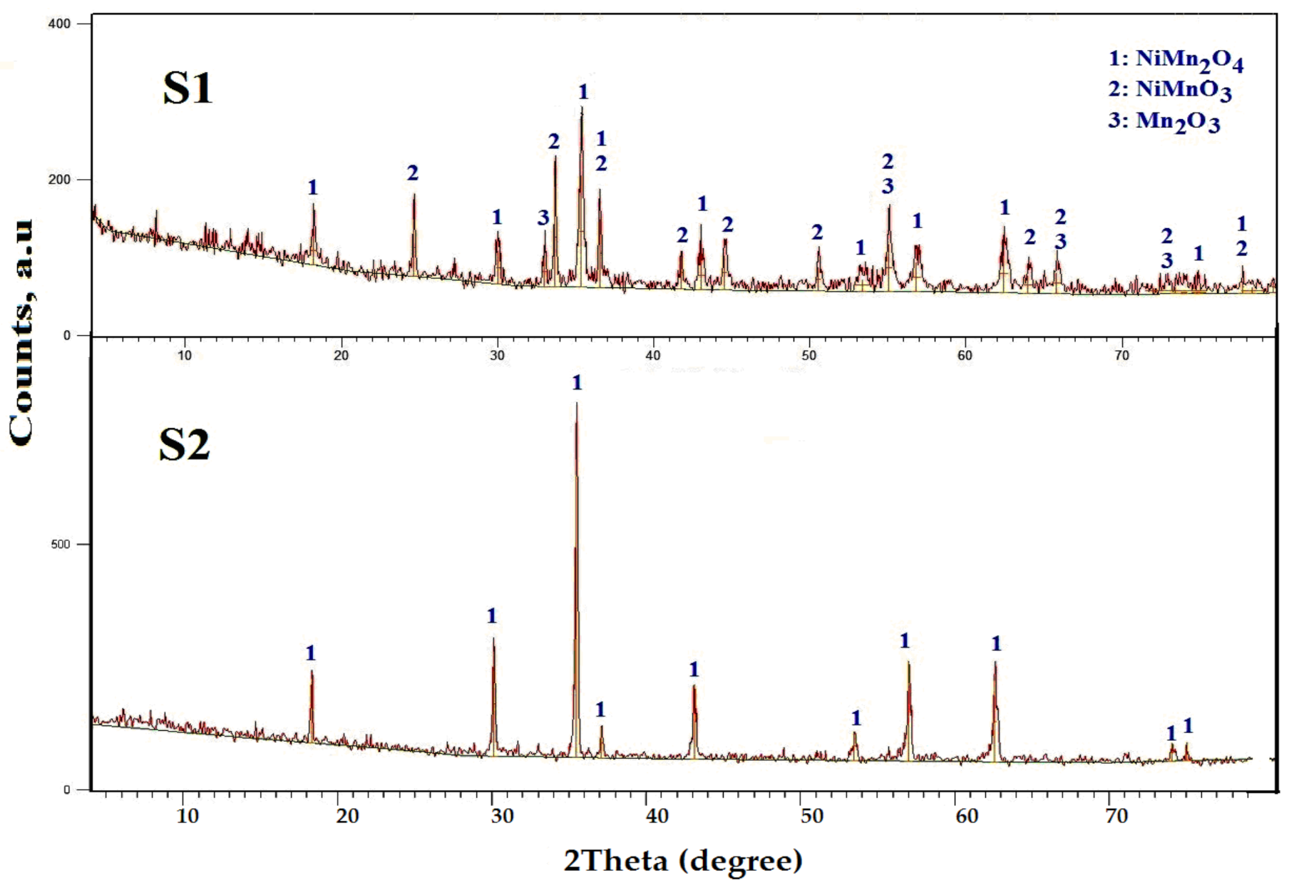
The S1 sample was exposed to the heat treatment in order to ascertain its heat stability and to understand its degradation pattern. TG–DTG measurements were used to study the thermal characteristics for the S1 sample as illustrated in Figure 1.
DTG thermogram of the S1 specimen shows endothermic peak located at 50 °C. This peak was followed by ~1.1% weight loss as a result of desorption or removal of moisture. The DTG peaks were observed at 250 °C, 325 °C and 430 °C with weight loss ~7.7% which corresponds to the complete formation of Mn2O3 and NiMnO3. The formation process of this composite may be expressed as:
2Ni(NO3)2·6H2O + 4Mn(NO3)2·4H2O → Mn2O3 + 2NiMnO3 + 12NO2 + 1.5O2 + 28H2O
Further DTG peak was located at 525 °C with weight loss ~8.3%. This weight loss could be due to partially solid-state reaction between the thermal products of the mixed oxides (Mn2O3 and NiMnO3) and/or phase transformation process yielding an excess amount of NiMn2O4 with liberation of oxygen according to the following reaction [23]:
Mn2O3 + 2NiMnO3 → 2NiMn2O4 + 0.5O2
To the alternative, the weight loss (9.3%) in the range between 850–1000 °C could be due to the complete transformation of Mn2O3 and NiMnO3 to NiMn2O4. Indeed, the value of the theoretical mass loss for the synthesized mixed oxides was 26.4%. This value is close to those found experimentally (27.35%). Thus, Mn2O3/NiMnO3/NiMn2O4 and NiMn2O4 hybrid nano particles can be synthesized by ceramic method in the thermal range of 450–850 °C and 850–1000 °C, respectively [21]. Similar results were reported in our previous work on the production of spinel NiMn2O4 NPs by adding calculated amounts of both nickel and manganese nitrates with and without a certain amount of egg white, immediately heated at 950 °C in air for 2 h [21].
In continuation of our previous work, we seek in this study to decrease the preparation temperature of NiMn2O4 solid by altering the fuel material from egg white to glycine. This is what made us also dealt with the traditional ceramic method for making a comparative study with the combustion method based on glycine. The ceramic method at 450–850 °C was used to build a composite containing Mn2O3/NiMnO3/NiMn2O4 NPs in this sample. XRD analysis will validate this result.

3.2. XRD Analysis

The identification and distinction between the crystalline lattices in the as synthesized solids carried out using the XRD analysis. XRD patterns of the S1 and S2 samples calcined at 700 °C for 2 h were illustrated in Figure 2, respectively. Examining this figure led to the following results: (i) the existence of NiMn2O4, NiMnO3 and Mn2O3 phases are registered in the XRD pattern of the S1 sample.
In other words, the hybrid oxides (NiMn2O4/NiMnO3/Mn2O3) rather than single-phase ones can be achieved during pyrolysis of the precursors without glycine at 700 °C. Indeed, XRD analysis of the S1 sample displays the construction of well crystalline NiMn2O4 as a major phase with cubic spinel like structure and space group Fd3m (PDF 01-1110). However, the Powder Diffraction File is a structured file that includes inorganic diffraction data for crystals and powders that can be quickly searched for unknown phase recognition. These findings relied on the diffraction lines of NiMn2O4 own of the families of crystalline planes (111), (220), (311), (222), (400), (422), (511), (440), (533), (622) and (441). However, the peaks own of the families of crystalline planes (012), (104), (110), (113), (202), (024), (116), (214), (300), (119) and (217) belong to NiMnO3 with a rhombohedral structure of the R 3̅ space group (PDF 12-269) [24]. On the other side, XRD pattern of the S1 sample displays formation of small amount of moderate crystalline cubic Mn2O3 lattice with space group Ia3̅ (PDF 41-1442). The peaks of Mn2O3 were observed with the planes of (222), (440) and (622). (ii) Based on the height of the characteristic diffraction peaks of NiMn2O4 (311), NiMnO3 (104) and Mn2O3 (222) planes can be used for calculation of the relative content or the calculated fraction (F) of these phases in the S1 sample by using the following relation:
F = Iinvest/Itotal
where I invest is the count of I222 or I311 or I104 of the studied phase and I total is the sum of the counts of I222, I311and I104 of cubic Mn2O3, NiMn2O4 and rhombohedra NiMnO3 phases detected in the XRD pattern, respectively. The fraction of NiMn2O4, NiMnO3 and Mn2O3 phases present in porous NiMn2O4/NiMnO3/Mn2O3 nanocomposite were found to be 48%, 41% and 12%, respectively. (iii) On other hand, glycine assisted combustion method followed by heating at 700 °C for 2 h resulted in solid-solid interaction between NiMnO3 and Mn2O3 yielding NiMn2O4. To put it more simply, heat treatment of as synthesized precursors containing a certain amount of glycine at 700 °C resulted in complete conversion of the investigated materials to NiMn2O4 as a single phase without the existence of any second phase. Using the glycine-assisted combustion process, however, the relative intensities of diffraction peaks indexed to NiMn2O4 increase.

3.3. Fourier-Transform Infrared (FTIR) Investigation

The study of both the position and vibration modes of the ions in the crystal structure of the compounds can be determined by FTIR analysis. In other words, the FTIR technique enabled us to study the different ordering positions of ions on the structural characteristics of materials. However, FTIR spectroscopy is an important tool to identify the functional groups, phase and purity of samples. The FTIR spectral study of the as synthesized nickel-manganese mixed oxides were recorded between 4000 and 400 cm−1 as shown in Figure 3. It is known that the specified bands of inorganic solids in the region of 1000 and 400 cm−1 are usually determined to the vibration of metallic ions in the crystal lattice [25].
Based on the group theoretical calculations, the manganite-based materials are known to exhibit two fundamentals IR active modes in the vibration spectra, which were observed around 600 cm−1 for tetrahedral (A) site and around 400 cm−1 for octahedral (B) site [26]. In this study, the absorption bands for spinel nickel manganites are in the expected range. The as synthesized manganites show strong absorption bands at 599–589 cm−1 and 535–525 cm−1. Indeed, the bands, γ1, at 599–589 cm−1 and the bands, γ2, at 535–525 cm−1 are a result of the A and B complexes of spinel manganite type structure, respectively. A broad and week bands, γ2*, located at 450–425 cm−1 is assigned to the divalent octahedral metal ion and oxygen ion complexes. In other words, the octahedral bands exhibit splitting of absorption bands including few small subsidiary bands. Detected subsidiary bands are mainly due to John-Teller distortion evolved by the presence of Mn2+ ions [27]. Moreover, the absorption bands located at 3445–3430 cm−1 and 1639–1630 cm−1 were a result of the stretching and bending mode of the OH group of absorbed water molecules on the Ni–Mn–O lattice since the nano crystalline materials have a high surface-volume ratio and thus absorb moisture [28,29,30]. However, using of glycine-based combustion method resulted in slightly change in the positions and intensities of detected bands due to the surface and quantum size effects of nanomaterials [31].

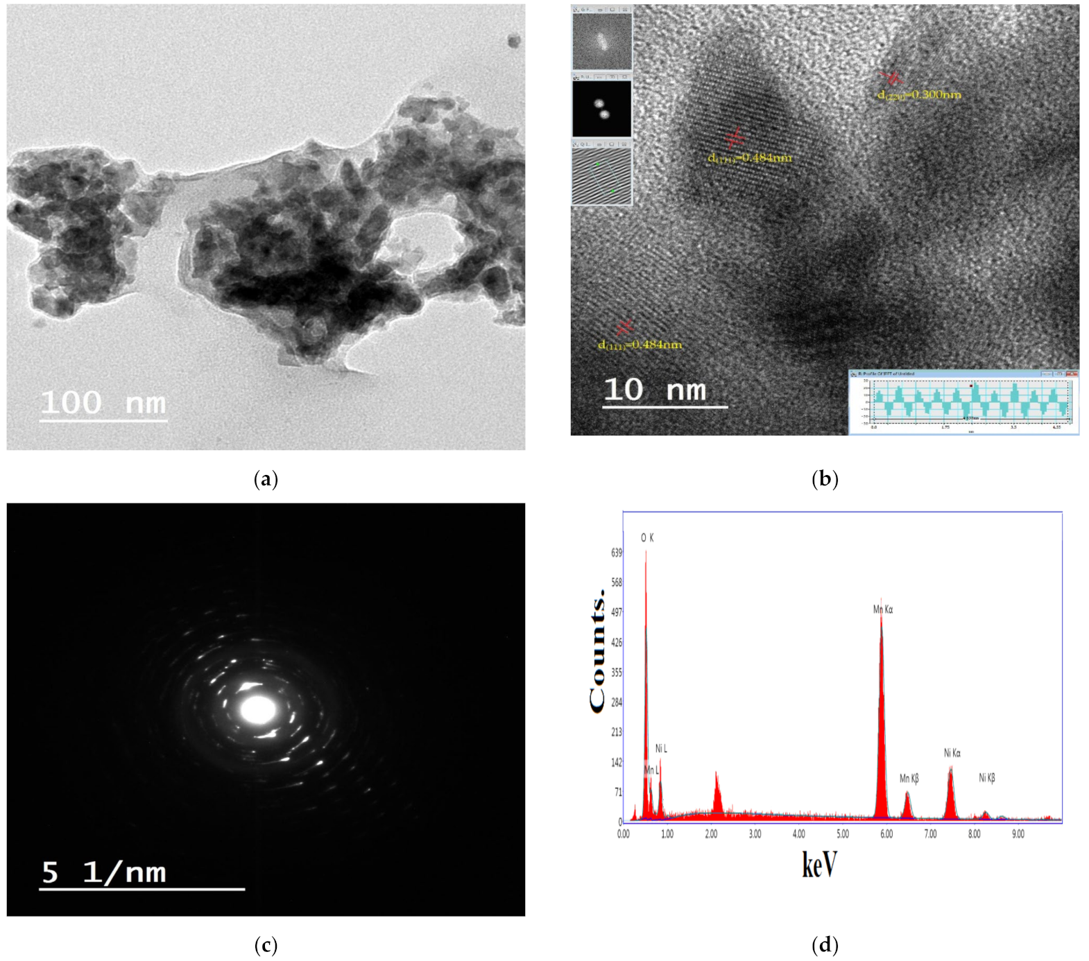
3.4. HRTEM and EDX Analyses

In this study, we found that both the presence of glycine and the magnetic nature of the resultant materials had a clear effect on the morphological characteristics of the as prepared samples. High-resolution TEM (HRTEM) images of the S1 and S2 are clear Figure 4a and Figure 5a and also the distinct fringes are depicted in Figure 4b and Figure 5b. On the other hand, the data of these fringes were analyzed with the software of Digital Micrograph using the fast Fourier transform (FFT) as shown in, Fourier mask filtering tools and inverse Fourier transform (IFFT) are described as inserts on the left and the right of Figure 4b and Figure 5b.
As shown in Figure 5a, uniformly dispersed porous and polyhedron-structured particles with an average diameter of about 22 nm were successfully synthesized by glycine-assisted combustion method. On the other side, the ceramic method resulted in formation numerous small sizes of particles (referred by red arrows) on the uppermost layers for the relatively large size type particles (average size ≈ 55 nm) accumulated with porous structure as noted in Figure 4a.
This observation confirms the XRD results of the S1 sample which consisted of NiMn2O4 (major phase) besides both Mn2O3 and NiMnO3 phases. One cannot ignore the action of the magnetic nature of the as prepared nano particles, which led to slightly agglomerations. The difference in the shape, size and aggregation of the grains of the prepared materials was clearly observed in the TEM images. This is probably because of the different fabrication mechanisms depending upon the difference in the energy during the glycine combustion. In the case of S2 sample, escaping large amounts of gases during the process of auto-combustion for the used precursors brought about appearance of various porous and voids yielding very fine particles. The lattice spaces of 0.484 nm or 0.465 nm and 0.300 nm for NiMn2O4 are in good agreement with the inter-plane space of the spinel-type NiMn2O4 (111) and (220) planes (PDF 01-1110), respectively. Figure 4c and Figure 5c display the selected area electron diffraction (SAED) patterns of the prepared samples. The SAED patterns are made up of a series of light diffraction circles with a variety of spots that reflect the presence of porous materials with a polycrystalline structure. These observations are consistent with the TG and XRD results and also confirm the solid–solid interaction between the constituents of the as prepared solids and/or phase transformation.
Identification of the elements present in the as prepared solid can be occurred by using EDX analysis. However, this technique can provide us the concentrations of the elements on the solid surface. In addition, the redistribution of the elements at top layers of the solid surface can be detected by carrying out the analysis at different points or various areas at the solid surface. Figure 4d and Figure 5d display representative EDX analyses for the S1 and S2 samples prepared by ceramic and glycine-assisted combustion methods, respectively. From the EDX results, the presence of Ni, Mn and O elements in different samples were confirmed. However, the relative atomic abundance of Ni, Mn and O elements at three different areas are much closer to each other of the prepared solids as determined in Table 1. The close values of each element in different regions indicate the homogeneity of the distribution of this element in the prepared sample. This was observed for all elements in different samples. Moreover, the glycine based green synthesis of nickel manganite brought about synthesis of homogeneously distributed particles.

3.5. Magnetic Characteristics

The magnetization of the synthesized solids was measured as a function of applied magnetic field in the range of −20,000 to +20,000 G using VSM at room temperature. Figure 6 shows the magnetization curves of the as prepared samples.
However, the values of coercive field (Hc), remanent magnetization (Mr), saturation magnetization (Ms), squareness (Mr/Ms), anisotropy constant (Ka) and magnetic moment (μm) per unit formula in Bohr magnetron of the synthesized specimens are listed in Table 2. This table showed that the values of Hc, Mr, Ms, Mr/Ms, μm and Ka for the S2 sample are greater than that of S1 sample.

4. Discussion

4.1. Formation of Perovskite-Spinel Nickel Manganites

Polycrystalline samples containing Mn2O3/NiMnO3/NiMn2O4 and NiMn2O4 have been synthesized using both the ceramic route and the combustion synthetic route using glycine as fuel followed by heating at elevated temperatures, respectively. Generally, NixMn3−xO4 materials crystallize in air to produce a cubic spinel NiMn2O4 solid at 730–875 °C. Below and above this temperature range, mixture of NiMnO3 and Mn2O3 were permanent phases depending on single-phase of NiMn2O4 cannot be formed [32]. However, in our previous work, we reported to formation of NiMn2O4 as a single phase [21]. This was achieved by heating at 950 °C for the mixture of precursors containing equimolar ratio of nickel and manganese nitrates with and without a certain amount of egg white [21]. In other words, these findings were opposite the results which obtained by Wickham [32]. This difference was extended in this study, when the mixture of Mn2O3 and perovskite NiMnO3, in addition, spinel NiMn2O4 were obtained from heating equimolar ratio of Ni- Mn nitrates at 700 °C for 2 h. Moreover, we were able to inhibit formation of both Mn2O3 and NiMnO3 at 700 °C and their transformation completely into single phase of NiMn2O4 by using of glycine. XRD results confirmed formation of NiMn2O4 (48%), NiMnO3 (41%) as major phases and Mn2O3 (12%) as a minor phase at 700 °C with crystallite size of 22 nm, 38 nm and 27 nm, respectively. In other words, the heat treatment at 700 °C is insufficient for complete conversion of Ni and Mn oxides to NiMn2O4 solid as single phase depending on the following equation:
2NiMnO3 + 1.5Mn2O3 → NiMn2O4 + NiMnO3 + Mn2O3 + 0.5O2

4.2. Formation of Spinel Nickel Manganite

The previous findings necessitated adding a certain amount of glycine followed by heating of the materials at 700 °C, which led to the acquisition of NiMn2O4 as a single phase. At the starting point, the solid-state reaction between manganese and nickel oxides brought about thin film of manganite which covers the grains surfaces of reacting oxides and acts as energy barrier of manganite formation. However, the glycine treatment followed by heating at 700 °C enhanced the thermal diffusion of the reacting oxides through the previous thin film of NiMn2O4 with subsequent complete conversion of NiMnO3 and Mn2O3 yielding single phase of NiMn2O4. Indeed, the results of XRD for the S2 sample showed disappearance of all peaks related to NiMnO3 and Mn2O3 with subsequent presence of sharp peaks related to crystalline of NiMn2O4. Finally, the nickel manganite studied experienced a transformation from perovskite to spinel phase and also cation redistribution by using a glycine-based combustion method at 700 °C.

4.3. Cation Distribution of Spinel Nickel Manganite

Spinel nickel manganite (NiMn2O4) has different physical and chemical behaviors depending on the type and amount of cations occupying A and B sites [33]. In other words, the variability of the Ni and Mn lattice positions resulted in the interesting structural and magnetic characteristics of spinel NiMn2O4 yielding different applications. As reported earlier Mn2+ ions have a strong preference to occupy A sites while Ni2+ and Mn3+ have preferentially occupied the B site [21,33,34]. According to one study, Ni2+ can be found in the A site depending on various factors such as calcination temperature and preparation route [34]. Ni and Mn cations can also occupy tetrahedral and octahedral crystal sites, which are interstitial sites within the cubic closed packed oxygen sub-lattice of the spinel structure, according to literature. Cation distribution mechanism of NiMn2O4 spinel may be explained in light of the migration of cations between B and A sites followed by a change in their valences in order to suppress the Jahn Teller distortion. This distortion resulted in lattice instability of solid due to the departure from ideal interactions among bonding orbital [35]. Indeed, this distortion can be examined from the XRD pattern of the S1 and S2 samples depending upon the study of the peaks observed at (2 2 0) and (4 4 0) reflection planes. The intensities of these planes are more sensitive to the cations on tetrahedral and octahedral sites, respectively [36]. It can be observed from Table 3 that the ratio of I220/I440 changes with and without glycine indicating to the cation redistribution.
Some authors speculated that a portion of the Ni2+ cations can be moved from A to B sites, then a corresponding proportion of Mn3+ cations in B sites disproportionate to Mn2+ and Mn4+ [37,38,39]. However, the Mn2+ cations move to A- sites to compensate for the Ni2+ vacancies. In this study, the maximum increase in the intensity of (2 2 0) and (4 4 0) planes, due to the glycine treatment followed by heating at 700 °C, attained 61% and 20%, respectively. This proves that the presence of cations at A site is greater than that at B site. Based on IR measurements, the intensity of peak band related to A site for the S2 sample is greater than that for the S1 sample depending upon NiMn2O4 has various states of manganese ions (Mn2+, Mn3+ and Mn4+) that distributed between A and B sites.
Thus, the authors confirm that the glycine treatment followed by heating at 700 °C of Ni-Mn mixed oxides brought about changes in related structural properties namely, the crystallite size (d), lattice constant (a), X-ray density (Dx), distance between the reacting ions (LA and LB), ionic radii (rA, rB) and bond lengths (A–O and B–O) on tetrahedral (A) sites and octahedral (B) sites of NiMn2O4 spinel as shown in Table 3 [40,41,42]. These findings could be attributed to the enhancement effect of glycine in formation nanosized particles depending upon redistribution of reacting cations and liberation of different gases during the preparation process.
Nickel manganite in the cubic spinel structure exhibits some oxygen vacancies or defects with release oxygen at the temperatures elevated. The vacancy formation was explained by the defect equation in Kroger–Vink notations as follows [43,44]:
Oox ↔ Vo•• + 2e + 0.5O2
where Oox is the oxide ion in the lattice, Vo•• is doubly charged oxygen vacancy, e is the electron in the conduction band. This finding brought about conversion of some Mn3+ to Mn4+ ions. However, the electrons formed during the previous reduction convert equivalent amount of Mn3+ to Mn2+ ions and lead to a distortion of the local symmetry in the crystals. These changes resulted in change in the bond length (A-O and B-O) and overall lattice parameters [45]. However, these parameters decrease as the size of the particles decreases as shown in case of the S2 sample.

4.4. Magnetic Properties

In fact, it has been referred that NiMn2O4 is not a simple ferromagnet because it contains both ferromagnetic and antiferromagnetic sublattices [14,31,46,47,48]. In addition, the exchange between Ni2+ and Mn3+ has antiferromagnetic character while the exchange between Mn2+ and Mn3+ has ferromagnetic behavior. This magnetic behavior is caused by strong coupling between A-B sites [49]. In this research, the glycine-based combustion route led to enhancement in the ferromagnetism of the manganite studied. This increase could be due to the transformation of Mn2O3/NiMnO3 to NiMn2O4 with subsequent decrease in the crystallite size, hole density, and the Zener double exchange (ZDE) mechanism of NiMn2O4 spinel [31,47,48]. Indeed, the glycine-based combustion route compared to the ceramic method led to a decrease in the crystallite size of NiMn2O4 spinel from 55 nm to 22 nm, respectively. However, the preparation of solid by glycine-assisted combustion route brought about appearance of ZDE mechanism leading to enhanced hole density. This is achieved by migration of some Ni2+ ions from B site to A site with subsequent conversion of some Mn3+ ions to both Mn2+ ions and Mn4+ ions which is adopted to B site while the Mn2+ ion will be adopted to tetrahedral site [50]. On the other hand, the super exchange interaction between Mn3+-O2−-Ni2+ (A–B interaction) became weak due to reduction of some Mn3+ ions.
In addition, the changes in the bond lengths between the cation-cation (M–M) and cation-anion (M–O) alter the overall strength of the magnetic interactions (A-A, B-B and A-B) in A and B sublattices. The strength of the magnetic interaction is inversely related to the bond length. Table 3 displays that the values of LA, LB, A–O and B–O for the S2 sample are smaller than that of the S1 sample. Mr/Ms values are found to be around 0.5 for the S2 sample which is the expected value for randomly packed single domain particles [49]. Coercivity of a manganite system is known to depend on various parameters, like magnetocrystalline anisotropy, lattice imperfections, grain size and shape, porosity and secondary phases. The low-coercive component may be due to other phases apparent in the XRD pattern ordering of the maganite nanoparticles affecting the nanoparticles’ anisotropy. Preparation of NiMn2O4 spinel using glycine-assisted combustion method resulted in an increase in the anisotropy constant with subsequent decrease in the value of rA and rB yielding large coercivity.

5. Conclusions

The ceramic technique and also the combustion method based on glycine, followed by heating at 700 °C for 2 h, were used to form both Mn2O3/NiMnO3/ NiMn2O4 nanocomposite and NiMn2O4 nanoparticles, respectively. The combustion method has a number of advantages, including cost effectiveness, scalability and faster synthesis of NiMn2O4 nanoparticles with a cubic spinel structure. Indeed, this method resulted in phase transformation of manganite from perovskite structure to spinel structure. An important observation of the work is formation of NiMn2O4 as prominent phase formation with complete suppression of NiMnO3 and Mn2O3 phases. The suppression of these oxides is attributed to complete conversion of these oxides to NiMn2O4 phase via the solid-state reaction. The combustion method led to modification of cation distribution in NiMn2O4 spinel yielding enhanced ferromagnetism. The elemental and morphological investigations of the prepared samples carried out using EDX and HRTEM techniques, respectively. The as-prepared NiMn2O4 NPs by using the combustion method had coercivity (229.50 Oe) and saturation magnetization (5.0216 emu/g). Finally, the glycine-assisted combustion method led to formation of manganites having best properties compared to the ceramic method at the same of preparation temperature.

Author Contributions

N.M.D., G.M.A.-S., N.S.A.-K. as well as O.H.A.-E.; technique, N.M.D., G.M.A.-S., N.S.A.-K. and O.H.A.-E.; program, O.H.A.-E.; validation N.M.D., G.M.A.-S., N.S.A.-K. and O.H.A.-E.; formal analysis, N.M.D., G.M.A.-S. and O.H.A.-E.; systematic evaluation, N.M.D., G.M.A.-S. and O.H.A.-E.; inquiry, N.M.D., G.M.A.-S.; resources, N.M.D., G.M.A.-S. and O.H.A.-E.; curation of results, N.M.D., G.M.A.-S. and O.H.A.-E.; writing—original preparation of the draft, N.M.D. and O.H.A.-E.; writing—review and editing, N.M.D., G.M.A.-S. and O.H.A.-E.; visualization, N.M.D. and O.H.A.-E.; supervision, N.M.D. and O.H.A.-E.; project management, N.M.D., G.M.A.-S. and O.H.A.-E.; procurement of financing, G.M.A.-S. and N.S.A.-K. All authors have read and agreed to the published version of the manuscript.

Funding

This research was funded by the Deanship of Scientific Research at Princess Nourah bint Abdulrahman University through the fast-track research-funding program.

Institutional Review Board Statement

Not applicable.

Informed Consent Statement

Not applicable.

Data Availability Statement

The data presented in this study are available on request from the corresponding author.

Acknowledgments

The authors would like to extend their sincere appreciation to Deanship of Scientific Research at Princess Nourah bint Abdulrahman University through the fast-track research-funding program.

Conflicts of Interest

The authors declare no conflict of interest.

References

  1. Deraz, N.M.; El-Aiashy, M.K.; Ali, S.A. Novel Preparation and Physicochemical Characterization of a Nanocrystalline Cobalt Ferrite System. Adsorp. Sci. Technol. 2009, 27, 797–810. [Google Scholar] [CrossRef]
  2. Deraz, N.M. Tailoring the Physicochemical and Magnetic Properties of an Mn Substituted Cobalt Ferrite System. Interceram 2018, 67, 14–19. [Google Scholar] [CrossRef]
  3. Shao, L.; Wang, P.X.; Zhang, N.Q.; Fan, L.; Zhang, N.; Sun, K.N. Nanostructured CuCo2O4 cathode for intermediate temperature solid oxide fuel cells via an impregnation technique. J. Power Sour. 2017, 343, 268–274. [Google Scholar] [CrossRef]
  4. Lu, B.; Cao, D.; Wang, P.; Gao, Y. Oxygen evolution reaction on Ni-substituted Co3O4 nanowire array electrodes. Int. J. Hydrogen Energy 2011, 36, 72–78. [Google Scholar] [CrossRef]
  5. Jung, J.; Töpfer, J.; Mürbe, J.; Feltz, A. Microstructure and phase development in NiMn2O4 spinel ceramics during isothermal sintering. J. Eur. Ceram. Soc. 1990, 6, 351–359. [Google Scholar] [CrossRef]
  6. Schmidt, R.; Basu, A.; Brinkman, A.W. Small polaron hopping in spinel manganates. Phys. Rev. B 2005, 72, 115101–115109. [Google Scholar] [CrossRef] [Green Version]
  7. Chengjian, M.; Hong, G. Preparation and characterization of single-phase NiMn2O4 NTC ceramics by two-step sintering method. J. Mater. Sci. Mater. Electron. 2017, 28, 6699–6703. [Google Scholar]
  8. Tsuda, N.; Nasu, K.; Fujimori, A.; Siratori, K. Electronic Conduction in Oxides, 2nd ed.; Springer Series in Solid-State Sciences; Springer: Berlin, Germany, 2000. [Google Scholar]
  9. Shuling, L.; Jin, X.; Zhong, M.; Shiming, Z.; Xiufang, W.; Xuebin, Y.; Yang, J.; Zi-Feng, M.; Xianxia, T. NiMn2O4 as an efficient cathode catalyst for rechargeable lithium–air batteries. Chem. Commun. 2017, 53, 8164–8167. [Google Scholar]
  10. Brabers, V.A.M.; Terhell, J.C.J.M. Electrical conductivity and cation valencies in nickel manganite. Phys. Status Solidi 1982, 69, 325–331. [Google Scholar] [CrossRef]
  11. Zener, C. Interaction between the d shells in the transition metals. Phys. Rev. 1951, 81, 440–444. [Google Scholar] [CrossRef]
  12. Feteira, A. Negative temperature coefficient resistance (NTCR) ceramic thermistors: An industrial perspective. J. Am. Ceram. Soc. 2009, 92, 967–983. [Google Scholar] [CrossRef]
  13. Hosseini, S.A.; Niaei, A.; Salari, D.; Nabavi, S.R. Nanocrystalline AMn2O4 (A = Co, Ni, Cu) spinels for remediation of volatile organic compounds—Synthesis, characterization and catalytic performance. Ceram. Int. 2012, 38, 1655–1661. [Google Scholar] [CrossRef]
  14. Cheeseman, N.B.B.; Chopdekar, R.V.; Iwata, J.M.; Toney, M.F.; Arenholz, E.; Suzuki, Y. Modified magnetic ground state in NiMn2O4 thin films. Phys. Rev. B 2010, 82, 144419. [Google Scholar] [CrossRef] [Green Version]
  15. Takayama, S.; Fukushima, J.; Nishijo, J.; Saito, M.; Sano, S.; Sato, M. Sintering of soft magnetic material under microwave magnetic field. Phys. Res. Int. 2014, 2102, 165849. [Google Scholar] [CrossRef] [Green Version]
  16. Nan, H.; Ma, W.; Gu, Z.; Geng, B.; Zhang, X. Hierarchical NiMn2O4@CNT nanocomposites for high-performance asymmetric supercapacitors. RSC Adv. 2015, 5, 24607–24614. [Google Scholar] [CrossRef]
  17. Selim, M.M.; Deraz, N.M.; El-Asmy, A.A.; El-Shafey, O. Synthesis, characterization and physicochemical properties of nanosized Zn/Mn oxides system. J. Alloy. Compd. 2010, 506, 541–547. [Google Scholar] [CrossRef]
  18. Schmidt, R.; Brinkman, A.W. Preparation and characterization of NiMn2O4 films. Int. J. Inorg. Mater. 2001, 3, 1215–1217. [Google Scholar] [CrossRef]
  19. Pena, O.; Moure, C.; Bodenez, V.; Caileaux, X.; Piriou, B.; Ortiz, J.; Zuniga, G.; Gautier, J.L.; Fliho, J.L.G. Magnetic properties of spinel-type oxides NiMn2−xMexO4. J. Chil. Chem. Soc. 2005, 50, 617–623. [Google Scholar] [CrossRef]
  20. Durán, P.; Tartaj, J.; Rubio, F.; Peña, O.; Moure, C. Preparation and sintering behaviour of spinel-type CoxNiMn2−xO4(0.2 ≤ x ≤ 1.2) by the ethylene glycol–metal nitrate polymerized complex process. J. Eur. Ceram. Soc. 2005, 15, 3021–3025. [Google Scholar] [CrossRef]
  21. Deraz, N.M.; Abd-Elkader, O.H.; Yassin, M. Impacts of egg white assisted combustion and ceramic methods on structural, morphological and magnetic properties of Nickel Manganite system. Crystals 2020, 10, 489. [Google Scholar] [CrossRef]
  22. Cullity, B.D. Elements of X-Ray Diffraction; Addison-Wesly Publishing Co. Inc.: Singapore, 1976. [Google Scholar]
  23. Sánchez-España, J.; Yusta, I. Coprecipitation of Co2+, Ni2+ and Zn2+ with Mn(III/IV) Oxides Formed in Metal-Rich Mine Waters. Minerals 2019, 9, 226. [Google Scholar] [CrossRef] [Green Version]
  24. Yu, J.; Yan, Q.; Chen, W.; Jain, A.; Neaton, J.B.; Persson, K.A. First-principles study of electronic structure and photocatalytic properties of MnNiO3 as an alkaline oxygen-evolution photocatalyst. Chem. Commun. 2015, 51, 2867–2870. [Google Scholar] [CrossRef] [PubMed] [Green Version]
  25. Ouaguenouni, M.H.S.; Benadda, A.; Kiennemann, A.; Barama, A. Preparation and catalytic activity of nickel–manganese oxide catalysts in the reaction of partial oxidation of methane. C. R. Chim. 2009, 12, 740–747. [Google Scholar] [CrossRef]
  26. Waldron, R.D. Infrared Spectra of Ferrites. Phys. Rev. 1955, 99, 1725–1727. [Google Scholar] [CrossRef]
  27. Berchmans, L.J.; Sevan, R.K.; Kumar, P.N.S.; Augustin, C.O. Structural and electrical properties of Ni1−xMgxFe2O4 synthesized by citrate gel process. J. Magn. Magn. Mater. 2004, 279, 103–110. [Google Scholar] [CrossRef]
  28. Nakamoto, K. Infrared and Raman Spectra of Inorganic and Coordination Compounds; John Wiley and Sons, Ltd.: Hoboken, NJ, USA, 1986. [Google Scholar]
  29. Jokanović, V.; Izvonar, D.; Dramićanin, M.D.; Jokanović, B.; Živojinović, V.; Marković, D.; Dačić, B. Hydrothermal synthesis and nanostructure of carbonated calcium hydroxyapatite. J. Mater. Sci. Mater. Med. 2006, 17, 539–546. [Google Scholar] [CrossRef]
  30. Kaniyoor, A.; Baby, T.T.; Ramaprabhu, S. Graphene synthesis via hydrogen induced low temperature exfoliation of graphite oxide. J. Mater. Chem. 2010, 20, 8467–8469. [Google Scholar] [CrossRef]
  31. Kloprogge, J.T.; Hickey, L.; Forst, R.L. FT-Raman and FT-IR spectroscopic study of synthetic Mg/Zn/Al-hydrotalcites. J. Raman Spectrosc. 2004, 35, 967–974. [Google Scholar] [CrossRef] [Green Version]
  32. Wickham, D.G.J. Solid-phase equilibria in the system NiO-Mn2O3-O2. Inorg. Nucl. Chem. 1964, 26, 1369–1377. [Google Scholar] [CrossRef]
  33. Shahjahan, M.; Talukder, S.M.; Hossain, M.S.; Begum, M.H.A.; Warnock, R.L.; Haque, M.A.; Hossain, M.A.; Ahmed, N.A. Synthesis and Characterization of Structural and Electrical Properties of Mg0.25xCu0.25xZn1−0.5xFe2O4 Ferrites by sol Gel Method. Ukr. J. Phys. 2019, 64, 861–869. [Google Scholar] [CrossRef] [Green Version]
  34. Deraz, N.M.; Shaban, S. Optimization of catalytic, surface and magnetic properties of nanocrystalline manganese ferrite. J. Anal. Appl. Pyrol. 2009, 86, 173–179. [Google Scholar] [CrossRef]
  35. Hem, J.D.; Lind, C.J.; Roberson, C.E. Coprecipitation mechanisms and products in manganese oxidation in the presence of cadmium. Geochim. Cosmochim. Acta 1989, 53, 2435–2451. [Google Scholar] [CrossRef]
  36. Alarifi, A.; Deraz, N.M.; Shaban, S. Structural, morphological and magnetic properties of NiFe2O4 nano-particles. J. Alloy. Compd. 2009, 486, 501–506. [Google Scholar] [CrossRef]
  37. Macklen, E.D. Electrical conductivity and cation distribution in nickel manganite. J. Phys. Chem. Solids 1986, 11, 1073–1079. [Google Scholar] [CrossRef]
  38. Devale, A.B.; Kulkarni, D.K. Structural, electrical and X-ray spectroscopic investigations of MnNi2O4 spinel. J. Phys. C Solid State Phys. 1982, 15, 899–905. [Google Scholar] [CrossRef]
  39. Jung, H.R.; Lee, S.G.; Kim, K.M.; Kwon, M.S.; Kim, Y.G. Preparation and electrical properties of nickel manganite Ni0.79Mn2.21O4 ceramics for NTC thermistors. J. Ceram. Process. Res. 2017, 18, 357–360. [Google Scholar]
  40. Mays, C.W.; Vermaak, J.S.; Wilsdorf, D.K. On surface stress and surface tension: II. Determination of the surface stress of gold. Surf. Sci. 1968, 12, 134–140. [Google Scholar] [CrossRef]
  41. Tsunekawa, S.; Sahara, R.; Kawazoe, Y.; Ishikawa, K. Lattice relaxation of monosize CeO2−x nanocrystalline particles. Appl. Surf. Sci. 1999, 152, 53–56. [Google Scholar] [CrossRef]
  42. Tsunekawa, S.; Ishikawa, K.; Li, Z.Q.; Kawazoe, Y.; Kasuya, Y. Origin of anomalous lattice expansion in oxide nanoparticles. Phys. Rev. Lett. 2000, 85, 3440–3443. [Google Scholar] [CrossRef]
  43. Sun, C.; Li, H.; Chen, L. Nanostructured ceria-based materials: Synthesis, properties, and applications. Energy Environ. Sci. 2012, 5, 8475–8505. [Google Scholar] [CrossRef]
  44. Mai, H.; Sun, D.; Zhang, Y.; Si, R.; Feng, W.; Zhang, H.; Liu, H.; Yan, C. Shape-selective synthesis and oxygen storage behavior of ceria nanopolyhedra, nanorods, and nanocubes. J. Phys. Chem. B 2005, 109, 24380–24385. [Google Scholar] [CrossRef]
  45. Kurian, M.; Kunjachan, C. Investigation of size dependency on lattice strain of nanoceria particles synthesized by wet chemical methods. Int. Nano. Lett. 2014, 4, 73–80. [Google Scholar] [CrossRef] [Green Version]
  46. Filho, P.N.L.; Bahout, M.; Barahona, P.; Moure, C.; Peña, O. Oxygen stoichiometry effects in spinel-type NiMn2O4−δ samples. J. Phys. Chem. Solids 2005, 66, 1206–1212. [Google Scholar] [CrossRef]
  47. Shen, Y.; Nakayama, T.; Arai, M.; Yanagisawa, O.; Izumi, M. Magnetic phase transition and physical properties of spinel-type nickel manganese oxide. J. Phys. Chem. Solids 2002, 63, 947–950. [Google Scholar] [CrossRef]
  48. Savić, S.M.; Tadić, M.; Jagličić, Z.; Vojisavljević, K.; Mancić, L.; Branković, G. Structural, electrical and magnetic properties of nickel manganite obtained by a complex polymerization method. Ceram. Int. 2014, 40, 15515–15521. [Google Scholar] [CrossRef]
  49. Yafet, Y.; Kittel, C. Antiferromagnetic Arrangements in Ferrites. Phys. Rev. 1952, 87, 290–294. [Google Scholar] [CrossRef]
  50. Wang, J.; Gu, B.; Sang, H.; Ni, G.; Du, Y. Magnetocaloric effect in perovskite manganites La0.7−xNdxCa0.3MnO3 and La0.7Ca0.3MnO3. J. Magn. Magn. Mater. 2001, 223, 50–54. [Google Scholar] [CrossRef]
Figure 1. TG-DTG pattern of the S1 sample.
Crystals 11 00487 g001
Figure 2. XRD patterns of the S1 and S2 samples.
Crystals 11 00487 g002
Figure 3. FT-IR patterns of the S1 and S2 samples.
Crystals 11 00487 g003
Figure 4. ((a): HRTEM, (b): FFT, (c): SAED and (d): EDX) images of the S1.
Crystals 11 00487 g004
Figure 5. ((a): HRTEM, (b): FFT, (c): SAED and (d): EDX) images of the S2 samples.
Crystals 11 00487 g005
Figure 6. M-H curve of the S1 and S2 samples.
Crystals 11 00487 g006
Table 1. Atomic abundance of elements measured at 20 keV and different areas over the as prepared solids.
SolidsElementsArea 1Area 2Area 3
S1O42.6153.7650.31
Mn38.0831.3332.34
Ni17.9214.2316.44
O50.3151.2554.94
Mn33.3432.1529.24
S2Ni15.4416.6015.81
Table 2. Magnetic properties (Ms, Mr, Mr/Ms, Ka, μm and Hc) of the as-prepared solids.
SolidsMs (emu/g)Mr (emu/g)Mr/Ms (emu/g)Hc (Oe)μmAnisotropy Constant (Ka)
S10.682419.26 × 10−328.24 × 10−3146.74102.1792.864 × 10−2
S25.02162.50340.4985229.51152.45721.074 × 10−2
Table 3. Lattice parameters of nanocrystalline NiMn2O4 involved in the as prepared solids.
SolidsS1S2
Parameters
Crystal StructureCubic SpinelCubic Spinel
d, nm5522
a, nm0.84160.8387
V, nm30.59610.5899
Dx, g/cm35.2225.276
LA, nm0.36440.3632
LB, nm0.29710.2961
A-O, nm0.19240.1917
B-O, nm0.21710.2160
rA, nm0.06040.0597
rB, nm0.08510.0844
I220/I4400.961.29
d—crystallite size, a—lattice constant of cubic cell, V—volume of unit cell, Dx—X-ray density, LA and LB—distances between the reacting ions, A-O and B-O—Bond length, rA and rB—ionic radii, I220/I440—Intensity ratio of two planes.
Publisher’s Note: MDPI stays neutral with regard to jurisdictional claims in published maps and institutional affiliations.

