Growth of Lu2O3 and HfO2 Based High Melting Temperature Single Crystals by Indirect Heating Method Using Arc Plasma

 , ,
, ,
Abstract
:1. Introduction
2. Experimental
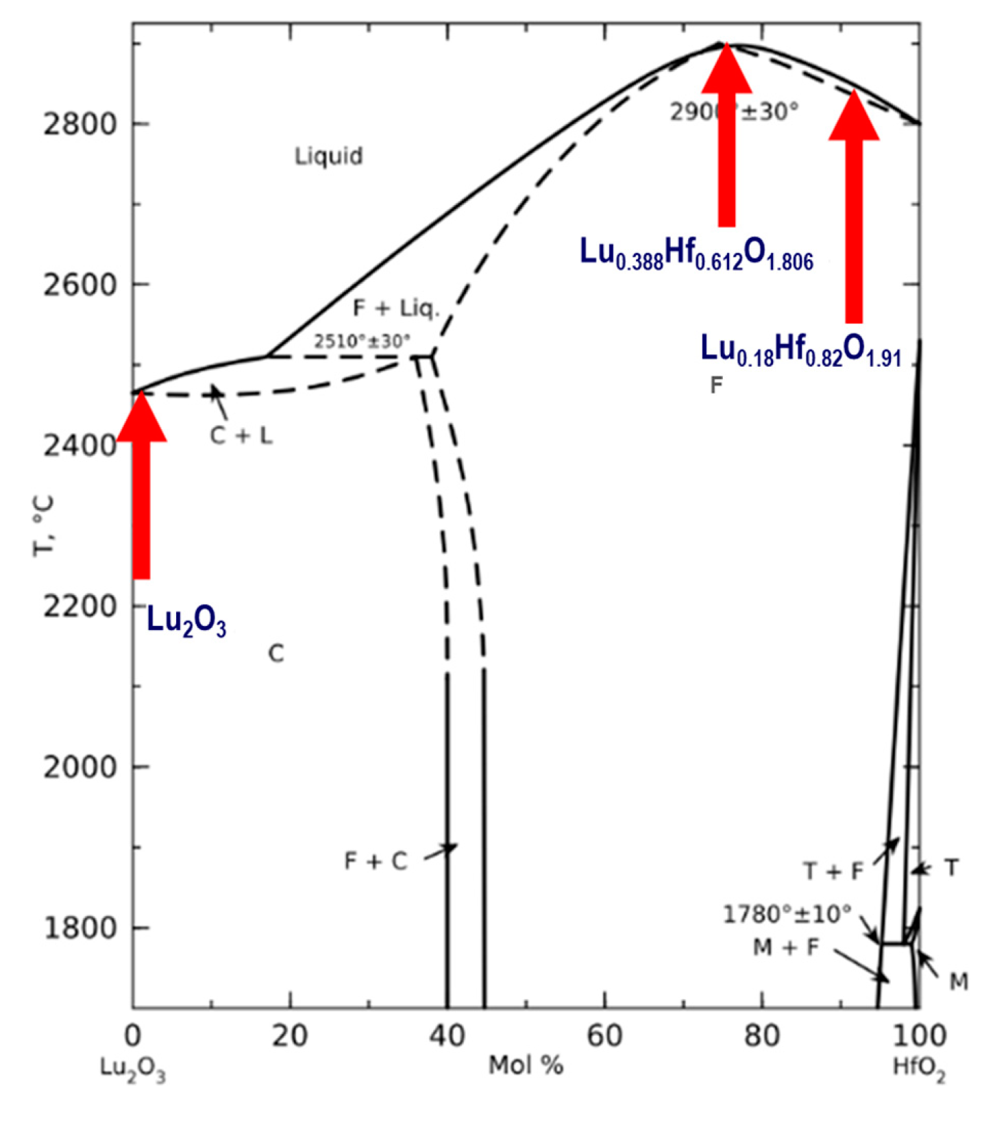
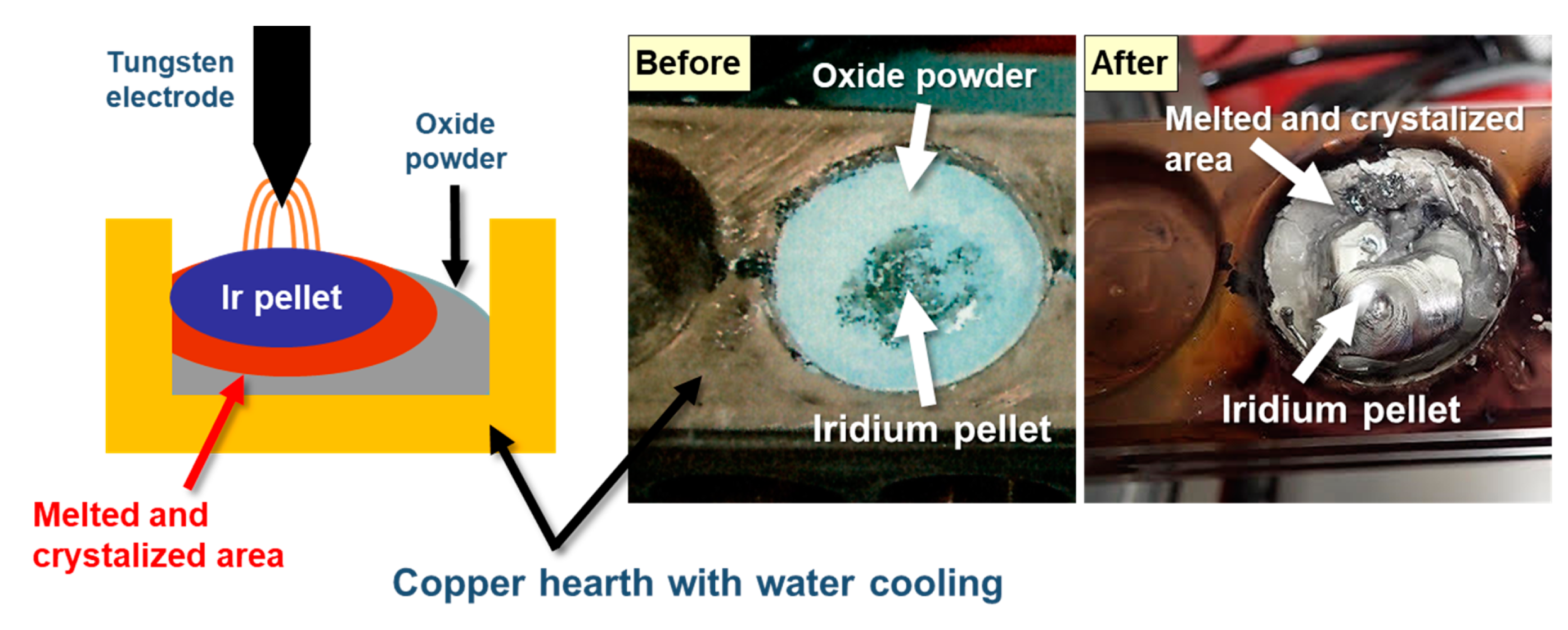
2.1. Crystal Growth
2.2. Characterizations
3. Results and Discussion
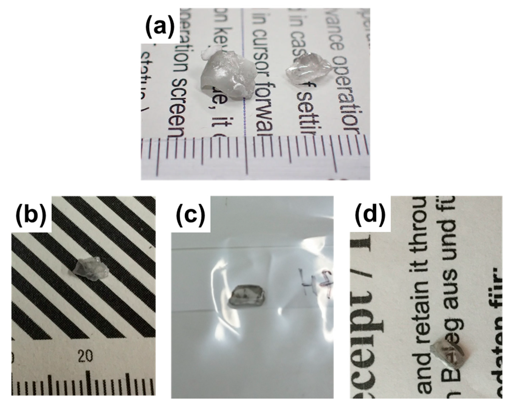
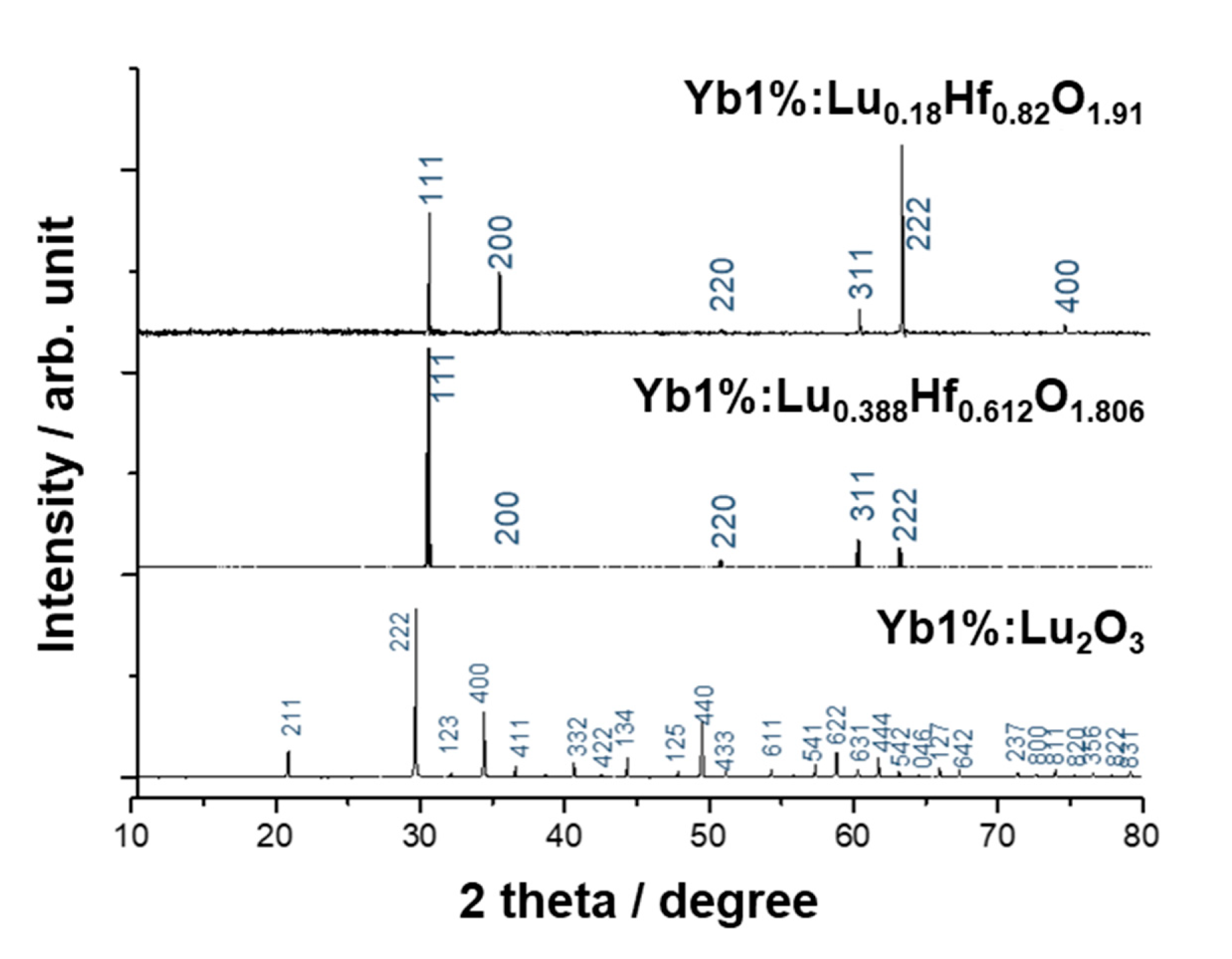
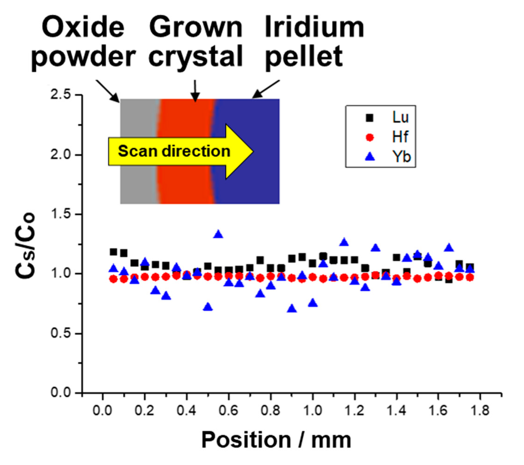
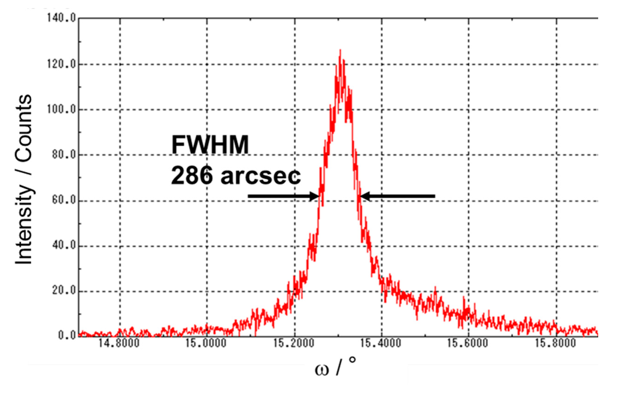
3.1. Crystal Growth
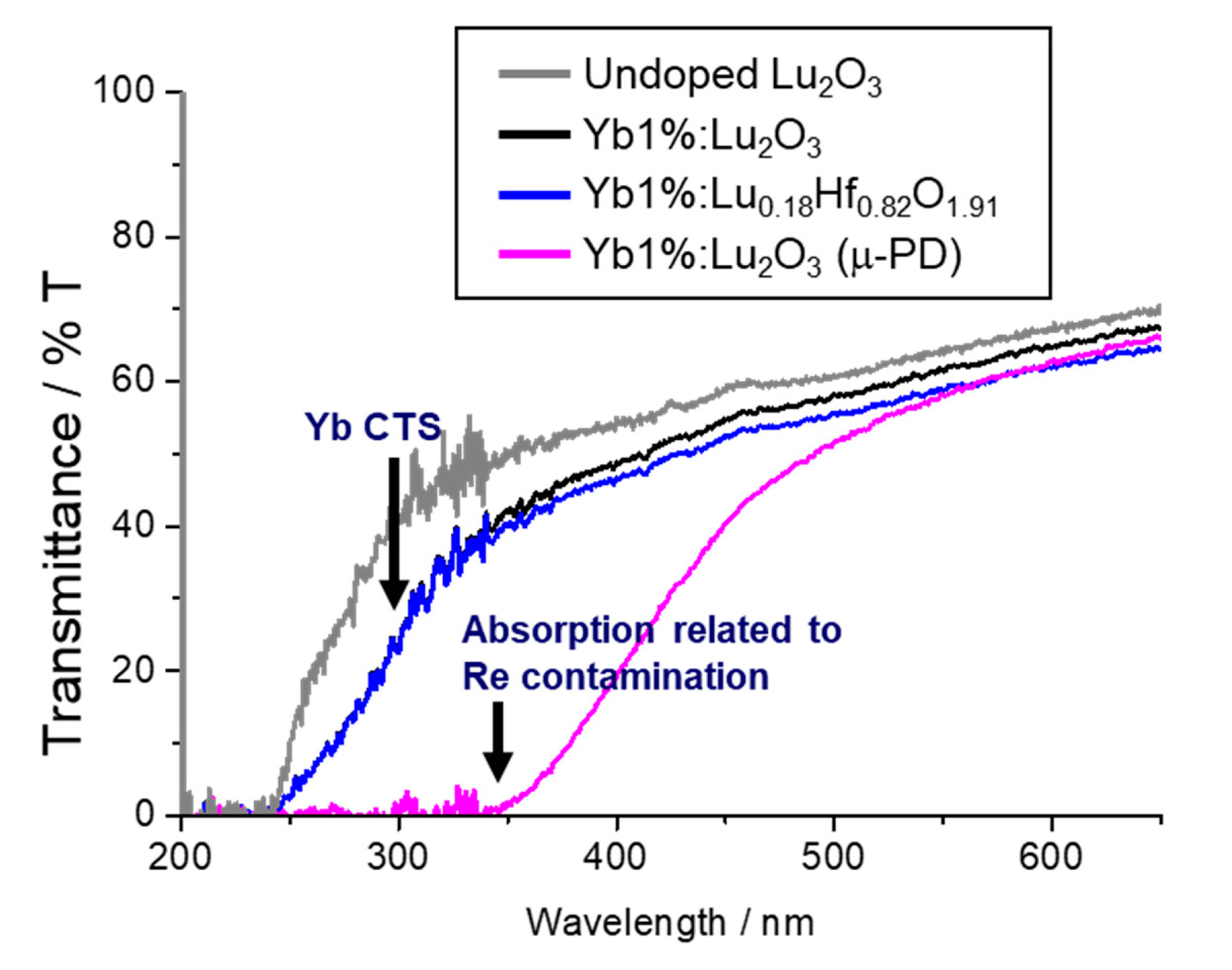
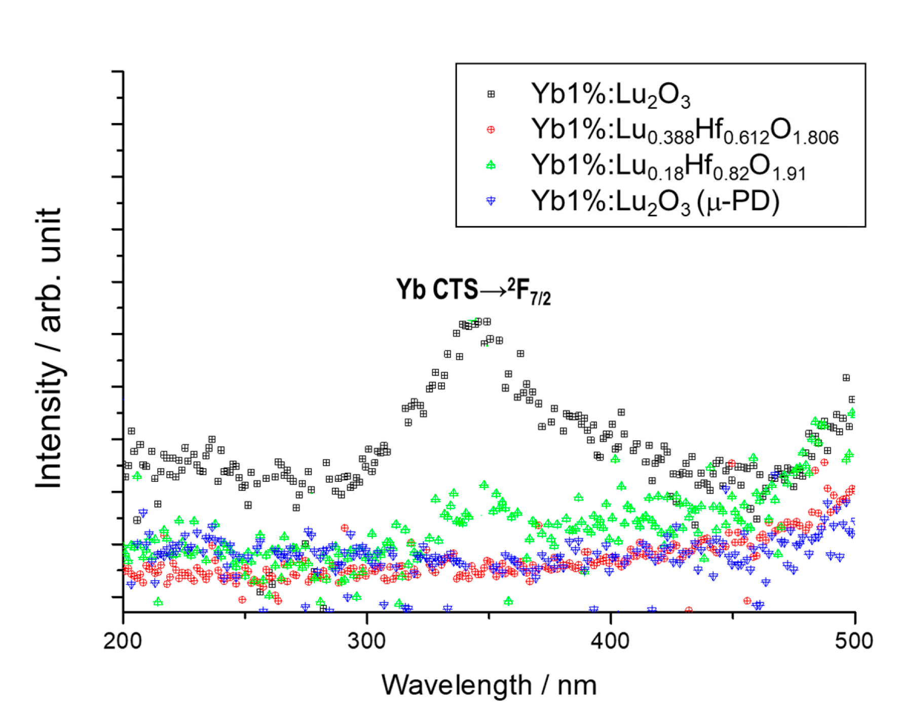
3.2. Optical and Luminescence Measurements
4. Conclusions
Author Contributions
Funding
Conflicts of Interest
References
- Shevchenko, A.V.; Lopato, L.M.; Kir’yakova, I.E. Interaction of HfO₂ with Y₂O₃, Ho₂O₃, Er₂O₃, Tm₂O₃, Yb₂O₃, and Lu₂O₃ at high temperatures. Inorg. Mater. 1984, 20, 1731–1736. [Google Scholar]
- Fukabori, A.; Chani, V.; Kamada, K.; Moretti, F.; Yoshikawa, A. Growth of Tm3+-Doped Y2O3, Sc2O3, and Lu2O3 Crystals by the Micro pulling down Technique and Their Optical and Scintillation Characteristics. Cryst. Growth Des. 2011, 11, 2404–2411. [Google Scholar] [CrossRef]
- Fukabori, A.; Chani, V.; Kamada, K.; Yanagida, T.; Yokota, Y.; Moretti, F.; Kawaguchi, N.; Yoshikawa, A. Growth of Y2O3, Sc2O3 and Lu2O3 crystals by the micro-pulling-down method and their optical and scintillation characteristics. J. Cryst. Growth 2011, 318, 823–827. [Google Scholar] [CrossRef]
- Fukabori, A.; Chani, V.; Kamada, K.; Yoshikawa, A. Growth of Yb-doped Y2O3, Sc2O3, and Lu2O3 single crystals by the micro-pulling-down technique and their optical and scintillation characterization. J. Cryst. Growth 2012, 352, 124–128. [Google Scholar] [CrossRef]
- Hou, W.; Zhao, H.; Li, N.; Xue, Y.; Shi, J.; Xu, X.; Xu, J. Growth and spectroscopic properties of Er:Lu2O3 crystal grown by floating zone method. Mater. Res. Express 2019, 6, 066203. [Google Scholar] [CrossRef]
- Wang, J.; Li, H.P.; Stevens, R. Hafnia and hafnia-toughened ceramics. J. Mater. Sci. 1992, 27, 5397–5430. [Google Scholar] [CrossRef]
- Shannon, R.D. Revised effective ionic radii and systematic studies of interatomic distances in halides and chalcogenides. Acta Cryst. 1976, A32, 751–767. [Google Scholar] [CrossRef]


| Ions | Cu (wt. ppm) | Re (wt. ppm) | Ir (wt. ppm) | W (wt. ppm) | |
|---|---|---|---|---|---|
| Growth Method | |||||
| CH | 14.1 | N.D. | 8.70 | 475 | |
| μ-PD | 0.40 | 61.4 | N.D. | 590 | |
© 2020 by the authors. Licensee MDPI, Basel, Switzerland. This article is an open access article distributed under the terms and conditions of the Creative Commons Attribution (CC BY) license (http://creativecommons.org/licenses/by/4.0/).
Share and Cite
Kim, K.J.; Kamada, K.; Murakami, R.; Horiai, T.; Ishikawa, S.; Kochurikhin, V.V.; Yoshino, M.; Yamaji, A.; Shoji, Y.; Kurosawa, S.; et al. Growth of Lu2O3 and HfO2 Based High Melting Temperature Single Crystals by Indirect Heating Method Using Arc Plasma. Crystals 2020, 10, 619. https://doi.org/10.3390/cryst10070619
Kim KJ, Kamada K, Murakami R, Horiai T, Ishikawa S, Kochurikhin VV, Yoshino M, Yamaji A, Shoji Y, Kurosawa S, et al. Growth of Lu2O3 and HfO2 Based High Melting Temperature Single Crystals by Indirect Heating Method Using Arc Plasma. Crystals. 2020; 10(7):619. https://doi.org/10.3390/cryst10070619
Chicago/Turabian StyleKim, Kyoung Jin, Kei Kamada, Rikito Murakami, Takahiko Horiai, Shiori Ishikawa, Vladimir V. Kochurikhin, Masao Yoshino, Akihiro Yamaji, Yasuhiro Shoji, Shunsuke Kurosawa, and et al. 2020. "Growth of Lu2O3 and HfO2 Based High Melting Temperature Single Crystals by Indirect Heating Method Using Arc Plasma" Crystals 10, no. 7: 619. https://doi.org/10.3390/cryst10070619