Article

Effect of Ultrasound Irradiation on the Synthesis of Hydroxyapatite/Titanium Oxide Nanocomposites

by
A. K. Sánchez-Hernández
1,
J. Martínez-Juárez
1,
J. J. Gervacio-Arciniega
2,
R. Silva-González
3 and
M. J. Robles-Águila
1,*
1
Instituto de Ciencias, Benemérita Universidad Autónoma de Puebla, Edificio 105 C, Boulevard 14 Sur y Av. San Claudio, Col. San Manuel, Puebla 72570, Mexico
2
Conacyt-Facultad de Ciencias Físico Matemáticas, Benemérita Universidad Autónoma de Puebla, Av. San Claudio y Av. 18 Sur, Col. San Manuel Ciudad Universitaria, Puebla 72570, Mexico
3
Instituto de Física, Benemérita Universidad Autónoma de Puebla, Apartado Postal J-48, Puebla 72570, Mexico
*
Author to whom correspondence should be addressed.
Crystals 2020, 10(11), 959; https://doi.org/10.3390/cryst10110959
Submission received: 27 August 2020 / Revised: 11 October 2020 / Accepted: 19 October 2020 / Published: 22 October 2020
(This article belongs to the Special Issue Hydroxyapatite Base Nanocomposites)

Abstract

:
Bioceramic materials, such as hydroxyapatite, Ca10(PO4)6(OH)2, (HAp), can be chemically bound to bone tissue; since they are bioactive and biocompatible. HAp, titanium oxide (TiO2), and hydroxyapatite/titanium oxide (HAp/TiO2) nanocomposite nanoparticles were obtained by ultrasound irradiation assisted by sol-gel and co-precipitation methods at different time intervals, using Ca(NO3)2•4H2O, (NH4)2HPO4, and TiOSO4•xH2O as calcium, phosphorus, and titanium sources, respectively. HAp, TiO2, and HAp/TiO2 nanocomposite powders were characterized by X-ray Diffraction (XRD) and Raman Spectroscopy. The percentages of anatase phase for TiO2 and of monoclinic and hexagonal phases for HAp were quantified by Rietveld refinement. Furthermore, sample crystallinity in each material was enhanced by increasing the ultrasound irradiation time. The nanoparticle shape was semi-spherical, agglomerated, and between 17 and 20 nm in size. The agglomeration of particles in the samples was corroborated with a Field Emission Scanning Electron Microscope (FESEM).

Graphical Abstract

1. Introduction

An example of a natural composite material is bone tissue in vertebrates and humans, whose main components are inorganic (e.g., CaPO4, HPO42−, CO32−, Cl, and metallic ions) and bioinorganic (e.g., fibrous protein collagen, polysaccharides, and primary bone cells) phases, and which resembles the structure of hydroxyapatite [1]. HAp is a bioceramic material that can be chemically bound to bone tissue, making it bioactive and biocompatible. Its origin can be from biological tissue (derived from coral or bovine bone tissue) or chemical synthesis [2,3] Recent research has been focused on improving the mechanical properties of HAp by using oxide semiconductors and ceramics, such as zirconia, alumina, pyrolytic carbon, and bioglass, to enhance the resistance and stability in specific parts of the human body that require further support [4,5,6]. Previous studies have shown the interaction between metallic oxides, such as titanium oxide (TiO2) and hydroxyapatite as an effective alternative to improve the mechanical and anticorrosive properties of the composite (HAp/TiO2) [7,8,9,10]. Titanium and its alloys are known for their use as biomaterials since they are resistant to erosion and have excellent mechanical and biocompatible properties [11,12,13]. The interaction between HAp and TiO2 has an influence on biological aspects, such as enhanced osteoblast adhesion, stimulated cell growth, biocompatibility, corrosion resistance, and mechanical properties. On the other hand, TiO2 is the most commonly used semiconductor due to its affordable synthesis, chemical stability, and innocuous, non-toxic and biocompatible properties. This semiconductor can be found in three phases: anatase, rutile, and brookite [14]. The anatase phase is more effective than the rutile phase in inducing apatite nucleation. Furthermore, Pushpakanth et al. reported that TiO2 has a tendency to adsorb water on surfaces, which promotes the presence of titanium hydroxide groups that are responsible for inducing apatite nucleation [15]. Several reports have studied the photocatalytic properties of semiconductor materials (TiO2, ZnO, Al2O3, ZrO2, and SiO2), while the use of HAp as a photocatalytic material has been recently taken into account for the degradation of certain polluting compounds in water and air. Mohamed et al. synthesized Pd-TiO2-hydroxyapatite nanoparticles with photocatalytic activity under visible light [16]. Márquez et al. reported the photocatalytic activity of the HAp/TiO2 system even with low content of TiO2 (1 wt.%) [17]. Similarly, Khamova [18] et al. reported the preparation of HAp/TiO2 core-shell composite with photoactivity properties. Currently, research on photodegradation of emerging contaminants using HAp/TiO2 composites has been reported [19,20].
Various methods have been used to prepare composites using hydroxyapatite and titanium oxide, such as the microwave [15], solid state reaction [21,22], ball milling, mechanochemistry [23], sputtering [8], spark plasma [12], precipitation [24], sol-gel [5,18,25], hydrothermal [6], and sonochemistry [26] methods.
Synthesis by the sol-gel and precipitation methods for HAp and TiO2, respectively, has been widely used since the use of advanced equipment is not required, and the cost is lower than other techniques that offer several advantages, such as the low processing temperature technique, which is a more effective manner to control chemical composition [7,25,27]. However, the precipitation process generates wide particle size distributions with sizes ranging from sub-microns to microns, whereas the sol-gel process has been studied for the synthesis of functional materials, and both processes present some difficulties associated with the control of particle size and the morphology of samples. Therefore, in this work, the coupling of ultrasound irradiation to co-precipitation and sol-gel processes was used to decrease particle size and study the effect of ultrasound irradiation time on the structural and morphological properties of the materials. Dodds et al. show that ultrasound has an effect on nucleation by shifting the particle size distribution towards small particles and by modifying the morphology or the synthesis of polymorphs due to segregation in a liquid mixture through pressure gradients that induce cavitation bubbles [28]. On the other hand, the physical phenomenon responsible for the ultrasonic processing is acoustic cavitation since ultrasound irradiation generates an implosive collapse of bubbles that increases temperature (from 5000 to 25,000 K) and high pressure (reaching approximately 1000 bar). Therefore, ultrasound can break substance binding and cause reaction times to become shorter than conventional heating. This process starts with the creation of the nucleation center for later growth until nanocrystalline materials are obtained [29,30]. Ultrasound irradiation promotes high reaction performance, high crystallinity and more efficient and homogeneous materials. Sonochemistry offers a considerable decrease in time reaction ranging from days to minutes and is a low-cost technique in comparison with others, so it has been used more frequently in recent years [31,32]. Currently, research efforts on HAp are focused on improving its mechanical properties and adherence to surfaces, as well as on optimizing process obtention by composite materials. This work studied the effect of ultrasound irradiation on the structural and morphological properties of HAp, TiO2 and HAp/TiO2 nanocomposites.
The effect of ultrasound irradiation on the synthesis of HAp and TiO2 powders has been reported by other researchers [26,33,34,35]. The use of ultrasound irradiation after the precipitation process is efficient for dispersing and deagglomerating the particles of the sample since ultrasonic processing in liquids is predominant in acoustic cavitations that increase the reaction speed. Rouhani et al. reported obtaining HAp nanoparticles at a smaller size and surface area by ultrasound irradiation, whereas other researchers have reported improvements in obtaining ultrafine powders of HAp with a similar crystal structure, morphology and particle size [26]. On the other hand, ultrasound irradiation is also used to decrease particle size and increase the surface area in TiO2 nanoparticles. Poiner et al. have indicated that the use of ultrasound irradiation during the synthesis process has an impact on chemical interactions among the reacting species [27,31]. Therefore, ultrasound irradiation was used in this work to obtain HAp/TiO2 nanocomposites in contrast to other researchers, who have obtained HAp/TiO2 nanocomposites using other methods [22,36,37].

2. Materials and Methods

2.1. Hydroxyapatite Preparation

Each chemical reagent used in the experiments was obtained from commercial sources as guaranteed-grade reagents and used without further purification. The synthesis of HAp and TiO2 was carried out using soft chemistry techniques and co-precipitation and sol-gel methods assisted by ultrasound irradiation to obtain the nanocrystalline materials at time intervals of 15, 30, 45, and 60 min.
Ca(NO3)2•4H2O at 99.9% purity (brand Fermont), (NH4)2HPO4 at 99.995% purity (brand Fermont), (NH4OH) at 28% purity (brand Aldrich), and Trizma base (NH2C(CH2OH)3 (brand Aldrich) were used as reactants for the synthesis of HAp. The solution of Ca(NO3)2•4H2O 0.04 M was prepared with a Trizma base solution 0.1 M to maintain the pH stable during the co-precipitation method. The hydroxyapatite precursor was obtained by adding 25 mL of Ca(NO3)2•4H2O solution dropwise into 25 mL of an aqueous solution of (NH4)2HPO4 0.04 M under constant stirring. Immediately after that, small nucleation centers appeared, showing a milky/white solution. The pH value was maintained in a range from 9 to 11 by adding NH4OH. Next, the solution obtained was assisted by ultrasound irradiation using the Ultrasonic Processor equipment (Sonics and Materials Inc., Newton, CT, USA) and applying a 20-kHz frequency and a 750 W power with 1 pulsation per 2 s. Time variations of ultrasound irradiation were made at time intervals of 15, 30, 45, and 60 min. The energy generated after ultrasound irradiation was 10,887; 11,344; 12,667; and 13,835 Joules for each time interval in the reaction system. The resulting homogeneous solutions were filtered, and the products were washed several times with deionized water and ethanol to eliminate salts obtained from the co-precipitation processes. The gel obtained was maintained at room temperature to allow gelification over 24 h. After that, the samples were dried at 150 °C for 6 h to remove water until a xerogel was obtained. Then, the samples were ground in an agate mortar and heated in air in alumina crucibles in a muffle furnace (Prendo model M1) at 550 °C for 6 h.

2.2. Titanium Oxide Preparation

TiOSO4•xH2O (≥29% Ti, Sigma-Aldrich, St. Louis, MO, USA) and CH3(CH2)15N(Br)(CH3)3 (CTAB, 99.9% purity, Sigma-Aldrich) were used as precursors, and NH4OH (28% purity, J.T. Baker) was used as reactant for the synthesis of TiO2. A solution of TiOSO4•xH2O 0.2 M under constant stirring was prepared by adjusting the pH to 8 by adding NH4OH dropwise. Subsequently, the CTAB solution 0.05 M was added and stirred vigorously and constantly. The process to synthesize TiO2 was assisted by ultrasound irradiation at the same time intervals described above. The precipitate was separated from the mother liquor by microfiltration. Then, it was washed with deionized water and ethanol to eliminate any soluble by-products and excessive precursor materials. This gel was maintained at room temperature for 24 h and dried at 150 °C until a xerogel was obtained. The dried gel was ground into fine powder in an agate mortar and heated in air in alumina crucibles in a muffle furnace (Prendo model M1) at 550 °C for 6 h.

2.3. HAp/TiO2 Nanocomposite Preparation

Grigorieva et al. reported that the mechanical activation of mixtures of two or more samples improved the preparation of nanocomposites [38]. The mixture of HAp and TiO2 was ground in an agate mortar with an agate pestle at room temperature with no solvent in a 1:1 ratio for 30 min. The nanocomposites obtained were thermally treated at 550 °C for 6 h. A schematic diagram of the synthesis is illustrated in Figure 1. The HAp and TiO2 nanoparticles and the HAp/TiO2 nanocomposite nanoparticles prepared were labelled as HAp, TiO2, and HT, respectively. A numeral was placed on the label corresponding to the ultrasound irradiation time used for that sample; for example, HAp-15 corresponds to hydroxyapatite with an ultrasound irradiation time of 15 min.

2.4. Characterization

To carry out the X-ray diffraction technique, the equipment used was a Panalytical diffractometer with CuKα1 radiation of 1.5406 Å. Data were collected over the 2θ range from 10 to 80° with a step size of 0.00417. The identification phase of the samples was determined by using the Powder Diffraction File PDF-4+2018 from the International Centre of Diffraction Data (ICDD) [39]. The input data for the Rietveld refinement (space group, cell parameters, and atomic positions) were taken from the database format PDF-4+2018. A background was modeled by a polynomial approach, and a pseudo-Voigt function was selected for the profile form. Then, a zero shift; a scale factor; the unit cell parameters; the U, W, V profile coefficients; and the shape parameters were refined. The Micro-Raman Spectroscopy was measured with a LabRAM HR-Olympus Micro Raman System (HORIBA Jobin Yvon Inc., Edison, NJ, USA) using the line 633 nm of He-Ne laser as excitation source. The compositional powders were determined by wavelength-dispersive X-ray fluorescence (WDXRF), using a Bruker S8 Tiger spectrometer. The compositional analysis was carried out using the Quant-Express method and a previous calibration of the equipment. This measurement was performed after preparing pellets of 5 mm in diameter with an applied pressure of 4 ton/cm2. A morphological evaluation of the samples was carried out with a Field Emission Scanning Electron Microscopy (FESEM) measurement (JEOL JSM-7800 F).

3. Results and Discussion

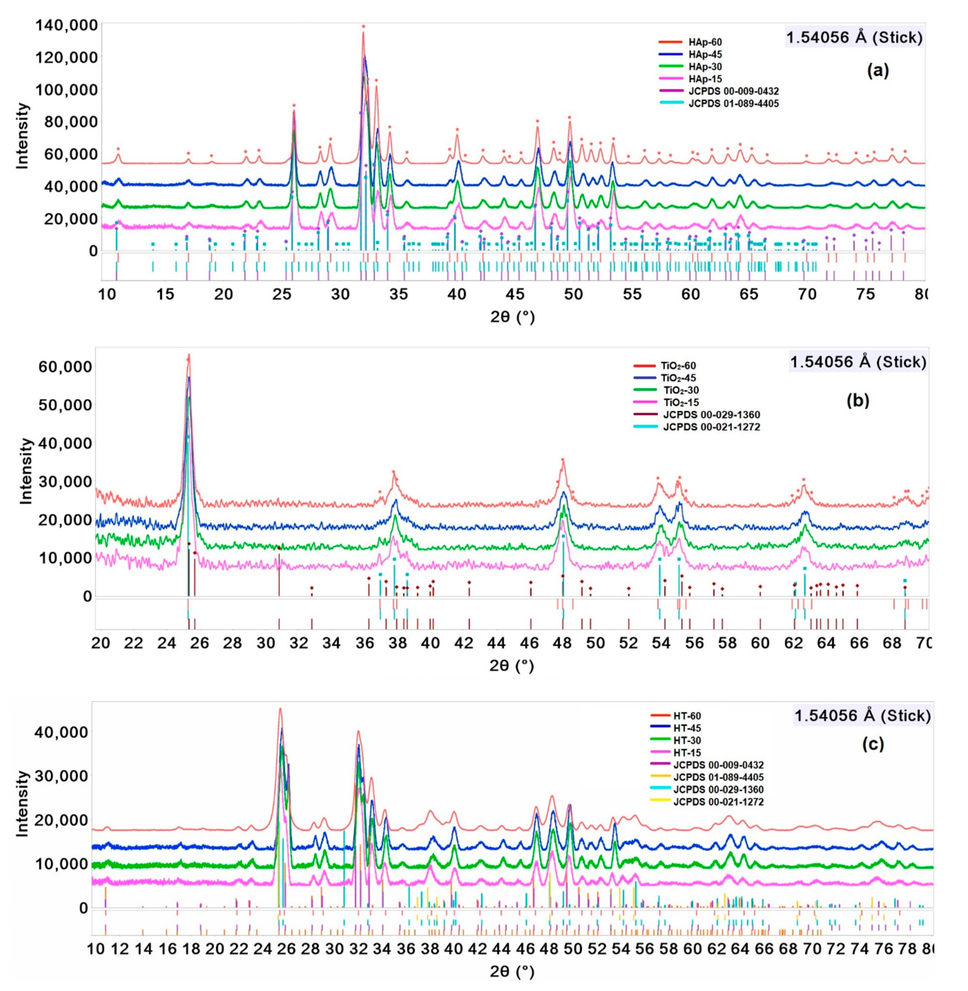
3.1. X-ray Diffraction

The XRD patterns of HAp, TiO2, and HAp/TiO2 nanocomposite nanoparticles were obtained, and the samples were synthesized at different time intervals of ultrasound irradiation at 550 °C. Other works have reported temperatures between 600 and 1500 °C, high pressures and long reaction times [5,40,41]. Two crystalline phases were detected in the HAp samples, shown in Figure 2a: a hexagonal phase (JCPDS 00-009-0432) and a monoclinic phase (JCPDS 01-089-4405). The TiO2 samples were mainly anatase phase (JCPDS 00-021-1272), shown in Figure 2b. The diffraction patterns were analyzed for the brookite phase (JCPDS 00-029-1360), and it was found not to have presence of this phase. On the other hand, the diffraction patterns of nanocomposites showed a mixture of these phases, and a small peak shift was observed towards values smaller than 2θ. Both the hexagonal and monoclinic phases were found for HAp, whereas the anatase phase was found for TiO2, shown in Figure 2c. The results indicated that ultrasound irradiation time induces more interaction among the chemical species in the solution to obtain the expected precursors in a shorter reaction time. Moreover, sample crystallinity in each sample was enhanced by increasing the ultrasound irradiation time [31,34,42,43].
Significant changes in the characteristic peaks of HAp, TiO2, and HAp/TiO2 as a function of ultrasound irradiation were observed in the main results from the XRD analysis. Figure 3 shows a short range of diffraction patterns of the samples. The XRD patterns show slight shifts in the peaks, a wider peak size and different relative intensities. The main (hkl) indices are (002), (102), (210), (211), (112), (300), (202), and (301) for HAp, and (101) and (004) for TiO2, but it was difficult to evaluate the main reflections for HAp/TiO2 due to a peak overlapping. A Rietveld refinement was carried out to evaluate and quantify the phases in each sample. For HAp/TiO2, the diffraction peak width may be attributed to the substitution of Ti4+ by calcium sites in hydroxyapatite as it has been proposed by several authors [12,16,31,44]. Furthermore, Figure 3c displays the presence of reflections related to TiO2 (001) and (004) and to HAp (002), (102), (210), (211), (300), and (200), which suggests a mixture of phases in the nanocomposites.
In addition, a Rietveld refinement of cell parameters for HAp, TiO2, and HAp/TiO2 nanocomposites was carried out, considerer the hexagonal phase to HAp and anatase phase to TiO2, which was consistent with the results reported by Zhou [44]. The quantitative analysis resulted in different percentages and phases present in each sample as a function of the ultrasound irradiation time, shown in Table 1. These results agree with Giardina et al. [45], who have reported that the increased crystallinity is related to the time of ultrasonic processing, generating chemical species that favor the reaction speed in the solution [34]. Table 1 shows only the results of the main phases: monoclinic and anatase.
From the Rietveld refinement results, small differences can be observed in the cell parameters for the monoclinic and hexagonal phases for HAp, anatase phase for TiO2, However, in the case of nanocomposites, the interaction between HAp and TiO2 was found to have no effect on the crystalline structure of the hydroxyapatite.
The crystallite sizes (coherent scattering domain (CSD) sizes) were estimated with Scherrer’s equation considering the full width at half maximum (FWHM) of the prominent reflection (101) for TiO2, while the reflection selected for HAp and HAp/TiO2 nanocomposite was (002) to avoid a peak overlap between 25.3° and 25.8° (2θ). The average sizes were found to change due to an increased nucleation rate that improved the sample crystallinity obtained at the maximum time interval of ultrasound irradiation [46,47], shown in Figure 4. As a result, the diffraction patterns showed a decrease in the full width at half maximum, where the crystallite size was expected to increase by the ultrasound irradiation time.

3.2. Raman Spectroscopy

A Raman spectroscopy was performed on all the samples to confirm the mixture of phases in the HAp/TiO2 nanocomposites. The Raman spectra showed an increase in the intensity of the phononic modes characteristic of each material in relation to the increase in the ultrasound irradiation time, confirming the results obtained by X-ray diffraction. Figure 5a shows an intense band at 142.7 cm−1 (E1g), as well as the characteristic bands at 196 cm−1 (Eg), 396 cm−1 (B1g), 517 cm−1 (A1g) and 639 cm−1 (Eg), which were attributed to the anatase phase of TiO2 [14]. The Raman analysis also confirmed a band at 964.6 cm−1 attributed to the symmetrical stretching mode of phosphate (ν1). On the other hand, the bands situated at 430.2 and 451 cm−1 correspond to the bending mode of the O-P-O bond in phosphate (ν2), while the bands at 582.6 and 595.1 cm−1 were assigned to the asymmetrical bending mode (ν4) [48,49,50]. The bands at 493 and 709.8 cm−1 were not assigned in these spectra because they were attributed to another phosphate group, shown in Figure 5b. Raman spectra show the same phononic modes of HAp and TiO2 for the nanocomposite only with small position shifts (2–4 cm−1). Figure 5c shows the main HAp bands at 429.5 and 964.8 cm−1 in the HAp/TiO2 nanocomposite, while there are slight band position shifts in comparison with the HAp as a result of the interaction between HAp and TiO2. The main TiO2 bands at 146, 199.4, 401.1, 518.1, and 638.7 cm−1 were observed in the nanocomposite, indicating the presence of TiO2 in the anatase phase. The presence of HAp, TiO2 anatase and HAp/TiO2 nanocomposite bands was demonstrated by Rempel et al. [51], whose samples contained from 10 to 20 wt.% of TiO2 into HAp.

3.3. Morphological Analysis

Other researchers have reported that the size and morphology of the hydroxyapatite particles are strongly related to synthetic methods and parameters such as pH, temperature and use of chelating agents and surfactants [52,53,54]. A FESEM micrograph of HAp, shown in Figure 6a, obtained at 60 min of ultrasound irradiation shows a semi-spherical shape with agglomerates and a size between 17 and 20 nm. These results agree with those of Nathanael et al., who state that the synthesis of hydroxyapatite crystals at nanometric scale shows particle agglomeration [55]. Nevertheless, TiO2 also exhibits a spherical shape with an average particle size of 18 nm in diameter and a nanoparticle agglomeration due to aggregation of primary particles, shown in Figure 6b. Lin et al. have reported a study where they indicate that the agglomeration and size distribution are related to fast mixing and synthesis methods, such as the mechanochemical, microwave, chemical precipitation, hydrothermal and sol-gel methods, due to fast nucleation and particle aggregation, which avoid Ostwald ripening [56]. Figure 6c shows the HAp/TiO2 nanocomposite nanoparticles, where the formation of agglomerates with a spherical shape and an average particle size of 20 nm can be observed. The interaction between HAp and TiO2 can favor the growth of spherical particles. An alternative to avoid particle agglomeration is to increase the concentration of surfactants that inhibit it.

3.4. Compositional Analysis

Quantitative WDXRF data of the samples show the nominal values of the sample composition confirmed by the presence of Ca2+, P5+, Ti4+ and other elements occurring at trace levels. Table 2 shows the values for each element and the presence of trace elements that occur due to possible contamination. The Ca/P ratio of hydroxyapatite varied from 1.80, 1.64, 1.88, and 1.76 for each sample. A semi-quantitative analysis presents values higher than the stoichiometric Ca/P ratio of 1.67 in the hydroxyapatite. McConnell et al. reported that this phenomenon may be attributed to volatile compounds formed during the thermal treatment or to the presence of other compounds containing calcium (e.g., Ca(PO4)2) or carbonates (e.g., CaCO3 and carbonate hydroxyapatite). Another reason for the Ca/P ratio to exceed this value may be due to the substitution of tetrahedral hydroxyls or phosphate groups in the hydroxyapatite [57]. Accordingly, the results described above show that a non-stoichiometric hydroxyapatite was obtained.
The results described above show that the TiO2 was incorporated into the HAp network as a nanocomposite. It should be emphasized that the time intervals of ultrasound irradiation are directly associated with the crystalline phase percentages obtained. Furthermore, the methodology promotes the formation of nanomaterials in shorter reaction time intervals than other methods, where high temperatures, high pressures and long reaction times are used. However, it is important to adjust the thermal treatment to reach a suitable stoichiometry in future research experiments.

4. Conclusions

In this work, HAp, TiO2, and HAp/TiO2 nanocomposites were successfully obtained by the sol-gel and co-precipitation methods assisted by ultrasound irradiation at different time intervals of 15, 30, 45, and 60 min at 550 °C, revealing a mixture of phases and an increased crystallinity as a result of the ultrasound irradiation time. The percentages of the crystalline phases were quantified and the effect on the network parameters were observed by Rietveld refinement. All of the materials showed a morphological shape ranging from semi-spherical to spherical particles and an average size between 17 and 20 nm. The agglomeration of particles was caused by fast nucleation in the co-precipitation process. Consequently, it is convenient to encourage the nanoparticle dispersion by increasing the surfactant concentration in order to reduce the aggregation of nanoparticles. The results obtained indicate that sample crystallinity can be controlled and modified by managing the ultrasound irradiation time.

Author Contributions

M.J.R.-Á. carried out experiments and characterizations, analyzed the data, and wrote the paper; A.K.S.-H. developed the methodology and carried out structural analysis; J.M.-J. participated in the project administration; R.S.-G. performed a formal morphological analysis and reviewed the manuscript; J.J.G.-A. participated in the writing and final editing of the manuscript. All authors have read and agreed to the published version of the manuscript.

Funding

This research was funded by Vicerrectoría de Investigación y Estudios de Posgrado grant number 100524279-VIEP2019 and the APC was funded by Sciences Institute of the Benemérita Universidad Autónoma de Puebla.

Acknowledgments

The authors are grateful to Laboratorio de Difracción de Rayos X and Laboratorio Central at the Instituto de Física (IFUAP) of the Benemérita Universidad Autónoma de Puebla for the X-ray diffraction and Raman spectroscopy measurements. A.K.S.H. wishes to thank CONACyT for Scholarship Grant No. 629604.

Conflicts of Interest

The authors declare no conflict of interest in this paper.

References

  1. Von Euw, S.; Wang, Y.; Laurent, G.; Drouet, C.; Babonneau, F.; Nassif, N.; Azaïs, T. Bone mineral: New insights into its chemical composition. Sci. Rep. 2019, 9, 1–11. [Google Scholar] [CrossRef] [PubMed] [Green Version]
  2. Koutsopoulos, S. Synthesis and characterization of hydroxyapatite crystals: A review study on the analytical methods. Willie Period. Inc. 2002, 62, 31–34. [Google Scholar] [CrossRef] [PubMed]
  3. Hench, L.L. Bioceramics. J. Am. Ceram. Soc. 1998, 28, 1705–1728. [Google Scholar] [CrossRef]
  4. Aminzare, M.; Eskandari, A.; Baroonian, M.H.; Berenov, A.; Razavi Hesabi, Z.; Taheri, M.; Sadrnezhaad, S.K. Hydroxyapatite nanocomposites: Synthesis, sintering and mechanical properties. Ceram. Int. 2013, 39, 2197–2206. [Google Scholar] [CrossRef]
  5. Salarian, M.; Xu, W.Z.; Wang, Z.; Sham, T.K.; Charpentier, P.A. Hydroxyapatite-TiO2-based Nanocomposites Synthesized in Supercritical CO2 for Bone Tissue Engineering: Physical and Mechanical Properties. ACS Appl. Mater. Interfaces 2014, 6, 16918–16931. [Google Scholar] [CrossRef]
  6. Joseph Nathanael, A.; Mangalaraj, D.; Chen, P.C.; Ponpandian, N. Mechanical and photocatalytic properties of hydroxyapatite/titania nanocomposites prepared by combined high gravity and hydrothermal process. Compos. Sci. Technol. 2010, 70, 419–426. [Google Scholar] [CrossRef]
  7. Enayati-Jazi, M.; Solati-Hashjin, M.; Nemati, A.; Bakhshi, F. Synthesis and characterization of hydroxyapatite/titania nanocomposites using in situ precipitation technique. Superlattices Microstruct. 2012, 51, 877–885. [Google Scholar] [CrossRef]
  8. Lee, B.-H.; Koshizaki, N. Nanostructured hydroxyapatite/TiO2 composite coating applied to commercially pure titanium by a co-sputtering technique. Nanotechnology 2008, 19, 415303. [Google Scholar] [CrossRef]
  9. Oktar, F.N. Hydroxyapatite-TiO2 composites. Mater. Lett. 2006, 60, 2207–2210. [Google Scholar] [CrossRef]
  10. Li, Z.; Yang, X.; Guo, H.; Yang, X.; Sun, L.; Dong, S. Hydroxyapatite additive influenced the bioactivity of bioactive nano-titania ceramics and new bone-forming capacity. J. Nanoparticle Res. 2012, 14. [Google Scholar] [CrossRef]
  11. Martinez, C.; Krieke, G.; Ločs, J.; Bērziņa-Cimdiņa, L.; Garrido, L.; Gilabert, U.; Ozols, A. Natural Hydroxyapatite-Ti-Dioxide Bone Substitutes. Procedia Mater. Sci. 2015, 8, 324–331. [Google Scholar] [CrossRef] [Green Version]
  12. Kumar, A.; Biswas, K.; Basu, B. Hydroxyapatite-titanium bulk composites for bone tissue engineering applications. J. Biomed. Mater. Res. Part A 2015, 103, 791–806. [Google Scholar] [CrossRef] [PubMed]
  13. Sidane, D.; Rammal, H.; Beljebbar, A.; Gangloff, S.C.; Chicot, D.; Velard, F.; Khireddine, H.; Montagne, A.; Kerdjoudj, H. Biocompatibility of sol-gel hydroxyapatite-titania composite and bilayer coatings. Mater. Sci. Eng. C 2017. [Google Scholar] [CrossRef] [PubMed] [Green Version]
  14. Reyes-Coronado, D.; Rodríguez-Gattorno, G.; Espinosa-Pesqueira, M.E.; Cab, C.; De Coss, R.; Oskam, G. Phase-pure TiO2 nanoparticles: Anatase, brookite and rutile. Nanotechnology 2008, 19. [Google Scholar] [CrossRef]
  15. Pushpakanth, S.; Srinivasan, B.; Sreedhar, B.; Sastry, T.P. An in situ approach to prepare nanorods of titania-hydroxyapatite (TiO2-HAp) nanocomposite by microwave hydrothermal technique. Mater. Chem. Phys. 2008, 107, 492–498. [Google Scholar] [CrossRef]
  16. Mohamed, R.M.; Baeissa, E.S. Preparation and characterisation of Pd-TiO2-hydroxyapatite nanoparticles for the photocatalytic degradation of cyanide under visible light. Appl. Catal. A Gen. 2013, 464–465, 218–224. [Google Scholar] [CrossRef]
  17. Márquez Brazón, E.; Piccirillo, C.; Moreira, I.S.; Castro, P.M.L. Photodegradation of pharmaceutical persistent pollutants using hydroxyapatite-based materials. J. Environ. Manage. 2016, 182, 486–495. [Google Scholar] [CrossRef]
  18. Khamova, T.V.; Frank-Kamenetskaya, O.V.; Shilova, O.A.; Chelibanov, V.P.; Marugin, A.M.; Yasenko, E.A.; Kuz’mina, M.A.; Baranchikov, A.E.; Ivanov, V.K. Hydroxyapatite/Anatase Photocatalytic Core–Shell Composite Prepared by Sol‒Gel Processing. Crystallogr. Reports 2018, 63, 254–260. [Google Scholar] [CrossRef]
  19. Murgolo, S.; Moreira, I.S.; Piccirillo, C.; Castro, P.M.L.; Ventrella, G.; Cocozza, C.; Mascolo, G. Photocatalytic degradation of diclofenac by hydroxyapatite-TiO2 composite material: Identification of transformation products and assessment of toxicity. Materials 2018, 11, 1779. [Google Scholar] [CrossRef] [Green Version]
  20. Bystrov, V.S.; Piccirillo, C.; Tobaldi, D.M.; Castro, P.M.L.; Coutinho, J.; Kopyl, S.; Pullar, R.C. Oxygen vacancies, the optical band gap (Eg) and photocatalysis of hydroxyapatite: Comparing modelling with measured data. Appl. Catal. B Environ. 2016, 196, 100–107. [Google Scholar] [CrossRef]
  21. Pramanik, S.; Agarwal, A.K.; Rai, K.N.; Garg, A. Development of high strength hydroxyapatite by solid-state-sintering process. Ceram. Int. 2007, 33, 419–426. [Google Scholar] [CrossRef]
  22. Monmaturapoj, N.; Thepsuwan, W.; Mai-Ngam, K.; Ngernpimai, S.; Klinsukhon, W.; Prahsarn, C. Preparation and properties of hydroxyapatite/titania composite for microbial filtration application. Adv. Appl. Ceram. 2014. [Google Scholar] [CrossRef]
  23. Zakeri, M.; Hasani, E.; Tamizifar, M. Mechanical properties of TiO2-hydroxyapatite nanostructured coatings on Ti-6Al-4V substrates by APS method. Int. J. Miner. Metall. Mater. 2013, 20, 397–402. [Google Scholar] [CrossRef]
  24. Ono, Y.; Rachi, T.; Okuda, T.; Yokouchi, M.; Kamimoto, Y.; Ono, H.; Nakajima, A.; Okada, K. An aqueous synthesis of photocatalyst by selective dissolution of titanium oxide/hydroxyapatite composite. Ceram. Int. 2011, 37, 1563–1568. [Google Scholar] [CrossRef]
  25. Li, P. Bioactive Ca10(PO4)6(OH)2-TiO2 Composite Coating Prepared by Sol-Gel Process. J. Sol-Gel Sci. Technol. 1996, 7, 27–34. [Google Scholar] [CrossRef]
  26. Rouhani, P.; Taghavinia, N.; Rouhani, S. Rapid growth of hydroxyapatite nanoparticles using ultrasonic irradiation. Ultrason. Sonochem. 2010, 17, 853–856. [Google Scholar] [CrossRef]
  27. Fidancevska, E.; Ruseska, G.; Bossert, J.; Lin, Y.M.; Boccaccini, A.R. Fabrication and characterization of porous bioceramic composites based on hydroxyapatite and titania. Mater. Chem. Phys. 2007, 103, 95–100. [Google Scholar] [CrossRef]
  28. Dodds, J.; Espitalier, F.; Louisnard, O.; Grossier, R.; David, R.; Hassoun, M.; Baillon, F.; Gatumel, C.; Lyczko, N. The effect of ultrasound on crystallisation-precipitation processes: Some examples and a new segregation model. Part. Part. Syst. Charact. 2007. [Google Scholar] [CrossRef]
  29. Mettin, R.; Cairós, C.; Troia, A. Sonochemistry and bubble dynamics. Ultrason. Sonochem. 2015, 25, 24–30. [Google Scholar] [CrossRef]
  30. Gedanken, A. Using sonochemistry for the fabrication of nanomaterials. Ultrason. Sonochem. 2004, 11, 47–55. [Google Scholar] [CrossRef]
  31. Nimmy, E.; Wilson, P. Investigations on sonofragmentation of hydroxyapatite crystals as a function of strontium incorporation. Ultrason. Sonochem. 2019, 50, 188–199. [Google Scholar] [CrossRef]
  32. Mason, T.J. Some neglected or rejected paths in sonochemistry—A very personal view. Ultrason. Sonochem. 2015, 25, 89–93. [Google Scholar] [CrossRef] [PubMed]
  33. Arami, H.; Mazloumi, M.; Khalifehzadeh, R.; Sadrnezhaad, S.K. Sonochemical preparation of TiO2 nanoparticles. Mater. Lett. 2007. [Google Scholar] [CrossRef]
  34. Fang, Y.; Agrawal, D.K.; Roy, D.M.; Roy, R.; Brown, P.W. Ultrasonically accelerated synthesis of hydroxyapatite. J. Mater. Res. 1992, 7, 2294–2298. [Google Scholar] [CrossRef]
  35. Oh, C.W.; Seong, G.D.L.; Park, S.; Ju, C.S.; Hong, S.S. Synthesis of nanosized TiO2 particles via ultrasonic irradiation and their photocatalytic activity. React. Kinet. Catal. Lett. 2005. [Google Scholar] [CrossRef]
  36. Sassoni, E.; D’Amen, E.; Roveri, N.; Scherer, G.W.; Franzoni, E. Photocatalytic hydroxyapatite-titania nanocomposites for preventive conservation of marble. IOP Conf. Ser. Mater. Sci. Eng. 2018, 364, 012073. [Google Scholar] [CrossRef]
  37. Nathanael, A.J.; Mangalaraj, D.; Ponpandian, N. Controlled growth and investigations on the morphology and mechanical properties of hydroxyapatite/titania nanocomposite thin films. Compos. Sci. Technol. 2010, 70, 1645–1651. [Google Scholar] [CrossRef]
  38. Grigorieva, T.F.; Barinova, A.P.; Lyakhov, N.Z. Mechanosynthesis of Nanocomposites. J. Nanoparticle Res. 2003, 5, 439–453. [Google Scholar] [CrossRef]
  39. Kabekkodu, S.N.; Faber, J.; Fawcett, T. New Powder Diffraction File (PDF-4) in relational database format: Advantages and data-mining capabilities. Acta Crystallogr. Sect. B Struct. Sci. 2002, 58, 333–337. [Google Scholar] [CrossRef] [Green Version]
  40. Hannora, A.E.; Ataya, S. Structure and compression strength of hydroxyapatite/titania nanocomposites formed by high energy ball milling. J. Alloys Compd. 2016, 658, 222–233. [Google Scholar] [CrossRef]
  41. Horiuchi, N.; Madokoro, K.; Nozaki, K.; Nakamura, M.; Katayama, K.; Nagai, A.; Yamashita, K. Electrical conductivity of polycrystalline hydroxyapatite and its application to electret formation. Solid State Ionics 2018, 315, 19–25. [Google Scholar] [CrossRef]
  42. Poinern, G.E.; Brundavanam, R.K.; Mondinos, N.; Jiang, Z.T. Synthesis and characterisation of nanohydroxyapatite using an ultrasound assisted method. Ultrason. Sonochem. 2009, 16, 469–474. [Google Scholar] [CrossRef] [PubMed] [Green Version]
  43. Nikolaev, A.L.; Gopin, A.V.; Severin, A.V.; Rudin, V.N.; Mironov, M.A.; Dezhkunov, N.V. Ultrasonic synthesis of hydroxyapatite in non-cavitation and cavitation modes. Ultrason. Sonochem. 2018, 44, 390–397. [Google Scholar] [CrossRef] [PubMed]
  44. Nie, J.; Zhou, J.; Huang, X.; Wang, L.; Liu, G.; Cheng, J. Effect of TiO2 doping on densification and mechanical properties of hydroxyapatite by microwave sintering. Ceram. Int. 2019, 45, 13647–13655. [Google Scholar] [CrossRef]
  45. Giardina, M.A.; Fanovich, M.A. Synthesis of nanocrystalline hydroxyapatite from Ca(OH)2 and H3PO4 assisted by ultrasonic irradiation. Ceram. Int. 2010, 36, 1961–1969. [Google Scholar] [CrossRef]
  46. Jordens, J.; Canini, E.; Gielen, B.; Van Gerven, T.; Braeken, L. Ultrasound assisted particle size control by continuous seed generation and batch growth. Crystals 2017, 7, 195. [Google Scholar] [CrossRef]
  47. Kim, H.N.; Suslick, K.S. The effects of ultrasound on crystals: Sonocrystallization and sonofragmentation. Crystals 2018, 8, 280. [Google Scholar] [CrossRef] [Green Version]
  48. Slepko, A.; Demkov, A.A. Hydroxyapatite: Vibrational spectra and monoclinic to hexagonal phase transition. J. Appl. Phys. 2016, 074701. [Google Scholar] [CrossRef]
  49. Anwar, A.; Akbar, S. Continuous microwave assisted flow synthesis and characterization of calcium deficient hydroxyapatite nanorods. Adv. Powder Technol. 2018, 29, 1493–1498. [Google Scholar] [CrossRef]
  50. Blakeslee, K.C.; Condrate, R.A., Sr. Vibrational Spectra of Hydrothermally Prepared Hydroxyapatites. J. Am. Ceram. Soc. 1971, 54, 559–563. [Google Scholar] [CrossRef]
  51. Rempel, S.V.; Eselevich, D.A.; Valeeva, A.A.; Rempel, A.A. Structure of a HAp/TiOy Nanocomposite Studied by Vibrational Spectroscopy Techniques. Inorg. Mater. 2018, 54, 898–903. [Google Scholar] [CrossRef]
  52. Swain, S.K.; Sarkar, D. A comparative study: Hydroxyapatite spherical nanopowders and elongated nanorods. Ceram. Int. 2011, 37, 2927–2930. [Google Scholar] [CrossRef]
  53. Zhang, C.; Yang, J.; Quan, Z.; Yang, P.; Li, C.; Hou, Z.; Lin, J. Hydroxyapatite nano- and microcrystals with multiform morphologies: Controllable synthesis and luminescence properties. Cryst. Growth Des. 2009, 9, 2725–2733. [Google Scholar] [CrossRef]
  54. Mary, I.R.; Sonia, S.; Viji, S.; Mangalaraj, D.; Viswanathan, C.; Ponpandian, N. Novel multiform morphologies of hydroxyapatite: Synthesis and growth mechanism. Appl. Surf. Sci. 2016, 361, 25–32. [Google Scholar] [CrossRef]
  55. Nathanael, A.J.; Lee, J.H.; Mangalaraj, D.; Hong, S.I.; Rhee, Y.H. Multifunctional properties of hydroxyapatite/titania bio-nano-composites: Bioactivity and antimicrobial studies. Powder Technol. 2012, 228, 410–415. [Google Scholar] [CrossRef]
  56. Lin, K.; Wu, C.; Chang, J. Advances in synthesis of calcium phosphate crystals with controlled size and shape. Acta Biomater. 2014, 10, 4071–4102. [Google Scholar] [CrossRef] [PubMed]
  57. McConnell, D. Crystal chemistry of hydroxyapatite. Its relation to bone mineral. Arch. Oral Biol. 1965. [Google Scholar] [CrossRef]
Figure 1. Diagram of the synthesis of HAp, TiO2, and HT nanocomposite.
Crystals 10 00959 g001
Figure 2. X-ray powder diffraction patterns for the different time intervals of ultrasound irradiation (15, 30, 45 and 60 min) and the JCPDS obtained from the Powder Diffraction File (PDF)-4+2018 database corresponding to the International Centre of Diffraction Data (ICDD) for (a) HAp, (b) TiO2, and (c) HT.
Crystals 10 00959 g002
Figure 3. Characteristic diffraction peaks of the (a) HAp monoclinic and hexagonal phases (b) TiO2 anatase phase, and (c) HT monoclinic, hexagonal and anatase phases.
Crystals 10 00959 g003
Figure 4. Coherent scattering domain (CSD) sizes (a) HAp, (b) TiO2, and (c) HT determined by Scherrer’s equation.
Crystals 10 00959 g004
Figure 5. Raman spectra at different time intervals of ultrasound irradiation (15, 30, 45, and 60 min) (a) HAp, (b) TiO2, and (c) HT.
Crystals 10 00959 g005
Figure 6. Field Emission Scanning Electron Microscopy (FESEM) images of (a) HAp, (b) TiO2, and (c) HT nanocomposite.
Crystals 10 00959 g006
Table 1. Main crystalline parameters of HAp, TiO2, and HAp/TiO2 nanocomposite samples.
SamplePhases, %Relevant Parameters of the Monoclinic and Anatase Phases
HexagonalMonoclinicAnatasea, Åb, Åc, ÅV, 106 pm3Rwp, %
HAp-1516.183.9----9.3887(5)6.8858(2)18.8142(5)1057.2074.97
HAp-3015.384.7----9.4022 (1)6.8931 (2)18.8303 (1)1059.16210.60
HAp-4513.886.2----9.3989 (3)6.8908 (5)18.7288 (5)1054.4075.24
HAp-6011.588.5----9.4480 (1)6.8818 (1)18.835 (1)1056.5223.16
TIO2-15--------1003.7845 (4)3.7845 (4)9.5050 (8)136.13911.90
TIO2-30--------1003.7837 (1)3.7837 (1)9.4860 (1)136.13912.43
TIO2-45--------1003.7852 (3)3.7852 (3)9.4863 (1)135.9204.35
TIO2-60--------1003.7862 (5)3.7862 (5)9.5035 (1)136.2364.22
HT-156.953.839.39.4005 (5)6.8871 (2)18.7590 (8)1055.4053.88
HT-306.151.442.59.4057 (1)6.8901 (6)18.7761 (6)1057.1704.07
HT-457.448.444.29.3983 (4)6.8925 (9)18.7315 (1)1054.8094.13
HT-605.843.550.79.3846 (8)6.8882 (6)18.8184 (7)1056.9244.08
Table 2. Compositional analysis of nanoparticles obtained by the sol-gel method assisted by ultrasound irradiation.
Composition, wt.%
SampleCaPCH2TiOther Traces, ppm
HAp-1524.89.7165------O, Cl
HAp-3026.012.3061-----Cl, Si, O
HAp-4526.49.2665------Si, O
HAp-6025.010.464------Cl, Si, O
TiO2-15---------------26.9S, Ca, O
TiO2-30---------------27.8S, Ca, Nb, O
TiO2-45---------------27.2S, Nb, O
TiO2-60---------------28.5Nb, Ca, O
HT-1533.3011.6-----29.8Si, O
HT-3035.99.46-----24.6Nb, S, Si, O
HT-4529.411.3-----35.0S, Nb, O, Cl
HT-6031.611.6-----32.5S, Cl, Nb, O
Publisher’s Note: MDPI stays neutral with regard to jurisdictional claims in published maps and institutional affiliations.

