Article

The Potential Applications of Bacillus sp. and Pseudomonas sp. Strains with Antimicrobial Activity against Phytopathogens, in Waste Oils and the Bioremediation of Hydrocarbons

by
Mariana-Gratiela Soare (Vladu)
1,2,
Elena Simina Lakatos
3,*,
Nicoleta Ene
2,4,
Nereida Malo (Dalanaj)
5,
Ovidiu Popa
1 and
Narcisa Babeanu
1
1
Faculty of Biotechnologies, University of Agronomic Sciences and Veterinary Medicine, Mărăști Blv. 59, 011464 Bucharest, Romania
2
National Institute for Chemical Pharmaceutical Research and Development-ICCF, Vitan Avenue 112, 031299 Bucharest, Romania
3
Institute for Research in Circular Economy and Environment “Ernest Lupan”, Dorobantilor Avenue 71–73, 400808 Cluj-Napoca, Romania
4
Biology Department, University of Bucharest, Regina Elisabeta Blv. 4–13, 050663 Bucharest, Romania
5
Research Institute of the University of Bucharest-ICUB, Regina Elisabeta Blv. 4–12, 030018 Bucharest, Romania
*
Author to whom correspondence should be addressed.
Catalysts 2019, 9(11), 959; https://doi.org/10.3390/catal9110959
Submission received: 27 September 2019 / Revised: 7 November 2019 / Accepted: 11 November 2019 / Published: 15 November 2019

Abstract

:
Biodegradation is one of the primary mechanisms for the elimination of petroleum and other hydrocarbon pollutants from the environment. This study presents the results obtained with two newly isolated microorganisms and their potential applications in bioremediation, agriculture, and industrial fields. Twenty-five strains of microorganisms were isolated from plant materials and were subject to a selection process on the basis of antimicrobial activity. Two bacterial strains, respectively Bacillus mycoides (Bm) and Pseudomonas putida (B1), were selected for further experiments, based on the largest inhibition zones against the phytopathogens Erwinia carotovora and Xanthomonas campestris. The production of biosurfactants and enzymes was evaluated in specific media. In order to assess the production of biosurfactants, submerged bioprocesses were carried out on Yeast Malt Peptone Glucose (YMPG), M44, Luria-Bertani (LB), and King B media (KB); the supernatants were used to form emulsions with heptane, octane, and sunflower oil, and the emulsifying indices were determined.

1. Introduction

The resistance of phytopathogens to a wide range of chemical synthesis compounds is currently a widespread phenomenon, and finding solutions to replace these substances with new, natural compounds is an ongoing concern in the field of research [1]. Many studies and experiments have been carried out over time, both in the laboratory and in the field, to combat the phytopathogens with the help of microbial antagonists. These antagonists, also known as biocontrol agents, are commonly occurring nonpathogenic microbes such as fungi, bacteria, or yeast. Antagonists act directly on phytopathogens through different mechanisms such as hyperparasitism, competition for a substrate, and the secretion of metabolites with an antibiotic effect. Many of the bacteria belonging to the Bacillus and Pseudomonas genera have been studied for their use in phytopathogens’ biocontrol [2]. These types of bacteria are known for their versatility and their ability to populate various niches and to grow on numerous substrates [3]. This is due to the fact that they are rich in enzymes and also to the metabolites with antimicrobial properties they excrete as biosurfactants [4,5]. It is known that, under normal conditions, biosurfactants are bioactive compounds with the property to decrease the superficial tension at the interface between immiscible liquids. The biosurfactant producing microorganisms have the advantage of being able to survive and grow on water immiscible substrates [6]. The Pseudomonas, Bacillus, Rhodococcus, and Candida genera are the most widely studied microorganisms as producers of different types of biosurfactants [7]. Having different molecular structures and surface activities, the biosurfactants are divided into different classes: glycolipids (Candida sp., Pseudomonas sp.), polymeric surfactants (Candida sp., Bacillus sp.), lipopeptides (Bacillus sp., Candida lipolytica, Pseudomonas fluorescens), fatty acids, particulate surfactants (Pseudomonas marginalis), and phospholipids [6]. For example, Bacillus sp. are known to produce surfactins, iturins, fengycins, and lichenysins; Pseudomonas sp. produce mostly rhamnolipids; Rhodococcus sp., trehalolipids; and Candida sp. liposans and sophorolipids.
Many studies aim to demonstrate the connections between compounds with an antimicrobial effect and their biosurfactant properties [8,9]. According to some researchers, the production of peptides with antimicrobial activity is characteristic for Bacillus sp. [10]. Grossman suggests that peptide antibiotics play an essential role in sporulation [11]. These peptides, commonly known as lipopeptides, are capable of damaging the plasma membrane of the target microorganisms (e.g., R. solanacearum, X. oryzae), forming pores and leading to their death [12]. Other studies indicate that biosurfactants have an antagonistic effect against other microorganisms [13,14], are involved in adherence to different surfaces [15], accelerate a composting process, hence providing favorable conditions for beneficial microbes [16], and are involved in bioremediation of contaminated environments.
Bioremediation is related to the acceleration of natural biodegradation processes in contaminated environments. It is often a cost effective method to treat oily soils and petroleum wastes containing biodegradable hydrocarbons and involves mostly indigenous microbes. The high diversity of biosurfactants produced by numerous microorganisms is noteworthy [17]. In this sense, the chemical nature of a biosurfactant/bioemulsifier plays an important role in its function. Many researchers have established direct connections between the emulsification capacity of a biosurfactant and bioremediation. The biosurfactants that registered good emulsification indexes proved to be most capable of making the insoluble substrate accessible for biodegradation. Such biosurfactants can be successfully used for Microbial Enhanced Oil Recovery (MEOR). For example, Silva et al. found that Pseudomonas aeruginosa UCP 0992 and the biosurfactant produced by this strain achieved degradation rates higher than 90% for oil present in sand and seawater [18]. Other researchers have shown that, due to their remarkable characteristics (such as dispersion, emulsification, and de-emulsification), the addition of biosurfactants has improved the bioremediation of oil contaminated sites, by improving the biodegradation activity of indigenous microorganisms [19,20].
The fact that biosurfactants have a biological origin implies both better biocompatibility and good microbial biodegradability; consequently, there is a large number of potential applications for this type of surfactants, especially when there is extensive interference with the environment, for example for tertiary petroleum recovery, decontamination of oil polluted areas, crop protection, and for the cosmetic and pharmaceutical industries [21]. Therefore, it is not surprising that a number of investigations have been carried out, both in the laboratory [22,23] and in the field [24,25,26], aiming to reveal the production of such compounds with potential applications in bioremediation, agriculture, and industry.
The present paper is structured as follows: Section 2 presents the results and discussions related to the production of biosurfactants and enzymes, in specific media. Section 3 provides the concept of the study, and methodological remarks are presented in Section 3. Section 4 gives the conclusions of the study.

2. Results and Discussion

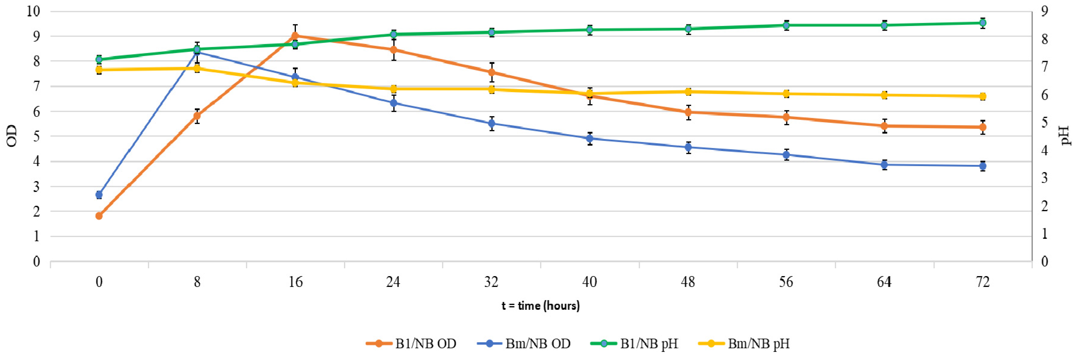
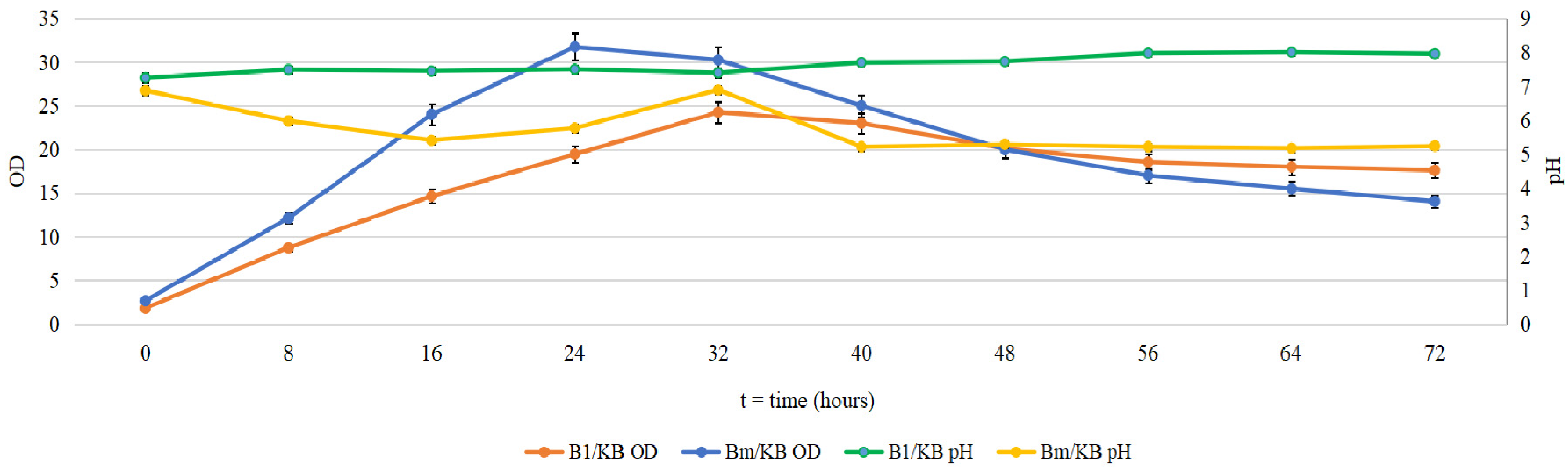
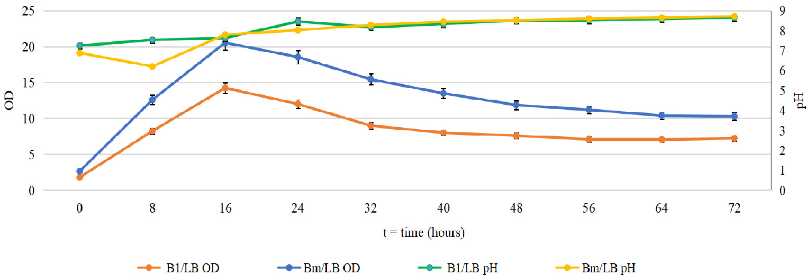
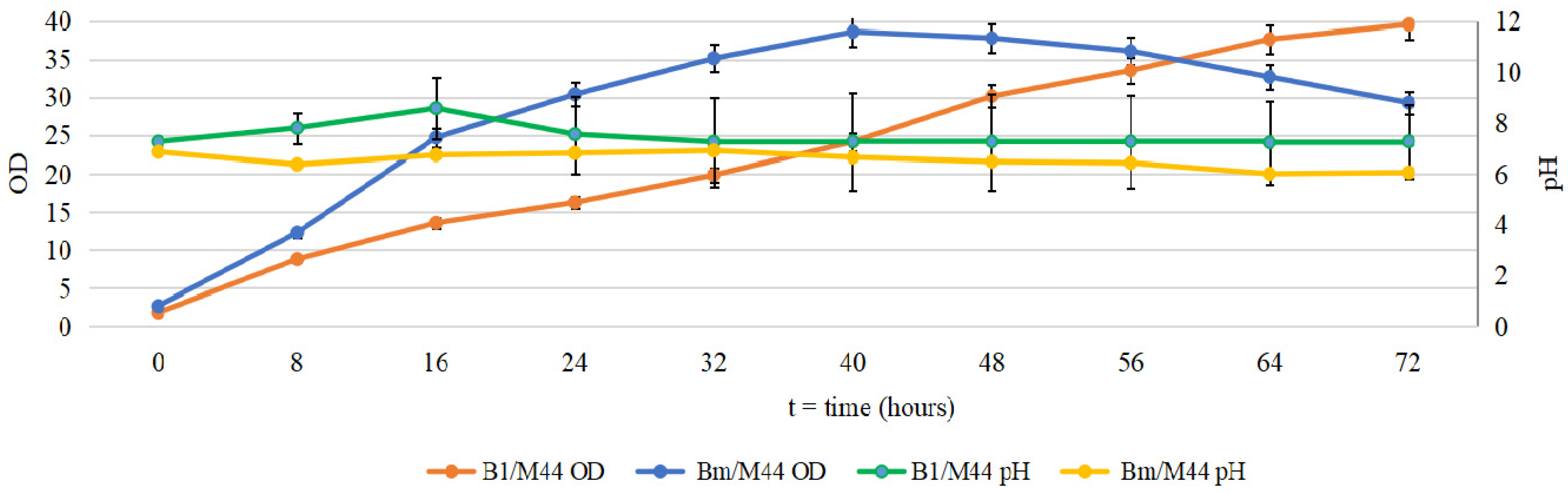
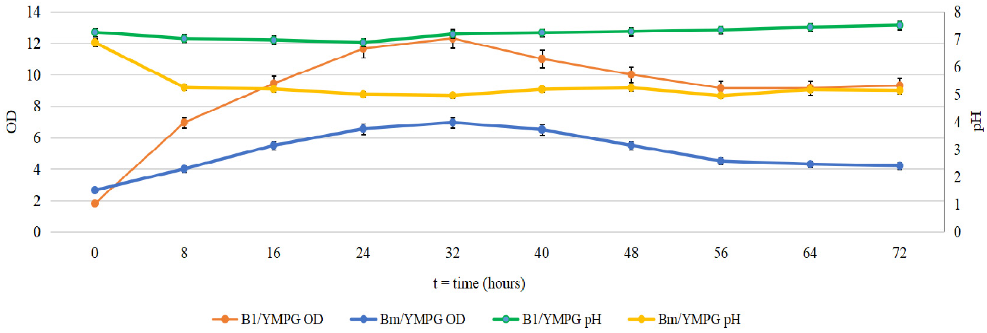
2.1. Cultivation of B. mycoides and P. putida in Several Media

Newly isolated microorganisms were grown submerged in the above mentioned media, in order to be tested for antimicrobial activity and for enzyme and biosurfactant production. In the figures below, the growth results for B. mycoides (Bm) and P. putida (B1) are presented.
In the NB medium (Figure 1), the strain P. putida (B1) reached the maximum growth after 16 h; then, a slow decrease was registered, measured in optical density.
The overall trend for pH value was a slight increase from 7.25 to 8.57. For B. mycoides (Bm), the maximum growth was achieved after 8 h of fermentation, when the pH value also reached a higher value. Then, both the OD and the pH started to decrease slowly.
The slight disruptions of the general trend in OD were probably due to the shift from one source of growth to another.
In King B (KB) medium (Figure 2), the strain P. putida (B1) achieved its maximum biomass growth at 32 h after inoculation, and then, a slow decrease in OD value was observed. During the growth period, the pH value was in the range of 7.2–7.5 and, after that, rose slightly to eight. The strain B. mycoides (Bm) showed significant growth until 24 h, coinciding with a slight decrease in pH value.
The significant decrease in OD between 32 and 48 h was probably due to the fact that the strain depleted the peptone from the medium and switched to glycerol metabolism. In the next 16 h, when glycerol was almost exhausted, the OD value began to decrease as a result of cell death because of the small amount of nutrients, remaining relatively constant between 56 and 72 h.
During fermentation in the LB medium (Figure 3), the strains’ growth was similar: Maximum growth was achieved at 16 h for both strains. However, B. mycoides (Bm) registered higher values for OD. Almost similar values were registered for the pH, with small differences in the first 12 h, as the general tendency for the Bm strain was the pH decrease in the logarithmic phase of growth.
In the M44 medium (Figure 4), both strains registered the best values for OD, when compared with the results obtained for the other media. Maximum growth for Bm was achieved at 44 h, then OD started to slowly decrease, probably because of the lack of a nitrogen source and an excess of glycerol. B1 exhibited a different behavior: The growth curve for this strain remained ascendant at 72 h, probably because the glycerol amount did not act as an inhibitor.
The maximum growth for B1 was reached at 32 h in the YMPG medium (Figure 5). In the first 24 h, the pH value slowly decreased to 6.88 and then slowly increased to 7.51 at the end of the bioprocess. The cellular growth for the Bm strain was greatest at 32 h and significantly decreased between 40 h and 48 h, as a result of cell death, remaining relatively constant between 48 and 72 h.

2.2. Antimicrobial Activity

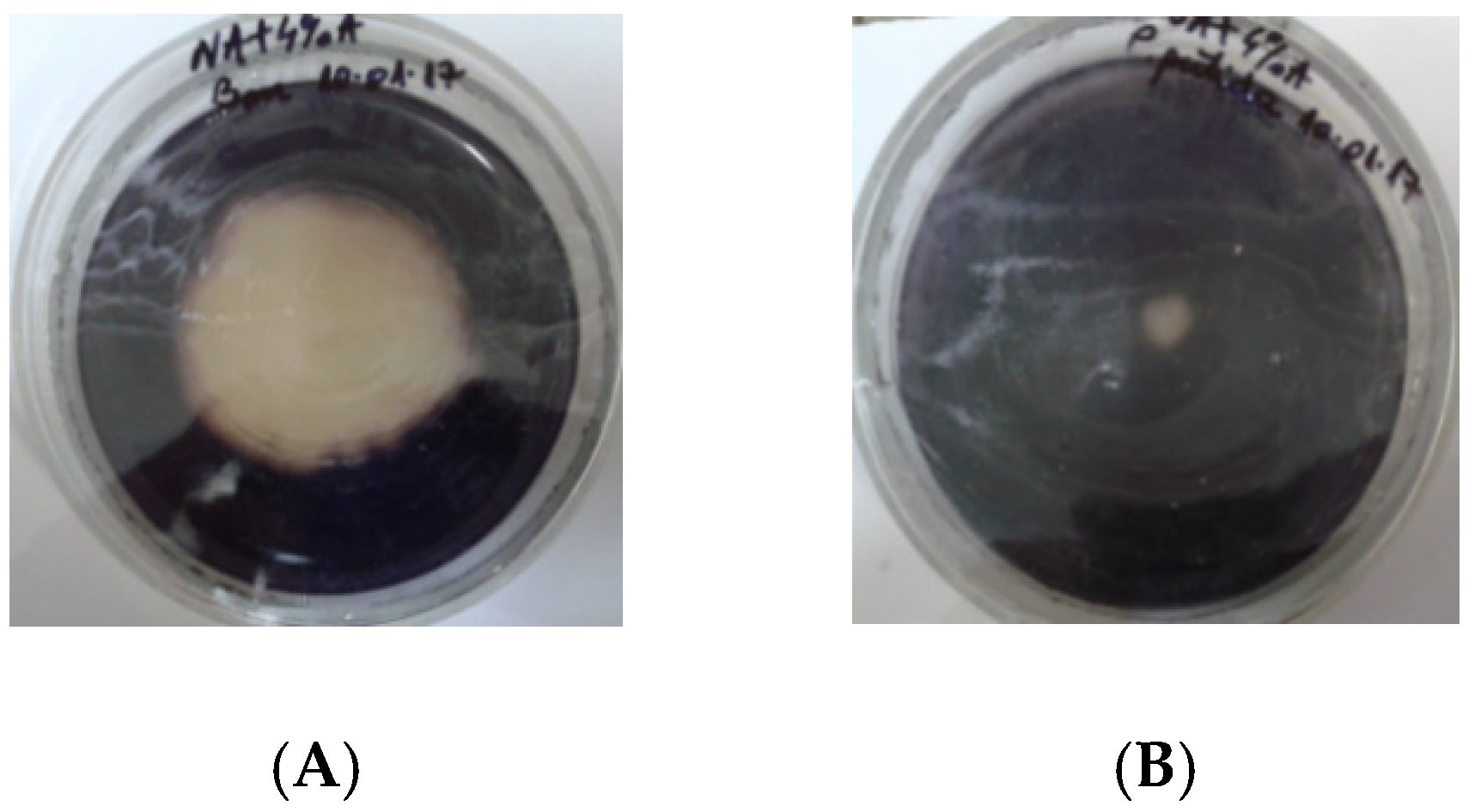
2.2.1. Antimicrobial Activity of P. putida and B. mycoides against E. carotovora and X. campestris

The results obtained during the experiments confirmed that, in both cases, the antagonists exerted antimicrobial action against the phytopathogens E. carotovora ICCF 138 and X. campestris ICCF 274. A better antimicrobial activity (Table 1) was noticed in Experiment I, where the diameters of the inhibition zones were larger than those registered in Experiment II.

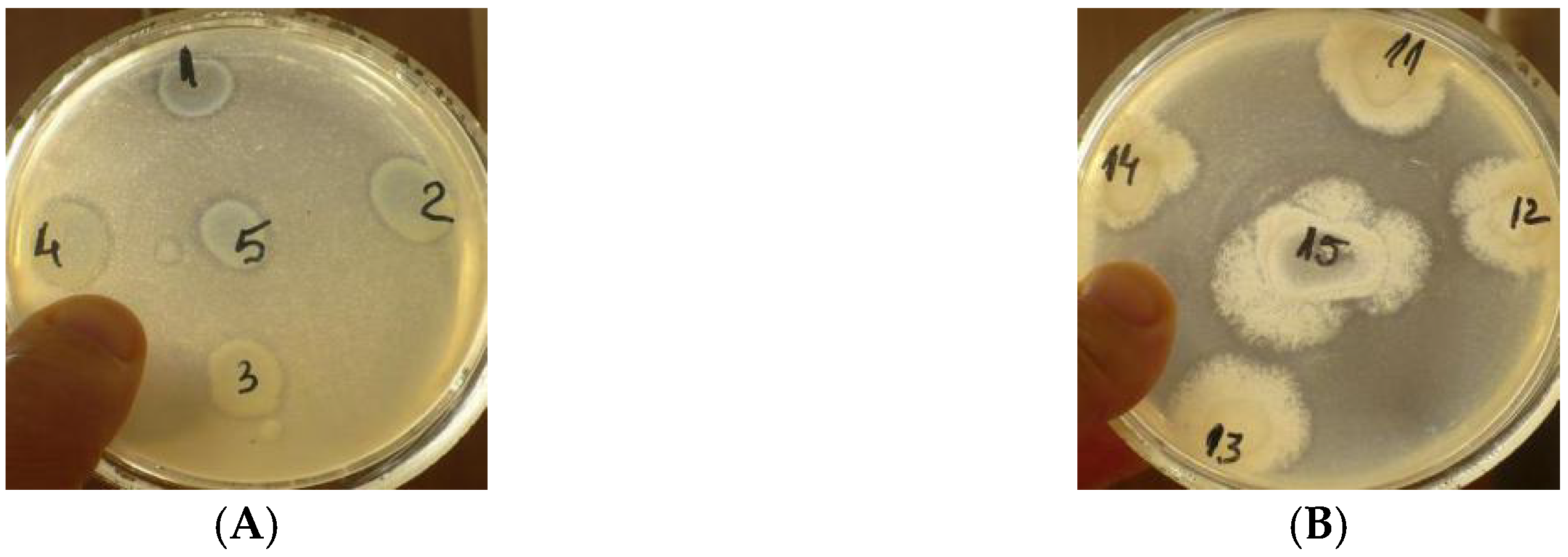
2.2.2. Antimicrobial Activity of the Biosurfactants from P. putida and B. mycoides Supernatants against E. carotovora and X. campestris

In Table 2, the results are presented as the average of the values together with the standard errors, from three independent replications.
According to some researchers [27], the production of biosurfactants is influenced by the type of carbon source present in the medium. The results obtained during this study confirmed their observations, especially in the case of the B. mycoides (Bm) strain, which grew better in the M44 medium containing glycerol.

2.3. Enzyme Production

Extracellular enzyme and metabolite production is often associated with antagonism properties [28]. Most of the species of the Bacillus and Pseudomonas genera have the ability to produce extracellular enzymes and secondary metabolites [29]. For this reason, the selected strains were tested in several media in order to study the production of such compounds. Enzyme production was revealed on agarized media, containing the appropriate carbon sources. Tests results revealed that B. mycoides (Bm) produced amylases, P. putida (B1) produced catalase (Figure 6 and Figure 7), and both strains produced lipases (Figure 8).
The inoculum obtained in all five media produced halos around the colonies, which is an indicator of lipase production and potential application of the strains in the bioremediation of waste oils.
The enzymatic activities measured for amylase (enzymatic assay of α-Amylase EC 3.2.1.1, with DNS, [30]), lipase (enzymatic assay of lipase EC 3.1.1.3, titrimetric method, [31]), and catalase (enzymatic assay of catalase EC 1.11.1.6, [32]), following the Sigma-Aldrich protocols, revealed that:
After 72 h of development in SNA medium, B. mycoides (Bm) registered 0.75 U/mL, and the amylase activity of the P. putida (B1) strain was insignificant.
After 48 h of development in LA medium, B. mycoides (Bm) registered 0.39 U/mL, and the lipase activity of the P. putida (B1) strain registered 0.36 U/mL.
After 72 h of development on M44 solid medium, P. putida (B1) registered 2.3 U/mL, and the catalase activity of the B. mycoides (Bm) strain was insignificant.

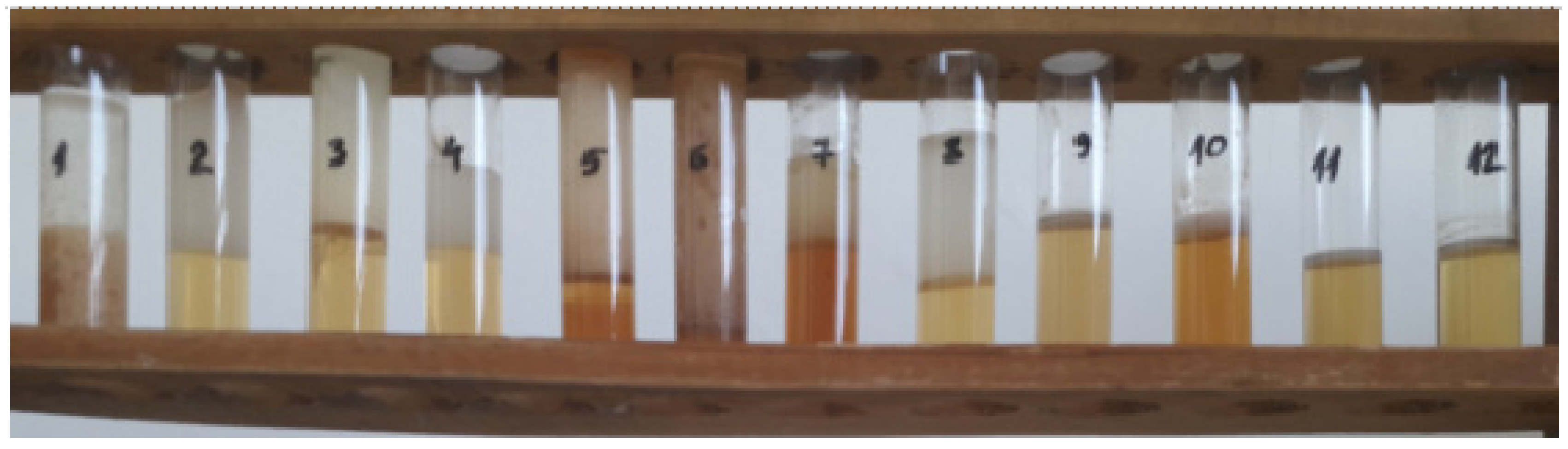
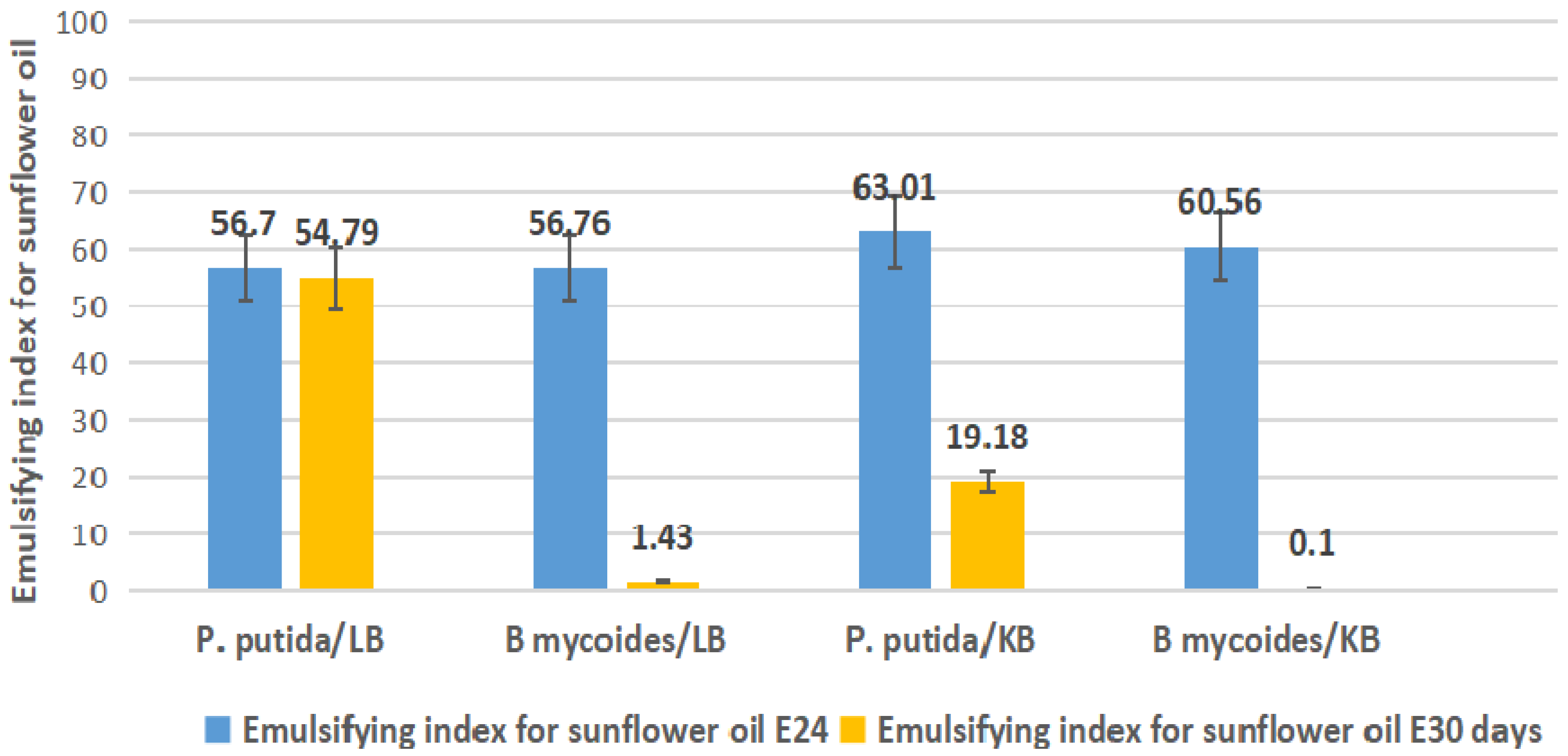
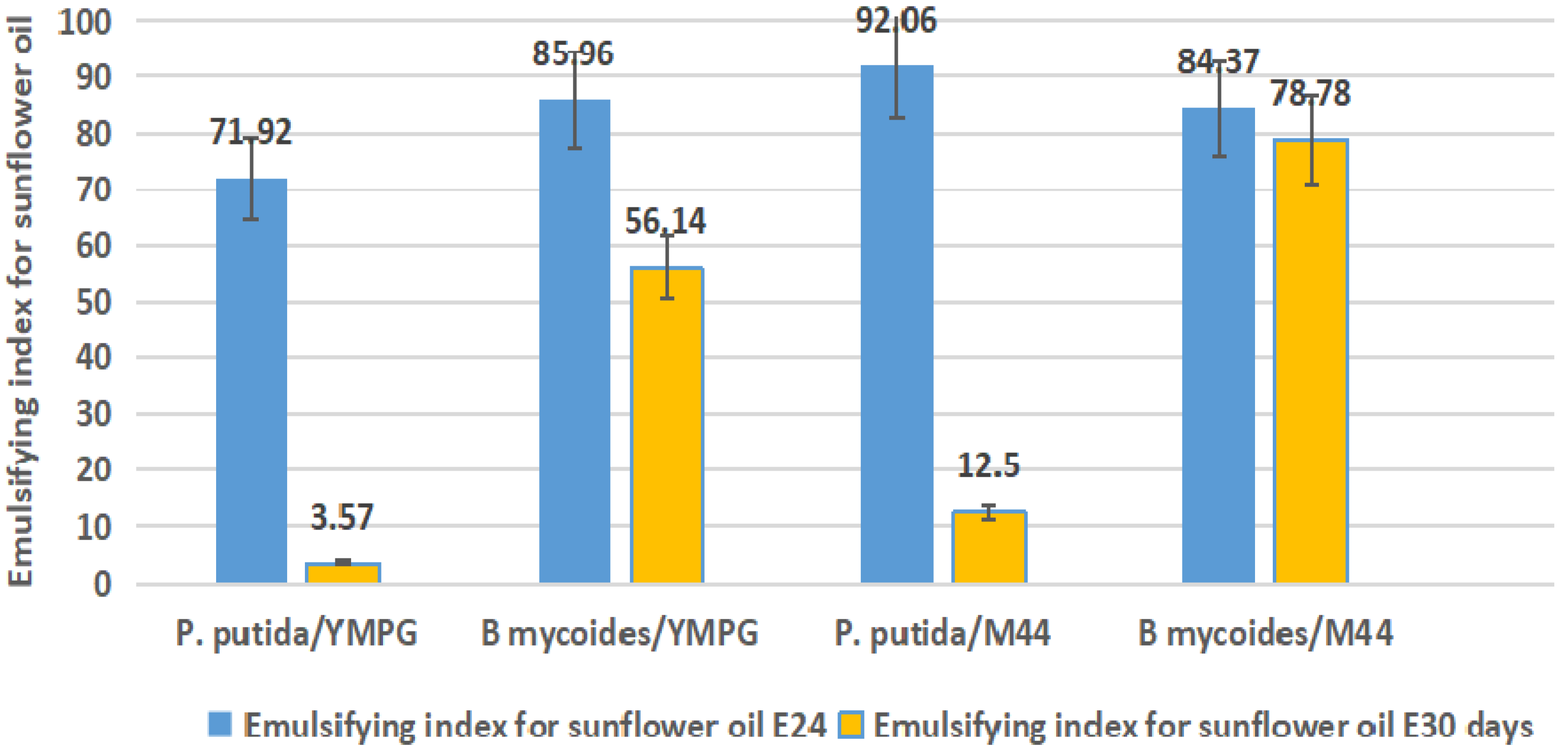
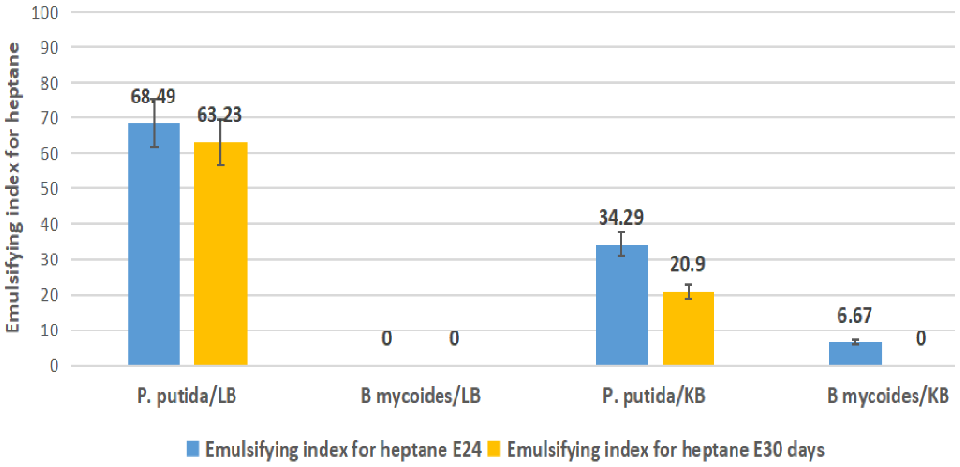
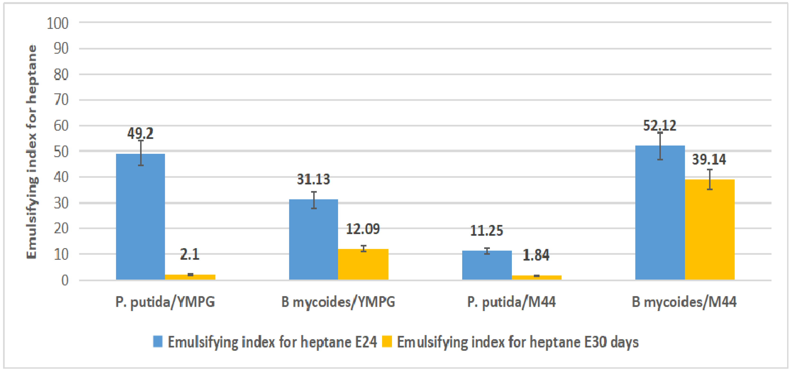
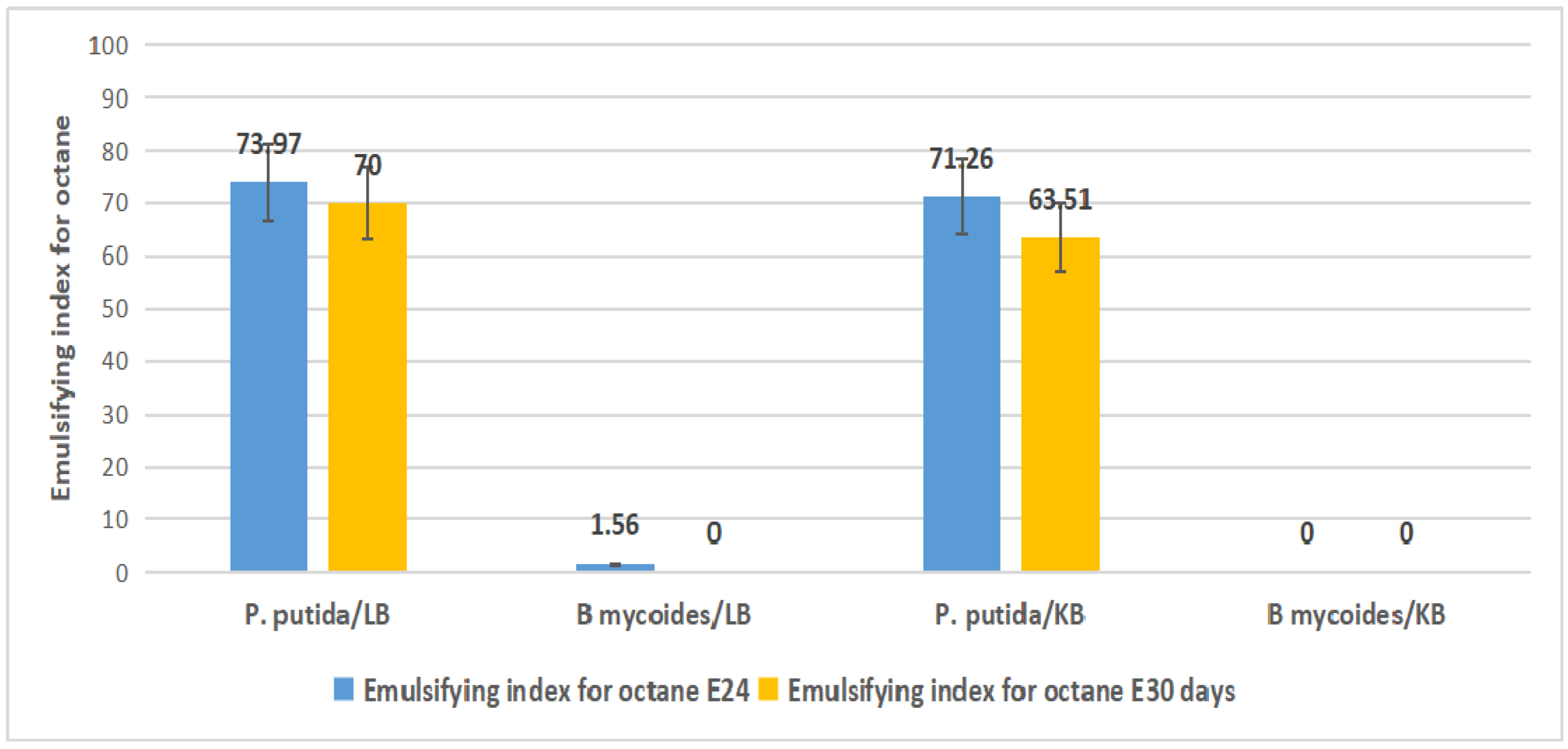
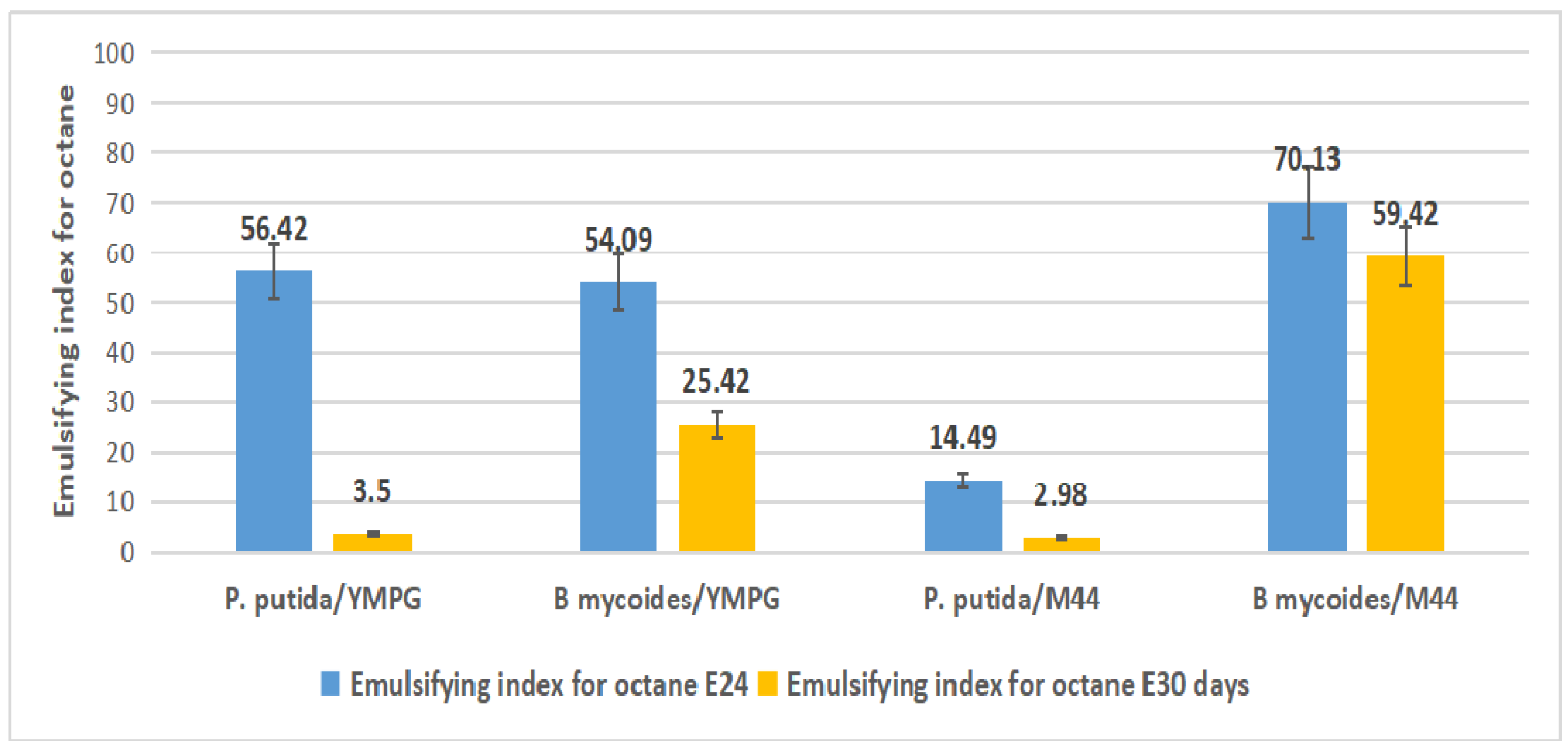
2.4. Emulsification Index

The cultivation dynamics of the selected strains, tested on KB, LB, M44, and YMPG media for biosurfactant production, was monitored by regular pH and ODλ = 550nm measurements. After 72 h of fermentation, the media were centrifuged, and the supernatants were used for the following experiments. In the Figure 9 are presented the emulsions obtained by these supernatants with sunflower oil, heptane and octane.
The figures below present the values of the emulsifying index obtained after 24 h and 30 days, respectively, following the vigorous stirring of supernatants with sunflower oil, heptane, and octane.
Supernatants of the P. putida (B1) strain grown in the LB medium produced stable emulsions with sunflower oil (Figure 10); the value of the emulsifying index was 56.7 after 24 h and 54.76 after 30 days, respectively. In the KB medium, the strain produced biosurfactants that emulsified the oil, but the emulsion formed was less stable; the emulsifying index was 19.18 after 30 days. Supernatants of the B. mycoides (Bm) strain grown on LB and KB media produced emulsions with sunflower oil (Figure 10) that remained stable for 24 h.
P. putida (B1) produced biosurfactants in the YMPG and M44 media (Figure 11), which formed emulsions with sunflower oil that remained stable for 24 h, while those of B. mycoides (Bm) remained stable for 30 days, when the strain was grown on M44.
The emulsifying indices registered for the P. putida (B1) strain grown in the LB medium were higher in the case of heptane (Figure 12) and were also stable: After 24 h, it was 68.49 and 63.23 after 30 days, respectively. The biosurfactants produced in the KB medium also formed relatively stable emulsions with heptane; the emulsifying indices were 34.29 after 24 h and 20.9 after 30 days, respectively.
The emulsions of the supernatants from B. mycoides (Bm) grown in both the LB and KB media (Figure 12) with heptane were not stable for even 24 h.
The emulsions of the biosurfactants from the P. putida (B1) strain (on YMPG and M44) with heptane (Figure 13) remained stable for 24 h, while the Bm supernatants from the fermentations in the M44 medium (Figure 13) formed emulsions that remained stable for 30 days.
Biosurfactants of the P. putida (B1) strain grown in both the KB and LB media (Figure 14) registered higher values of emulsifying indices with octane at 24 h and 30 days, respectively. Biosurfactants produced by B. mycoides (Bm) in the KB and LB media (Figure 14) did not seem to have the ability to emulsify the octane, but those produced on M44 (Figure 15) registered, at 24 h and 30 days, good values for emulsifying indices.
The emulsions of the biosurfactants from the P. putida (B1) strain (on YMPG and M44) with heptane (Figure 13) and octane (Figure 15) remained stable for 24 h, while the Bm supernatants from the fermentations in the M44 medium (Figure 13 and Figure 15) formed emulsions that remained stable for 30 days.
The values of the emulsifying index obtained with sunflower oil indicated that the biosurfactants produced by B. mycoides (Bm) could have applications in the preparation of oil based cosmetics and foods, as well as in the bioremediation of oil waste.
Some authors have shown that Pseudomonas sp. strains are good producers of biosurfactants that can be used in many industrial applications [33,34,35]. According to Chong and Lee, the key enzymes for rhamnolipids biosynthesis are almost exclusively limited to Pseudomonas sp. and Burkholderia sp. Although many authors have claimed that P. aeruginosa is the best producer of biosurfactants, there remain some concerns about its pathogenicity in the case of large scale production and applications.
Our results showed that the P. putida (B1) strain was a good producer of biosurfactants, and in addition, the strain did not present a danger for future applications. The emulsions formed by supernatants of the newly isolated strains with sunflower oil were stable, and many authors considered that the emulsion stability is one of the most important properties of a biosurfactant. In their opinion, the emulsion remains stable, and the biosurfactant could have numerous potential applications, if its E24 corresponds to 50% or more [36,37,38,39].
The values of the emulsifying indices obtained for octane were better than those obtained for heptane. This observation is in accordance with the observations made by Pathak and Keharia (2014) [40], who obtained an emulsifying index of 50.0 for octane and an emulsifying index of 40.0 for heptane, with the emulsions remaining stable for two days.
Many researchers agree with the observation of Pathak and Keharia that the emulsification index decreases with the reduction of hydrocarbon chain length [40,41].

3. Materials and Methods

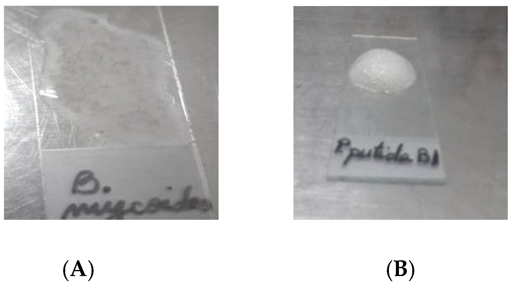
3.1. Biologic Material

The microorganisms of interest were isolated from various plant materials (hay, beans, potatoes, cabbage). The plant materials were collected from different areas of Romania (Valcea, Bucharest, Ilfov) in sterile receptacles, kept in a refrigerator until processing, and prepared according to the protocol described elsewhere: approximately 1 g of each material was sterile inoculated in 500 mL flakes with 100 mL liquid medium, specific for bacteria, yeasts, and fungi [42]. After 24 h of incubation at 30 ± 1 °C and 220 rpm, the microorganisms were isolated as a pure culture using the serial dilution method and the streak plate method, on specific media: bacteria on Nutrient Agar (NA), yeasts on Yeast Malt Peptone Glucose (YMPG), and fungi on Potato Dextrose Agar (PDA). Twenty-five strains of microorganisms were isolated from the plant collected and the samples processed: 20 strains of bacteria, 3 of yeasts, and 2 of fungi.
These microorganisms were tested for their antimicrobial activity against two phytopathogens, from the collection of the National Institute for Chemical Pharmaceutical Research and Development-ICCF, Erwinia carotovora ICCF 138 and Xanthomonas campestris ICCF 274.
Two bacterial strains were the most interesting, being identified as belonging to Pseudomonas putida and Bacillus mycoides, by Maldi Biotyper MSP Identification Standard Method 1.1 and Rep-PCR.

3.2. Culture Media

The following media were used in the experiments [43]:
NA (Nutrient Agar) % (g/v): 0.50 peptone, 0.30 yeast extract, 1.50 agar, 0.50 NaCl;
Bacillus thuringiensis medium is denoted here as NB (Nutrient Broth) % (g/v): 0.10 beef extract, 0.20 yeast extract, 0.50 peptone, 0.50 NaCl;
YMPG (Yeast malt peptone glucose) % (g/v): 0.30 yeast extract, 0.30 malt extract, 0.50 peptone, 1.00 glucose, 2.00 agar;
PDA % (g/v): 20.00 potato infusion, 2.00 dextrose, 2.00 agar;
LA (lipase Agar) % (g/v): 0.50 peptone, 0.30 beef extract, 1.00 tributyrin, 2.00 agar;
M44 (and M44 broth without agar) % (g/v): 1.00 yeast extract, 1.00 bacteriological peptone, 5.00 glycerol, 2.00 agar;
LB (Luria-Bertani) broth % (g/v): 1.00 tryptone, 0.50 yeast extract, 0.50 NaCl;
KB (King’s Medium B) broth % (g/v): 2.00 proteose-peptone, 1.00 glycerol, 0.15 K2HPO4, 0.15 MgSO4 * 7H2O;
SNA (Starch Nutrient Agar) % (g/v): 0.30 beef extract, 0.10 peptone, 0.50 NaCl, 1.00 starch, 2.00 agar.
The chemicals were purchased from Sigma and Difco. All culture media were prepared with distilled water, adjusted to a pH range of 6.5–7.2, and sterilized for 20 min at 121 °C. For the submerged bioprocesses, Erlenmeyer flasks of 500 mL capacity, with 100 mL medium, were used.

3.3. Preservation of Strains and Cultivation Conditions

The phytopathogen strains used in experiments, X. campestris ICCF 274 and E. carotovora ICCF 138, were from the collection of the National Institute for Chemical Pharmaceutical Research and Development-ICCF. The microorganisms of interest, B. mycoides (Bm) and P. putida (B1), were isolated from various plant materials collected from different areas of Romania (Valcea, Bucharest, Ilfov).
Except for the X. campestris ICCF 274 strain, which was maintained on YMPG medium, all the other strains used in experiments, respectively E. carotovora ICCF 138, B. mycoides (Bm), and P. putida (B1), were grown on M44 medium.
The pre-inoculum consisted of bacterial strains incubated at 28–30 °C for 48–72 h on YMPG, respectively M44 agarized medium. The inoculum medium (YMPG for X. campestris ICCF 274 and M44 for all the other strains) was seeded with 2.0 mL pre-inoculum containing 9 × 10⁸ CFU/mL (McFarland Standard No. 3), and the fermentation medium was inoculated with 10.0 mL of inoculum. The strains were cultivated 24–48 h for inoculum and 48–72 h for bioproduction, at 28–30 °C and 220 rpm.
Bacterial cell growth was expressed as Optical Density (OD) at 550 nm.

3.4. Antimicrobial Activity Assay

The isolated strains and their supernatants were tested for their antimicrobial activity in two parallel experiments, conducted in triplicate. The antimicrobial activity assay was performed by the double culture method, in Petri plates [42] (Soare et al., 2017). YMPG medium in the case of Xanthomonas campestris ICCF 274 and M44 medium for Erwinia carotovora ICCF 138 were used, being the best growth media for these strains. The first experiment (Experiment I) followed an approach described by Soare et al.: One millimeter of inoculum from the broth culture of phytopathogens (containing 3 × 10⁸ UFC/mL, McFarland Standard No. 1) was added by pipette to the center of the Petri dish, over the agar medium (cooled, but still molten) and rotated gently, till they mixed. After solidification, 20 µL of inoculum containing 9 × 10⁸ CFU/mL (McFarland Standard No. 3), from the broth culture of B. mycoides (Bm), respectively P. putida (B1), were put in the middle of the plate, allowed to be adsorbed in the medium, and the plates were incubated at 30 ± 1 °C. The inhibition zones were checked daily.
In the second experiment (Experiment II), the phytopathogens (1 mL of inoculum from the broth culture of phytopathogens, containing 3 × 10⁸ UFC/mL) were first added to the culture medium according to the same method. The plates were maintained at room temperature (20 ± 1 °C) for 24 h, and then, the antagonists (B. mycoides (Bm), respectively P. putida (B1)) were inoculated. The antagonists were added in the center of the Petri dishes containing the phytopathogen and were incubated for 48 h at 30 ± 1 °C. 1 m L of the phytopathogen suspension (containing 3 × 10⁸ UFC/mL), and 20 µL of the antagonists inoculum, containing 3 × 10⁸ UFC/mL were added in every Petri plate. The antimicrobial activity was checked daily, and the inhibition zones were recorded. The Zones Of Inhibition (ZOI) were calculated using the formula:
ZOI = colony diameter + 2xd
where ZOI = Zones Of Inhibition and d = distance between the edge of the antagonist’s colony and the edge of the zone of inhibition.
For the antimicrobial activity assay of the biosurfactants, the strains of B. mycoides (Bm) and P. putida (B1) were grown submerged, for 72 h, at 28–30 °C and 220 rpm, on five media: NB, KB, LB, M44, and YMPG. At the end of the fermentations, the broths were centrifuged for 40 min at 4 °C and 9000 rpm.
The supernatants used in experiments were sterilized for 30 min at 115 °C, and after sterilization, no significant changes were registered in the emulsification activity. Many authors mention that some biosurfactants are heat resistant [44], while some are not, and the main criterion in their tests was verifying the emulsification activity after autoclaving the samples [45]. The antimicrobial activity of the biosurfactants was evaluated using the agar diffusion method against the aforementioned phytopathogens. For each sample, 0.25 mL of supernatant were added in metal cylinders. After 24 h of incubation at 30 ± 1 °C, the inhibition zones were measured from the edge of the cylinder to the edge of the inhibition zone.

3.5. Enzyme Production

The ability of the two bacterial strains to produce certain enzymes was tested on differential media, according to the protocols described below; then, the enzymatic activities were measured for: amylase (enzymatic assay of α-amylase EC 3.2.1.1, with DNS, [30]), lipase (enzymatic assay of lipase EC 3.1.1.3, titrimetric method, [31]), and catalase (enzymatic assay of catalase EC 1.11.1.6, [32]), following the Sigma-Aldrich protocols.
In order to investigate amylases’ production, B. mycoides (Bm) and P. putida (B1) were grown on Petri plates with Starch Nutrient Agar (SNA) medium. The medium was spot inoculated and incubated for 48 h at 30 °C. Following incubation, the plates were flooded with 2 mL of Gram’s iodine, which forms a dark blue-colored complex in the presence of starch. Colorless zones around the colonies revealed starch hydrolysis.
For amylase activity determination, the strains were cultivated in SNA broth medium, for 72 h, at 28–30 °C and 220 rpm, till B. mycoides (Bm) reached an OD of 8.15 and P. putida (B1) reached an OD of 4.25. After centrifugation of the culture broths (30 min at 4 °C and 8000 rpm), the supernatants were used for the determination of the enzymatic activity, by a spectrophotometric method, based on starch hydrolysis into maltose [30].
One unit of amylase activity is defined as the amount of enzyme that liberates 1.0 mg of maltose from starch in 3 min, at pH 6.9, at 20 °C.
The production of catalase is important in neutralizing the bactericidal effects of hydrogen peroxide, which can harm the microorganisms. To check the ability of B. mycoides (Bm) and P. putida (B1) to produce catalase, a small amount of bacterial isolate was mixed with 3% hydrogen peroxide solution. The bacterial isolates were considered as catalase positive if oxygen bubbles occurred.
To investigate catalase production at a quantitative level, B. mycoides (Bm) and P. putida (B1) were grown on M44 solid medium, for 72 h, at 30 °C. After that, the surfaces of the media containing the cultures of the strains (6 × 10⁸ UFC/mL) were washed with 3 mL of distilled water. The cell suspensions were subjected to ultrasonication to extract the enzyme (35 kHz frequency ultrasound, Bandelin Sonorex digital 10 p ultrasonic bath dk 255 P), then centrifuged, and the supernatant was analyzed for spectrophotometric determination of the enzymatic activity [32], based on the following reaction:
Catalysts 09 00959 i001
One unit of catalase will decompose 1.0 µm of H2O2 per minute at pH 7.0 at 25 °C, while the H2O2 concentration falls from 10.3 mM to 9.2 mM. The rate of disappearance of H2O2 was followed by observing the rate of decrease in the absorbance at 240 nm.
For testing the microorganisms’ ability to produce lipases, the strains were grown in submerged culture for 72 h at 30 °C and 220 rpm, on several media (NB, KB, LB, M44, YMPG). These constituted the inoculum used in the lipase assay. To show lipase production, the strains were then cultivated (spot inoculated) on Petri plates with LA medium containing tributyrin. After 48 h of incubation, the plates were analyzed as regards the diameters of the clear zones around the colonies, which signifies lipase production.
Catalysts 09 00959 i002
One unit of enzyme activity is defined as that amount of enzyme that liberates the equivalent of 1 μm of fatty acid per minute from the substrate emulsion (olive oil in the presence of thymolphthalein indicator) under the described assay conditions.

3.6. Emulsification Index Test

In order to evaluate the production of biosurfactants by the selected strains, submerged bioprocesses in 500 mL flasks with 100 mL medium on a rotary shaker at 220 rpm were performed. The strains were grown for 72 h at 30 °C, in YMPG, KB, LB, and M44 media. After centrifugation of the culture broths (30 min at 4 °C and 9000 rpm), the supernatants were used for the determination of the emulsifying indexes of heptane, octane, and sunflower oil.
Emulsions were prepared as described by Pathak and Keharia (2014) [40]. Six milliliters of sunflower oil/heptane/octane and 4 mL of supernatant were added in the test tubes.
The tubes were vigorously stirred, and the height of the emulsions formed were measured after 24 h and 30 days, respectively. The emulsifying index was calculated using the following formula:
E24 = (Height of emulsion layer/Height of total liquid column) × 100
The experiments were performed in triplicate.

4. Conclusions

Regarding the growth of the B. mycoides (Bm) and the P. putida (B1) strains, the best results were obtained in the M44 medium as the ODs were 40.35 and 24.35, respectively, after 72 h of the bioprocess.
Newly isolated strains and their supernatants exerted antimicrobial activity against the phytopathogens Erwinia carotovora ICCF 138 and Xanthomonas campestris ICCF 274.
Initial tests on solid media showed that B. mycoides (Bm) produced the enzymes amylase and lipase. After 72 h of submerged development in SNA medium and 48 h in LA medium, B. mycoides (Bm) produced small amounts of amylases (as the amylase activity was 0.75 U/mL) and lipases (0.39 U/mL), which means that the fermentation media needed to be optimized. The amylase activity of P. putida (B1) was insignificant: the lipase activity of the strain registered 0.36 U/mL, and the catalase activity registered 2.3 U/mL.
The biosurfactants of both strains formed emulsions with sunflower oil. The emulsions formed by the supernatants of P. putida (B1), grown in the LB medium, presented the most significant values in both cases (after 24 and 30 days, respectively). The emulsifying indices (E24) for octane reached the highest values: 73.97% in the LB medium and 71.26% in the KB medium.
Although supernatants from the B. mycoides (Bm) strain grown in the KB and LB media formed emulsions with sunflower oil (with an emulsifying index of 60.56 and 56.76, respectively), they remained stable for only 24 h, while those from M44 and YMPG remained stable for 30 days.
These results suggest the possibility of using these newly isolated strains in agriculture (for preventing the diseases caused by several phytopathogens) and in the bioremediation of waste oils and hydrocarbons, as well as in the food, cosmetic, chemical, and pharmaceutical industries. However, further investigations are required to improve the media, mainly with waste oils and hydrocarbons. Being able to grow on these kinds of substrates, the strains could be used both to reduce the pollutants and to produce biosurfactants with many potential applications.

Author Contributions

M.-G.S. (Vladu) conceived of and designed the research and drafted and finalized the paper; E.S.L. was responsible for the supervision and methodology and was the project coordinator; N.E. performed the research and the analysis; N.M. (Dalanaj) performed the analysis; O.P. performed the writing, review, and editing; N.B. performed the writing, review, and editing; all authors contributed to discussing the research, writing parts of the paper, and commenting on draft versions.

Funding

This research was funded by Erasmus +, Grant Number 2019-1-RO01-KA105-062720. Research: “Act for sustainable consumption and production in the context of circular economy in line with sustainable development goals–ACT4SCP”.

Acknowledgments

We would like to express our gratitude to Diana Smarandache (Pelinescu) for her valuable work to identify the surfactant producing strains.

Conflicts of Interest

The authors declare that there is no conflict of interest regarding the publication of this article.

References

  1. Bubici, G.; Kaushal, M.; Prigigallo, M.I.; Cabanás, G.-L.C.; Mercado-Blanco, J. Biological Control Agents Against Fusarium Wilt of Banana. Front. Microbiol. 2019, 10, 616. [Google Scholar] [CrossRef]
  2. Caulier, S.; Gillis, A.; Colau, G.; Licciardi, F.; Liépin, M.; Desoignies, N.; Modrie, P.; Legrève, A.; Mahillon, J.; Bragard, C. Versatile Antagonistic Activities of Soil-Borne Bacillus spp. and Pseudomonas spp. against Phytophthora infestans and Other Potato Pathogens. Front. Microbiol. 2018, 9, 143. [Google Scholar] [CrossRef]
  3. Raaijmakers, J.M.; De Bruijn, I.; Nybroe, O.; Ongena, M. Natural functions of lipopeptides from Bacillus and Pseudomonas: More than surfactants and antibiotics. FEMS Microbiol. Rev. 2010, 34, 1037–1062. [Google Scholar] [CrossRef]
  4. Fira, D.; Dimkic, I.; Beric, T.; Lozo, J.; Stankovic, S. Biological control of plant pathogens by Bacillus species. J. Biotechnol. 2018, 285, 44–55. [Google Scholar] [CrossRef]
  5. Gross, H.; Loper, J.E. Genomics of secondary metabolite production by Pseudomonas spp. Nat. Prod. Rep. 2009, 26, 1408–1446. [Google Scholar] [CrossRef]
  6. Santos, D.K.; Rufino, R.D.; Luna, J.M.; Santos, V.A.; Sarubbo, L.A. Biosurfactants: Multifunctional Biomolecules of the 21st Century. Int. J. Mol. Sci. 2016, 17, 401. [Google Scholar] [CrossRef]
  7. Singh, P.; Patil, Y.; Rale, V. Biosurfactant production: Emerging trends and promising strategies. J. Appl. Microbiol. 2018, 126, 2–13. [Google Scholar] [CrossRef]
  8. Gomaa, E.Z. Antimicrobial activity of a biosurfactant produced by Bacillus licheniformis strain M104 grown on whey. Braz. Arch. Biol. Technol. 2013, 56, 259–268. [Google Scholar] [CrossRef]
  9. Ndlovu, T.; Rautenbach, M.; Vosloo, J.A.; Khan, S.; Khan, W. Characterisation and antimicrobial activity of biosurfactant extracts produced by Bacillus amyloliquefaciens and Pseudomonas aeruginosa isolated from a wastewater treatment plant. AMB Express 2017, 7, 108. [Google Scholar] [CrossRef]
  10. Katz, E.; Demain, A.L. The peptide antibiotics of Bacillus: Chemistry, biogenesis and possible functions. Bacteriol. Rev. 1977, 41, 449. [Google Scholar]
  11. Grossman, A.D. Genetic networks controlling the initiation of sporulation and the development of genetic competence in Bacillus subtilis. Annu. Rev. Genet. 1995, 29, 477–508. [Google Scholar] [CrossRef] [PubMed]
  12. Caulier, S.; Nannan, C.; Gillis, A.; Licciardi, F.; Bragard, C.; Mahillon, J. Overview of the antimicrobial compounds produced by members of the Bacillus subtilis group. Front. Microbiol. 2019, 10, 302. [Google Scholar] [CrossRef] [PubMed]
  13. Nihorimbere, V.; Ongena, M.; Smargiassi, M.; Thonart, P. Beneficial effect of the rhizosphere microbial community for plant growth and health. Biotechnol. Agron. Soc. Environ. 2011, 15, 327–337. [Google Scholar]
  14. Sachdev, D.P.; Cameotra, S.S. Biosurfactants in agriculture. Appl. Microbiol. Biotechnol. 2013, 97, 1005–1016. [Google Scholar] [CrossRef] [PubMed]
  15. Rendueles, O.; Ghigo, J.M. Multi-species biofilms: How to avoid unfriendly neighbors. FEMS Microbiol. Rev. 2012, 36, 972–989. [Google Scholar] [CrossRef]
  16. Zhang, F.; Gu, W.; Xu, P.; Tang, S.; Xie, K.; Huang, X.; Huang, Q. Effects of alkyl polyglycoside (APG) on composting of agricultural wastes. Waste Manag. 2011, 31, 1333–1338. [Google Scholar] [CrossRef]
  17. Rosenberg, E.; Ron, E.Z. High- and low-molecular-mass microbial surfactants. Appl. Microbiol. Biotechnol. 1999, 52, 154–162. [Google Scholar] [CrossRef]
  18. Silva, E.J.; Correa, P.F.; Almeida, D.G.; Luna, J.M.; Rufino, R.D.; Sarubbo, L.A. Recovery of contaminated marine environments by biosurfactant-enhanced bior-emediation. Colloids Surf. B Biointerfaces 2018, 172, 127–135. [Google Scholar] [CrossRef]
  19. Chandankere, R.; Yao, J.; Cai, M.; Masakorala, K.; Jain, A.K.; Choi, M.M. Properties and characterization of biosurfactant in crude oil biodegradation by bacterium Bacillus methylotrophicus USTBa. Fuel 2014, 122, 140–148. [Google Scholar] [CrossRef]
  20. Desai, J.D.; Banat, I.M. Microbial production of surfactants and their commercial potential. Microbiol. Mol. Biol. Rev. 1997, 61, 47–64. [Google Scholar]
  21. Banat, I.M.; Makkar, R.S.; Cameotra, S.S. Potential commercial applications of microbial surfactants. Appl. Microbiol. Biotechnol. 2000, 53, 495–508. [Google Scholar] [CrossRef] [PubMed]
  22. Banat, I.M. Biosurfactants production and possible uses in microbial enhanced oil recovery and oil pollution remediation: A review. Bioresour. Technol. 1995, 51, 1–12. [Google Scholar] [CrossRef]
  23. Barkay, T.; Navon-Venezia, S.; Ron, E.Z.; Rosenberg, E. Enhancement of solubilization and biodegradation of polyaromatic hydrocarbons by the bioemulsifier alasan. Appl. Environ. Microbiol. 1991, 65, 2697–2702. [Google Scholar]
  24. Christofi, N.; Ivshina, I.B. Microbial surfactants and their use in field studies of soil remediation. J. Appl. Microbiol. 2002, 93, 915–929. [Google Scholar] [CrossRef] [PubMed]
  25. Kosaric, N. Biosurfactants and their application for soil bioremediation. Food Technol. Biotechnol. 2001, 39, 295–304. [Google Scholar]
  26. Calvo, C.; Manzanera, M.; Silva-Castro, G.A.; Uad, I.; González-López, J. Application of bioemulsifiers in soil oil bioremediation processes. Future prospects. Sci. Total Environ. 2009, 407, 3634–3640. [Google Scholar] [CrossRef]
  27. Mouafo, T.H.; Mbawala, A.; Ndjouenkeu, R. Effect of Different Carbon Sources on Biosurfactants’ Production by Three Strains of Lactobacillus spp. Biomed Res. Int. 2018, 2018, 5034783. [Google Scholar] [CrossRef]
  28. Nielsen, T.H.; Sørensen, D.; Tobiasen, C.; Andersen, J.B.; Christophersen, C.; Givskov, M.; Sørensen, J. Antibiotic and Biosurfactant Properties of Cyclic Lipopeptides Produced by Fluorescent Pseudomonas spp. from the Sugar Beet Rhizosphere. Appl. Environ. Microbiol. 2002, 68, 3416–3423. [Google Scholar] [CrossRef]
  29. Petatán-Sagahón, I.; Anducho-Reyes, M.A.; Silva-Rojas, H.V.; Arana-Cuenca, A.; Tellez-Jurado, A.; Cárdenas-Álvarez, I.O.; Mercado-Flores, Y. Isolation of Bacteria with Antifungal Activity against the Phytopathogenic Fungi Stenocarpella maydis and S macrospora. Int. J. Mol. Sci. 2011, 12, 5522–5537. [Google Scholar] [CrossRef]
  30. Available online: https://www.sigmaaldrich.com/technical-documents/protocols/biology/enzymatic-assay-of-a-amylase.html (accessed on 5 November 2019).
  31. Available online: https://www.sigmaaldrich.com/content/dam/sigma-aldrich/docs/Sigma/General_Information/lipase_-_titrimetric_ph_72.pdf (accessed on 4 November 2019).
  32. Available online: https://www.sigmaaldrich.com/technical-documents/protocols/biology/enzymatic-assay-of-catalase.html (accessed on 5 November 2019).
  33. Das, P.; Yang, X.P.; Ma, L.Z. Analysis of biosurfactants from industrially viable Pseudomonas strain isolated from crude oil suggests how rhamnolipids congeners affect emulsification property and antimicrobial activity. Front. Microbiol. 2014, 5, 696. [Google Scholar] [CrossRef]
  34. El-Sheshtawy, H.S.; Doheim, M.M. Selection of Pseudomonas aeruginosa for biosurfactant production and studies of its antimicrobial activity. Egypt. J. Pet. 2014, 23, 1–6. [Google Scholar] [CrossRef]
  35. Chong, H.; Li, Q. Microbial production of rhamnolipids: Opportunities, challenges and strategies. Microb. Cell Fact. 2017, 16, 137. [Google Scholar] [CrossRef] [PubMed]
  36. Gnanamani, A.; Kavitha, V.; Radhakrishnan, N.; Mandal, A.B. Bioremdiation of crude oil contamination using microbial surface-active agents isolation production and characterization. J. Bioremed. Biodegrad. 2010, 1, 107. [Google Scholar] [CrossRef]
  37. Lima, T.M.S.; Fonseco, A.F.; Leao, B.A.; Houndeer, A.H.; Totala, M.R.; Borges, A.C. Oil recovery from oil fuel oil storage tank sludges using biosurfactants. J. Bioremed. Biodegrad. 2011, 2, 125–137. [Google Scholar]
  38. Anyanwu, C.U.; Obi, S.K.C.; Okolo, B.M. Lipopeptide biosurfactant produced by Serratia marcescens NSK-1 strain isolated from petroleum contaminated soil. J. Appl. Sci. Res. 2011, 7, 79–87. [Google Scholar]
  39. Diab, A.; Sami, S.; Diab, A. Production, Characterization and Application of a New Biosurfactant Derived from Egyptian Sunflower Seeds. Int. J. Sci. Res. 2016, 5, 602–612. [Google Scholar]
  40. Pathak, K.V.; Keharia, H. Application of extracellular lipopeptide biosurfactant produced by endophytic Bacillus subtilis K1 isolated from aerial roots of banyan (Ficus benghalensis) in microbially enhanced oil recovery (MEOR). 3 Biotech 2014, 4, 41–48. [Google Scholar] [CrossRef]
  41. Sharma, R.; Singh, J.; Verma, N. Production, characterization and environmental applications of biosurfactants from Bacillus amyloliquefaciens and Bacillus subtilis. Biocatal. Agric. Biotechnol. 2018, 16, 132–139. [Google Scholar] [CrossRef]
  42. Available online: http://cfarm.ncpri.ro/files/cmii-iccf/CMII-ICCF.pdf (accessed on 27 September 2019).
  43. Soare, M.G.; Tomulescu, C.; Petrescu, M.M.; Lupescu, I.; Moscovici, M.; Popa, O.; Băbeanu, N. Antimicrobial activity of newly isolated Bacillus sp. and Pseudomonas sp. strains and their potential use as biocontrol agents. Sci. Bull. Ser. F Biotechnol. 2017, XXI, 81–86. [Google Scholar]
  44. Nitschke, M.; Pastore, G.M. Production and properties of a surfactant obtained from Bacillus subtilis grown on cassava wastewater. Bioresour. Technol. 2006, 97, 336–341. [Google Scholar] [CrossRef]
  45. Moro, G.V.; Almeida, R.T.R.; Napp, A.P.; Porto, C.; Pilau, E.J.; Lüdtke, D.S.; Moro, A.V.; Vainstein, M.H. Identification and ultra-high-performance liquid chromatography coupled with high-resolution mass spectrometry characterization of biosurfactants, including a new surfactin, isolated from oil-contaminated environments. Microb. Biotechnol. 2018, 11, 759–769. [Google Scholar] [CrossRef] [PubMed]
Figure 1. Growth of P. putida (B1) and B. mycoides (Bm) strains in NB medium.
Catalysts 09 00959 g001
Figure 2. Growth of P. putida (B1) and B. mycoides (Bm) strains in King B (KB) medium.
Catalysts 09 00959 g002
Figure 3. Growth of P. putida (B1) and B. mycoides (Bm) strains in LB medium.
Catalysts 09 00959 g003
Figure 4. Growth of P. putida (B1) and B. mycoides (Bm) strains in M44 medium.
Catalysts 09 00959 g004
Figure 5. Growth of P. putida (B1) and B. mycoides (Bm) strains in YMPG medium.
Catalysts 09 00959 g005
Figure 6. Amylase production: (A) B. mycoides (Bm) positive; (B) P. putida (B1) negative.
Catalysts 09 00959 g006
Figure 7. Catalase production: (A) B. mycoides (Bm) insignificant; (B) P. putida (B1) positive.
Catalysts 09 00959 g007
Figure 8. Lipase production with strains P. putida (B1) (A) and B. mycoides (Bm) (B) (inocula from media: NA (1 and 11), KB (2 and 12), LB (3 and 13), M44 (4 and 14), YMPG (5 and 15)).
Catalysts 09 00959 g008
Figure 9. Emulsions obtained with the supernatants of the two strains cultivated on LB and KB media: supernatants of P. putida with: sunflower oil 1 and 2, heptane 3 and 4, octane 5 and 6; supernatants of B. mycoides with sunflower oil 7 and 8, heptane 9 and 10, octane 11 and 12.
Catalysts 09 00959 g009
Figure 10. Emulsifying index for sunflower oil (LB and KB).
Catalysts 09 00959 g010
Figure 11. Emulsifying index for sunflower oil (YMPG and M44).
Catalysts 09 00959 g011
Figure 12. Emulsifying index for heptane (LB and KB).
Catalysts 09 00959 g012
Figure 13. Emulsifying index for heptane (YMPG and M44).
Catalysts 09 00959 g013
Figure 14. Emulsifying index for octane (LB and KB).
Catalysts 09 00959 g014
Figure 15. Emulsifying index for octane (YMPG and M44).
Catalysts 09 00959 g015
Table 1. Antimicrobial activity of antagonists after 48 h of growth together with phytopathogens E. carotovora ICCF 138 and X. campestris ICCF 274.
Bacterial StrainInhibition Zones (mm) against E. carotovora ICCF 138Inhibition Zones (mm) against X. campestris ICCF 274
Experiment IExperiment IIExperiment IExperiment II
P. putida (B1)35 ± 0.5824 ± 0.2936 ± 0.5822 ± 0.29
B. mycoides (Bm)60 ± 0.5827 ± 0.5843 ± 0.5830 ± 0.29
Control----
Note: Experiment I, antagonists added in the same day with phytopathogen; Experiment II, antagonists added 24 h after phytopathogen inoculation. Data are the means of three replicates (n = 3) ± standard error.
Table 2. Antimicrobial activity (as zones of inhibition) of the biosurfactants after 48 h of growth together with the phytopathogens.
Supernatants of the StrainsInhibition Zone Diameters (in mm) Against E. c. ICCF138Inhibition Zone Diameters (in mm) Against X. c. ICCF274
P. putida (B1)/Medium *1 *7 ± 0.878 ± 0.29
2 *10 ± 0.5810 ± 0.29
3 *10 ± 0.589 ± 0.58
4 *11 ± 0.5812 ± 0.29
5 *8 ± 0.589 ± 0.58
B. mycoides (Bm)/Medium *1 *8 ± 0.587 ± 0.58
2 *10 ± 0.299 ± 0.58
3 *9 ± 0.5810 ± 0.58
4 *10 ± 0.5811 ± 0.87
5 *8 ± 0.587 ± 0.58
Medium *: 1 *, NB; 2 *, KB; 3 *, LB; 4 *, M44; 5 *, YMPG. Data are the means of three replicates (n = 3) ± standard error.

